Article

Mn3O4/NiO Nanoparticles Decorated on Carbon Nanofibers as an Enzyme-Free Electrochemical Sensor for Glucose Detection

1
College of Sciences, Shanghai University, Shanghai 200444, China
2
Key Laboratory of Hunan Forest Products and Chemical Industry Engineering, Jishou University, Jishou 416000, China
*
Authors to whom correspondence should be addressed.
Biosensors 2023, 13(2), 264; https://doi.org/10.3390/bios13020264
Submission received: 31 December 2022 / Revised: 9 February 2023 / Accepted: 11 February 2023 / Published: 13 February 2023
(This article belongs to the Special Issue Electrochemical Biosensors for Disease Detection)

Abstract

:
Transition metal oxides have garnered a lot of attention in the field of electrocatalysis along with their unique crystal structure and excellent catalytic properties. In this study, carbon nanofibers (CNFs) decorated with Mn3O4/NiO nanoparticles were made using electrospinning and calcination. The conductive network constructed by CNFs not only facilitates electron transport, but also provides landing sites for nanoparticles, thus reducing nanoparticle aggregation and exposing more active sites. Additionally, the synergistic interaction between Mn3O4 and NiO improved electrocatalytic capacity for glucose oxidation. The Mn3O4/NiO/CNFs modified glassy carbon electrode shows satisfactory results in terms of linear range and anti-interference capability for glucose detection, suggesting that the constructed enzyme-free sensor has a promising application in clinical diagnosis.

Graphical Abstract

1. Introduction

Electrochemical glucose sensors perform an indispensable role in monitoring glucose concentrations, which not only can be employed to control the intake of glucose in diet, but also to predict diseases associated with high or low glucose levels [1,2]. Enzyme-based sensors show excellent sensitivity and accuracy for glucose detection, but they are sensitive to temperature and pH which limits their storage and transportation [3]. In contrast, enzyme-free sensors have the advantage of being cheap, stable, and sensitive [4], thus attracting considerable attention in recent years.
Noble metals and their alloys have shown excellent catalytic properties and stability [5,6] and are often used as enzyme-free sensors for glucose detection [7,8]. However, the high cost and scarcity of resources limit wide applications of noble metals. In this regard, transition metal can overcome the drawbacks mentioned above for noble metals and be applied as catalysts for many reactions [9], such as CO2 utilization [10], oxygen evolution reaction [11], and oxygen reduction reaction [12,13]. Generally speaking, composite transition metal oxides exhibit higher catalytic capacity and lower catalytic potential than single transition metal oxide due to the synergistic effect between different metal oxides. Wang et al. synthesized CuCo2O4/NiO nanoneedle arrays, modified on carbon cloth by hydrothermal method, which were used as self-supporting electrodes for the glucose detection [14]. Xu et al. fabricated CuO-NiO nanocomposites for the detection of glucose which exhibited excellent selectivity, reproducibility, and long-time stability [15].
One-dimensional nanofibers possess low density, large aspect ratio, and high charge carrier directed mobility [16]. Thomas et al. grew α-MoO3 nanorods on glass substrates by thermal evaporation for ammonia sensing at room temperature [17]. Zhang et al. used a hydrothermal method with low temperature growth to synthesize Co9S8 nanotubes @CuS nanosheets, which were modified on glassy carbon electrode (GCE) for the detection of glucose and H2O2 [18]. Kokulnathan et al. synthesized TiO/Au nanofibers by electrospinning for the determination of diphenylamine in food [19]. Among the above methods, electrospinning is a simple, efficient, and inexpensive way to synthesize one-dimensional nanofibers [16,20]. Transition metal oxides/carbon nanofibers composites fabricated by electrospinning have the following advantages: (i) CNFs with large aspect ratio can form a conductive network, which facilitates the electron transport [21]; (ii) the mesh structure can serve as a substrate to support the nanoparticles which allows more active sites of the nanoparticles to be exposed; and, (iii) the synergistic interaction between transition metal oxides and carbon nanofibers can facilitate the electrocatalytic performance.
In this study, CNFs decorated with Mn3O4/NiO nanoparticles (Mn3O4/NiO/CNFs) were synthesized using electrospinning and subsequent heat treatment. The presence of CNFs not only increases the conductivity of the material, but also expands the active surface area of the catalysts. Additionally, the multivalence of transition metals facilitates the redox reaction at the electrode surface. The synergistic effect between Mn3O4 and NiO is beneficial to improve the catalytic performance of glucose oxidation. It is worth noting that the constructed sensor has a good range of linearity, low detection limit, and outstanding interference immunity, indicating that the constructed enzyme-free sensor is promising in clinical diagnosis.

2. Experimental Section

2.1. Materials

Polyacrylonitrile (PAN) was supplied by Sigma-Aldrich (St. Louis, MO, USA). Mn(Ac)2∙4H2O, N,N-dimethylformamide (DMF), NaOH, glycocoll (Gly), Ni(Ac)2∙4H2O, and glucose (Glu) were supported by Sinopharm Chemical Reagent Co., Ltd. (Shanghai, China). Sucrose (Suc), dopamine (DA), maltose (Mal), ascorbic acid (AA), fructose (Fru), uric acid (UA), and NaCl were bought from Alfa Aesar Co., Ltd. (Shanghai, China). Millipore water (18.25 MΩ cm) was used.

2.2. Instrumentations

The scanning electron microscope (SEM, SU5000, HITACHI, Tokyo, Japan) was utilized to observe the morphology and size of materials. The high-resolution transmission electron microscope (HRTEM; JEM 2100F, JEOL, Tokyo, Japan) was further used to investigate the microscopic crystal structure of samples. X-ray diffraction (XRD, D/MAX2200, Rigaku, Tokyo, Japan) was employed to characterize the crystal structure of the materials. Fourier transform infrared spectroscopy (FTIR, AVATAR370, Thermo Scientific, Waltham, MA, USA) was performed to study chemical structure. The degree of graphitization of CNFs was studied by Raman spectrum (INVIA, Renishaw, London, UK). X-ray photoelectron spectrometer (XPS, ESCALAB-250XI, Thermo Scientific, Waltham, MA, USA) was applied to measure the surface components of materials in qualitative detail.

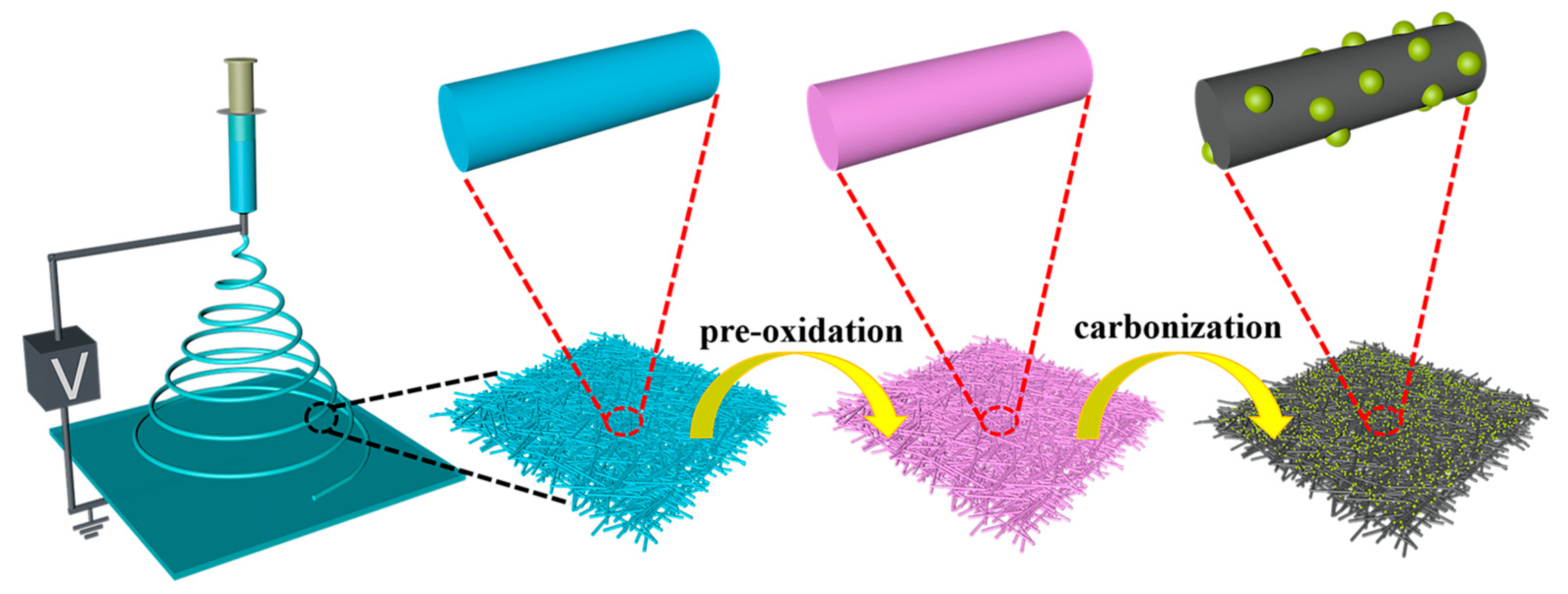
2.3. Synthesis of Mn3O4/NiO/CNFs

The synthesis process of Mn3O4/NiO/CNFs is illustrated in Scheme 1. Firstly, 0.5 g of PAN was mixed with 10 mL DMF and agitated to form a transparent solution. Then, 0.068 g Mn(Ac)2·4H2O and 0.017 g Ni(Ac)2·4H2O were added to the above solution and stirred continuously. Afterwards, the obtained brown precursor solution was moved to a 10 mL plastic injection syringe, and delivered to the metal needle at a constant stream rate of 0.6 mL h−1 under control of an injection pump. The grounded aluminum plate was used as the collector and kept away from the needle at a distance of 12 cm. After electrospinning, the collected precursor fibers were dried overnight at 60 °C under vacuum. The dried precursor fibers were pre-oxidized in air at 250 °C for 2 h, and then carbonized at 800 °C for another 2 h in an Ar stream. For comparison, Mn3O4/CNFs and NiO/CNFs were synthesized by the same method.

2.4. Electrochemical Measurements

The electrochemical measurements were carried out in a three-electrode system consisting of a platinum wire as counter electrode, Ag/AgCl (saturated KCl) as reference electrode, and Mn3O4/NiO/CNFs/GCE as working electrode. The three-electrode system was connected to an electrochemical workstation (CHI 842D, CH Instruments, Shanghai, China). The Mn3O4/NiO/CNFs/GCE was prepared in following steps: the bare GCE was polished with aluminum powder (0.5 μm and 0.01 μm), followed by cleaning with deionized water. Then, 5 μL of Mn3O4/NiO/CNFs uniform dispersion was dropped on GCE and then dried under infrared light to obtain Mn3O4/NiO/CNFs/GCE for glucose detection. The supporting electrolyte was 0.1 M NaOH.

3. Results and Discussion

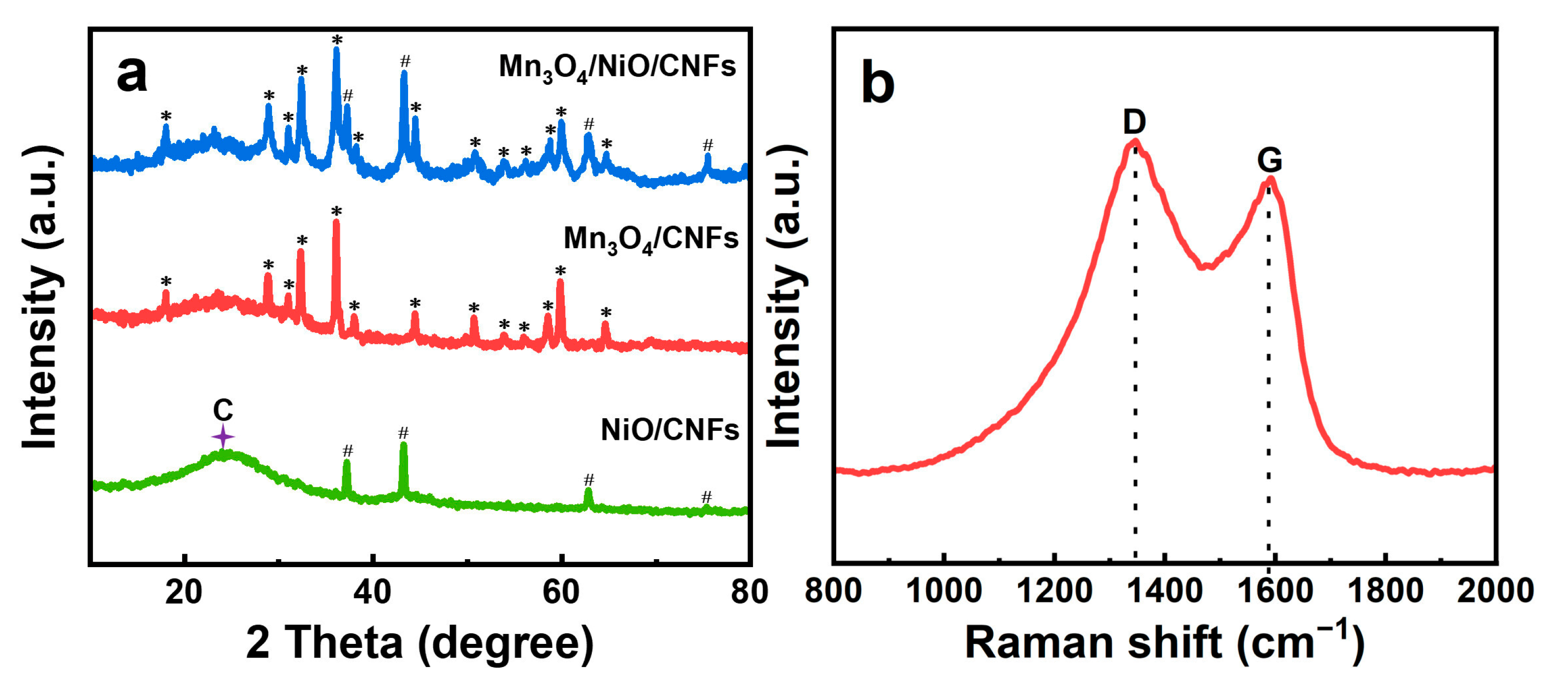
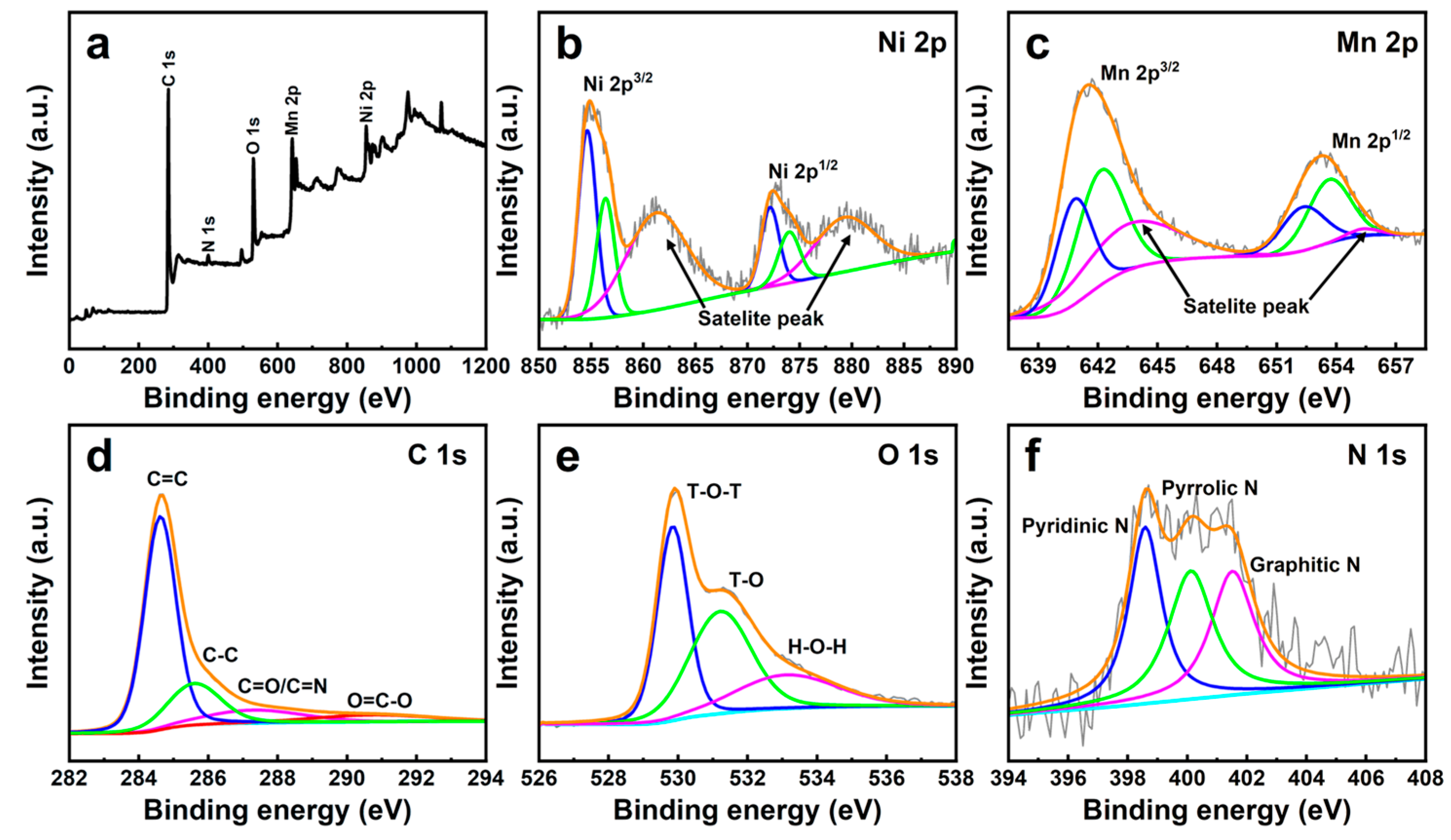
3.1. XRD, Raman, FT-IR, and XPS Characterization of Mn3O4/NiO/CNFs

The crystal structure and purity of Mn3O4/NiO/CNFs were characterized with XRD. Figure 1a shows the XRD patterns of Mn3O4/CNF, NiO/CNF, and Mn3O4/NiO/CNF. The obtained material contained a set of diffraction peaks that matched with Mn3O4, NiO, and CNF. In addition, there were no other impure phases. The peak at 24.2° could be ascribed to the (200) plane of CNF. The diffraction peaks at 37.2°, 43.2°, 62.8°, and 75.4° displayed the crystal planes of (111), (200), (220), and (311) plane for NiO [22]. The diffraction peaks at 17.9°, 28.9°, 31.0°, 32.3°, 36.1°, 38.0°, 44.4°, 50.7°, 53.1°, 56.0°, 58.5°, 59.9°, and 64.5° were well indexed to the (101), (112), (200), (103), (211), (004), (220), (105), (312), (303), (321), (224), and (400) plane of Mn3O4 with a cubic spinel [23].
The Raman spectrum was applied to evaluate the degree of graphitization of CNFs. In Figure 1b, the Raman spectrum shows a couple of significant peaks at 1350 and 1590 cm−1 which were directly related to the D band and G band, respectively [24]. The intensity of the D band reflected the carbon defects, while the intensity of the G band gave information about the graphitic structure. The intensity ratio between the D and G band, R = ID/IG, can be used to reflect the degree of graphitization of CNFs [25]. The R value is 1.07, which means that there are still defects in the material under calcination at 800 °C. The explanation for the defect is that the N element in PAN has not been completely removed. As shown in Figure S1, an FT-IR spectrum of Mn3O4/NiO/CNFs was performed. The band at 3444 cm−1 is identified as the stretching vibration of O-H, which is related to the adsorption of water. The peak at 1620 cm−1 can correspond to the C=C bond. The weak peak observed at 1383 cm−1 can be attributed to the C-H bond. The band at about 1169 cm−1 is caused by C-O [26]. The peaks which appear at 618 cm−1 and 523 cm−1 can be traced back to Mn3O4 and NiO, respectively [27].
The elemental composition and valence state of the Mn3O4/NiO/CNFs were reflected using XPS technique. The fully scanned spectrum (Figure 2a) demonstrates that the synthesized materials contained elements of C, N, O, Ni, and Mn. The peaks located at 854.7 eV and 872.1 eV were associated with Ni 2p3/2 and Ni 2p1/2, which were derived from Ni2+ (Figure 2b). In addition, two satellite peaks accompanying the main peaks were observed at 861.5 eV and 879.8 eV, proving the presence of Ni2+ from NiO [28]. The peaks located at 856.5 eV and 874.0 eV in Ni 2p spectrum corresponded to Ni 2p3/2 and Ni 2p1/2 from Ni3+, respectively. Here, more energy was required to form Ni3+ than Ni2+, since the compensation of large amounts of charge leads to the introduction of Ni vacancies and oxygen gaps, which in turn cause the formation of Ni3+ [29,30]. As shown in Figure 2c, the Mn 2p spectrum consists of a pair of major peaks located at 653.3 eV and 641.6 eV. The deconvolution peaks at the binding energies of 640.9 eV/652.4 eV and 642.3 eV/653.7 eV were caused by Mn2+ and Mn3+ species, respectively. The peaks located at about 645.0 eV and 655.0 eV belong to satellite peaks. Four peaks with energies of 284.6 eV, 285.6 eV, 287.4 eV, and 290.1 eV, corresponding to C=C, C-C, C=O/C=N, and O=C-O bonds, respectively, were fitted from the high-resolution C 1s peak (Figure 2d) [31,32]. The O 1s spectrum can be divided into three characteristic peaks, where the peaks of T-O-T and T-OH (T symbolizes transition metal) were observed at 529.9 eV and 531.2 eV, respectively. Moreover, the binding energy value of O 1s located at 533.2 eV represents the water-adsorbed on the surface (H-O-H) (Figure 2e). In the spectrogram of N 1s (Figure 2f), three peaks located at 398.8 eV, 400.1 eV, and 402.0 eV can be considered as pyridinic N, pyrrolic N, and graphitic N, respectively. Among them, pyridinic N and pyrrolic N are the two main doping forms in CNF [33].

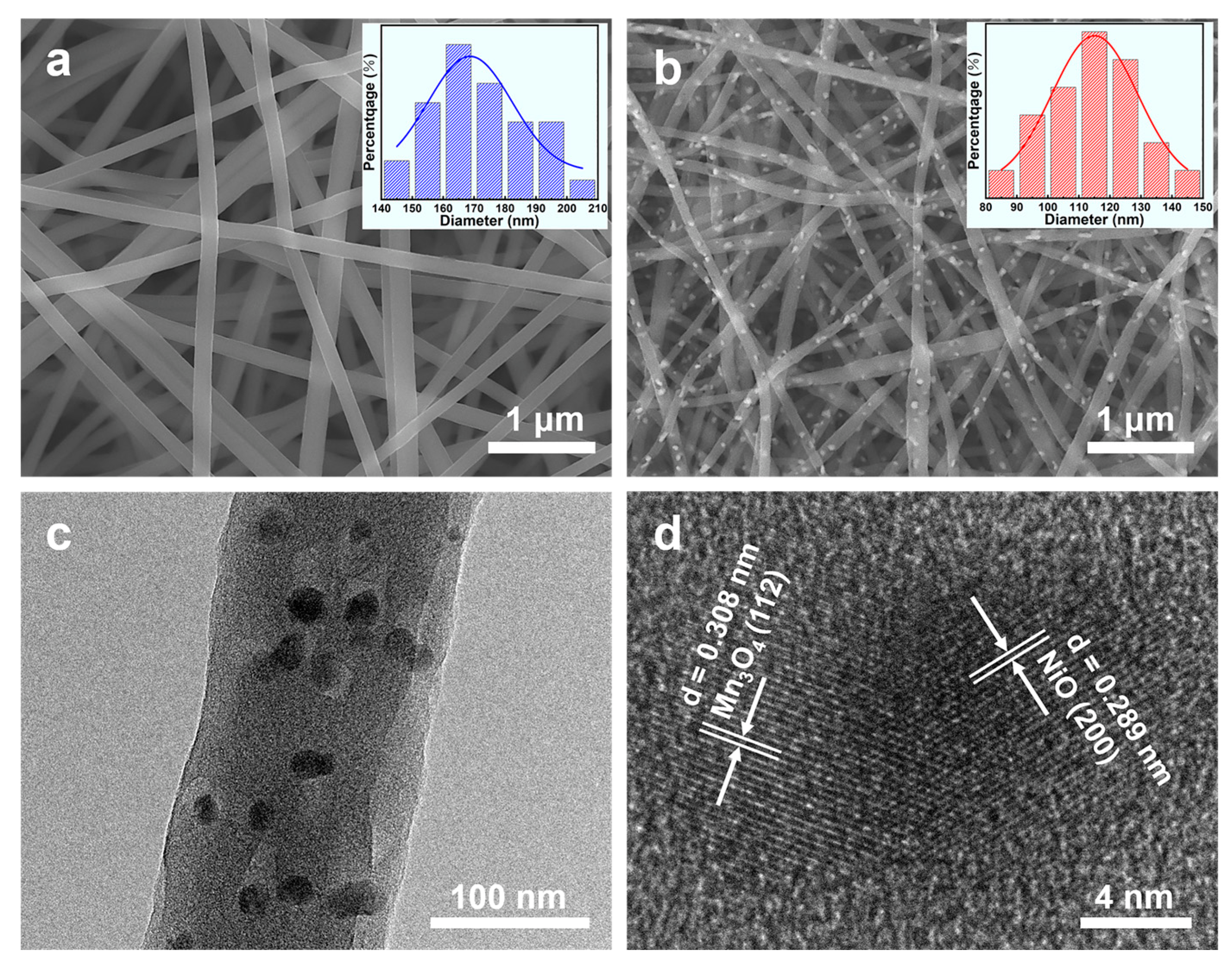
3.2. SEM and TEM of Mn3O4/NiO/CNFs

The surface morphologies of Mn(Ac)2/Ni(Ac)2/PAN precursor nanofibers and Mn3O4/NiO/CNFs were observed through SEM. In Figure 3a, the surface of uncalcined precursor nanofibers was smooth due to the amorphous structure of Ni(Ac)2 and Mn(Ac)2. After calcination at 800 °C, the nanoparticles were uniformly dispersed on the nanofiber surface (Figure 3b). The previous XRD result confirmed that the nanoparticle consisted of Mn3O4 and NiO. The diameter statistics and Gaussian distribution of the precursor nanofibers and Mn3O4/NiO/CNFs are shown in the insets of Figure 3a,b. Obviously, the average diameter of the nanofibers decreased from 168 to 115 nm after calcination, a phenomenon which was ascribed to the reduction of huge amount of H, O, and N elements during the carbonization process. TEM and HRTEM were carried out to further reveal the microscopic crystal structure of Mn3O4/NiO/CNF. As shown in Figure 3c, Mn3O4/NiO nanoparticles were uniformly adhered to the CNF surface. Lattice fringes with spacing of 0.308 nm and 0.289 nm corresponds to the (112) plane of Mn3O4 and (200) plane of NiO, respectively (Figure 3d) [34,35].

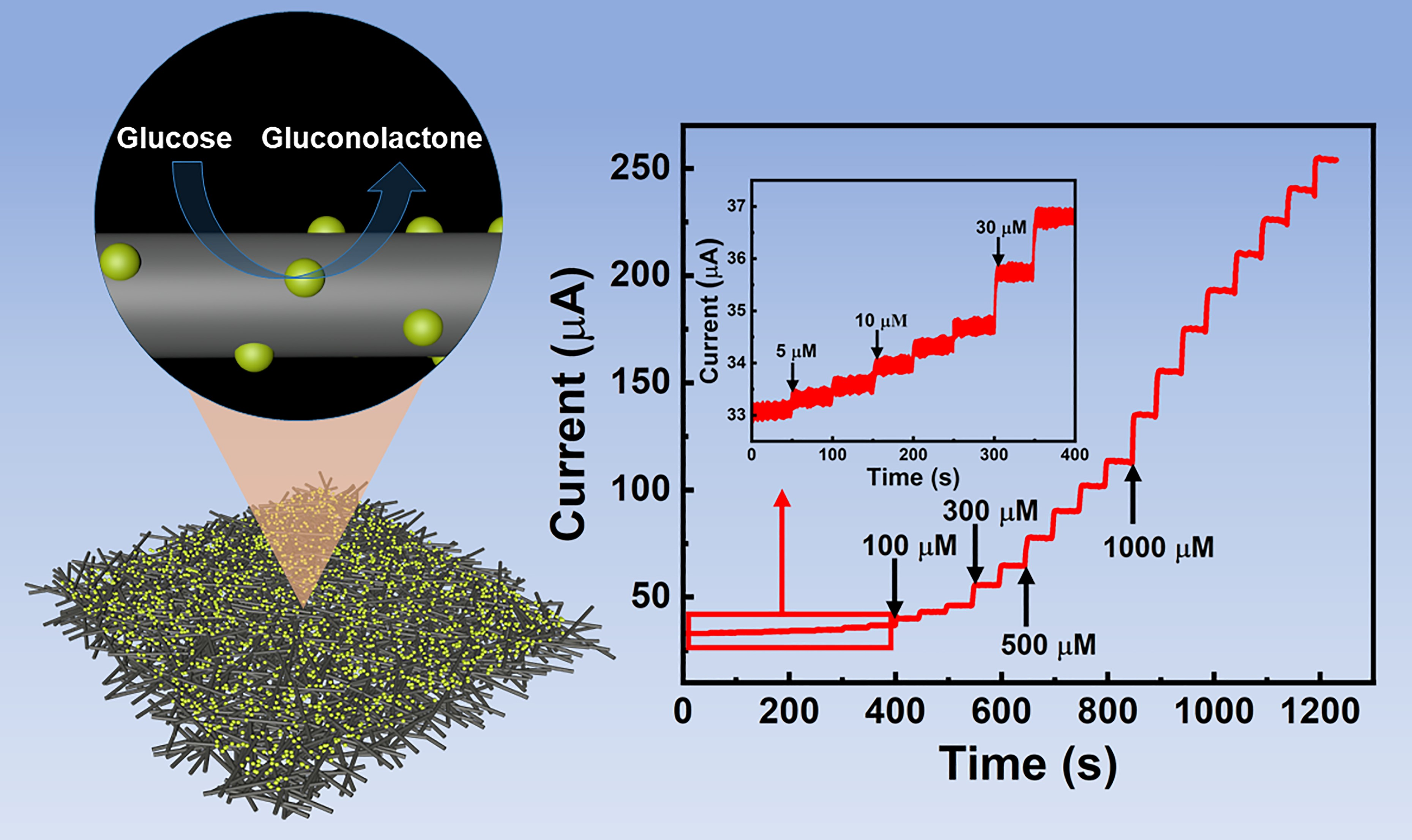
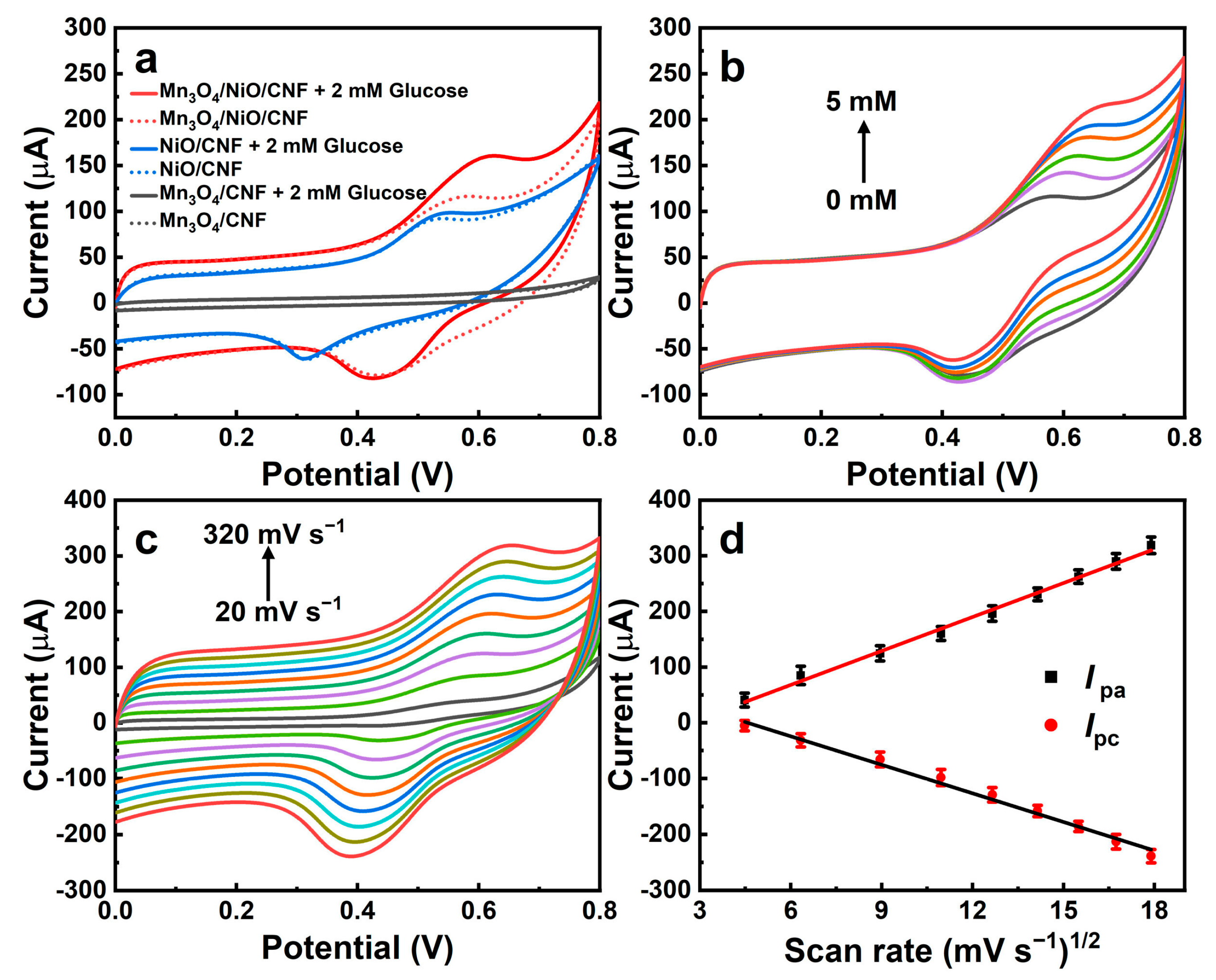
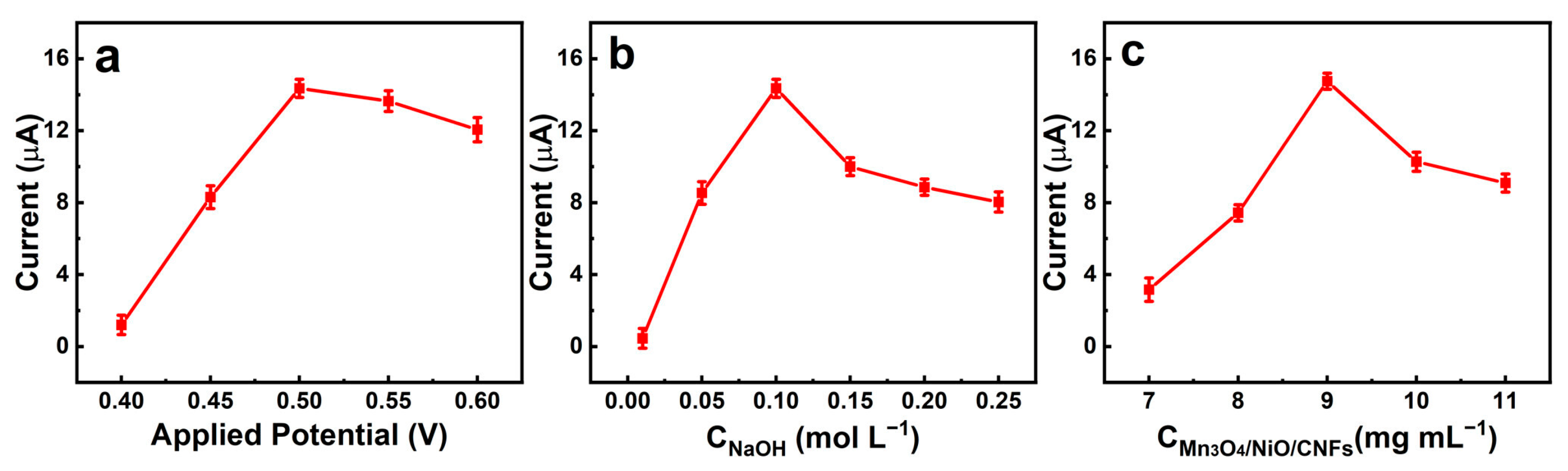
3.3. Electrochemical Characterization

The cyclic voltammetry (CV) and electrochemical impedance spectroscopy (EIS) were used to study the electrochemical performance of Mn3O4/NiO/CNFs. EIS tests of different modified electrodes were conducted in the solution with 5 mM [Fe(CN)6]3−/4− and 0.1 M KCl. The outcome of the EIS test is reflected by the Nyquist plot, where the semicircular part of the high frequency region relates to the electron transfer restriction process and the linear part of the low frequency region reflects the diffusion process. The decrease in the radius of the semicircle represents a decrease in the electron transfer resistance at the electrode interface (Ret). In Figure S2, compared to Mn3O4/CNFs/GCE and NiO/CNFs/GCE, Mn3O4/NiO/CNFs/GCE shows the smallest diameter, indicating the fastest charge transfer capability. To investigate the electrochemical performance of Mn3O4/NiO/CNFs, CV was employed. Figure 4a shows the CV curves of Mn3O4/CNFs/GCE, NiO/CNFs/GCE, and Mn3O4/NiO/CNFs/GCE in 0.1 M NaOH solution with and without 2 mM glucose. The oxidation current that changed for Mn3O4/CNFs/GCE in the presence of glucose can be neglected. Meanwhile, NiO/CNFs/GCE exhibited a positive electrocatalytic oxidation peak current for glucose detection which can be ascribed to the transition between Ni2+ and Ni3+. Notably, Mn3O4/NiO/CNFs/GCE exhibited the highest oxidation current. The mechanism of glucose oxidation on Mn3O4/NiO/CNFs can be explained by the following equations [36]:
NiO + H2O → Ni(OH)2
Ni(OH)2 + OH → NiOOH + H2O + e
NiOOH + C6H12O6 → Ni(OH)2 + C6H10O6 (Gluconolactone)
Originally, in the alkaline medium, NiO strongly adsorbed water molecules to form Ni(OH)2 (Equation (1)). Next, Ni(OH)2 was oxidized to NiOOH as electron mediator (Equation (2)). Then, NiOOH obtained electrons from glucose to form Ni(OH)2, and glucose was oxidized to gluconolactone (Equation (3)). Previous studies have indicated that Mn-based materials have a wide range for enzyme-free detection of glucose [37,38]. In this work, by virtue of the synergistic interaction between Mn and Ni, the composite exhibits more significant electrocatalytic activity for the oxidation of glucose [39]. The results of the EIS also confirm that the introduction of Mn3O4 can increase the electrical conductivity of the composites.
Figure 4b describes the CV curves of Mn3O4/NiO/CNFs/GCE in different glucose concentrations (from 0 mM to 5 mM). The oxidation current signal increased with increasing glucose concentration, indicating that Mn3O4/NiO/CNFs are suitable as electrocatalytic media for the construction of glucose sensors. To further investigate the kinetic process of glucose oxidation on the electrode surface, the CV curves of Mn3O4/NiO/CNFs/GCE were tested in the range of scan rate from 20 mV s−1 to 320 mV s−1 (Figure 4c). As illustrated in Figure 4d, the currents of the anodic peak (Ipa) and cathodic peak (Ipc) maintained a good linear connection with the square root of the scanning speed, indicating that the oxidation of glucose on Mn3O4/NiO/CNFs/GCE is a diffusion-controlled process.
To improve the electrocatalytic effect of Mn3O4/NiO/CNFs on glucose detection, the time-current method was used to optimize the parameters during the experiments, including the applied voltage and the concentrations of NaOH and Mn3O4/NiO/CNFs. Figure 5a shows that the response current increases with increasing applied voltage at the beginning, and then decreases when the applied voltage is higher than 0.50 V. Thus, 0.50 V is the optimum applied potential. In Figure 5b, the response current initially increases with increasing concentration of NaOH, and then starts to decrease when the concentration of NaOH exceeds 0.10 M. Therefore, 0.1 M NaOH was chosen as the optimum concentration of electrolyte. Figure 5c shows that the response current reaches its maximum when the concentration of Mn3O4/NiO/CNFs is 9 mg mL−1; therefore, 9 mg mL−1 was selected as the optimum modification material concentration. Figure 6a shows the current response of Mn3O4/NiO/CNFs/GCE for continuous addition of glucose under known optimal experimental conditions. The relationship between response current and glucose concentration shows linearly in the range of 5–3000 μM (R2 = 0.997) and 3000–12,000 μM (R2 = 0.993), and could be well-fitted to the equation: I (μA) = 27.33 C (mM) + 1.343 and I (μA) = 17.22 C (mM) + 35.015. The detection limit of Mn3O4/NiO/CNFs/GCE was calculated to be 0.73 μM (S/N = 3). The sensitivity was calculated to be 386.84 μA mM−1 cm−2 (5–3000 μM) and 243.74 μA mM−1 cm−2 (3000–12,000 μM). Additionally, the response time is a key factor in weighing up the practical application of the sensor. Figure S3 shows that the Mn3O4/NiO/CNFs/GCE at different glucose concentrations reaches its steady-state current within 2–4 s after the addition of glucose. Table 1 lists the electrocatalytic effects of some reported enzyme-free glucose sensors and enzymatic glucose sensors. Compared with them, our proposed Mn3O4/NiO/CNFs/GCE has a wider linear range as well as lower detection limit as glucose sensor.

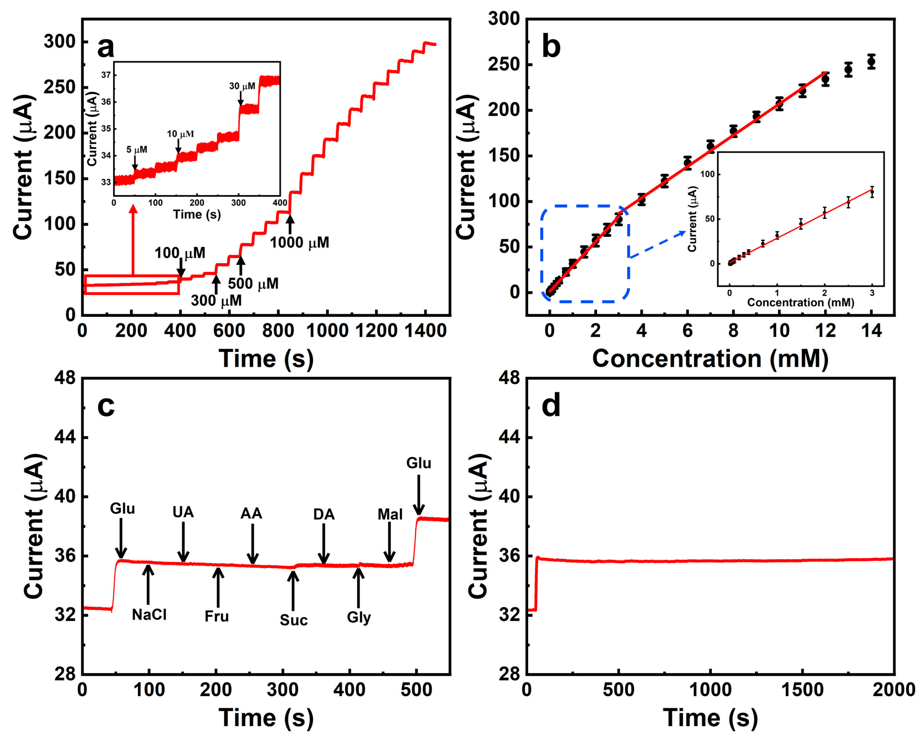
3.4. Selectivity, Stability, Repeatability, and Reproducibility of Mn3O4/NiO/CNFs/GCE

The presence of other easily oxidized molecules in the actual sample may affect the accuracy of glucose detection. In Figure 6c, the addition of 5 μM of some interferences to 100 μM glucose solution at the ratio of 1:20 was investigated by a time-current test. The outcomes showed that the current responses caused by the addition of interferences were virtually negligible, indicating that Mn3O4/NiO/CNFs/GCE had good selectivity. Figure 6d shows the amperometric response of Mn3O4/NiO/CNFs/GCE to 100 μM glucose. The current kept stable during 2000 s, suggesting that the electrode had an excellent stability as glucose sensor. Besides, repeatability and reproducibility are also key to determine whether the synthesized materials can be used for actual sample detection. The five identical modified electrodes used to detect 0.1 mM glucose were found to have a relative standard deviation (RSD) of 6.38% (Figure S4), indicating that the electrodes have good reproducibility. The modified electrode was repeated five times to detect 0.1 mM glucose, and the RSD was calculated to be 1.32% (Figure S5), meaning that the electrode has a good repeatability. To evaluate the long-term stability of the sensor, the Mn3O4/NiO/CNFs/GCE was stored at room temperature for 30 days. The results show that the response current remains at 95.3% of the initial current after 30 days (Figure S6), indicating that our sensors have satisfactory long-term stability.

3.5. Actual Sample Analysis

To evaluate the practicality of Mn3O4/NiO/CNFs/GCE, serum samples with different concentrations of glucose were detected using standard addition method. The serum samples used in the experiment were from the School of Medicine, Shanghai University. The recoveries of the three samples were 100.09%, 100.20%, and 99.91%, respectively (Table 2), indicating that the built sensor can be accepted for glucose detection in serum samples.

4. Conclusions

In this work, Mn3O4/NiO/CNFs were synthesized by electrospinning and following calcination, and were used as an enzyme-free sensor for glucose detection. The intricate network structure of CNFs provides the backbone for the nanoparticles and facilitates electron transport. In addition, the synergistic effect between Mn3O4 and NiO is beneficial to improve the catalytic performance for glucose detection. The constructed Mn3O4/NiO/CNFs/GCE sensor has good linear range, low detection limit, and anti-interference ability, indicating that Mn3O4/NiO/CNFs have some application prospects as electrochemical sensors in clinical diagnosis.

Supplementary Materials

The following supporting information can be downloaded at: https://www.mdpi.com/article/10.3390/bios13020264/s1, Figure S1: FT-IR spectrum of Mn3O4/NiO/CNFs.; Figure S2: EIS tests of the Mn3O4/CNFs/GCE, NiO/CNFs/GCE and Mn3O4/NiO/CNFs/GCE in 5.0 mM [Fe(CN)6]3−/4− containing 0.1 M KCl.; Figure S3: Response time of (a) low glucose concentration and (b) high glucose concentration.; Figure S4: The reproducibility of the Mn3O4/NiO/CNFs/GCE.; Figure S5: The repeatability of the Mn3O4/NiO/CNFs/GCE.; Figure S6: The long-time stability.

Author Contributions

Conceptualization, M.L. and S.L.; methodology, X.Y. and S.L.; software, J.D. and X.O.; validation, M.L., J.D. and X.O.; formal analysis, J.D., X.O., S.L. and X.Y.; investigation, M.L. and S.L.; resources, L.L. and D.D.; data curation, M.L., J.D. and X.O.; writing—original draft preparation, M.L.; writing—review and editing, L.L., D.D. and X.Y.; visualization, S.L. and X.Y.; supervision, D.D. and L.L.; project administration, D.D. and L.L.; funding acquisition, D.D. and L.L. All authors have read and agreed to the published version of the manuscript.

Funding

This research was funded by the National Natural Science Foundation of China (Nos. 61971274 and 62171268).

Institutional Review Board Statement

Not applicable.

Informed Consent Statement

Not applicable.

Data Availability Statement

Date will be made available on request.

Conflicts of Interest

The authors declare that they have no known competing financial interests or personal relationships that could have appeared to influence the work reported in this paper.

References

  1. Wang, D.; Chang, Y. The Construct CoSe2 on Carbon Nanosheets as High Sensitivity Catalysts for Electrocatalytic Oxidation of Glucose. Nanomaterials 2022, 12, 572. [Google Scholar] [CrossRef] [PubMed]
  2. Wang, S.; Li, S.P.; Wang, W.W.; Zhao, M.T.; Liu, J.F.; Feng, H.F.; Chen, Y.M.; Du, Y.; Hao, W.C. A Non-Enzymatic Photoelectrochemical Glucose Sensor Based on BiVO4 Electrode under Visible Light. Sens. Actuators B 2019, 291, 34–41. [Google Scholar] [CrossRef]
  3. Cao, F.H.; Zhou, Y.; Wu, J.; Li, W.; Zhang, C.L.; Ni, G.; Cui, P.; Song, C.J. Electrospinning One-dimensional Surface-phosphorized CuCo/C Nanofibers for Enzyme-free Glucose Sensing. New J. Chem. 2022, 46, 11531–11539. [Google Scholar] [CrossRef]
  4. Adeel, M.; Rahman, M.M.; Caligiuri, I.; Canzonieri, V.; Rizzolio, F.; Daniele, S. Recent Advances of Electrochemical and Optical Enzyme-Free Glucose Sensors Operating at Physiological Conditions. Biosens. Bioelectron. 2020, 165, 112331. [Google Scholar] [CrossRef]
  5. Liu, S.; Qiu, Y.Z.; Liu, Y.F.; Zhang, W.F.; Dai, Z.; Srivastava, D.; Kumar, A.; Pan, Y.; Liu, J.Q. Recent Advances in Bimetallic Metal-organic Frameworks (BMOFs): Synthesis, Applications and Challenges. New J. Chem. 2022, 46, 13818–13837. [Google Scholar] [CrossRef]
  6. Li, L.T.; Zou, J.F.; Han, Y.T.; Liao, Z.H.; Lu, P.F.; Nezamzadeh-Ejhieh, A.; Liu, J.Q.; Peng, Q.P. Recent Advances in Al(iii)/In(iii)-based MOFs for the Detection of Pollutants. New J. Chem. 2022, 46, 19577–19592. [Google Scholar] [CrossRef]
  7. Gao, H.; Xiao, F.; Ching, C.B.; Duan, H. One-Step Electrochemical Synthesis of PtNi Nanoparticle-Graphene Nanocomposites for Nonenzymatic Amperometric Glucose Detection. ACS Appl. Mater. Interfaces 2011, 3, 3049–3057. [Google Scholar] [CrossRef]
  8. Li, Y.Y.; Tang, L.; Deng, D.M.; He, H.B.; Yan, X.X.; Wang, J.H.; Luo, L.Q. Hetero-Structured MnO-Mn3O4@rGO Composites: Synthesis and Nonenzymatic Detection of H2O2. Mater. Sci. Eng. C 2021, 118, 111443. [Google Scholar] [CrossRef]
  9. Liu, X.Z.; Yang, H.P.; Diao, Y.Y.; He, Q.; Lu, C.Y.; Singh, A.; Kumar, A.; Liu, J.Q.; Lan, Q. Recent Advances in the Electrochemical Applications of Ni-based Metal Organic Frameworks (Ni-MOFs) and Their Derivatives. Chemosphere 2022, 307, 135729. [Google Scholar] [CrossRef]
  10. Saxena, A.; Liyanage, W.P.R.; Kapila, S.; Nath, M. Nickel Selenide as an Efficient Electrocatalyst for Selective Reduction of Carbon Dioxide to Carbon-rich Products. Catal. Sci. Technol. 2022, 12, 4727–4739. [Google Scholar] [CrossRef]
  11. Li, M.R.; Zheng, K.T.; Zhang, J.J.; Li, X.M.; Xu, C.J. Design and Construction of 2D/2D Sheet-on-sheet Transition Metal Sulfide/Phosphide Heterostructure for Efficient Oxygen Evolution Reaction. Appl. Surf. Sci. 2021, 565, 150510. [Google Scholar] [CrossRef]
  12. Sivanantham, A.; Shanmugam, S. Graphitic Carbon-NiCo Nanostructures as Efficient Non-precious-metal Electrocatalysts for the Oxygen Reduction Reaction. ChemElectroChem 2018, 5, 1937–1943. [Google Scholar] [CrossRef]
  13. Hernandez-Ramirez, D.; Mendoza-Huizar, L.H.; Galan-Vidal, C.A.; Aguilar-Lira, G.Y.; Alvarez-Romero, G.A. Development of a Non-Enzymatic Glucose Sensor Based on Fe2O3 Nanoparticles-carbonve and Analytica Paste Electrodes. J. Electrochem. Soc. 2022, 169, 067507. [Google Scholar] [CrossRef]
  14. Wang, H.; Li, Y.Y.; Deng, D.M.; Li, M.J.; Zhang, C.Y.; Luo, L.Q. NiO-Coated CuCo2O4 Nanoneedle Arrays on Carbon Cloth for Nonenzymatic Glucose Sensing. ACS Appl. Nano Mater. 2021, 4, 9821–9830. [Google Scholar] [CrossRef]
  15. Xu, Y.H.; Ding, Y.P.; Zhang, L.H.; Zhang, X.X. Highly Sensitive Enzyme-free Glucose Sensor Based on CuO-NiO Nanocomposites by Electrospinning. Compos. Commun. 2021, 25, 100687. [Google Scholar] [CrossRef]
  16. Zhang, R.; Zhou, T.T.; Zhang, T. Functionalization of Hybrid 1D SnO2-ZnO Nanofibers for Formaldehyde Detection. Adv. Mater. Interfaces 2018, 5, 1800967. [Google Scholar] [CrossRef]
  17. Thomas, T.; Jayababu, N.; Shruthi, J.; Mathew, A.; Cerdan-Pasaran, A.; Hernandez-Magallanes, J.A.; Sanal, K.C.; Reshmi, R. Room Temperature Ammonia Sensing of Alpha-MoO3 Nanorods Grown on Glass Substrates. Thin Solid Films 2021, 722, 138575. [Google Scholar] [CrossRef]
  18. Zhang, C.Y.; Li, Y.Y.; Wang, H.; Niu, X.Y.; Deng, D.M.; Qin, X.; Yan, X.X.; He, H.B.; Luo, L.Q. Low Temperature Growth of CuS Nanosheets on Hollow Co9S8 Nanotubes: Synthesis and Analytical Application. Microchem. J. 2022, 183, 108037. [Google Scholar] [CrossRef]
  19. Kokulnathan, T.; Vishnuraj, R.; Chen, S.M.; Pullithadathil, B.; Ahmed, F.; Hasan, P.M.Z.; Bilgrami, A.L.; Kumar, S. Tailored Construction of One-Dimensional TiO2/Au Nanofibers: Validation of an Analytical Assay for Detection of Diphenylamine in Food Samples. Food Chem. 2022, 380, 132052. [Google Scholar] [CrossRef] [PubMed]
  20. Wang, C.H.; Kim, J.; Kim, M.; Lim, H.; Zhang, M.; You, J.; Yun, J.H.; Bando, Y.; Li, J.S.; Yamauchi, Y. Nanoarchitectured Metal-Organic Framework-Derived Hollow Carbon Nanofiber Filters for Advanced Oxidation Processes. J. Mater. Chem. A 2019, 7, 13743–13750. [Google Scholar] [CrossRef]
  21. Zhang, Z.X.; Wang, Y.X.; Leng, X.X.; Crespi, V.H.; Kang, F.Y.; Lv, R.T. Controllable Edge Exposure of MoS2 for Efficient Hydrogen Evolution with High Current Density. ACS Appl. Energy Mater. 2018, 1, 1268–1275. [Google Scholar] [CrossRef]
  22. Sui, L.L.; Yu, T.T.; Zhao, D.; Cheng, X.L.; Zhang, X.F.; Wang, P.; Xu, Y.M.; Gao, S.; Zhao, H.; Gao, Y.; et al. In Situ Deposited Hierarchical CuO/NiO Nanowall Arrays Film Sensor with Enhanced Gas Sensing Performance to H2S. J. Hazard. Mater. 2020, 385, 121570. [Google Scholar] [CrossRef]
  23. Liu, S.Y.; Xie, J.; Zheng, Y.X.; Cao, G.S.; Zhu, T.J.; Zhao, X.B. Nanocrystal Manganese Oxide (Mn3O4, MnO) Anchored on Graphite Nanosheet with Improved Electrochemical Li-Storage Properties. Electrochim. Acta 2012, 66, 271–278. [Google Scholar] [CrossRef]
  24. Niu, H.T.; Zhang, J.; Xie, Z.L.; Wang, X.G.; Lin, T. Preparation, Structure and Supercapacitance of Bonded Carbon Nanofiber Electrode Materials. Carbon 2011, 49, 2380–2388. [Google Scholar] [CrossRef]
  25. Abe, J.; Takahashi, K.; Kawase, K.; Kobayashi, Y.; Shiratori, S. Self-Standing Carbon Nanofiber and SnO2 Nanorod Composite as a High-Capacity and High-Rate-Capability Anode for Lithium-Ion Batteries. ACS Appl. Nano Mater. 2018, 1, 2982–2989. [Google Scholar] [CrossRef]
  26. Yang, G.; Li, Y.H.; Ji, H.M.; Wang, H.Y.; Gao, P.; Wang, L.; Liu, H.D.; Pinto, J.; Jiang, X.F. Influence of Mn Content on the Morphology and Improved Electrochemical Properties of Mn3O4 Vertical Bar MnO@Carbon Nanofiber as Anode Material for Lithium Batteries. J. Power Source 2012, 216, 353–362. [Google Scholar] [CrossRef]
  27. Rani, B.J.; Rathika, S.; Ravi, G.; Yuvakkumar, R. Synthesis of MnNiO3/Mn3O4 Nanocomposites for the Water Electrolysis Process. J. Sol-Gel Sci. Technol. 2019, 92, 1–11. [Google Scholar] [CrossRef]
  28. Liu, M.M.; An, M.L.; Xu, J.Q.; Liu, T.; Wang, L.L.; Liu, Y.Y.; Zhang, J.J. Three-Dimensional Carbon Foam Supported NiO Nanosheets as Non-Enzymatic Electrochemical H2O2 sensors. Appl. Surf. Sci. 2021, 542, 148699. [Google Scholar] [CrossRef]
  29. Pan, J.Q.; Li, S.; Ou, W.; Liu, Y.Y.; Li, H.L.; Wang, J.J.; Song, C.S.; Zheng, Y.Y.; Li, C.R. The Photovoltaic Conversion Enhancement of NiO/Tm:CeO2/SnO2 Transparent P-N Junction Device with Dual-Functional Tm:CeO2 Quantum Dots. Chem. Eng. J. 2020, 393, 124802. [Google Scholar] [CrossRef]
  30. Zhang, Y.Q.; Wang, Y.Z.; Jia, J.B.; Wang, J.G. Nonenzymatic Glucose Sensor Based on Graphene Oxide and Electrospun NiO Nanofibers. Sens. Actuators B 2012, 171, 580–587. [Google Scholar] [CrossRef]
  31. Alegre, C.; Busacca, C.; Di Blasi, A.; Di Blasi, O.; Arico, A.S.; Antonucci, V.; Modica, E.; Baglio, V. Electrospun Carbon Nanofibers Loaded with Spinel-Type Cobalt Oxide as Bifunctional Catalysts for Enhanced Oxygen Electrocatalysis. J. Energy Storage 2019, 23, 269–277. [Google Scholar] [CrossRef]
  32. Busacca, C.; Di Blasi, O.; Giacoppo, G.; Briguglio, N.; Antonucci, V.; Di Blasi, A. High Performance Electrospun Nickel Manganite on Carbon Nanofibers Electrode for Vanadium Redox Flow Battery. Electrochim. Acta 2020, 355, 136755. [Google Scholar] [CrossRef]
  33. Huan, K.; Li, Y.Y.; Deng, D.M.; Wang, H.; Wang, D.J.; Li, M.J.; Luo, L.Q. Composite-Controlled Electrospinning of CuSn Bimetallic Nanoparticles/Carbon Nanofibers for Electrochemical Glucose Sensor. Appl. Surf. Sci. 2022, 573, 151528. [Google Scholar] [CrossRef]
  34. Hao, Z.Y.; Meng, Z.S.; Li, X.Y.; Sun, X.C.; Xu, J.; Nan, H.; Shi, W.; Qi, G.J.; Hu, X.Y.; Tian, H.W. Two-Step Fabrication of Lanthanum Nickelate and Nickel Oxide Core-Shell Dandelion-Like Materials for High-Performance Supercapacitors. J. Colloid Interface Sci. 2022, 617, 430–441. [Google Scholar] [CrossRef] [PubMed]
  35. Zhang, W.J.; Guo, X.L.; Zhao, J.J.; Zheng, Y.M.; Xie, H.; Zhang, Z.; Wang, S.H.; Xu, Q.; Fu, Q.P.; Zhang, T. High Performance Flower-Like Mn3O4/rGO Composite for Supercapacitor Applications. J. Electroanal. Chem. 2022, 910, 116170. [Google Scholar] [CrossRef]
  36. Singer, N.; Pillai, R.G.; Johnson, A.I.D.; Harris, K.D.; Jemere, A.B. Nanostructured Nickel Oxide Electrodes for Non-Enzymatic Electrochemical Glucose Sensing. Microchim. Acta 2020, 187, 196. [Google Scholar] [CrossRef]
  37. Dong, C.J.; Tao, Y.; Chang, Q.; Liu, Q.H.; Guan, H.T.; Chen, G.; Wang, Y.D. Direct Growth of MnCO3 on Ni Foil for a Highly Sensitive Nonenzymatic Glucose Sensor. J. Alloys Compd. 2018, 762, 216–221. [Google Scholar] [CrossRef]
  38. Zhang, Y.Y.; Huang, Y.Q.; Gao, P.P.; Yin, W.; Yin, M.; Pu, H.C.; Sun, Q.Q.; Liang, X.L.; Huan, B.H. Bimetal-organic Frameworks MnCo-MOF-74 Derived Co/MnO@HC for the Construction of a Novel Enzyme-free Glucose sensor. Microchem. J. 2022, 175, 107097. [Google Scholar] [CrossRef]
  39. Wang, L.J.; Li, Y.Y.; Zhang, C.Y.; Deng, D.M.; He, H.B.; Yan, X.X.; Li, Z.G.; Luo, L.Q. Hierarchical NiMn Layered Double Hydroxide Nanostructures on Carbon Cloth for Electrochemical Detection of Hydrogen Peroxide. ACS Appl. Nano Mater. 2022, 5, 17741–17749. [Google Scholar] [CrossRef]
  40. Dong, Q.Y.; He, Z.Y.; Tang, X.; Zhang, Y.; Yang, L.; Huang, K.; Jiang, X.; Xiong, X.L. One-Step Synthesis of Mn3O4@ZIF-67 on Carbon Cloth: As an Effective Non-Enzymatic Glucose Sensor. Microchem. J. 2022, 175, 107203. [Google Scholar] [CrossRef]
  41. Pu, F.Z.; Miao, H.R.; Lu, W.J.; Zhang, X.J.; Yang, Z.M.; Kong, C.C. High-Performance Non-Enzymatic Glucose Sensor Based on Flower-Like Cu2O-Cu-Au Ternary Nanocomposites. Appl. Surf. Sci. 2022, 581, 152389. [Google Scholar] [CrossRef]
  42. Wei, H.G.; Xu, Q.Z.; Li, A.; Wan, T.; Huang, Y.; Cui, D.P.; Pan, D.; Dong, D.D.; Wei, R.B.; Naik, N.; et al. Dendritic Core-Shell Copper-Nickel Alloy@Metal Oxide for Efficient Non-Enzymatic Glucose Detection. Sens. Actuators B 2021, 337, 129687. [Google Scholar] [CrossRef]
  43. Rani, S.D.; Ramachandran, R.; Sheet, S.; Aziz, M.A.; Lee, Y.S.; Al-Sehemi, A.G.; Pannipara, M.; Xia, Y.; Tsai, S.Y.; Ng, F.L.; et al. NiMoO4 Nanoparticles Decorated Carbon Nanofiber Membranes for the Flexible and High Performance Glucose Sensors. Sens. Actuators B 2020, 312, 127886. [Google Scholar] [CrossRef]
  44. He, L.; Li, J.W.; Cao, J.; Li, X.; Feng, X.F.; Zhang, J.; Yang, Y. High Performance of Non-Enzymatic Glucose Biosensors Based on the Design of Microstructure of Ni2P/Cu3P Nanocomposites. Appl. Surf. Sci. 2022, 593, 153395. [Google Scholar] [CrossRef]
  45. Myndrul, V.; Coy, E.; Babayevska, N.; Zahorodna, V.; Balitskyi, V.; Baginskiy, I.; Gogotsi, O.; Bechelany, M.; Giardi, M.T.; Iatsunskyi, I. MXene Nanoflakes Decorating ZnO Tetrapods for Enhanced Performance of Skin-Attachable Stretchable Enzymatic Electrochemical Glucose Sensor. Biosens. Bioelectron. 2022, 207, 114141. [Google Scholar] [CrossRef]
  46. Kong, X.Y.; Xia, B.; Xiao, Y.W.; Chen, H.H.; Li, H.F.; Chen, W.Z.; Wu, P.; Shen, Y.; Wu, J.S.; Li, S.; et al. Regulation of Cobalt-Nickel LDHs’ Structure and Components for Optimizing the Performance of an Electrochemical Sensor. ACS Appl. Nano Mater. 2019, 2, 6387–6396. [Google Scholar] [CrossRef]
  47. Wang, Y.; Cui, J.W.; Wang, Y.; Yu, D.B.; Cheng, S.; Zheng, H.M.; Shu, X.; Zhang, Y.; Wu, Y.C. Decorating Mn3O4 Nanoparticle on NiO Nanoflake Arrays for High-performance Electrochemical Biosensors. J. Solid State Electrochem. 2019, 23, 135–142. [Google Scholar] [CrossRef]
  48. Sinha, L.; Pakhira, S.; Bhojane, P.; Mali, S.; Hong, C.K.; Shirage, P.M. Hybridization of Co3O4 and alpha-MnO2 Nanostructures for High-Performance Nonenzymatic Glucose Sensing. ACS Sustain. Chem. Eng. 2018, 6, 13248–13261. [Google Scholar] [CrossRef]
  49. Tai, N.H.; Lin, M.H.; Gupta, S.; Chang, C.; Lee, C.Y. Carbon Nanotubes/Polyethylenimine/Glucose Oxidase as a Non-invasive Electrochemical Biosensor Performs High Sensitivity for Detecting Glucose in Saliva. Microchem. J. 2022, 180, 107547. [Google Scholar] [CrossRef]
  50. Murugan, P.; Annamalai, J.; Atchudan, R.; Govindasamy, M.; Nallaswamy, D.; Ganapathy, D.; Reshetilov, A.; Sundramoorthy, A.K. Electrochemical Sensing of Glucose Using Glucose Oxidase/PEDOT:4-Sulfocalix 4 arene/MXene Composite Modified Electrode. Micromachines 2022, 13, 304. [Google Scholar] [CrossRef]
  51. Dong, S.; Niu, H.W.; Sun, L.W.; Zhang, S.X.; Wu, D.Q.; Yang, Z.; Xiang, M. Highly Dense Ni-MOF Nanoflake Arrays Supported on Conductive Graphene/Carbon Fiber Substrate as Flexible Microelectrode for Electrochemical Sensing of Glucose. J. Electroanal. Chem. 2022, 911, 116219. [Google Scholar] [CrossRef]
Scheme 1. Schematic diagram of synthesis procedure of Mn3O4/NiO/CNFs.
Biosensors 13 00264 sch001
Figure 1. (a) XRD patterns of Mn3O4/CNFs, NiO/CNFs, and Mn3O4/NiO/CNFs; (b) Raman spectrum of Mn3O4/NiO/CNFs at 800 °C.
Biosensors 13 00264 g001
Figure 2. XPS spectra of Mn3O4/NiO/CNFs: (a) full wide-scan; (b) Ni 2p; (c) Mn 2p; (d) C 1s; (e) O 1s; and (f) N 1s.
Biosensors 13 00264 g002
Figure 3. SEM images of electrospun: (a) Mn(Ac)2/Ni(Ac)2/PAN nanofibers and (b) Mn3O4/NiO/CNFs; (c) TEM image of Mn3O4/NiO/CNFs; (d) HRTEM image of Mn3O4/NiO.
Biosensors 13 00264 g003
Figure 4. (a) CVs of Mn3O4/CNFs, NiO/CNFs, and Mn3O4/NiO/CNFs electrodes in the absence and presence of 2 mM glucose; (b) CVs of Mn3O4/NiO/CNFs electrode with different concentrations of glucose in 0.1 M NaOH at the scan rate of 100 mV s−1; (c) CVs of Mn3O4/NiO/CNFs electrode in the presence of 1 mM glucose at different scan rates; (d) peak currents in relationship to the square root of the scanning speed scan rate.
Biosensors 13 00264 g004
Figure 5. Optimization of parameters on the current response of 0.5 mM glucose: (a) applied potential; (b) the concentration of NaOH; (c) the content of Mn3O4/NiO/CNFs.
Biosensors 13 00264 g005
Figure 6. (a) Amperometric response of Mn3O4/NiO/CNFs electrode for different concentrations of glucose (inset is current response at low concentrations); (b) calibration curves corresponding to (a); (c) current response of Mn3O4/NiO/CNFs electrode to 100 μM glucose and different interferences; (d) the stability test of Mn3O4/NiO/CNFs electrode during the 2000 s. All the above experiments were conducted under optimal parameters.
Biosensors 13 00264 g006
Table 1. Performance comparison of the proposed Mn3O4/NiO/CNFs/GCE with other glucose sensors.
Sensing MaterialLinear Range
(μM)
Detection Limit
(μM)
Sensitivity
(μA mM−1 cm−2)
Ref.
Mn3O4@ZIF-67/CC 10.8–60000.243421.0[40]
CuCo2O4/NiO/CC0.5–60000.284140[14]
Cu2O-Cu-Au0–45001.711082[41]
Cu-Ni/NF1–600211,340.25[42]
NiMoO4/CNF0.3–45000.05301.77[43]
Ni2P-Cu3P4–50000.14700[44]
ZnO/MXene50–7001729[45]
Co0.33Ni0.67-HLDH 210–20003.1242.9[46]
Mn3O4/NiO9.9–36651.0226.2[47]
MnO2/Co3O460–70000.03127[48]
FTO-CNTs/PEI/GOX70–7007063.38[49]
PEDOT: SCX/MXene/GOX/GCE500–800022.5-[50]
Ni-MOF 3/rGO 4/CF 56–20900.6852[51]
Mn3O4/NiO/CNFs5–3000
3000–12,000
0.73386.84
243.74
This work
1 Carbon cloth. 2 Hollow layered double hydroxides. 3 Metal organic frameworks. 4 Reduced graphene oxide. 5 Carbon fiber.
Table 2. Analysis of glucose in human blood serum.
SampleDetected
(mM)
Commercial
Method (mM)
RSD (%)Recovery (%)
14.724.591.91100.09
24.834.691.47100.2
38.007.672.3499.91
Disclaimer/Publisher’s Note: The statements, opinions and data contained in all publications are solely those of the individual author(s) and contributor(s) and not of MDPI and/or the editor(s). MDPI and/or the editor(s) disclaim responsibility for any injury to people or property resulting from any ideas, methods, instructions or products referred to in the content.

