Next Issue
Volume 13, March
Previous Issue
Volume 13, January


Biosensors, Volume 13, Issue 2 (February 2023) – 152 articles

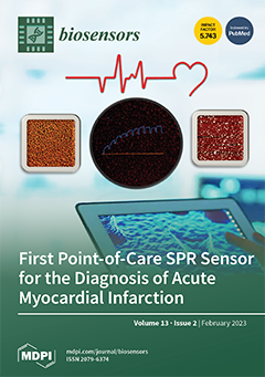
Cover Story (view full-size image): In this work, Altintas et al. developed a biomimetic sensor for the detection of cardiac troponin-I (cTnI) biomarker using a portable angular SPR and epitope-imprinted synthetic receptors. The novel SPR technique offers unique advantages, such as being rapid, cost-efficient, real-time, user-friendly, and label-free. It does not require complex assay strategies, such as sandwich or competitive assays, for obtaining high sensitivity. The sensor has shown good reproducibility, sensitivity, specificity, and affinity. The as-synthesized receptors as well as sensor fabrication steps were successfully characterized using DLS, AFM, fluorescence microscopy, CV, SWV, and EIS techniques. The developed sensor has great potential to be further enhanced for point-of-care testing and the early diagnosis of acute myocardial infarction. View this paper
  • Issues are regarded as officially published after their release is announced to the table of contents alert mailing list.
  • You may sign up for e-mail alerts to receive table of contents of newly released issues.
  • PDF is the official format for papers published in both, html and pdf forms. To view the papers in pdf format, click on the "PDF Full-text" link, and use the free Adobe Reader to open them.
Order results
Result details
Section
Select all
Export citation of selected articles as:
12 pages, 2746 KiB  
Article
Nanobody-Based Sandwich Immunoassay for Pathogenic Escherichia coli F17 Strain Detection
by Asma Dhehibi, Abdelmounaaim Allaoui, Amal Raouafi, Mohammed Terrak, Balkiss Bouhaouala-Zahar, Mohamed Hammadi, Noureddine Raouafi and Imed Salhi
Biosensors 2023, 13(2), 299; https://doi.org/10.3390/bios13020299 - 20 Feb 2023
Cited by 4 | Viewed by 1938
Abstract
Rapid and specific detection of pathogenic bacteria in fecal samples is of critical importance for the diagnosis of neonatal diarrhea in veterinary clinics. Nanobodies are a promising tool for the treatment and diagnosis of infectious diseases due to their unique recognition properties. In [...] Read more.
Rapid and specific detection of pathogenic bacteria in fecal samples is of critical importance for the diagnosis of neonatal diarrhea in veterinary clinics. Nanobodies are a promising tool for the treatment and diagnosis of infectious diseases due to their unique recognition properties. In this study, we report the design of a nanobody-based magnetofluorescent immunoassay for the sensitive detection of pathogenic Escherichia coli F17-positive strains (E. coli F17). For this, a camel was immunized with purified F17A protein from F17 fimbriae and a nanobody library was constructed by phage display. Two specific anti-F17A nanobodies (Nbs) were selected to design the bioassay. The first one (Nb1) was conjugated to magnetic beads (MBs) to form a complex capable of efficiently capturing the target bacteria. A second horseradish peroxidase (HRP)-conjugated nanobody (Nb4) was used for detection by oxidizing o-phenylenediamine (OPD) to fluorescent 2,3-diaminophenazine (DAP). Our results show that the immunoassay recognizes E. coli F17 with high specificity and sensitivity, with a detection limit of 1.8 CFU/mL in only 90 min. Furthermore, we showed that the immunoassay can be applied to fecal samples without pretreatment and remains stable for at least one month when stored at 4 °C. Full article
(This article belongs to the Section Biosensor and Bioelectronic Devices)
Show Figures

Figure 1

29 pages, 5955 KiB  
Review
Review of HIV Self Testing Technologies and Promising Approaches for the Next Generation
by Amanda Bacon, Weijing Wang, Hankeun Lee, Saurabh Umrao, Prima Dewi Sinawang, Demir Akin, Kodchakorn Khemtonglang, Anqi Tan, Sabina Hirshfield, Utkan Demirci, Xing Wang and Brian T. Cunningham
Biosensors 2023, 13(2), 298; https://doi.org/10.3390/bios13020298 - 20 Feb 2023
Cited by 5 | Viewed by 7456
Abstract
The ability to self-test for HIV is vital to preventing transmission, particularly when used in concert with HIV biomedical prevention modalities, such as pre-exposure prophylaxis (PrEP). In this paper, we review recent developments in HIV self-testing and self-sampling methods, and the potential future [...] Read more.
The ability to self-test for HIV is vital to preventing transmission, particularly when used in concert with HIV biomedical prevention modalities, such as pre-exposure prophylaxis (PrEP). In this paper, we review recent developments in HIV self-testing and self-sampling methods, and the potential future impact of novel materials and methods that emerged through efforts to develop more effective point-of-care (POC) SARS-CoV-2 diagnostics. We address the gaps in existing HIV self-testing technologies, where improvements in test sensitivity, sample-to-answer time, simplicity, and cost are needed to enhance diagnostic accuracy and widespread accessibility. We discuss potential paths toward the next generation of HIV self-testing through sample collection materials, biosensing assay techniques, and miniaturized instrumentation. We discuss the implications for other applications, such as self-monitoring of HIV viral load and other infectious diseases. Full article
(This article belongs to the Special Issue Smart Materials for Chemical and Biosensing)
Show Figures

Figure 1

14 pages, 2840 KiB  
Article
Bioluminescent RIPoptosome Assay for FADD/RIPK1 Interaction Based on Split Luciferase Assay in a Human Neuroblastoma Cell Line SH-SY5Y
by Parisa Ghanavatian, Hossein Salehi-Sedeh, Farangis Ataei and Saman Hosseinkhani
Biosensors 2023, 13(2), 297; https://doi.org/10.3390/bios13020297 - 20 Feb 2023
Cited by 2 | Viewed by 2463
Abstract
Different programed cell death (PCD) modalities involve protein–protein interactions in large complexes. Tumor necrosis factor α (TNFα) stimulated assembly of receptor-interacting protein kinase 1 (RIPK1)/Fas-associated death domain (FADD) interaction forms Ripoptosome complex that may cause either apoptosis or necroptosis. The present study addresses [...] Read more.
Different programed cell death (PCD) modalities involve protein–protein interactions in large complexes. Tumor necrosis factor α (TNFα) stimulated assembly of receptor-interacting protein kinase 1 (RIPK1)/Fas-associated death domain (FADD) interaction forms Ripoptosome complex that may cause either apoptosis or necroptosis. The present study addresses the interaction of RIPK1 and FADD in TNFα signaling by fusion of C-terminal (CLuc) and N-terminal (NLuc) luciferase fragments to RIPK1-CLuc (R1C) or FADD-NLuc (FN) in a caspase 8 negative neuroblastic SH-SY5Y cell line, respectively. In addition, based on our findings, an RIPK1 mutant (R1C K612R) had less interaction with FN, resulting in increasing cell viability. Moreover, presence of a caspase inhibitor (zVAD.fmk) increases luciferase activity compared to Smac mimetic BV6 (B), TNFα -induced (T) and non-induced cell. Furthermore, etoposide decreased luciferase activity, but dexamethasone was not effective in SH-SY5Y. This reporter assay might be used to evaluate basic aspects of this interaction as well as for screening of necroptosis and apoptosis targeting drugs with potential therapeutic application. Full article
(This article belongs to the Special Issue Cell-Based Biosensors for Rapid Detection and Monitoring)
Show Figures

Figure 1

23 pages, 5454 KiB  
Review
Multiplex Surface-Enhanced Raman Scattering: An Emerging Tool for Multicomponent Detection of Food Contaminants
by Qingyi Wei, Qirong Dong and Hongbin Pu
Biosensors 2023, 13(2), 296; https://doi.org/10.3390/bios13020296 - 19 Feb 2023
Cited by 6 | Viewed by 3835
Abstract
For survival and quality of human life, the search for better ways to ensure food safety is constant. However, food contaminants still threaten human health throughout the food chain. In particular, food systems are often polluted with multiple contaminants simultaneously, which can cause [...] Read more.
For survival and quality of human life, the search for better ways to ensure food safety is constant. However, food contaminants still threaten human health throughout the food chain. In particular, food systems are often polluted with multiple contaminants simultaneously, which can cause synergistic effects and greatly increase food toxicity. Therefore, the establishment of multiple food contaminant detection methods is significant in food safety control. The surface-enhanced Raman scattering (SERS) technique has emerged as a potent candidate for the detection of multicomponents simultaneously. The current review focuses on the SERS-based strategies in multicomponent detection, including the combination of chromatography methods, chemometrics, and microfluidic engineering with the SERS technique. Furthermore, recent applications of SERS in the detection of multiple foodborne bacteria, pesticides, veterinary drugs, food adulterants, mycotoxins and polycyclic aromatic hydrocarbons are summarized. Finally, challenges and future prospects for the SERS-based detection of multiple food contaminants are discussed to provide research orientation for further. Full article
(This article belongs to the Special Issue Application of Biosensors in Food Safety Analysis)
Show Figures

Figure 1

24 pages, 5328 KiB  
Review
Molecularly Imprinted Polymer-Based Luminescent Chemosensors
by Ruoyang Liu and Chi-Chiu Ko
Biosensors 2023, 13(2), 295; https://doi.org/10.3390/bios13020295 - 19 Feb 2023
Cited by 6 | Viewed by 2355
Abstract
Molecularly imprinted polymer (MIP)-based luminescent chemosensors combine the advantages of the highly specific molecular recognition of the imprinting sites and the high sensitivity with the luminescence detection. These advantages have drawn great attention during the past two decades. Luminescent molecularly imprinted polymers (luminescent [...] Read more.
Molecularly imprinted polymer (MIP)-based luminescent chemosensors combine the advantages of the highly specific molecular recognition of the imprinting sites and the high sensitivity with the luminescence detection. These advantages have drawn great attention during the past two decades. Luminescent molecularly imprinted polymers (luminescent MIPs) towards different targeted analytes are constructed with different strategies, such as the incorporation of luminescent functional monomers, physical entrapment, covalent attachment of luminescent signaling elements on the MIPs, and surface-imprinting polymerization on the luminescent nanomaterials. In this review, we will discuss the design strategies and sensing approaches of luminescent MIP-based chemosensors, as well as their selected applications in biosensing, bioimaging, food safety, and clinical diagnosis. The limitations and prospects for the future development of MIP-based luminescent chemosensors will also be discussed. Full article
(This article belongs to the Special Issue Activatable Probes for Biosensing, Imaging, and Photomedicine)
Show Figures

Figure 1

28 pages, 5314 KiB  
Review
A Review of Detection Methods for Vancomycin-Resistant Enterococci (VRE) Genes: From Conventional Approaches to Potentially Electrochemical DNA Biosensors
by Nor Dyana Zakaria, Hairul Hisham Hamzah, Ibrahim Luqman Salih, Venugopal Balakrishnan and Khairunisak Abdul Razak
Biosensors 2023, 13(2), 294; https://doi.org/10.3390/bios13020294 - 18 Feb 2023
Cited by 3 | Viewed by 3572
Abstract
Vancomycin-resistant Enterococci (VRE) genes are bacteria strains generated from Gram-positive bacteria and resistant to one of the glycopeptides antibiotics, commonly, vancomycin. VRE genes have been identified worldwide and exhibit considerable phenotypic and genotypic variations. There are six identified phenotypes of vancomycin-resistant genes: VanA, [...] Read more.
Vancomycin-resistant Enterococci (VRE) genes are bacteria strains generated from Gram-positive bacteria and resistant to one of the glycopeptides antibiotics, commonly, vancomycin. VRE genes have been identified worldwide and exhibit considerable phenotypic and genotypic variations. There are six identified phenotypes of vancomycin-resistant genes: VanA, VanB, VanC, VanD, VanE, and VanG. The VanA and VanB strains are often found in the clinical laboratory because they are very resistant to vancomycin. VanA bacteria can pose significant issues for hospitalized patients due to their ability to spread to other Gram-positive infections, which changes their genetic material to increase their resistance to the antibiotics used during treatment. This review summarizes the established methods for detecting VRE strains utilizing traditional, immunoassay, and molecular approaches and then focuses on potential electrochemical DNA biosensors to be developed. However, from the literature search, no information was reported on developing electrochemical biosensors for detecting VRE genes; only the electrochemical detection of vancomycin-susceptible bacteria was reported. Thus, strategies to create robust, selective, and miniaturized electrochemical DNA biosensor platforms to detect VRE genes are also discussed. Full article
(This article belongs to the Special Issue DNA Based Biosensors)
Show Figures

Figure 1

12 pages, 6728 KiB  
Article
A CRISPR-Cas and Tat Peptide with Fluorescent RNA Aptamer System for Signal Amplification in RNA Imaging
by Heng Tang, Junran Peng, Xin Jiang, Shuang Peng, Fang Wang, Xiaocheng Weng and Xiang Zhou
Biosensors 2023, 13(2), 293; https://doi.org/10.3390/bios13020293 - 18 Feb 2023
Cited by 1 | Viewed by 2733
Abstract
We reported on an efficient RNA imaging strategy based on a CRISPR-Cas and Tat peptide with a fluorescent RNA aptamer (TRAP-tag). Using modified CRISPR-Cas RNA hairpin binding proteins fused with a Tat peptide array that recruits modified RNA aptamers, this simple and sensitive [...] Read more.
We reported on an efficient RNA imaging strategy based on a CRISPR-Cas and Tat peptide with a fluorescent RNA aptamer (TRAP-tag). Using modified CRISPR-Cas RNA hairpin binding proteins fused with a Tat peptide array that recruits modified RNA aptamers, this simple and sensitive strategy is capable of visualizing endogenous RNA in cells with high precision and efficiency. In addition, the modular design of the CRISPR-TRAP-tag facilitates the substitution of sgRNAs, RNA hairpin binding proteins, and aptamers in order to optimize imaging quality and live cell affinity. With CRISPR-TRAP-tag, exogenous GCN4, endogenous mRNA MUC4, and lncRNA SatIII were distinctly visualized in single live cells. Full article
(This article belongs to the Section Nano- and Micro-Technologies in Biosensors)
Show Figures

Figure 1

5 pages, 207 KiB  
Editorial
Investigation of Biomolecule Interactions: Optical-, Electrochemical-, and Acoustic-Based Biosensors
by Ieva Plikusiene and Almira Ramanaviciene
Biosensors 2023, 13(2), 292; https://doi.org/10.3390/bios13020292 - 18 Feb 2023
Cited by 5 | Viewed by 1433
Abstract
Today, optical, electrochemical, and acoustic affinity biosensors; immunosensors; and immunoanalytical systems play an important role in the detection and characterization of a number of biological substances, including viral antigens, specific antibodies, and clinically important biomarkers [...] Full article
18 pages, 5371 KiB  
Review
Covalent Organic Frameworks-Based Electrochemical Sensors for Food Safety Analysis
by Zhenyu Lu, Yingying Wang and Gongke Li
Biosensors 2023, 13(2), 291; https://doi.org/10.3390/bios13020291 - 17 Feb 2023
Cited by 7 | Viewed by 2624
Abstract
Food safety is a key issue in promoting human health and sustaining life. Food analysis is essential to prevent food components or contaminants causing foodborne-related illnesses to consumers. Electrochemical sensors have become a desirable method for food safety analysis due to their simple, [...] Read more.
Food safety is a key issue in promoting human health and sustaining life. Food analysis is essential to prevent food components or contaminants causing foodborne-related illnesses to consumers. Electrochemical sensors have become a desirable method for food safety analysis due to their simple, accurate and rapid response. The low sensitivity and poor selectivity of electrochemical sensors working in complex food sample matrices can be overcome by coupling them with covalent organic frameworks (COFs). COFs are a kind of novel porous organic polymer formed by light elements, such as C, H, N and B, via covalent bonds. This review focuses on the recent progress in COF-based electrochemical sensors for food safety analysis. Firstly, the synthesis methods of COFs are summarized. Then, a discussion of the strategies is given to improve the electrochemistry performance of COFs. There follows a summary of the recently developed COF-based electrochemical sensors for the determination of food contaminants, including bisphenols, antibiotics, pesticides, heavy metal ions, fungal toxin and bacterium. Finally, the challenges and the future directions in this field are discussed. Full article
Show Figures

Figure 1

20 pages, 22442 KiB  
Article
Microfluidic Wound-Healing Assay for ECM and Microenvironment Properties on Microglia BV2 Cells Migration
by Ehsan Yazdanpanah Moghadam, Nahum Sonenberg and Muthukumaran Packirisamy
Biosensors 2023, 13(2), 290; https://doi.org/10.3390/bios13020290 - 17 Feb 2023
Cited by 3 | Viewed by 1975
Abstract
Microglia cells, as the resident immune cells of the central nervous system (CNS), are highly motile and migratory in development and pathophysiological conditions. During their migration, microglia cells interact with their surroundings based on the various physical and chemical properties in the brain. [...] Read more.
Microglia cells, as the resident immune cells of the central nervous system (CNS), are highly motile and migratory in development and pathophysiological conditions. During their migration, microglia cells interact with their surroundings based on the various physical and chemical properties in the brain. Herein, a microfluidic wound-healing chip is developed to investigate microglial BV2 cell migration on the substrates coated with extracellular matrixes (ECMs) and substrates usually used for bio-applications on cell migration. In order to generate the cell-free space (wound), gravity was utilized as a driving force to flow the trypsin with the device. It was shown that, despite the scratch assay, the cell-free area was created without removing the extracellular matrix coating (fibronectin) using the microfluidic assay. It was found that the substrates coated with Poly-L-Lysine (PLL) and gelatin stimulated microglial BV2 migration, while collagen and fibronectin coatings had an inhibitory effect compared to the control conditions (uncoated glass substrate). In addition, the results showed that the polystyrene substrate induced higher cell migration than the PDMS and glass substrates. The microfluidic migration assay provides an in vitro microenvironment closer to in vivo conditions for further understanding the microglia migration mechanism in the brain, where the environment properties change under homeostatic and pathological conditions. Full article
(This article belongs to the Section Optical and Photonic Biosensors)
Show Figures

Figure 1

13 pages, 3195 KiB  
Article
Horseradish Peroxidase-Encapsulated Fluorescent Bio-Nanoparticle for Ultra-Sensitive and Easy Detection of Hydrogen Peroxide
by Myeong-Jun Lee, Ji-Ae Song, Jin-Ha Choi, Jeong-Hyeop Shin, Ji-Woon Myeong, Ki-Ppeum Lee, Taehwan Kim, Ki-Eob Park and Byung-Keun Oh
Biosensors 2023, 13(2), 289; https://doi.org/10.3390/bios13020289 - 17 Feb 2023
Cited by 6 | Viewed by 1978
Abstract
Hydrogen peroxide (H2O2) has been a fascinating target in various chemical, biological, clinical, and industrial fields. Several types of fluorescent protein-stabilized gold nanoclusters (protein-AuNCs) have been developed for sensitive and easy detection of H2O2. However, [...] Read more.
Hydrogen peroxide (H2O2) has been a fascinating target in various chemical, biological, clinical, and industrial fields. Several types of fluorescent protein-stabilized gold nanoclusters (protein-AuNCs) have been developed for sensitive and easy detection of H2O2. However, its low sensitivity makes is difficult to measure negligible concentrations of H2O2. Therefore, to overcome this limitation, we developed a horseradish peroxidase-encapsulated fluorescent bio-nanoparticle (HEFBNP), comprising bovine serum albumin-stabilized gold nanoclusters (BSA-AuNCs) and horseradish peroxidase-stabilized gold nanoclusters (HRP-AuNCs). The fabricated HEFBNP can sensitively detect H2O2 owing to its two properties. The first is that HEFBNPs have a continuous two-step fluorescence quenching mechanism, which comes from the heterogenous fluorescence quenching mechanism of HRP-AuNCs and BSA-AuNCs. Second, the proximity of two protein-AuNCs in a single HEFBNP allows a reaction intermediate (•OH) to rapidly reach the adjacent protein-AuNCs. As a result, HEFBNP can improve the overall reaction event and decrease the loss of intermediate in the solution. Due to the continuous quenching mechanism and effective reaction event, a HEFBNP-based sensing system can measure very low concentrations of H2O2 up to 0.5 nM and show good selectivity. Furthermore, we design a glass-based microfluidic device to make it easier use HEFBNP, which allowed us to detect H2O2 with the naked eye. Overall, the proposed H2O2 sensing system is expected to be an easy and highly sensitive on-site detection tool in chemistry, biology, clinics, and industry fields. Full article
(This article belongs to the Special Issue Nanomaterials and Their Applications in Sensing and Biosensing)
Show Figures

Figure 1

15 pages, 3159 KiB  
Article
PEDOT-Polyamine-Based Organic Electrochemical Transistors for Monitoring Protein Binding
by Marjorie Montero-Jimenez, Francisco L. Amante, Gonzalo E. Fenoy, Juliana Scotto, Omar Azzaroni and Waldemar A. Marmisolle
Biosensors 2023, 13(2), 288; https://doi.org/10.3390/bios13020288 - 17 Feb 2023
Cited by 3 | Viewed by 2216
Abstract
The fabrication of efficient organic electrochemical transistors (OECTs)-based biosensors requires the design of biocompatible interfaces for the immobilization of biorecognition elements, as well as the development of robust channel materials to enable the transduction of the biochemical event into a reliable electrical signal. [...] Read more.
The fabrication of efficient organic electrochemical transistors (OECTs)-based biosensors requires the design of biocompatible interfaces for the immobilization of biorecognition elements, as well as the development of robust channel materials to enable the transduction of the biochemical event into a reliable electrical signal. In this work, PEDOT-polyamine blends are shown as versatile organic films that can act as both highly conducting channels of the transistors and non-denaturing platforms for the construction of the biomolecular architectures that operate as sensing surfaces. To achieve this goal, we synthesized and characterized films of PEDOT and polyallylamine hydrochloride (PAH) and employed them as conducting channels in the construction of OECTs. Next, we studied the response of the obtained devices to protein adsorption, using glucose oxidase (GOx) as a model system, through two different strategies: The direct electrostatic adsorption of GOx on the PEDOT-PAH film and the specific recognition of the protein by a lectin attached to the surface. Firstly, we used surface plasmon resonance to monitor the adsorption of the proteins and the stability of the assemblies on PEDOT-PAH films. Then, we monitored the same processes with the OECT showing the capability of the device to perform the detection of the protein binding process in real time. In addition, the sensing mechanisms enabling the monitoring of the adsorption process with the OECTs for the two strategies are discussed. Full article
Show Figures

Figure 1

17 pages, 2950 KiB  
Article
Difunctional Hydrogel Optical Fiber Fluorescence Sensor for Continuous and Simultaneous Monitoring of Glucose and pH
by Yangjie Li, Site Luo, Yongqiang Gui, Xin Wang, Ziyuan Tian and Haihu Yu
Biosensors 2023, 13(2), 287; https://doi.org/10.3390/bios13020287 - 17 Feb 2023
Cited by 8 | Viewed by 2719
Abstract
It is significant for people with diabetes to know their body’s real-time glucose level, which can guide the diagnosis and treatment. Therefore, it is necessary to research continuous glucose monitoring (CGM) as it gives us real-time information about our health condition and its [...] Read more.
It is significant for people with diabetes to know their body’s real-time glucose level, which can guide the diagnosis and treatment. Therefore, it is necessary to research continuous glucose monitoring (CGM) as it gives us real-time information about our health condition and its dynamic changes. Here, we report a novel hydrogel optical fiber fluorescence sensor segmentally functionalized with fluorescein derivative and CdTe QDs/3-APBA, which can continuously monitor pH and glucose simultaneously. In the glucose detection section, the complexation of PBA and glucose will expand the local hydrogel and decrease the fluorescence of the quantum dots. The fluorescence can be transmitted to the detector by the hydrogel optical fiber in real time. As the complexation reaction and the swelling–deswelling of the hydrogel are all reversible, the dynamic change of glucose concentration can be monitored. For pH detection, the fluorescein attached to another segment of the hydrogel exhibits different protolytic forms when pH changes and the fluorescence changes correspondingly. The significance of pH detection is compensation for pH errors in glucose detection because the reaction between PBA and glucose is sensitive to pH. The emission peaks of the two detection units are 517 nm and 594 nm, respectively, so there is no signal interference between them. The sensor can continuously monitor glucose in 0–20 mM and pH in 5.4–7.8. The advantages of this sensor are multi-parameter simultaneous detection, transmission-detection integration, real-time dynamic detection, and good biocompatibility. Full article
(This article belongs to the Special Issue Advanced Optical Fiber Sensors for Chemical and Biological Detection)
Show Figures

Graphical abstract

19 pages, 7759 KiB  
Review
Recent Advancements in Novel Sensing Systems through Nanoarchitectonics
by Karthick Velu, Rekha Goswami Shrestha, Lok Kumar Shrestha and Katsuhiko Ariga
Biosensors 2023, 13(2), 286; https://doi.org/10.3390/bios13020286 - 16 Feb 2023
Cited by 3 | Viewed by 1735
Abstract
The fabrication of various sensing devices and the ability to harmonize materials for a higher degree of organization is essential for effective sensing systems. Materials with hierarchically micro- and mesopore structures can enhance the sensitivity of sensors. Nanoarchitectonics allows for atomic/molecular level manipulations [...] Read more.
The fabrication of various sensing devices and the ability to harmonize materials for a higher degree of organization is essential for effective sensing systems. Materials with hierarchically micro- and mesopore structures can enhance the sensitivity of sensors. Nanoarchitectonics allows for atomic/molecular level manipulations that create a higher area-to-volume ratio in nanoscale hierarchical structures for use in ideal sensing applications. Nanoarchitectonics also provides ample opportunities to fabricate materials by tuning pore size, increasing surface area, trapping molecules via host–guest interactions, and other mechanisms. Material characteristics and shape significantly enhance sensing capabilities via intramolecular interactions, molecular recognition, and localized surface plasmon resonance (LSPR). This review highlights the latest advancements in nanoarchitectonics approaches to tailor materials for various sensing applications, including biological micro/macro molecules, volatile organic compounds (VOC), microscopic recognition, and the selective discrimination of microparticles. Furthermore, different sensing devices that utilize the nanoarchitectonics concept to achieve atomic-molecular level discrimination are also discussed. Full article
(This article belongs to the Special Issue Nanomechanical Sensors for Gas Detection)
Show Figures

Figure 1

17 pages, 1256 KiB  
Review
Cortisol Immunosensors: A Literature Review
by Chrysoula-Evangelia Karachaliou, Georgios Koukouvinos, Dimitrios Goustouridis, Ioannis Raptis, Sotirios Kakabakos, Panagiota Petrou and Evangelia Livaniou
Biosensors 2023, 13(2), 285; https://doi.org/10.3390/bios13020285 - 16 Feb 2023
Cited by 8 | Viewed by 3581
Abstract
Cortisol is a steroid hormone that is involved in a broad range of physiological processes in human/animal organisms. Cortisol levels in biological samples are a valuable biomarker, e.g., of stress and stress-related diseases; thus, cortisol determination in biological fluids, such as serum, saliva [...] Read more.
Cortisol is a steroid hormone that is involved in a broad range of physiological processes in human/animal organisms. Cortisol levels in biological samples are a valuable biomarker, e.g., of stress and stress-related diseases; thus, cortisol determination in biological fluids, such as serum, saliva and urine, is of great clinical value. Although cortisol analysis can be performed with chromatography-based analytical techniques, such as liquid chromatography–tandem mass spectrometry (LC-MS/MS), conventional immunoassays (radioimmunoassays (RIAs), enzyme-linked immunosorbent assays (ELISAs), etc.) are considered the “gold standard” analytical methodology for cortisol, due to their high sensitivity along with a series of practical advantages, such as low-cost instrumentation, an assay protocol that is fast and easy to perform, and high sample throughput. Especially in recent decades, research efforts have focused on the replacement of conventional immunoassays by cortisol immunosensors, which may offer further improvements in the field, such as real-time analysis at the point of care (e.g., continuous cortisol monitoring in sweat through wearable electrochemical sensors). In this review, most of the reported cortisol immunosensors, mainly electrochemical and also optical ones, are presented, focusing on their immunosensing/detection principles. Future prospects are also briefly discussed. Full article
(This article belongs to the Special Issue Advances in Biosensors for Health-Care and Diagnostics)
Show Figures

Figure 1

17 pages, 4810 KiB  
Review
MOFs-Modified Electrochemical Sensors and the Application in the Detection of Opioids
by Jiaqi Zhao, Ying Kan, Zhi Chen, Hongmei Li and Weifei Zhang
Biosensors 2023, 13(2), 284; https://doi.org/10.3390/bios13020284 - 16 Feb 2023
Cited by 7 | Viewed by 2911
Abstract
Opioids are widely used in clinical practice, but drug overdoses can lead to many adverse reactions, and even endanger life. Therefore, it is essential to implement real-time measurement of drug concentrations to adjust the dosage given during treatment, keeping drug levels within therapeutic [...] Read more.
Opioids are widely used in clinical practice, but drug overdoses can lead to many adverse reactions, and even endanger life. Therefore, it is essential to implement real-time measurement of drug concentrations to adjust the dosage given during treatment, keeping drug levels within therapeutic levels. Metal-Organic frameworks (MOFs) and their composite materials modified bare electrode electrochemical sensors have the advantages of fast production, low cost, high sensitivity, and low detection limit in the detection of opioids. In this review, MOFs and MOFs composites, electrochemical sensors modified with MOFs for the detection of opioids, as well as the application of microfluidic chips in combination with electrochemical methods are all reviewed, and the potential for the development of microfluidic chips electrochemical methods with MOFs surface modifications for the detection of opioids is also prospected. We hope that this review will provide contributions to the study of electrochemical sensors modified with MOFs for the detection of opioids. Full article
(This article belongs to the Special Issue Microfluidics for Detection and Analysis)
Show Figures

Figure 1

13 pages, 3073 KiB  
Article
Functional Imaging and Inhibitor Screening of Human Pancreatic Lipase by a Resorufin-Based Fluorescent Probe
by Fan-Bin Hou, Na Zhang, Guang-Hao Zhu, Yu-Fan Fan, Meng-Ru Sun, Liang-Liang Nie, Guang-Bo Ge, Yue-Juan Zheng and Ping Wang
Biosensors 2023, 13(2), 283; https://doi.org/10.3390/bios13020283 - 16 Feb 2023
Cited by 3 | Viewed by 2007
Abstract
Human pancreatic lipase (hPL) is a crucial digestive enzyme responsible for the digestion of dietary lipids in humans, and inhibition of hPL is effective in reducing triglyceride intake, thereby preventing and treating obesity. In this study, a series of fatty acids with different [...] Read more.
Human pancreatic lipase (hPL) is a crucial digestive enzyme responsible for the digestion of dietary lipids in humans, and inhibition of hPL is effective in reducing triglyceride intake, thereby preventing and treating obesity. In this study, a series of fatty acids with different carbon chain lengths were constructed to the fluorophore resorufin based on the substrate preference of hPL. Among them, RLE was found to have the best combination of stability, specificity, sensitivity and reactivity towards hPL. Under physiological conditions, RLE can be rapidly hydrolyzed by hPL and released to resorufin, which triggered approximately 100-fold fluorescence enhancement at 590 nm. RLE was successfully applied for sensing and imaging of endogenous PL in living systems with low cytotoxicity and high imaging resolution. Moreover, a visual high-throughput screening platform was established using RLE, and the inhibitory effects of hundreds of drugs and natural products toward hPL were evaluated. Collectively, this study reports a novel and highly specific enzyme-activatable fluorogenic substrate for hPL that could serve as a powerful tool for monitoring hPL activity in complex biological systems and showcases the potential to explore physiological functions and rapid screening of inhibitors. Full article
(This article belongs to the Special Issue Enzyme-Based Biosensors and Their Applications)
Show Figures

Graphical abstract

17 pages, 2904 KiB  
Review
Approaches and Challenges for Biosensors for Acute and Chronic Heart Failure
by Sariye Irem Kaya, Ahmet Cetinkaya, Goksu Ozcelikay, Seyda Nur Samanci and Sibel A. Ozkan
Biosensors 2023, 13(2), 282; https://doi.org/10.3390/bios13020282 - 16 Feb 2023
Cited by 3 | Viewed by 2164
Abstract
Heart failure (HF) is a cardiovascular disease defined by several symptoms that occur when the heart cannot supply the blood needed by the tissues. HF, which affects approximately 64 million people worldwide and whose incidence and prevalence are increasing, has an important place [...] Read more.
Heart failure (HF) is a cardiovascular disease defined by several symptoms that occur when the heart cannot supply the blood needed by the tissues. HF, which affects approximately 64 million people worldwide and whose incidence and prevalence are increasing, has an important place in terms of public health and healthcare costs. Therefore, developing and enhancing diagnostic and prognostic sensors is an urgent need. Using various biomarkers for this purpose is a significant breakthrough. It is possible to classify the biomarkers used in HF: associated with myocardial and vascular stretch (B-type natriuretic peptide (BNP), N-terminal proBNP and troponin), related to neurohormonal pathways (aldosterone and plasma renin activity), and associated with myocardial fibrosis and hypertrophy (soluble suppression of tumorigenicity 2 and galactin 3). There is an increasing demand for the design of fast, portable, and low-cost biosensing devices for the biomarkers related to HF. Biosensors play a significant role in early diagnosis as an alternative to time-consuming and expensive laboratory analysis. In this review, the most influential and novel biosensor applications for acute and chronic HF will be discussed in detail. These studies will be evaluated in terms of advantages, disadvantages, sensitivity, applicability, user-friendliness, etc. Full article
(This article belongs to the Special Issue Emerging Biosensing Technologies for Healthcare Applications)
Show Figures

Figure 1

11 pages, 4986 KiB  
Communication
Low-Cost Impedance Camera for Cell Distribution Monitoring
by Bo Tang, Mengxi Liu and Andreas Dietzel
Biosensors 2023, 13(2), 281; https://doi.org/10.3390/bios13020281 - 16 Feb 2023
Cited by 1 | Viewed by 1973
Abstract
Electrical impedance spectroscopy (EIS) is widely recognized as a powerful tool in biomedical research. For example, it allows detection and monitoring of diseases, measuring of cell density in bioreactors, and characterizing the permeability of tight junctions in barrier-forming tissue models. However, with single-channel [...] Read more.
Electrical impedance spectroscopy (EIS) is widely recognized as a powerful tool in biomedical research. For example, it allows detection and monitoring of diseases, measuring of cell density in bioreactors, and characterizing the permeability of tight junctions in barrier-forming tissue models. However, with single-channel measurement systems, only integral information is obtained without spatial resolution. Here we present a low-cost multichannel impedance measurement set-up capable of mapping cell distributions in a fluidic environment by using a microelectrode array (MEA) realized in 4-level printed circuit board (PCB) technology including layers for shielding, interconnections, and microelectrodes. The array of 8 × 8 gold microelectrode pairs was connected to home-built electric circuitry consisting of commercial components such as programmable multiplexers and an analog front-end module which allows the acquisition and processing of electrical impedances. For a proof-of-concept, the MEA was wetted in a 3D printed reservoir into which yeast cells were locally injected. Impedance maps were recorded at 200 kHz which correlate well with the optical images showing the yeast cell distribution in the reservoir. Blurring from parasitic currents slightly disturbing the impedance maps could be eliminated by deconvolution using an experimentally determined point spread function. The MEA of the impedance camera can in future be further miniaturized and integrated into cell cultivation and perfusion systems such as organ on chip devices to augment or even replace light microscopic monitoring of cell monolayer confluence and integrity during the cultivation in incubation chambers. Full article
(This article belongs to the Special Issue Impedance-Spectroscopy-Based Biosensors)
Show Figures

Figure 1

19 pages, 2692 KiB  
Article
Proof of Concept for Sustainable Manufacturing of Neural Electrode Array for In Vivo Recording
by Szu-Ying Li, Hsin-Yi Tseng, Bo-Wei Chen, Yu-Chun Lo, Huai-Hsuan Shao, Yen-Ting Wu, Ssu-Ju Li, Ching-Wen Chang, Ta-Chung Liu, Fu-Yu Hsieh, Yi Yang, Yan-Bo Lai, Po-Chun Chen and You-Yin Chen
Biosensors 2023, 13(2), 280; https://doi.org/10.3390/bios13020280 - 16 Feb 2023
Viewed by 2623
Abstract
Increasing requirements for neural implantation are helping to expand our understanding of nervous systems and generate new developmental approaches. It is thanks to advanced semiconductor technologies that we can achieve the high-density complementary metal-oxide-semiconductor electrode array for the improvement of the quantity and [...] Read more.
Increasing requirements for neural implantation are helping to expand our understanding of nervous systems and generate new developmental approaches. It is thanks to advanced semiconductor technologies that we can achieve the high-density complementary metal-oxide-semiconductor electrode array for the improvement of the quantity and quality of neural recordings. Although the microfabricated neural implantable device holds much promise in the biosensing field, there are some significant technological challenges. The most advanced neural implantable device relies on complex semiconductor manufacturing processes, which are required for the use of expensive masks and specific clean room facilities. In addition, these processes based on a conventional photolithography technique are suitable for mass production, which is not applicable for custom-made manufacturing in response to individual experimental requirements. The microfabricated complexity of the implantable neural device is increasing, as is the associated energy consumption, and corresponding emissions of carbon dioxide and other greenhouse gases, resulting in environmental deterioration. Herein, we developed a fabless fabricated process for a neural electrode array that was simple, fast, sustainable, and customizable. An effective strategy to produce conductive patterns as the redistribution layers (RDLs) includes implementing microelectrodes, traces, and bonding pads onto the polyimide (PI) substrate by laser micromachining techniques combined with the drop coating of the silver glue to stack the laser grooving lines. The process of electroplating platinum on the RDLs was performed to increase corresponding conductivity. Sequentially, Parylene C was deposited onto the PI substrate to form the insulation layer for the protection of inner RDLs. Following the deposition of Parylene C, the via holes over microelectrodes and the corresponding probe shape of the neural electrode array was also etched by laser micromachining. To increase the neural recording capability, three-dimensional microelectrodes with a high surface area were formed by electroplating gold. Our eco-electrode array showed reliable electrical characteristics of impedance under harsh cyclic bending conditions of over 90 degrees. For in vivo application, our flexible neural electrode array demonstrated more stable and higher neural recording quality and better biocompatibility as well during the 2-week implantation compared with those of the silicon-based neural electrode array. In this study, our proposed eco-manufacturing process for fabricating the neural electrode array reduced 63 times of carbon emissions compared to the traditional semiconductor manufacturing process and provided freedom in the customized design of the implantable electronic devices as well. Full article
Show Figures

Figure 1

12 pages, 1666 KiB  
Article
A Multiple-Array SPRi Biosensor as a Tool for Detection of Gynecological–Oncological Diseases
by Beata Szymanska, Zenon Lukaszewski, Kinga Hermanowicz-Szamatowicz and Ewa Gorodkiewicz
Biosensors 2023, 13(2), 279; https://doi.org/10.3390/bios13020279 - 16 Feb 2023
Cited by 2 | Viewed by 1706
Abstract
Diagnostics based on the determination of biomarkers in body fluids will be more successful when several biomarkers are determined. A multiple-array SPRi biosensor for the simultaneous determination of CA125, HE4, CEA, IL-6 and aromatase has been developed. Five individual biosensors were placed on [...] Read more.
Diagnostics based on the determination of biomarkers in body fluids will be more successful when several biomarkers are determined. A multiple-array SPRi biosensor for the simultaneous determination of CA125, HE4, CEA, IL-6 and aromatase has been developed. Five individual biosensors were placed on the same chip. Each of them consisted of a suitable antibody covalently immobilized onto a gold chip surface via a cysteamine linker by means of the NHS/EDC protocol. The biosensor for IL-6 works in the pg mL−1 range, that for CA125 in the µg mL−1 range, and the other three within the ng mL−1 range; these are ranges suitable for the determination of biomarkers in real samples. The results obtained with the multiple-array biosensor are very similar to those obtained with a single biosensor. The applicability of the multiple biosensor was demonstrated using several examples of plasma from patients suffering from ovarian cancer and endometrial cyst. The average precision was 3.4% for the determination of CA125, 3.5% for HE4, 5.0% for CEA and IL-6, and 7.6% for aromatase. The simultaneous determination of several biomarkers may be an excellent tool for the screening of the population for earlier detection of diseases. Full article
(This article belongs to the Special Issue Biosensors for Determination of Protein Biomarkers)
Show Figures

Figure 1

17 pages, 4807 KiB  
Article
Detection of Rice Fungal Spores Based on Micro- Hyperspectral and Microfluidic Techniques
by Xiaodong Zhang, Houjian Song, Yafei Wang, Lian Hu, Pei Wang and Hanping Mao
Biosensors 2023, 13(2), 278; https://doi.org/10.3390/bios13020278 - 15 Feb 2023
Cited by 1 | Viewed by 2020
Abstract
As rice is one of the world’s most important food crops, protecting it from fungal diseases is very important for agricultural production. At present, it is difficult to diagnose rice fungal diseases at an early stage using relevant technologies, and there are a [...] Read more.
As rice is one of the world’s most important food crops, protecting it from fungal diseases is very important for agricultural production. At present, it is difficult to diagnose rice fungal diseases at an early stage using relevant technologies, and there are a lack of rapid detection methods. This study proposes a microfluidic chip-based method combined with microscopic hyperspectral detection of rice fungal disease spores. First, a microfluidic chip with a dual inlet and three-stage structure was designed to separate and enrich Magnaporthe grisea spores and Ustilaginoidea virens spores in air. Then, the microscopic hyperspectral instrument was used to collect the hyperspectral data of the fungal disease spores in the enrichment area, and the competitive adaptive reweighting algorithm (CARS) was used to screen the characteristic bands of the spectral data collected from the spores of the two fungal diseases. Finally, the support vector machine (SVM) and convolutional neural network (CNN) were used to build the full-band classification model and the CARS filtered characteristic wavelength classification model, respectively. The results showed that the actual enrichment efficiency of the microfluidic chip designed in this study on Magnaporthe grisea spores and Ustilaginoidea virens spores was 82.67% and 80.70%, respectively. In the established model, the CARS-CNN classification model is the best for the classification of Magnaporthe grisea spores and Ustilaginoidea virens spores, and its F1-core index can reach 0.960 and 0.949, respectively. This study can effectively isolate and enrich Magnaporthe grisea spores and Ustilaginoidea virens spores, providing new methods and ideas for early detection of rice fungal disease spores. Full article
(This article belongs to the Special Issue Development of Point-of-Care Diagnostic Tools and New Bioassays)
Show Figures

Figure 1

15 pages, 4009 KiB  
Article
Metal Coordination-Driven Supramolecular Nanozyme as an Effective Colorimetric Biosensor for Neurotransmitters and Organophosphorus Pesticides
by Preeti Bhatt, Manju Solra, Smarak Islam Chaudhury and Subinoy Rana
Biosensors 2023, 13(2), 277; https://doi.org/10.3390/bios13020277 - 15 Feb 2023
Cited by 4 | Viewed by 2118
Abstract
Analytical methods for detecting neurotransmitters (NTs) and organophosphorus (OP) pesticides with high sensitivity are vitally necessary for the rapid identification of physical, mental, and neurological illnesses, as well as to ensure food safety and safeguard ecosystems. In this work, we developed a supramolecular [...] Read more.
Analytical methods for detecting neurotransmitters (NTs) and organophosphorus (OP) pesticides with high sensitivity are vitally necessary for the rapid identification of physical, mental, and neurological illnesses, as well as to ensure food safety and safeguard ecosystems. In this work, we developed a supramolecular self-assembled system (SupraZyme) that exhibits multi-enzymatic activity. SupraZyme possesses the ability to show both oxidase and peroxidase-like activity, which has been employed for biosensing. The peroxidase-like activity was used for the detection of catecholamine NTs, epinephrine (EP), and norepinephrine (NE) with a detection limit of 6.3 µM and 1.8 µM, respectively, while the oxidase-like activity was utilized for the detection of organophosphate pesticides. The detection strategy for OP chemicals was based on the inhibition of acetylcholine esterase (AChE) activity: a key enzyme that is responsible for the hydrolysis of acetylthiocholine (ATCh). The corresponding limit of detection of paraoxon-methyl (POM) and methamidophos (MAP) was measured to be 0.48 ppb and 15.8 ppb, respectively. Overall, we report an efficient supramolecular system with multiple enzyme-like activities that provide a versatile toolbox for the construction of sensing platforms for the colorimetric point-of-care detection of both NTs and OP pesticides. Full article
(This article belongs to the Special Issue Nanozymes for Biosensing II)
Show Figures

Figure 1

10 pages, 2091 KiB  
Communication
AIEgens-Doped Photonic Crystals for High Sensitivity Fluorescence Detection of Tumor Markers
by Zhijun Liao, Qian Zhou and Bingbing Gao
Biosensors 2023, 13(2), 276; https://doi.org/10.3390/bios13020276 - 15 Feb 2023
Cited by 7 | Viewed by 1601
Abstract
Detection of tumor markers is of great significance to preliminarily judge whether patients have malignant tumors. Fluorescence detection (FD) is an effective means to achieve sensitive detection of tumor markers. Currently, the increased sensitivity of FD has attracted research interest worldwide. Here, we [...] Read more.
Detection of tumor markers is of great significance to preliminarily judge whether patients have malignant tumors. Fluorescence detection (FD) is an effective means to achieve sensitive detection of tumor markers. Currently, the increased sensitivity of FD has attracted research interest worldwide. Here, we have proposed a method of doping luminogens with aggregation-induced emission (AIEgens) into photonic crystals (PCs), which can significantly enhance the fluorescence intensity to achieve high sensitivity in the detection of tumor markers. PCs are made by scraping and self-assembling, which has the special effect of fluorescence enhancement. The combination of AIEgens and PCs can enhance the fluorescence intensity 4–7 times. These characteristics make it extremely sensitive. The limit of detection (LOD) for the detection of alpha-fetoprotein (AFP) in the AIE10 (Tetraphenyl ethylene-Br) doped PCs with a reflection peak of 520 nm is 0.0377 ng/mL. LOD for the detection of carcinoembryonic antigen (CEA) in the AIE25 (Tetraphenyl ethylene-NH2) doped PCs with a reflection peak of 590 nm is 0.0337 ng/mL. Our concept offers a good solution for highly sensitive detection of tumor markers. Full article
(This article belongs to the Special Issue Advances in Fluorescent Probe Biosensing)
Show Figures

Figure 1

12 pages, 1979 KiB  
Article
Validation of Rapid and Economic Colorimetric Nanoparticle Assay for SARS-CoV-2 RNA Detection in Saliva and Nasopharyngeal Swabs
by María Armesto, Mathias Charconnet, José M. Marimón, Cristina Lía Fernández Regueiro, Jia Jia, Tingdong Yan, Ane Sorarrain, Marek Grzelczak, María Sanromán, Mónica Vicente, Boris Klempa, Javier Zubiria, Yuan Peng, Lei Zhang, Jianhua Zhang and Charles H. Lawrie
Biosensors 2023, 13(2), 275; https://doi.org/10.3390/bios13020275 - 15 Feb 2023
Cited by 2 | Viewed by 2536
Abstract
Even with the widespread uptake of vaccines, the SARS-CoV-2-induced COVID-19 pandemic continues to overwhelm many healthcare systems worldwide. Consequently, massive scale molecular diagnostic testing remains a key strategy to control the ongoing pandemic, and the need for instrument-free, economic and easy-to-use molecular diagnostic [...] Read more.
Even with the widespread uptake of vaccines, the SARS-CoV-2-induced COVID-19 pandemic continues to overwhelm many healthcare systems worldwide. Consequently, massive scale molecular diagnostic testing remains a key strategy to control the ongoing pandemic, and the need for instrument-free, economic and easy-to-use molecular diagnostic alternatives to PCR remains a goal of many healthcare providers, including WHO. We developed a test (Repvit) based on gold nanoparticles that can detect SARS-CoV-2 RNA directly from nasopharyngeal swab or saliva samples with a limit of detection (LOD) of 2.1 × 10⁵ copies mL−1 by the naked eye (or 8 × 10⁴ copies mL−1 by spectrophotometer) in less than 20 min, without the need for any instrumentation, and with a manufacturing price of <$1. We tested this technology on 1143 clinical samples from RNA extracted from nasopharyngeal swabs (n = 188), directly from saliva samples (n = 635; assayed by spectrophotometer) and nasopharyngeal swabs (n = 320) from multiple centers and obtained sensitivity values of 92.86%, 93.75% and 94.57% and specificities of 93.22%, 97.96% and 94.76%, respectively. To our knowledge, this is the first description of a colloidal nanoparticle assay that allows for rapid nucleic acid detection at clinically relevant sensitivity without the need for external instrumentation that could be used in resource-limited settings or for self-testing. Full article
(This article belongs to the Special Issue Advances in Molecular Biosensors)
Show Figures

Figure 1

12 pages, 4925 KiB  
Article
Construction and Manipulation of Serial Gradient Dilution Array on a Microfluidic Slipchip for Screening and Characterizing Inhibitors against Human Pancreatic Lipase
by Junqiang Yang, Yanyan Deng, Min Zhang, Shilun Feng, Sheng Peng, Shijia Yang, Peirong Liu, Gaozhe Cai and Guangbo Ge
Biosensors 2023, 13(2), 274; https://doi.org/10.3390/bios13020274 - 15 Feb 2023
Cited by 1 | Viewed by 2408
Abstract
Obesity is one of the foremost public health concerns. Human pancreatic lipase (hPL), a crucial digestive enzyme responsible for the digestion of dietary lipids in humans, has been validated as an important therapeutic target for preventing and treating obesity. The serial dilution technique [...] Read more.
Obesity is one of the foremost public health concerns. Human pancreatic lipase (hPL), a crucial digestive enzyme responsible for the digestion of dietary lipids in humans, has been validated as an important therapeutic target for preventing and treating obesity. The serial dilution technique is commonly used to generate solutions with different concentrations and can be easily modified for drug screening. Conventional serial gradient dilution is often performed with tedious multiple manual pipetting steps, where it is difficult to precisely control fluidic volumes at low microliter levels. Herein, we presented a microfluidic SlipChip that enabled formation and manipulation of serial dilution array in an instrument-free manner. With simple slipping steps, the compound solution could be diluted to seven gradients with the dilution ratio of 1:1 and co-incubated with the enzyme (hPL)-substrate system for screening the anti-hPL potentials. To ensure complete mixing of solution and diluent during continuous dilution, we established a numerical simulation model and conducted an ink mixing experiment to determine the mixing time. Furthermore, we also demonstrated the serial dilution ability of the proposed SlipChip using standard fluorescent dye. As a proof of concept, we tested this microfluidic SlipChip using one marketed anti-obesity drug (Orlistat) and two natural products (1,2,3,4,6-penta-O-galloyl-β-D-glucopyranose (PGG) and sciadopitysin) with anti-hPL potentials. The IC50 values of these agents were calculated as 11.69 nM, 8.22 nM and 0.80 μM, for Orlistat, PGG and sciadopitysin, respectively, which were consistent with the results obtained by conventional biochemical assay. Full article
(This article belongs to the Special Issue Enzyme-Based Biosensors and Their Applications)
Show Figures

Figure 1

17 pages, 3348 KiB  
Article
SERS Determination of Oxidative Stress Markers in Saliva Using Substrates with Silver Nanoparticle-Decorated Silicon Nanowires
by Anastasia Kanioura, Georgia Geka, Ioannis Kochylas, Vlassis Likodimos, Spiros Gardelis, Anastasios Dimitriou, Nikolaos Papanikolaou, Sotirios Kakabakos and Panagiota Petrou
Biosensors 2023, 13(2), 273; https://doi.org/10.3390/bios13020273 - 14 Feb 2023
Cited by 4 | Viewed by 1643
Abstract
Glutathione and malondialdehyde are two compounds commonly used to evaluate the oxidative stress status of an organism. Although their determination is usually performed in blood serum, saliva is gaining ground as the biological fluid of choice for oxidative stress determination at the point [...] Read more.
Glutathione and malondialdehyde are two compounds commonly used to evaluate the oxidative stress status of an organism. Although their determination is usually performed in blood serum, saliva is gaining ground as the biological fluid of choice for oxidative stress determination at the point of need. For this purpose, surface-enhanced Raman spectroscopy (SERS), which is a highly sensitive method for the detection of biomolecules, could offer additional advantages regarding the analysis of biological fluids at the point of need. In this work, silicon nanowires decorated with silver nanoparticles made by metal-assisted chemical etching were evaluated as substrates for the SERS determination of glutathione and malondialdehyde in water and saliva. In particular, glutathione was determined by monitoring the reduction in the Raman signal obtained from substrates modified with crystal violet upon incubation with aqueous glutathione solutions. On the other hand, malondialdehyde was detected after a reaction with thiobarbituric acid to produce a derivative with a strong Raman signal. The detection limits achieved after optimization of several assay parameters were 50 and 3.2 nM for aqueous solutions of glutathione and malondialdehyde, respectively. In artificial saliva, however, the detection limits were 2.0 and 0.32 μM for glutathione and malondialdehyde, respectively, which are, nonetheless, adequate for the determination of these two markers in saliva. Full article
(This article belongs to the Special Issue Surface-Enhanced Raman Scattering Biosensors)
Show Figures

Figure 1

15 pages, 2593 KiB  
Article
Multi-Parameter Detection of Urine Based on Electropolymerized PANI: PSS/AuNPs/SPCE
by Dong Wang, Xiyu Mao, Yitao Liang, Yu Cai, Tingting Tu, Shanshan Zhang, Tianyu Li, Lu Fang, Yue Zhou, Zhaoyang Wang, Yu Jiang, Xuesong Ye and Bo Liang
Biosensors 2023, 13(2), 272; https://doi.org/10.3390/bios13020272 - 14 Feb 2023
Cited by 1 | Viewed by 2028
Abstract
Urine analysis is widely used in clinical practice to indicate human heathy status and is important for diagnosing chronic kidney disease (CKD). Ammonium ions (NH4+), urea, and creatinine metabolites are main clinical indicators in urine analysis of CKD patients. In [...] Read more.
Urine analysis is widely used in clinical practice to indicate human heathy status and is important for diagnosing chronic kidney disease (CKD). Ammonium ions (NH4+), urea, and creatinine metabolites are main clinical indicators in urine analysis of CKD patients. In this paper, NH4+ selective electrodes were prepared using electropolymerized polyaniline-polystyrene sulfonate (PANI: PSS), and urea- and creatinine-sensing electrodes were prepared by modifying urease and creatinine deiminase, respectively. First, PANI: PSS was modified on the surface of an AuNPs-modified screen-printed electrode, as a NH4+-sensitive film. The experimental results showed that the detection range of the NH4+ selective electrode was 0.5~40 mM, and the sensitivity reached 192.6 mA M−1 cm−2 with good selectivity, consistency, and stability. Based on the NH4+-sensitive film, urease and creatinine deaminase were modified by enzyme immobilization technology to achieve urea and creatinine detection, respectively. Finally, we further integrated NH4+, urea, and creatinine electrodes into a paper-based device and tested real human urine samples. In summary, this multi-parameter urine testing device offers the potential for point-of-care testing of urine and benefits the efficient chronic kidney disease management. Full article
Show Figures

Figure 1

13 pages, 3463 KiB  
Article
Applicability of a Green Nanocomposite Consisted of Spongin Decorated Cu2WO4(OH)2 and AgNPs as a High-Performance Aptasensing Platform in Staphylococcus aureus Detection
by Faezeh Shahdost-Fard, Shahin Faridfar, Amir Homayoun Keihan, Mohammad Aghaei, Iaroslav Petrenko, Farhad Ahmadi, Hermann Ehrlich and Mehdi Rahimi-Nasrabadi
Biosensors 2023, 13(2), 271; https://doi.org/10.3390/bios13020271 - 14 Feb 2023
Cited by 7 | Viewed by 1721
Abstract
This study reports the synthesis of a nanocomposite consisting of spongin and its applicability in the development of an aptasensing platform with high performance. The spongin was carefully extracted from a marine sponge and decorated with copper tungsten oxide hydroxide. The resulting spongin-copper [...] Read more.
This study reports the synthesis of a nanocomposite consisting of spongin and its applicability in the development of an aptasensing platform with high performance. The spongin was carefully extracted from a marine sponge and decorated with copper tungsten oxide hydroxide. The resulting spongin-copper tungsten oxide hydroxide was functionalized by silver nanoparticles and utilized in electrochemical aptasensor fabrication. The nanocomposite covered on a glassy carbon electrode surface amplified the electron transfer and increased active electrochemical sites. The aptasensor was fabricated by loading of thiolated aptamer on the embedded surface via thiol-AgNPs linkage. The applicability of the aptasensor was tested in detecting the Staphylococcus aureus bacterium as one of the five most common causes of nosocomial infectious diseases. The aptasensor measured S. aureus under a linear concentration range of 10–108 colony-forming units per milliliter and a limit of quantification and detection of 12 and 1 colony-forming unit per milliliter, respectively. The highly selective diagnosis of S. aureus in the presence of some common bacterial strains was satisfactorily evaluated. The acceptable results of the human serum analysis as the real sample may be promising in the bacteria tracking in clinical samples underlying the green chemistry principle. Full article
(This article belongs to the Special Issue Electrochemical (Bio-) Sensors in Biological Applications)
Show Figures

Figure 1

17 pages, 1791 KiB  
Review
Progress and Trends of Optical Microfiber-Based Biosensors
by Yasmin Mustapha Kamil, Muhammad Hafiz Abu Bakar, Nurul Hida Zainuddin, Mohd Hanif Yaacob and Mohd Adzir Mahdi
Biosensors 2023, 13(2), 270; https://doi.org/10.3390/bios13020270 - 14 Feb 2023
Cited by 2 | Viewed by 1609
Abstract
Biosensors are central to diagnostic and medicinal applications, especially in terms of monitoring, managing illness, and public health. Microfiber-based biosensors are known to be capable of measuring both the presence and behavior of biological molecules in a highly sensitive manner. In addition, the [...] Read more.
Biosensors are central to diagnostic and medicinal applications, especially in terms of monitoring, managing illness, and public health. Microfiber-based biosensors are known to be capable of measuring both the presence and behavior of biological molecules in a highly sensitive manner. In addition, the flexibility of microfiber in supporting a variety of sensing layer designs and the integration of nanomaterials with biorecognition molecules brings immense opportunity for specificity enhancement. This review paper aims to discuss and explore different microfiber configurations by highlighting their fundamental concepts, fabrication processes, and performance as biosensors. Full article
(This article belongs to the Special Issue New Progress in Optical Fiber-Based Biosensors)
Show Figures

Figure 1

Previous Issue
Next Issue
Back to TopTop