Biosensors Based on the Binding Events of Nitrilotriacetic Acid–Metal Complexes


Abstract
:1. Introduction
2. NTA–Metal Complexes−Based Biosensors
2.1. SPR Biosensors

2.2. Electrochemical Biosensors
2.3. Fluorescence Biosensors

2.4. Colorimetric Assays
2.5. Others
2.5.1. SERS
2.5.2. Chemiluminescence
2.5.3. Immunochromatic Rapid Diagnostic Tests (RDTs)

| Detection Techniques | Substrate | Biorecognition Elements | Metal Ions | Target | Linear Range | LOD | Ref. |
|---|---|---|---|---|---|---|---|
| SPR | NTA-modified gold-coated fiber-optic probe | His6-tagged scFv-33H1F7 | Co3+ | PAI-1 | 3.125~400 ng/mL | 0.20 ng/mL | [70] |
| Polypyrrole-NTA-modified graphene-gold chip | BiotinylatedCT cholera toxin | Cu2+ | Anti-CT | 4 × 10⁻³~4 ng/mL | 4 pg/mL | [101] | |
| TrisNTA-modified chip | His6-tagged S1 protein | Ni2+ | Anti-SARS-CoV-2 antibody | 0.5~96 μg/mL | 57 ng/mL | [111] | |
| TrisNTA-modified chip | His6-tagged protein G | Ni2+ | IgG | 0.5~20 μg/mL | 47 ng/mL | [113] | |
| NTA-modified gold-coated fiber-optic probe | His6-tagged ADAMTS13 | Co3+ | Anti-ADAMTS13 autoantibodies | 1.56~100 ng/mL | 0.24 ng/mL | [116] | |
| NTA-modified electrode | His6-tagged receptor | Cu2+ | Amyloid-beta16–23 | 1 × 10⁻³~1 μM | 1.43 nM | [82] | |
| EC | NTA-modified gold electrode | NTA–Cu2+ | Cu2+ | Lipopolysaccharide | 1 × 10⁻⁴~0.1 ng/mL | 0.1 pg/mL | [84] |
| NTA-modified carbon electrode | His6-tagged SOD | Ni2+ | O2•− | 0.1~100 μM | 21 nM | [132] | |
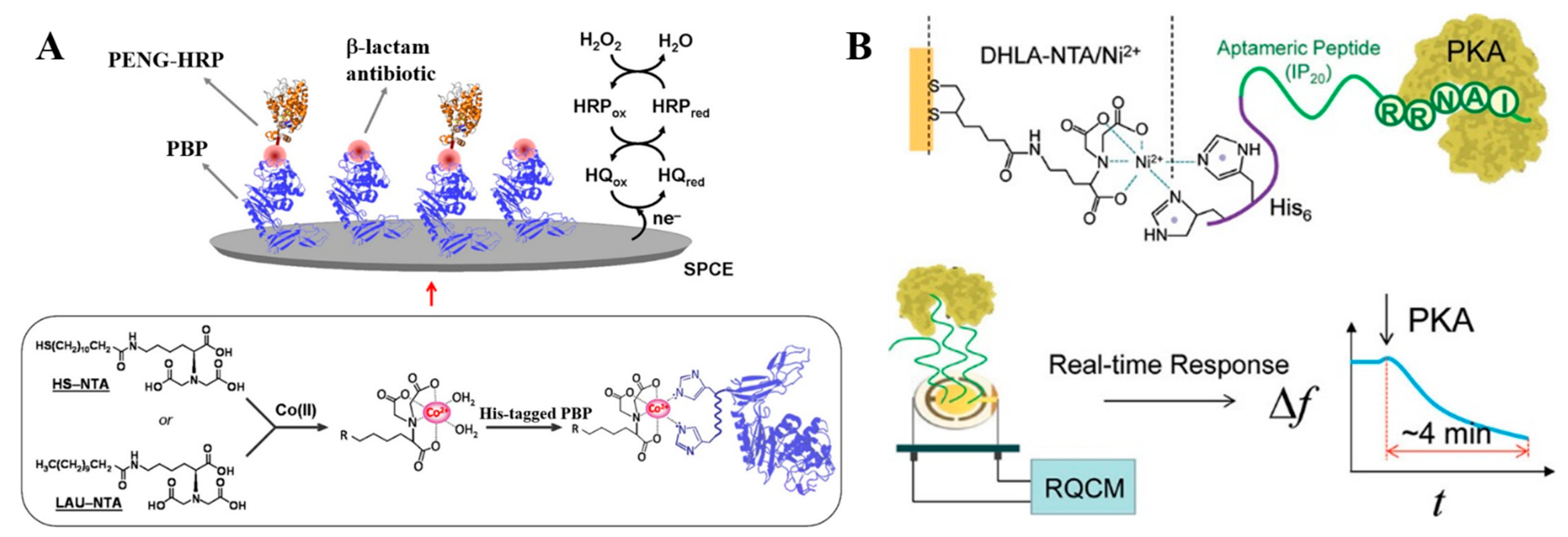
| NTA-modified SPCEs | His6-tagged PBP | Co2+ | Ampicillin | 1.3~9.9 ng/mL | 0.7 ng/mL | [133] | |
| NTA-modified Au-coated quartz electrode | His6-tagged peptide | Ni2+ | PKA | 0.64~22.33 mU/μL | 0.061 mU/μL | [134] | |
| Polypyrrole-NTA-modified electrode | NH2–His5-DNA | Cu2+ | HIV DNA | 1 × 10⁻⁶~10 nM | 1 fM | [135] | |
| Polypyrrole-NTA-modified electrode | His5-modified aptamer | Cu2+ | Thrombin | 4.7 × 10⁻³~0.5 nM | 4.4 pM | [136] | |
| Polypyrrole-NTA-modified electrode | His5-modified aptamer | Cu2+ | Bisphenol A | 1 × 10⁻⁵~1μM | 10 pM | [137] | |
| NTA-modified gold electrode | His6-tagged Ara h 2 | Ni2+ | Ara h 2 antibody | 1~10 pM | 1 pM | [138] | |
| Polypyrrole /NTA-modified electrode | Biotinylated CT B Subunit | Cu2+ | Anti-CT | 1 × 10⁻⁷~10 μg/mL | 0.1 pg/mL | [141] | |
| NTA-modified gold electrode | NTA | Cu2+ | 0.4~100 μM | 10 nM | [144] | ||
| NTA-modified thin-film transistor | NTA | Cu2+ | 0~15 μM | 0.51 μM | [145] | ||
| FL | Zr–NTA-modified MNPs | EGFP | Ni2+ | thrombin | 3 × 10⁻⁴~5 × 10⁻² U/mL | 0.3 mU/mL | [151] |
| Zr–NTA-modified MNPs | FITC-labeled peptide | Zr4+ | PKA | 0~1 U/μL | 0.5 mU/μL | [152] | |
| Color | Carboxy AuNPs | Dual His6-tagged peptide | Ni2+ | MMP-7 | 3~52 nM | 10 nM | [184] |
| NTA-modified chip | His6-tagged peptide | Ni2+ | MMP-7 | 0.1~100 ng/mL | 97 pg/mL | [188] | |
| SERS | NTA-modified AgNPs | NTA–Fe3+ | Fe3+ | Dopamine | 0.5~4 nM | 60 pM | [189] |
| NTA-modified AuNPs | NTA–Fe3+ | Fe3+ | Dopamine, norepinephrine and epinephrine | 0.556~10 μM; 0.125~10 μM; 0.2~9.09 μM | Notreported | [190] | |
| NTA-modified AgNPs | NTA–Ni2+ | Ni2+ | Histamine | 1~100 μM | 1 μM | [191] | |
| CL | Fe3O4@SiO2-NTA | His6-taggedCan f 1 | Ni2+ | SpecifcIgE | 2.52~10.02 ng/mL | 0.35 ng/mL | [192] |
3. Conclusions
Author Contributions
Funding
Data Availability Statement
Conflicts of Interest
References
- Wong, L.S.; Khan, F.; Micklefield, J. Selective covalent protein immobilization: Strategies and applications. Chem. Rev. 2009, 109, 4025–4053. [Google Scholar] [CrossRef]
- Trilling, A.K.; Beekwilder, J.; Zuilhof, H. Antibody orientation on biosensor surfaces: A minireview. Analyst 2013, 138, 1619–1627. [Google Scholar] [CrossRef] [PubMed]
- Steen Redeker, E.; Ta, D.T.; Cortens, D.; Billen, B.; Guedens, W.; Adriaensens, P. Protein engineering for directed immobilization. Bioconjug. Chem. 2013, 24, 1761–1777. [Google Scholar] [CrossRef]
- Samanta, D.; Sarkar, A. Immobilization of bio-macromolecules on self-assembled monolayers: Methods and sensor applications. Chem. Soc. Rev. 2011, 40, 2567–2592. [Google Scholar] [CrossRef]
- Li, Y.; Zhang, J.W.; Huang, X.R.; Wang, T.H. Construction and direct electrochemistry of orientation controlled laccase electrode. Biochem. Biophys. Res. Commun. 2014, 446, 201–205. [Google Scholar] [CrossRef]
- Kim, E.S.; Shim, C.K.; Lee, J.W.; Park, J.W.; Choi, K.Y. Synergistic effect of orientation and lateral spacing of protein g on an on-chip immunoassay. Analyst 2012, 137, 2421–2430. [Google Scholar] [CrossRef] [PubMed]
- Karyakin, A.A.; Presnova, G.V.; Rubtsova, M.Y.; Egorov, A.M. Oriented immobilization of antibodies onto the gold surfaces via their native thiol groups. Anal. Chem. 2000, 72, 3805–3811. [Google Scholar] [CrossRef]
- Della Ventura, B.; Schiavo, L.; Altucci, C.; Esposito, R.; Velotta, R. Light assisted antibody immobilization for bio-sensing. Biomed. Opt. Express. 2011, 2, 3223–3231. [Google Scholar] [CrossRef] [PubMed]
- Song, S.; Li, N.; Bai, L.; Gai, P.; Li, F. Photo-assisted robust anti-interference self-powered biosensing of MicroRNA based on Pt-s bonds and the inorganic-organic hybridization strategy. Anal. Chem. 2022, 94, 1654–1660. [Google Scholar] [CrossRef]
- Guesdon, J.L.; Ternynck, T.; Avrameas, S. The use of avidin-biotin interaction in immunoenzymatic techniques. J. Histochem. Cytochem. 2017, 27, 1131–1139. [Google Scholar] [CrossRef]
- Yu, C.C.; Kuo, Y.Y.; Liang, C.F.; Chien, W.T.; Wu, H.T.; Chang, T.C.; Jan, F.D.; Lin, C.C. Site-specific immobilization of enzymes on magnetic nanoparticles and their use in organic synthesis. Bioconjug. Chem. 2012, 23, 714–724. [Google Scholar] [CrossRef]
- Holland-Nell, K.; Beck-Sickinger, A.G. Specifically immobilised aldo/keto reductase akr1a1 shows a dramatic increase in activity relative to the randomly immobilised enzyme. Chembiochem 2007, 8, 1071–1076. [Google Scholar] [CrossRef]
- Cheung, R.C.; Wong, J.H.; Ng, T.B. Immobilized metal ion affinity chromatography: A review on its applications. Appl. Microbiol. Biotechnol. 2012, 96, 1411–1420. [Google Scholar] [CrossRef] [PubMed]
- Arnold, F.H. Metal-affinity separations: A new dimension in protein processing. Nat. Biotechnol. 1991, 9, 151–156. [Google Scholar] [CrossRef] [PubMed]
- Block, H.; Maertens, B.; Spriestersbach, A.; Brinker, N.; Kubicek, J.; Fabis, R.; Labahn, J.; Schāfer, F. Immobilized-metal affinity chromatography (imac): A review. Methods Enzymol. 2009, 463, 439–473. [Google Scholar]
- Gaberc-Porekar, V.; Menart, V. Perspectives of immobilized-metal affinity chromatography. J. Biochem. Biophys. Methods 2001, 49, 335–360. [Google Scholar] [CrossRef] [PubMed]
- Xu, J.J.; Ambrosini, S.; Tamahkar, E.; Rossi, C.; Haupt, K.; Tse Sum Bui, B. Toward a universal method for preparing molecularly imprinted polymer nanoparticles with antibody-like affinity for proteins. Biomacromolecules 2016, 17, 345–353. [Google Scholar] [CrossRef]
- Hochuli, E.; Dobeli, H.; Schacher, A. New metal chelate adsorbent selective for proteins and peptides containing neighbouring histidine residues. J. Chromatogr. 1987, 411, 177–184. [Google Scholar] [CrossRef]
- Ruan, M.; Nicolas, I.; Baudy-Floèh, M. New building blocks or dendritic pseudopeptides for metal chelating. Springerplus 2016, 5, 55–61. [Google Scholar] [CrossRef]
- Hsieh, Y.L.; Chen, C.W.; Lin, W.H.; Li, B.R. Construction of the nickel oxide nanocoral structure on microscope slides for total self-assembly-oriented probe immobilization and signal enhancement. ACS Appl. Bio Mater. 2020, 3, 3304–3312. [Google Scholar] [CrossRef]
- Hall, E.A.H.; Chen, S.; Chun, J.; Du, Y.; Zhao, Z.Y. A molecular biology approach to protein coupling at a biosensor interface. TrAC-Trend. Anal. Chem. 2016, 79, 247–256. [Google Scholar] [CrossRef]
- Zeng, X.Q.; Shen, Z.H.; Mernaugh, R. Recombinant antibodies and their use in biosensors. Anal. Bioanal. Chem. 2012, 402, 3027–3038. [Google Scholar] [CrossRef] [PubMed]
- Bellare, M.; Kadambar, V.K.; Bollella, P.; Gamella, M.; Katz, E.; Melman, A. Electrochemical signal-triggered release of biomolecules functionalized with His-tag units. Electroanalysis 2019, 31, 2274–2282. [Google Scholar] [CrossRef]
- Wang, W.; Wang, D.I.; Li, Z. Facile fabrication of recyclable and active nanobiocatalyst: Purification and immobilization of enzyme in one pot with Ni-NTA functionalized magnetic nanoparticle. Chem. Commun. 2011, 47, 8115–8117. [Google Scholar] [CrossRef]
- Bauer, W.S.; Kimmel, D.W.; Adams, N.M.; Gibson, L.E.; Scherr, T.F.; Richardson, K.A.; Conrad, J.A.; Matakala, H.K.; Haselton, F.R.; Wright, D.W. Magnetically-enabled biomarker extraction and delivery system: Towards integrated ASSURED diagnostic tools. Analyst 2017, 142, 1569–1580. [Google Scholar] [CrossRef] [PubMed]
- Rowinska-Zyrek, M.; Witkowska, D.; Potocki, S.; Remelli, M.; Kozlowski, H. His-rich sequences—Is plagiarism from nature a good idea? New J. Chem. 2013, 37, 58–70. [Google Scholar] [CrossRef]
- Jones, A.L.; Hulett, M.D.; Parish, C.R. Histidine-rich glycoprotein: A novel adaptor protein in plasma that modulates the immune, vascular and coagulation systems. Immunol. Cell Biol. 2005, 83, 106–118. [Google Scholar] [CrossRef]
- Sahal, D.; Kannan, R.; Sinha, A.; Babbarwal, V.; Gnana Prakash, B.; Singh, G.; Chauhan, V.S. Specific and instantaneous one-step chemodetection of histidine-rich proteins by pauly’s stain. Anal. Biochem. 2002, 308, 405–408. [Google Scholar] [CrossRef]
- Davis, K.M.; Gibson, L.E.; Haselton, F.R.; Wright, D.W. Simple sample processing enhances malaria rapid diagnostic test performance. Analyst 2014, 139, 3026–3031. [Google Scholar] [CrossRef]
- Ricks, K.M.; Adams, N.M.; Scherr, T.F.; Haselton, F.R.; Wright, D.W. Direct transfer of hrpii-magnetic bead complexes to malaria rapid diagnostic tests significantly improves test sensitivity. Malar. J. 2016, 15, 399–406. [Google Scholar] [CrossRef]
- Kantor, A.G.; Markwalter, C.F.; Nourani, A.; Wright, D.W. An antibody-free dual-biomarker rapid enrichment workflow (andrew) improves the sensitivity of malaria rapid diagnostic tests. Anal. Biochem. 2021, 612, 114020–114026. [Google Scholar] [CrossRef] [PubMed]
- Liu, T.H.; Huang, Y.T.; Cheng, H.W.; Chen, Y.W.; Lee, C.H.; Hsu, Y.D.; Pan, R.L.; Tseng, F.G. Single molecule take-and-place technique for positioning a membrane protein on a lipid bilayer. J. Phys. Chem. C 2015, 119, 21184–21190. [Google Scholar] [CrossRef]
- Kitai, T.; Watanabe, Y.; Toyoshima, Y.Y.; Kobayashi, T.; Murayama, T.; Sakaue, H.; Suzuki, H.; Takahagi, T. Simple method of synthesizing nickel–nitrilotriacetic acid gold nanoparticles with a narrow size distribution for protein labeling. Jpn. J. Appl. Phys. 2011, 50, 095002–095006. [Google Scholar] [CrossRef]
- Schmid, E.L.; Keller, T.A.; Dienes, Z.; Vogel, H. Reversible oriented surface immobilization of functional proteins on oxide surfaces. Anal. Chem. 1997, 69, 1979–1985. [Google Scholar] [CrossRef]
- Wasserberg, D.; Cabanas-Danes, J.; Prangsma, J.; O’Mahony, S.; Cazade, P.A.; Tromp, E.; Blum, C.; Thompson, D.; Huskens, J.; Subramaniam, V.; et al. Controlling protein surface orientation by strategic placement of oligo-histidine tags. ACS Nano 2017, 11, 9068–9083. [Google Scholar] [CrossRef]
- Hainfeld, J.F.; Liu, W.; Halsey, C.M.; Freimuth, P.; Powell, R.D. Ni-NTA-gold clusters target His-tagged proteins. J. Struct. Biol. 1999, 127, 185–198. [Google Scholar] [CrossRef] [PubMed]
- Yang, J.B.; Ni, K.F.; Wei, D.Z.; Ren, Y.H. One-step purification and immobilization of his-tagged protein via ni2+-functionalized Fe3O4@polydopamine magnetic nanoparticles. Biotechnol. Bioproc. Eng. 2015, 20, 901–907. [Google Scholar] [CrossRef]
- Johnson, D.L.; Martin, L.L. Controlling protein orientation at interfaces using histidine tags: An alternative to Ni/NTA. J. Am. Chem. Soc. 2005, 127, 2018–2019. [Google Scholar] [CrossRef]
- Ravikumar, R.; Chen, L.H.; Jayaraman, P.; Poh, C.L.; Chan, C.C. Chitosan-nickel film based interferometric optical fiber sensor for label-free detection of histidine tagged proteins. Biosens. Bioelectron. 2018, 99, 578–585. [Google Scholar] [CrossRef]
- Schmitt, L.; Dietrich, C.; Tampé, R. Synthesis and characterization of chelator-lipids for reversible immobilization of engineered proteins at self-assembled lipid interfaces. J. Am. Chem. Soc. 2002, 116, 8485–8491. [Google Scholar] [CrossRef]
- You, C.J.; Bhagawati, M.; Brecht, A.; Piehler, J. Affinity capturing for targeting proteins into micro and nanostructures. Anal. Bioanal. Chem. 2009, 393, 1563–1570. [Google Scholar] [CrossRef] [PubMed]
- Markwalter, C.F.; Kantor, A.G.; Moore, C.P.; Richardson, K.A.; Wright, D.W. Inorganic complexes and metal-based nanomaterials for infectious disease diagnostics. Chem. Rev. 2019, 119, 1456–1518. [Google Scholar] [CrossRef]
- Soler, M.; Lechuga, L.M. Biochemistry strategies for label-free optical sensor biofunctionalization: Advances towards real applicability. Anal. Bioanal. Chem. 2022, 414, 5071–5085. [Google Scholar] [CrossRef]
- Mu, B.; Zhang, J.Q.; McNicholas, T.P.; Reuel, N.F.; Kruss, S.; Strano, M.S. Recent advances in molecular recognition based on nanoengineered platforms. Acc. Chem. Res. 2014, 47, 979–988. [Google Scholar] [CrossRef]
- Wieneke, R.; Tampe, R. Multivalent chelators for in vivo protein labeling. Angew. Chem. Int. Ed. 2019, 58, 8278–8290. [Google Scholar] [CrossRef] [PubMed]
- You, C.J.; Piehler, J. Multivalent chelators for spatially and temporally controlled protein functionalization. Anal. Bioanal. Chem. 2014, 406, 3345–3357. [Google Scholar] [CrossRef]
- López-Laguna, H.; Voltà-Durán, E.; Parladé, E.; Villaverde, A.; Vázquez, E.; Unzueta, U. Insights on the emerging biotechnology of histidine-rich peptides. Biotechnol. Adv. 2022, 54, 107817–107831. [Google Scholar] [CrossRef]
- Bauer, W.S.; Richardson, K.A.; Adams, N.M.; Ricks, K.M.; Gasperino, D.J.; Ghionea, S.J.; Rosen, M.; Nichols, K.P.; Weigl, B.H.; Haselton, F.R.; et al. Rapid concentration and elution of malarial antigen histidine-rich protein II using solid phase Zn(II) resin in a simple flow-through pipette tip format. Biomicrofluidics 2017, 11, 034115–034130. [Google Scholar] [CrossRef] [PubMed]
- Hochuli, E.; Bannwarth, W.; Döbeli, H.; Gentz, R.; Stüber, D. Genetic approach to facilitate purification of recombinant proteins with a novel metal chelate adsorbent. Nat. Biotechnol. 1988, 6, 1321–1325. [Google Scholar] [CrossRef]
- Suh, J.K.; Poulsen, L.L.; Ziegler, D.M.; Robertus, J.D. Molecular cloning and kinetic characterization of a flavin-containing monooxygenase from saccharomyces cerevisiae. Arch. Biochem. Biophys. 1996, 336, 268–274. [Google Scholar] [CrossRef]
- Giusti, F.; Kessler, P.; Hansen, R.W.; Della Pia, E.A.; Le Bon, C.; Mourier, G.; Popot, J.L.; Martinez, K.L.; Zoonens, M. Synthesis of a polyhistidine-bearing amphipol and its use for immobilizing membrane proteins. Biomacromolecules 2015, 16, 3751–3761. [Google Scholar] [CrossRef]
- Knecht, S.; Ricklin, D.; Eberle, A.N.; Ernst, B. Oligohis-tags: Mechanisms of binding to Ni2+-NTA surfaces. J. Mol. Recognit. 2009, 22, 270–279. [Google Scholar] [CrossRef] [PubMed]
- Madoz-Gúrpide, J.; Abad, J.M.; Fernández-Recio, J.; Vélez, M.; Vázquez, L.; Gómez-Moreno, C.; Fernández, V.M. Modulation of electroenzymatic nadph oxidation through oriented immobilization of ferredoxin: NANP+ reductase onto modified gold electrodes. J. Am. Chem. Soc. 2000, 122, 9808–9817. [Google Scholar] [CrossRef]
- Schroper, F.; Baumann, A.; Offenhausser, A.; Mayer, D. Direct electrochemistry of novel affinity-tag immobilized recombinant horse heart cytochrome c. Biosens. Bioelectron. 2012, 34, 171–177. [Google Scholar] [CrossRef]
- Khan, F.; He, M.Y.; Taussig, M.J. Double-hexahistidine tag with high-affinity binding for protein immobilization, purification, and detection on ni-nitrilotriacetic acid surfaces. Anal. Chem. 2006, 78, 3072–3079. [Google Scholar] [CrossRef] [PubMed]
- Kapanidis, A.N.; Ebright, Y.W.; Ebright, R.H. Site-specific incorporation of fluorescent probes into protein: Hexahistidine-tag-mediated fluorescent labeling with (Ni2+:nitrilotriacetic acid)n-fluorochrome conjugates. J. Am. Chem. Soc. 2001, 123, 12123–12125. [Google Scholar] [CrossRef]
- Gershon, P.D.; Khilko, S. Stable chelating linkage for reversible immobilization of oligohistidine tagged proteins in the biacore surface plasmon resonance detector. J. Immunol. Methods 1995, 183, 65–76. [Google Scholar] [CrossRef]
- Le, T.T.; Wilde, C.P.; Grossman, N.; Cass, A.E. A simple method for controlled immobilization of proteins on modified SAMs. Phys. Chem. Chem. Phys. 2011, 13, 5271–5278. [Google Scholar] [CrossRef]
- Tinazli, A.; Tang, J.; Valiokas, R.; Picuric, S.; Lata, S.; Piehler, J.; Liedberg, B.; Tampe, R. High-affinity chelator thiols for switchable and oriented immobilization of histidine-tagged proteins: A generic platform for protein chip technologies. Chem. Eur. J. 2005, 11, 5249–5259. [Google Scholar] [CrossRef]
- Lata, S.; Piehler, J. Stable and functional immobilization of histidine-tagged proteins via multivalent chelator headgroups on a molecular poly(ethylene glycol) brush. Anal. Chem. 2005, 77, 1096–1105. [Google Scholar] [CrossRef]
- Knezevic, J.; Langer, A.; Hampel, P.A.; Kaiser, W.; Strasser, R.; Rant, U. Quantitation of affinity, avidity, and binding kinetics of protein analytes with a dynamically switchable biosurface. J. Am. Chem. Soc. 2012, 134, 15225–15228. [Google Scholar] [CrossRef] [PubMed]
- Huang, Z.H.; Hwang, P.; Watson, D.S.; Cao, L.M.; Szoka, F.C., Jr. Tris-nitrilotriacetic acids of subnanomolar affinity toward hexahistidine tagged molecules. Bioconjug. Chem. 2009, 20, 1667–1672. [Google Scholar] [CrossRef] [PubMed]
- Huang, Z.H.; Park, J.I.; Watson, D.S.; Hwang, P.; Szoka, F.C., Jr. Facile synthesis of multivalent nitrilotriacetic acid (NTA) and NTA conjugates for analytical and drug delivery applications. Bioconjug. Chem. 2006, 17, 1592–1600. [Google Scholar] [CrossRef] [PubMed]
- Lata, S.; Reichel, A.; Brock, R.; Tampe, R.; Piehler, J. High-affinity adaptors for switchable recognition of histidine-tagged proteins. J. Am. Chem. Soc. 2005, 127, 10205–10215. [Google Scholar] [CrossRef] [PubMed]
- Meredith, G.D.; Wu, H.Y.; Allbritton, N.L. Targeted protein functionalization using His-tags. Bioconjug. Chem. 2004, 15, 969–982. [Google Scholar] [CrossRef]
- Hintersteiner, M.; Weidemann, T.; Kimmerlin, T.; Filiz, N.; Buehler, C.; Auer, M. Covalent fluorescence labeling of His-tagged proteins on the surface of living cells. Chembiochem 2008, 9, 1391–1395. [Google Scholar] [CrossRef]
- Willard, F.S.; Siderovski, D.P. Covalent immobilization of histidine-tagged proteins for surface plasmon resonance. Anal. Biochem. 2006, 353, 147–149. [Google Scholar] [CrossRef]
- Chevalier, S.; Cuestas-Ayllon, C.; Grazu, V.; Luna, M.; Feracci, H.; de la Fuente, J.M. Creating biomimetic surfaces through covalent and oriented binding of proteins. Langmuir 2010, 26, 14707–14715. [Google Scholar] [CrossRef]
- Mateo, C.; Fernández-Lorente, G.; Cortés, E.; Garcia, J.L.; Fernández-Lafuente, R.; Guisan, J.M. One-step purification, covalent immobilization, and additional stabilization of poly-His-tagged proteins using novel heterofunctional chelate-epoxy supports. Biotechnol. Bioeng. 2001, 76, 269–276. [Google Scholar] [CrossRef]
- Qu, J.H.; Horta, S.; Delport, F.; Sillen, M.; Geukens, N.; Sun, D.W.; Vanhoorelbeke, K.; Declerck, P.; Lammertyn, J.; Spasic, D. Expanding a portfolio of (FO-) SPR surface chemistries with the Co(III)-NTA oriented immobilization of His6-tagged bioreceptors for applications in complex matrices. ACS Sens. 2020, 5, 960–969. [Google Scholar] [CrossRef]
- Mehlenbacher, M.R.; Bou-Abdallah, F.; Liu, X.X.; Melman, A. Calorimetric studies of ternary complexes of Ni(II) and Cu(II) nitrilotriacetic acid and N-acetyloligohistidines. Inorg. Chim. Acta 2015, 437, 152–158. [Google Scholar] [CrossRef]
- Li, X.M.; Song, S.Y.; Pei, Y.X.; Dong, H.; Aastrup, T.; Pei, Z.C. Oriented and reversible immobilization of His-tagged proteins on two- and three-dimensional surfaces for study of protein–protein interactions by a qcm biosensor. Sens. Actuat. B Chem. 2016, 224, 814–822. [Google Scholar] [CrossRef]
- Clow, F.; Fraser, J.D.; Proft, T. Immobilization of proteins to biacore sensor chips using Staphylococcus aureus sortase A. Biotechnol. Lett. 2008, 30, 1603–1607. [Google Scholar] [CrossRef]
- Pellis, A.; Vastano, M.; Quartinello, F.; Herrero Acero, E.; Guebitz, G.M. His-tag immobilization of cutinase 1 from thermobifida cellulosilytica for solvent-free synthesis of polyesters. Biotechnol. J. 2017, 12, 1700322–1700327. [Google Scholar] [CrossRef] [PubMed]
- Wegner, S.V.; Spatz, J.P. Cobalt(III) as a stable and inert mediator ion between NTA and his6-tagged proteins. Angew. Chem. Int. Ed. 2013, 52, 7593–7596. [Google Scholar] [CrossRef]
- Wegner, S.V.; Schenk, F.C.; Spatz, J.P. Cobalt(III)-mediated permanent and stable immobilization of histidine-tagged proteins on NTA-functionalized surfaces. Chem. Eur. J. 2016, 22, 3156–3162. [Google Scholar] [CrossRef]
- Tang, Y.J.; Mernaugh, R.; Zeng, X.Q. Nonregeneration protocol for surface plasmon resonance: Study of high-affinity interaction with high-density biosensors. Anal. Chem. 2006, 78, 1841–1848. [Google Scholar] [CrossRef]
- Lori, J.A.; Morrin, A.; Killard, A.J.; Smyth, M.R. Development and characterization of nickel-NTA-polyaniline modified electrodes. Electroanalysis 2006, 18, 77–81. [Google Scholar] [CrossRef]
- Kang, E.; Park, J.W.; McClellan, S.J.; Kim, J.M.; Holland, D.P.; Lee, G.U.; Franses, E.I.; Park, K.; Thompson, D.H. Specific adsorption of histidine-tagged proteins on silica surfaces modified with Ni2+/NTA-derivatized poly(ethylene glycol). Langmuir 2007, 23, 6281–6288. [Google Scholar] [CrossRef]
- Ley, C.; Holtmann, D.; Mangold, K.M.; Schrader, J. Immobilization of histidine-tagged proteins on electrodes. Colloids Surf. B 2011, 88, 539–551. [Google Scholar] [CrossRef]
- Tinazli, A.; Piehler, J.; Beuttler, M.; Guckenberger, R.; Tampé, R. Native protein nanolithography that can write, read and erase. Nat. Nanotechnol. 2007, 2, 220–225. [Google Scholar] [CrossRef] [PubMed]
- Zborowska, M.; Sulima, M.; Marszałek, I.; Wysłouch-Cieszyńska, A.; Radecka, H.; Radecki, J. Nitrilotriacetic acid–copper(II) monolayer deposited on a gold electrode for the immobilization of histidine tagged v domain of receptor for advanced glycation end products–the basis of amyloid–beta peptide sensing. Anal. Lett. 2014, 47, 1375–1391. [Google Scholar] [CrossRef]
- Cheng, F.; Gamble, L.J.; Castner, D.G. XPS, tof-sims, NEXAFS, and SPR characterization of nitrilotriacetic acid-terminated self-assembled monolayers for controllable immobilization of proteins. Anal. Chem. 2008, 80, 2564–2573. [Google Scholar] [CrossRef] [PubMed]
- Cho, M.; Chun, L.; Lin, M.; Choe, W.; Nam, J.; Lee, Y. Sensitive electrochemical sensor for detection of lipopolysaccharide on metal complex immobilized gold electrode. Sens. Actuat. B Chem. 2012, 174, 490–494. [Google Scholar] [CrossRef]
- Tran, Q.T.; de Sanoit, J.; Pierre, S.; Arnault, J.-C.; Bergonzo, P. Diamond electrodes for trace alpha pollutant sequestration via covalent grafting of nitrilotriacetic acid (NTA) ligand. Electrochim. Acta 2014, 136, 430–434. [Google Scholar] [CrossRef]
- Haddour, N.; Cosnier, S.; Gondran, C. Electrogeneration of a poly(pyrrole)-NTA chelator film for a reversible oriented immobilization of histidine-tagged proteins. J. Am. Chem. Soc. 2005, 127, 5752–5753. [Google Scholar] [CrossRef] [PubMed]
- Holzinger, M.; Baur, J.; Haddad, R.; Wang, X.; Cosnier, S. Multiple functionalization of single-walled carbon nanotubes by dip coating. Chem. Commun. 2011, 47, 2450–2452. [Google Scholar] [CrossRef]
- Singh, M.; Holzinger, M.; Biloivan, O.; Cosnier, S. 3D-nanostructured scaffold electrodes based on single-walled carbon nanotubes and nanodiamonds for high performance biosensors. Carbon 2013, 61, 349–356. [Google Scholar] [CrossRef]
- Osella, S.; Kiliszek, M.; Harputlu, E.; Unlu, C.G.; Ocakoglu, K.; Kargul, J.; Trzaskowski, B. Controlling the charge transfer flow at the graphene/pyrene–nitrilotriacetic acid interface. J. Mater. Chem. C 2018, 6, 5046–5054. [Google Scholar] [CrossRef]
- Jorde, L.; Li, Z.H.; Pöppelwerth, A.; Piehler, J.; You, C.J.; Meyer, C. Biofunctionalization of carbon nanotubes for reversible site-specific protein immobilization. J. Appl. Phys. 2021, 129, 094302–094311. [Google Scholar] [CrossRef]
- Mauriz, E.; García-Fernández, M.C.; Lechuga, L.M. Towards the design of universal immunosurfaces for SPR-based assays: A review. TrAC-Trend. Anal. Chem. 2016, 79, 191–198. [Google Scholar] [CrossRef]
- Pfeifer, P.; Aldinger, U.; Schwotzer, G.; Diekmann, S.; Steinrücke, P. Real time sensing of specific molecular binding using surface plasmon resonance spectroscopy. Sens. Actuat. B Chem. 1999, 54, 166–175. [Google Scholar] [CrossRef]
- Boonen, A.; Singh, A.K.; Hout, A.V.; Das, K.; Loy, T.V.; Noppen, S.; Schols, D. Development of a novel SPR assay to study cxcr4-ligand interactions. Biosensors 2020, 10, 150. [Google Scholar] [CrossRef] [PubMed]
- Maalouli, N.; Gouget-Laemmel, A.C.; Pinchemel, B.; Bouazaoui, M.; Chazalviel, J.N.; Ozanam, F.; Yang, Y.K.; Burkhard, P.; Boukherroub, R.; Szunerits, S. Development of a metal-chelated plasmonic interface for the linking of His-peptides with a droplet-based surface plasmon resonance read-off scheme. Langmuir 2011, 27, 5498–5505. [Google Scholar] [CrossRef]
- Wegner, G.J.; Lee, H.J.; Marriott, G.; Corn, R.M. Fabrication of histidine-tagged fusion protein arrays for surface plasmon resonance imaging studies of protein-protein and protein-DNA interactions. Anal. Chem. 2003, 75, 4740–4746. [Google Scholar] [CrossRef]
- Sigal, G.B.; Bamdad, C.; Barberis, A.; Strominger, J.; Whitesides, G.M. A self-assembled monolayer for the binding and study of histidine-tagged proteins by surface plasmon resonance. Anal. Chem. 1996, 68, 490–497. [Google Scholar] [CrossRef]
- Gautrot, J.E.; Huck, W.T.; Welch, M.; Ramstedt, M. Protein-resistant NTA-functionalized polymer brushes for selective and stable immobilization of histidine-tagged proteins. ACS Appl. Mater. Interfaces 2010, 2, 193–202. [Google Scholar] [CrossRef]
- Schartner, J.; Hoeck, N.; Guldenhaupt, J.; Mavarani, L.; Nabers, A.; Gerwert, K.; Kotting, C. Chemical functionalization of germanium with dextran brushes for immobilization of proteins revealed by attenuated total reflection fourier transform infrared difference spectroscopy. Anal. Chem. 2015, 87, 7467–7475. [Google Scholar] [CrossRef]
- Qu, J.H.; Leirs, K.; Escudero, R.; Strmsek, Z.; Jerala, R.; Spasic, D.; Lammertyn, J. Novel regeneration approach for creating reusable FO-SPR probes with NTA surface chemistry. Nanomaterials 2021, 11, 186. [Google Scholar] [CrossRef] [PubMed]
- Yi, X.Y.; Hao, Y.Q.; Xia, N.; Wang, J.X.; Quintero, M.; Li, D.; Zhou, F.M. Sensitive and continuous screening of inhibitors of beta-site amyloid precursor protein cleaving enzyme 1 (BACE1) at single SPR chips. Anal. Chem. 2013, 85, 3660–3666. [Google Scholar] [CrossRef]
- Singh, M.; Holzinger, M.; Tabrizian, M.; Winters, S.; Berner, N.C.; Cosnier, S.; Duesberg, G.S. Noncovalently functionalized monolayer graphene for sensitivity enhancement of surface plasmon resonance immunosensors. J. Am. Chem. Soc. 2015, 137, 2800–2803. [Google Scholar] [CrossRef]
- Yuan, P.X.; Deng, S.Y.; Yao, C.G.; Wan, Y.; Cosnier, S.; Shan, D. Polymerization amplified SPR-DNA assay on noncovalently functionalized graphene. Biosens. Bioelectron. 2017, 89, 319–325. [Google Scholar] [CrossRef] [PubMed]
- Wang, X.Y.; Liu, Q.H.; Tan, X.F.; Liu, L.Y.; Zhou, F.M. Covalent affixation of histidine-tagged proteins tethered onto Ni-nitrilotriacetic acid sensors for enhanced surface plasmon resonance detection of small molecule drugs and kinetic studies of antibody/antigen interactions. Analyst 2019, 144, 587–593. [Google Scholar] [CrossRef] [PubMed]
- Wang, X.Y.; Zhou, F.M. Dual-valve and counter-flow surface plasmon resonance. Anal. Chem. 2018, 90, 4972–4977. [Google Scholar] [CrossRef] [PubMed]
- Nieba, L.; Nieba-Axmann, S.E.; Persson, A.; Hamalainen, M.; Edebratt, F.; Hansson, A.; Lidholm, J.; Magnusson, K.; Karlsson, A.F.; Pluckthun, A. Biacore analysis of histidine-tagged proteins using a chelating NTA sensor chip. Anal. Biochem. 1997, 252, 217–228. [Google Scholar] [CrossRef] [PubMed]
- Fischer, M.; Leech, A.P.; Hubbard, R.E. Comparative assessment of different histidine-tags for immobilization of protein onto surface plasmon resonance sensorchips. Anal. Chem. 2011, 83, 1800–1807. [Google Scholar] [CrossRef]
- Liu, J.; Spulber, M.; Wu, D.; Talom, R.M.; Palivan, C.G.; Meier, W. Poly(N-isopropylacrylamide-co-tris-nitrilotriacetic acid acrylamide) for a combined study of molecular recognition and spatial constraints in protein binding and interactions. J. Am. Chem. Soc. 2014, 136, 12607–12614. [Google Scholar] [CrossRef]
- Tanner, P.; Ezhevskaya, M.; Nehring, R.; Van Doorslaer, S.; Meier, W.; Palivan, C. Specific his6-tag attachment to metal-functionalized polymersomes relies on molecular recognition. J. Phys. Chem. B 2012, 116, 10113–10124. [Google Scholar] [CrossRef]
- Rakickas, T.; Gavutis, M.; Reichel, A.; Piehler, J.; Liedberg, B.; Valiokas, R. Protein-protein interactions in reversibly assembled nanopatterns. Nano Lett. 2008, 8, 3369–3375. [Google Scholar] [CrossRef]
- Bhagawati, M.; You, C.J.; Piehler, J. Quantitative real-time imaging of protein-protein interactions by LSPR detection with micropatterned gold nanoparticles. Anal. Chem. 2013, 85, 9564–9571. [Google Scholar] [CrossRef]
- Jiang, M.; Dong, T.B.; Han, C.W.; Liu, L.Y.; Zhang, T.T.; Kang, Q.; Wang, P.C.; Zhou, F.M. Regenerable and high-throughput surface plasmon resonance assay for rapid screening of anti-SARS-CoV-2 antibody in serum samples. Anal. Chim. Acta. 2022, 1208, 339830–339838. [Google Scholar] [CrossRef] [PubMed]
- Reichel, A.; Schaible, D.; Al Furoukh, N.; Cohen, M.; Schreiber, G.; Piehler, J. Noncovalent, site-specific biotinylation of histidine-tagged proteins. Anal. Chem. 2007, 79, 8590–8600. [Google Scholar] [CrossRef] [PubMed]
- Liu, L.Y.; Han, C.W.; Jiang, M.; Zhang, T.T.; Kang, Q.; Wang, X.Y.; Wang, P.C.; Zhou, F.M. Rapid and regenerable surface plasmon resonance determinations of biomarker concentration and biomolecular interaction based on tris-nitrilotriacetic acid chips. Anal. Chim. Acta. 2021, 1170, 338625–338633. [Google Scholar] [CrossRef] [PubMed]
- Pires, M.M.; Chmielewski, J. Self-assembly of collagen peptides into microflorettes via metal coordination. J. Am. Chem. Soc. 2009, 131, 2706–2712. [Google Scholar] [CrossRef] [PubMed]
- Xu, L.; Cao, H.Y.; Huang, C.D.; Jia, L.Y. Oriented immobilization and quantitative analysis simultaneously realized in sandwich immunoassay via His-tagged nanobody. Molecules 2019, 24, 1890. [Google Scholar] [CrossRef]
- Horta, S.; Qu, J.H.; Dekimpe, C.; Bonnez, Q.; Vandenbulcke, A.; Tellier, E.; Kaplanski, G.; Delport, F.; Geukens, N.; Lammertyn, J.; et al. Co(III)-NTA mediated antigen immobilization on a fiber optic-SPR biosensor for detection of autoantibodies in autoimmune diseases: Application in immune-mediated thrombotic thrombocytopenic purpura. Anal. Chem. 2020, 92, 13880–13887. [Google Scholar] [CrossRef]
- Auer, S.; Azizi, L.; Faschinger, F.; Blazevic, V.; Vesikari, T.; Gruber, H.J.; Hytönen, V.P. Stable immobilisation of His-tagged proteins on BLI biosensor surface using cobalt. Sens. Actuat. B Chem. 2017, 243, 104–113. [Google Scholar] [CrossRef]
- Qu, J.H.; Leirs, K.; Maes, W.; Imbrechts, M.; Callewaert, N.; Lagrou, K.; Geukens, N.; Lammertyn, J.; Spasic, D. Innovative FO-SPR label-free strategy for detecting anti-rbd antibodies in COVID-19 patient serum and whole blood. ACS Sens. 2022, 7, 477–487. [Google Scholar] [CrossRef]
- Qu, J.H.; Peeters, B.; Delport, F.; Vanhoorelbeke, K.; Lammertyn, J.; Spasic, D. Gold nanoparticle enhanced multiplexed biosensing on a fiber optic surface plasmon resonance probe. Biosens. Bioelectron. 2021, 192, 113549–113557. [Google Scholar] [CrossRef]
- Yang, L.; Yin, X.; An, B.; Li, F. Precise capture and direct quantification of tumor exosomes via a highly efficient dual-aptamer recognition-assisted ratiometric immobilization-free electrochemical strategy. Anal. Chem. 2021, 93, 1709–1716. [Google Scholar] [CrossRef]
- Lu, L.; Su, H.; Li, F. Ultrasensitive homogeneous electrochemical detection of transcription factor by coupled isothermal cleavage reaction and cycling amplification based on Exonuclease III. Anal. Chem. 2017, 89, 8328–8334. [Google Scholar] [CrossRef] [PubMed]
- Ataka, K.; Giess, F.; Knoll, W.; Naumann, R.; Haber-Pohlmeier, S.; Richter, B.; Heberle, J. Oriented attachment and membrane reconstitution of His-tagged cytochrome c oxidase to a gold electrode: In situ monitoring by surface-enhanced infrared absorption spectroscopy. J. Am. Chem. Soc. 2004, 126, 16199–16206. [Google Scholar] [CrossRef]
- Aghamiri, Z.S.; Mohsennia, M.; Rafiee-Pour, H.A. Immobilization of cytochrome c and its application as electrochemical biosensors. Talanta 2018, 176, 195–207. [Google Scholar] [CrossRef] [PubMed]
- Akram, M.S.; Ur Rehman, J.; Hall, E.A. Engineered proteins for bioelectrochemistry. Annu. Rev. Anal. Chem. 2014, 7, 257–274. [Google Scholar] [CrossRef]
- Maly, J.; Di Meo, C.; De Francesco, M.; Masci, A.; Masojidek, J.; Sugiura, M.; Volpe, A.; Pilloton, R. Reversible immobilization of engineered molecules by Ni-NTA chelators. Bioelectrochemistry 2004, 63, 271–275. [Google Scholar] [CrossRef] [PubMed]
- Maly, J.; Ilie, M.; Foglietti, V.; Cianci, E.; Minotti, A.; Nardi, L.; Masci, A.; Vastarella, W.; Pilloton, R. Continuous flow micro-cell for electrochemical addressing of engineered bio-molecules. Sens. Actuat. B Chem. 2005, 111–112, 317–322. [Google Scholar] [CrossRef]
- Balland, V.; Hureau, C.; Cusano, A.M.; Liu, Y.; Tron, T.; Limoges, B. Oriented immobilization of a fully active monolayer of histidine-tagged recombinant laccase on modified gold electrodes. Chem. Eur. J. 2008, 14, 7186–7192. [Google Scholar] [CrossRef]
- Demin, S.; Hall, E.A. Breaking the barrier to fast electron transfer. Bioelectrochemistry 2009, 76, 19–27. [Google Scholar] [CrossRef]
- Campbell, W.H.; Henig, J.; Plumeré, N. Affinity binding via zinc(II) for controlled orientation and electrochemistry of histidine-tagged nitrate reductase in self-assembled monolayers. Bioelectrochemistry 2013, 93, 46–50. [Google Scholar] [CrossRef]
- Vallina-Garcia, R.; del Mar Garcia-Suarez, M.; Fernandez-Abedul, M.T.; Mendez, F.J.; Costa-Garcia, A. Oriented immobilisation of anti-pneumolysin fab through a histidine tag for electrochemical immunosensors. Biosens. Bioelectron. 2007, 23, 210–217. [Google Scholar] [CrossRef]
- Blankespoor, R.; Limoges, B.; Schollhorn, B.; Syssa-Magalé, J.L.; Yazidi, D. Dense monolayers of metal-chelating ligands covalently attached to carbon electrodes electrochemically and their useful application in affinity binding of histidine-tagged proteins. Langmuir 2005, 21, 3362–3375. [Google Scholar] [CrossRef]
- Wang, Z.; Liu, D.; Gu, H.; Zhu, A.W.; Tian, Y.; Shi, G.Y. NTA-modified carbon electrode as a general relaying substrate to facilitate electron transfer of SOD: Application to in vivo monitoring of O2- in a rat brain. Biosens. Bioelectron. 2013, 43, 101–107. [Google Scholar] [CrossRef]
- Conzuelo, F.; Gamella, M.; Campuzano, S.; Martinez-Ruiz, P.; Esteban-Torres, M.; de las Rivas, B.; Reviejo, A.J.; Munoz, R.; Pingarron, J.M. Integrated amperometric affinity biosensors using Co2+-tetradentate nitrilotriacetic acid modified disposable carbon electrodes: Application to the determination of beta-lactam antibiotics. Anal. Chem. 2013, 85, 3246–3254. [Google Scholar] [CrossRef] [PubMed]
- Xu, X.H.; Zhou, J.; Liu, X.; Nie, Z.; Qing, M.; Guo, M.L.; Yao, S.Z. Aptameric peptide for one-step detection of protein kinase. Anal. Chem. 2012, 84, 4746–4753. [Google Scholar] [CrossRef] [PubMed]
- Baur, J.; Gondran, C.; Holzinger, M.; Defrancq, E.; Perrot, H.; Cosnier, S. Label-free femtomolar detection of target DNA by impedimetric DNA sensor based on poly(pyrrole-nitrilotriacetic acid) film. Anal. Chem. 2010, 82, 1066–1072. [Google Scholar] [CrossRef] [PubMed]
- Xu, H.; Gorgy, K.; Gondran, C.; Le Goff, A.; Spinelli, N.; Lopez, C.; Defrancq, E.; Cosnier, S. Label-free impedimetric thrombin sensor based on poly(pyrrole-nitrilotriacetic acid)-aptamer film. Biosens. Bioelectron. 2013, 41, 90–95. [Google Scholar] [CrossRef]
- Kazane, I.; Gorgy, K.; Gondran, C.; Spinelli, N.; Zazoua, A.; Defrancq, E.; Cosnier, S. Highly sensitive bisphenol-a electrochemical aptasensor based on poly(pyrrole-nitrilotriacetic acid)-aptamer film. Anal. Chem. 2016, 88, 7268–7273. [Google Scholar] [CrossRef]
- Zaitouna, A.J.; Lai, R.Y. An electrochemical peptide-based Ara h 2 antibody sensor fabricated on a nickel(II)-nitriloacetic acid self-assembled monolayer using a His-tagged peptide. Anal. Chim. Acta. 2014, 828, 85–91. [Google Scholar] [CrossRef]
- Griesser, R.; Sigel, H.; Wright, L.D.; McCormick, D.B. Interactions of metal ions with biotin and biotin derivatives. Complexing and hydrogen-bond formation of the ureido group. Biochemistry 1973, 12, 1917–1922. [Google Scholar] [CrossRef] [PubMed]
- Baur, J.; Holzinger, M.; Gondran, C.; Cosnier, S. Immobilization of biotinylated biomolecules onto electropolymerized poly(pyrrole-nitrilotriacetic acid)–Cu2+ film. Electrochem. Commun. 2010, 12, 1287–1290. [Google Scholar] [CrossRef]
- Palomar, Q.; Gondran, C.; Holzinger, M.; Marks, R.; Cosnier, S. Controlled carbon nanotube layers for impedimetric immunosensors: High performance label free detection and quantification of anti-cholera toxin antibody. Biosens. Bioelectron. 2017, 97, 177–183. [Google Scholar] [CrossRef] [PubMed]
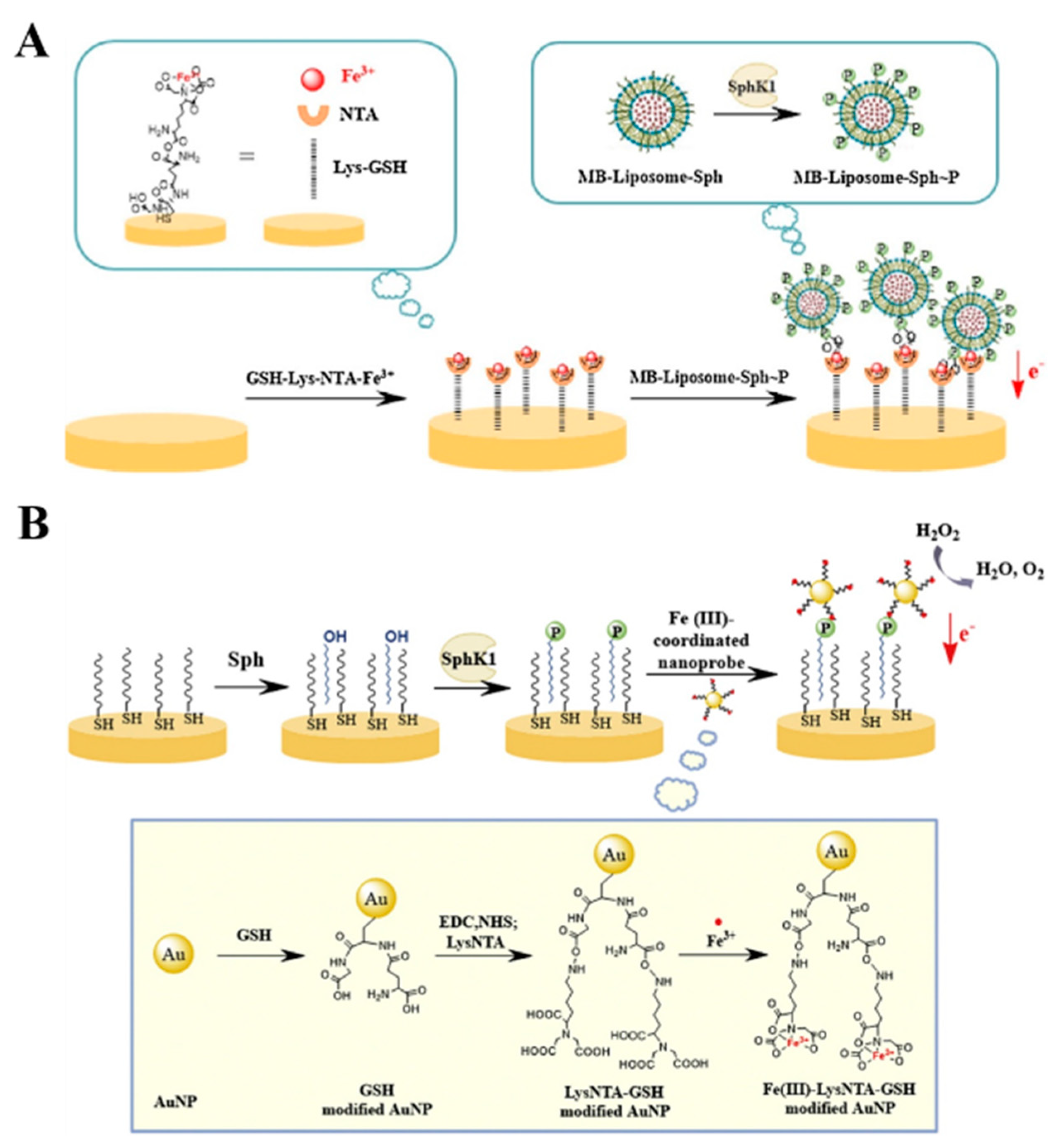
- Gao, T.; Gu, S.; Mu, C.; Zhang, M.; Yang, J.; Liu, P.; Li, G. Electrochemical assay of lipid kinase activity facilitated by liposomes. Electrochim. Acta 2017, 252, 362–367. [Google Scholar] [CrossRef]
- Gu, S.; Gao, T.; Yang, Y.; Zhi, J.; Li, J.; Xiang, Y.; Wang, K.; Yang, J. A bifunctional Fe(III)-coordinated nanoprobe for electrochemical detection of sphingosine kinase 1 activity. Electrochem. Commun. 2016, 72, 104–108. [Google Scholar] [CrossRef]
- Kerekovic, I.; Milardovic, S.; Palcic, M.; Grabaric, Z. Characterization of cysteamine self assembled on gold functionalized with nitrilotriacetic acid and evaluation of copper(II) binding capacity with adsorption transfer stripping voltammetry. J. Electroanal. Chem. 2014, 724, 103–110. [Google Scholar] [CrossRef]
- Sasaki, Y.; Minami, T.; Minamiki, T.; Tokito, S. An organic transistor-based electrical assay for copper(ii) in water. Electrochemistry 2017, 85, 775–778. [Google Scholar] [CrossRef]
- Yang, W.C.; Swartz, J.R. A filter microplate assay for quantitative analysis of DNA binding proteins using fluorescent DNA. Anal. Biochem. 2011, 415, 168–174. [Google Scholar] [CrossRef]
- Kim, S.H.; Ge, P.; Katzenellenbogen, J.A. A new quinoline sensitizer-centered lanthanide chelate and its use for protein labling on Ni-NTA beads for TR LRET assays. Chem. Commun. 2009, 45, 183–185. [Google Scholar] [CrossRef]
- Li, N.; Yi, L.; He, Z.; Zhang, W.; Li, H.; Lin, J.M. A DNA-directed covalent conjugation fluorescence probe for in vitro detection of functional matrix metalloproteinases. Analyst 2017, 142, 634–640. [Google Scholar] [CrossRef]
- Chao, A.L.; Jiang, N.; Yang, Y.; Li, H.Y.; Sun, H.Z. A Ni-NTA-based red fluorescence probe for protein labelling in live cells. J. Mater. Chem. B 2017, 5, 1166–1173. [Google Scholar] [CrossRef]
- Kim, S.H.; Jeyakumar, M.; Katzenellenbogen, J.A. Dual-mode fluorophore-doped nickel nitrilotriacetic acid-modified silica nanoparticles combine histidine-tagged protein purification with site-specific fluorophore labeling. J. Am. Chem. Soc. 2007, 129, 13254–13264. [Google Scholar] [CrossRef]
- Wang, M.; Lei, C.Y.; Nie, Z.; Guo, M.L.; Huang, Y.; Yao, S.Z. Label-free fluorescent detection of thrombin activity based on a recombinant enhanced green fluorescence protein and nickel ions immobilized nitrilotriacetic acid-coated magnetic nanoparticles. Talanta 2013, 116, 468–473. [Google Scholar] [CrossRef]
- Tan, P.L.; Lei, C.Y.; Liu, X.; Qing, M.; Nie, Z.; Guo, M.L.; Huang, Y.; Yao, S.Z. Fluorescent detection of protein kinase based on zirconium ions-immobilized magnetic nanoparticles. Anal. Chim. Acta. 2013, 780, 89–94. [Google Scholar] [CrossRef]
- Goldsmith, C.R.; Jaworski, J.; Sheng, M.; Lippard, S.J. Selective labeling of extracellular proteins containing polyhistidine sequences by a fluorescein-nitrilotriacetic acid conjugate. J. Am. Chem. Soc. 2006, 128, 418–419. [Google Scholar] [CrossRef]
- Peneva, K.; Mihov, G.; Herrmann, A.; Zarrabi, N.; Borsch, M.; Duncan, T.M.; Mullen, K. Exploiting the nitrilotriacetic acid moiety for biolabeling with ultrastable perylene dyes. J. Am. Chem. Soc. 2008, 130, 5398–5399. [Google Scholar] [CrossRef]
- Guignet, E.G.; Hovius, R.; Vogel, H. Reversible site-selective labeling of membrane proteins in live cells. Nat. Biotechnol. 2004, 22, 440–444. [Google Scholar] [CrossRef]
- Thai, H.B.; Yu, J.K.; Park, Y.J.; Ahn, D.R. A dual-responsive pH-sensor and its potential as a universal probe for assays of pH-changing enzymes. Analyst 2015, 140, 2804–2809. [Google Scholar] [CrossRef]
- Zhao, C.X.; Hellman, L.M.; Zhan, X.; Bowman, W.S.; Whiteheart, S.W.; Fried, M.G. Hexahistidine-tag-specific optical probes for analyses of proteins and their interactions. Anal. Biochem. 2010, 399, 237–245. [Google Scholar] [CrossRef]
- Glymenaki, E.; Kandyli, M.; Apostolidou, C.P.; Kokotidou, C.; Charalambidis, G.; Nikoloudakis, E.; Panagiotakis, S.; Koutserinaki, E.; Klontza, V.; Michail, P.; et al. Design and synthesis of porphyrin-nitrilotriacetic acid dyads with potential applications in peptide labeling through metallochelate coupling. ACS Omega 2022, 7, 1803–1818. [Google Scholar] [CrossRef]
- Lata, S.; Gavutis, M.; Tampe, R.; Piehler, J. Specific and stable fluorescence labeling of histidine-tagged proteins for dissecting multi-protein complex formation. J. Am. Chem. Soc. 2006, 128, 2365–2372. [Google Scholar] [CrossRef]
- Gatterdam, K.; Joest, E.F.; Gatterdam, V.; Tampé, R. The scaffold design of trivalent chelator heads dictates affinity and stability for labeling His-tagged proteins in vitro and in cells. Angew. Chem. Int. Ed. 2018, 57, 12395–12399. [Google Scholar] [CrossRef]
- Uchinomiya, S.H.; Nonaka, H.; Fujishima, S.H.; Tsukiji, S.; Ojida, A.; Hamachi, I. Site-specific covalent labeling of His-tag fused proteins with a reactive Ni(II)-NTA probe. Chem. Commun. 2009, 59, 5880–5882. [Google Scholar] [CrossRef]
- Hatai, J.; Prasad, P.K.; Lahav-Mankovski, N.; Oppenheimer-Low, N.; Unger, T.; Sirkis, Y.F.; Dadosh, T.; Motiei, L.; Margulies, D. Assessing changes in the expression levels of cell surface proteins with a turn-on fluorescent molecular probe. Chem. Commun. 2021, 57, 1875–1878. [Google Scholar] [CrossRef]
- Selvakumar, K.; Motiei, L.; Margulies, D. Enzyme-artificial enzyme interactions as a means for discriminating among structurally similar isozymes. J. Am. Chem. Soc. 2015, 137, 4892–4895. [Google Scholar] [CrossRef]
- Nissinkorn, Y.; Lahav-Mankovski, N.; Rabinkov, A.; Albeck, S.; Motiei, L.; Margulies, D. Sensing protein surfaces with targeted fluorescent receptors. Chem. Eur. J. 2015, 21, 15981–15987. [Google Scholar] [CrossRef]
- Peri-Naor, R.; Pode, Z.; Lahav-Mankovski, N.; Rabinkov, A.; Motiei, L.; Margulies, D. Glycoform differentiation by a targeted, self-assembled, pattern-generating protein surface sensor. J. Am. Chem. Soc. 2020, 142, 15790–15798. [Google Scholar] [CrossRef]
- Wieneke, R.; Laboria, N.; Rajan, M.; Kollmannsperger, A.; Natale, F.; Cardoso, M.C.; Tampe, R. Live-cell targeting of his-tagged proteins by multivalent N-nitrilotriacetic acid carrier complexes. J. Am. Chem. Soc. 2014, 136, 13975–13978. [Google Scholar] [CrossRef]
- Zhang, L.S.; Yin, Y.L.; Wang, L.; Xia, Y.; Ryu, S.; Xi, Z.; Li, L.Y.; Zhang, Z.S. Self-assembling nitrilotriacetic acid nanofibers for tracking and enriching His-tagged proteins in living cells. J. Mater. Chem. B 2021, 9, 80–84. [Google Scholar] [CrossRef]
- Glembockyte, V.; Lincoln, R.; Cosa, G. Cy3 photoprotection mediated by Ni2+ for extended single-molecule imaging: Old tricks for new techniques. J. Am. Chem. Soc. 2015, 137, 1116–1122. [Google Scholar] [CrossRef]
- Glembockyte, V.; Lin, J.; Cosa, G. Improving the photostability of red- and green-emissive single-molecule fluorophores via Ni2+ mediated excited triplet-state quenching. J. Phys. Chem. B 2016, 120, 11923–11929. [Google Scholar] [CrossRef]
- Glembockyte, V.; Wieneke, R.; Gatterdam, K.; Gidi, Y.; Tampé, R.; Cosa, G. Tris -N-nitrilotriacetic acid fluorophore as a self-healing dye for single-molecule fluorescence imaging. J. Am. Chem. Soc. 2018, 140, 11006–11012. [Google Scholar] [CrossRef]
- Brege, J.J.; Gallaway, C.; Barron, A.R. Fluorescence quenching of single-walled carbon nanotubes with transition-metal ions. J. Phys. Chem. C 2009, 113, 4270–4276. [Google Scholar] [CrossRef]
- Hendler-Neumark, A.; Bisker, G. Fluorescent single-walled carbon nanotubes for protein detection. Sensors 2019, 19, 5403. [Google Scholar] [CrossRef]
- Ahn, J.H.; Kim, J.H.; Reuel, N.F.; Barone, P.W.; Boghossian, A.A.; Zhang, J.; Yoon, H.; Chang, A.C.; Hilmer, A.J.; Strano, M.S. Label-free, single protein detection on a near-infrared fluorescent single-walled carbon nanotube/protein microarray fabricated by cell-free synthesis. Nano Lett. 2011, 11, 2743–2752. [Google Scholar] [CrossRef]
- Reuel, N.F.; Ahn, J.H.; Kim, J.H.; Zhang, J.Q.; Boghossian, A.A.; Mahal, L.K.; Strano, M.S. Transduction of glycan-lectin binding using near-infrared fluorescent single-walled carbon nanotubes for glycan profiling. J. Am. Chem. Soc. 2011, 133, 17923–17933. [Google Scholar] [CrossRef]
- Jana, B.; Mondal, G.; Biswas, A.; Chakraborty, I.; Saha, A.; Kurkute, P.; Ghosh, S. Dual functionalized graphene oxide serves as a carrier for delivering oligohistidine- and biotin-tagged biomolecules into cells. Macromol. Biosci. 2013, 13, 1478–1484. [Google Scholar] [CrossRef]
- Morales, D.P.; Morgan, E.N.; McAdams, M.; Chron, A.B.; Shin, J.E.; Zasadzinski, J.A.; Reich, N.O. Light-triggered genome editing: Cre recombinase mediated gene editing with near-infrared light. Small 2018, 14, e1800543–e1800550. [Google Scholar] [CrossRef]
- Lan, W.S.; Chen, G.P.; Cui, F.; Tan, F.; Liu, R.; Yushupujiang, M. Development of a novel optical biosensor for detection of organophosphorus pesticides based on methyl parathion hydrolase immobilized by metal-chelate affinity. Sensors 2012, 12, 8477–8490. [Google Scholar] [CrossRef]
- Chen, Y.Y.; Lian, H.T.; Liu, B.; Liu, G.M.; Wei, X.F. Ni-NTA resin-based multiplexed origami device for highly efficient sensing of allergen-specific IgE. Sens. Actuat. B Chem. 2023, 385, 133674–133684. [Google Scholar] [CrossRef]
- Breger, J.C.; Oh, E.; Susumu, K.; Klein, W.P.; Walper, S.A.; Ancona, M.G.; Medintz, I.L. Nanoparticle size influences localized enzymatic enhancement-a case study with phosphotriesterase. Bioconjug. Chem. 2019, 30, 2060–2074. [Google Scholar] [CrossRef]
- Hondred, J.A.; Breger, J.C.; Garland, N.T.; Oh, E.; Susumu, K.; Walper, S.A.; Medintz, I.L.; Claussen, J.C. Enhanced enzymatic activity from phosphotriesterase trimer gold nanoparticle bioconjugates for pesticide detection. Analyst 2017, 142, 3261–3271. [Google Scholar] [CrossRef]
- Moss, M.L.; Koller, G.; Bartsch, J.W.; Rakow, S.; Schlomann, U.; Rasmussen, F.H. A colorimetric-based amplification system for proteinases including MMP2 and ADAM8. Anal. Biochem. 2015, 484, 75–81. [Google Scholar] [CrossRef] [PubMed]
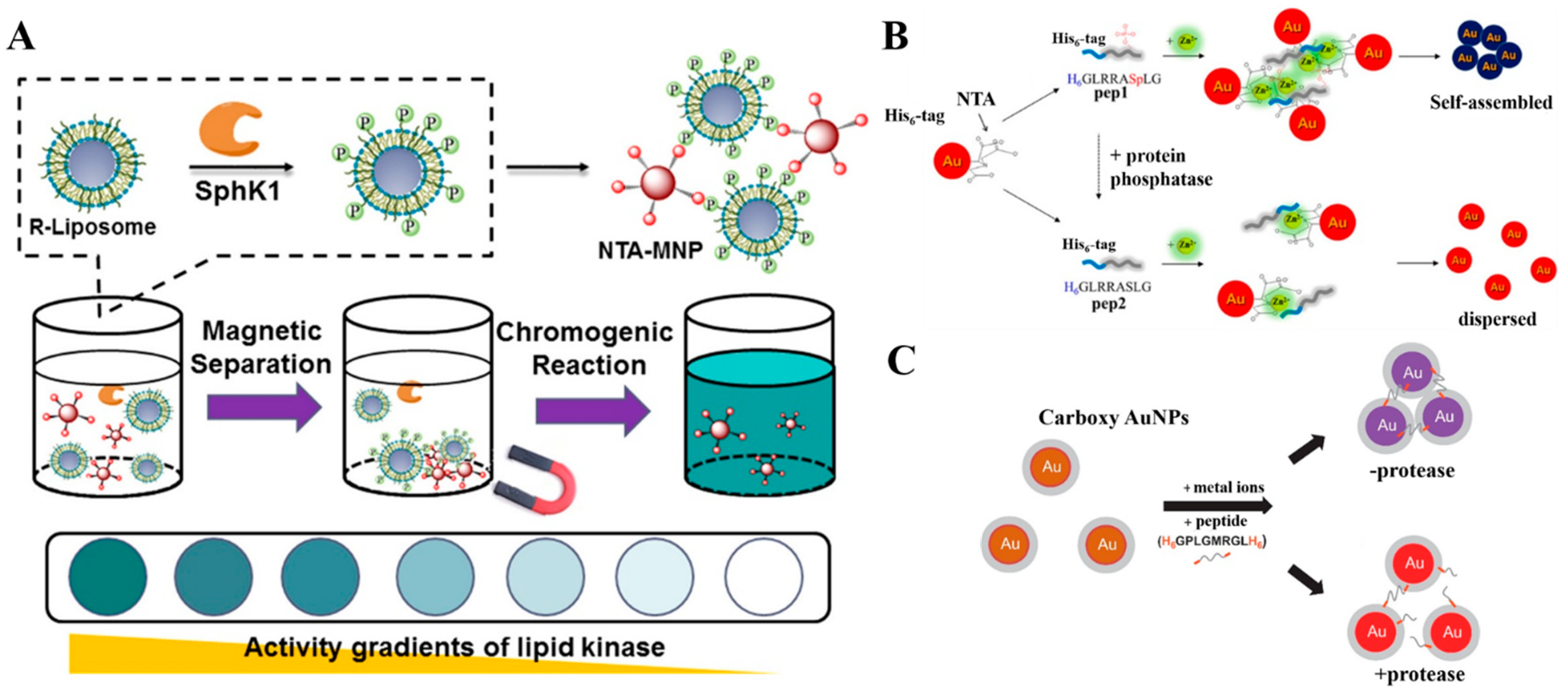
- Gao, T.; Mu, C.L.; Shi, H.; Shi, L.; Mao, X.X.; Li, G.X. Embedding capture-magneto-catalytic activity into a nanocatalyst for the determination of lipid kinase. ACS Appl. Mater. Interfaces 2018, 10, 59–65. [Google Scholar] [CrossRef] [PubMed]
- Lee, J.O.; Kim, E.J.; Lim, B.; Kim, T.W.; Kim, Y.P. Rapid detection of protein phosphatase activity using Zn(II)-coordinated gold nanosensors based on His-tagged phosphopeptides. Anal. Chem. 2015, 87, 1257–1265. [Google Scholar] [CrossRef] [PubMed]
- Kim, G.B.; Kim, K.H.; Park, Y.H.; Ko, S.; Kim, Y.P. Colorimetric assay of matrix metalloproteinase activity based on metal-induced self-assembly of carboxy gold nanoparticles. Biosens. Bioelectron. 2013, 41, 833–839. [Google Scholar] [CrossRef]
- Lee, S.K.; Maye, M.M.; Zhang, Y.B.; Gang, O.; van der Lelie, D. Controllable g5p-protein-directed aggregation of ssDNA-gold nanoparticles. Langmuir 2009, 25, 657–660. [Google Scholar] [CrossRef] [PubMed]
- Alsadig, A.; Vondracek, H.; Pengo, P.; Pasquato, L.; Posocco, P.; Parisse, P.; Casalis, L. Label-free, rapid and facile gold-nanoparticles-based assay as a potential spectroscopic tool for trastuzumab quantification. Nanomaterials 2021, 11, 3181. [Google Scholar] [CrossRef]
- Swartz, J.D.; Gulka, C.P.; Haselton, F.R.; Wright, D.W. Development of a histidine-targeted spectrophotometric sensor using Ni(II)NTA-functionalized Au and Ag nanoparticles. Langmuir 2011, 27, 15330–15339. [Google Scholar] [CrossRef] [PubMed]
- Cheng, W.; Chen, Y.L.; Yan, F.; Ding, L.; Ding, S.J.; Ju, H.X.; Yin, Y.B. Ultrasensitive scanometric strategy for detection of matrix metalloproteinases using a histidine tagged peptide-Au nanoparticle probe. Chem. Commun. 2011, 47, 2877–2879. [Google Scholar] [CrossRef]
- Kaya, M.; Volkan, M. New approach for the surface enhanced resonance Raman scattering (SERRS) detection of dopamine at picomolar (pM) levels in the presence of ascorbic acid. Anal. Chem. 2012, 84, 7729–7735. [Google Scholar] [CrossRef]
- Cao, X.M.; Qin, M.; Li, P.; Zhou, B.B.; Tang, X.H.; Ge, M.H.; Yang, L.B.; Liu, J.H. Probing catecholamine neurotransmitters based on iron-coordination surface-enhanced resonance Raman spectroscopy label. Sens. Actuat. B Chem. 2018, 268, 350–358. [Google Scholar] [CrossRef]
- Li, P.; Zhou, B.; Ge, M.; Jing, X.; Yang, L. Metal coordination induced SERS nanoprobe for sensitive and selective detection of histamine in serum. Talanta 2022, 237, 122913–122920. [Google Scholar] [CrossRef] [PubMed]
- Han, X.S.; Cao, M.D.; Zhou, B.C.; Yu, C.M.; Liu, Y.X.; Peng, B.; Meng, L.; Wei, J.F.; Li, L.; Huang, W. Specifically immobilizing His-tagged allergens to magnetic nanoparticles for fast and quantitative detection of allergen-specific IgE in serum samples. Talanta 2020, 219, 121301–121308. [Google Scholar] [CrossRef] [PubMed]
- Filchakova, O.; Dossym, D.; Ilyas, A.; Kuanysheva, T.; Abdizhamil, A.; Bukasov, R. Review of COVID-19 testing and diagnostic methods. Talanta 2022, 244, 123409–123440. [Google Scholar] [CrossRef] [PubMed]
- Davis, K.M.; Swartz, J.D.; Haselton, F.R.; Wright, D.W. Low-resource method for extracting the malarial biomarker histidine-rich protein ii to enhance diagnostic test performance. Anal. Chem. 2012, 84, 6136–6142. [Google Scholar] [CrossRef]
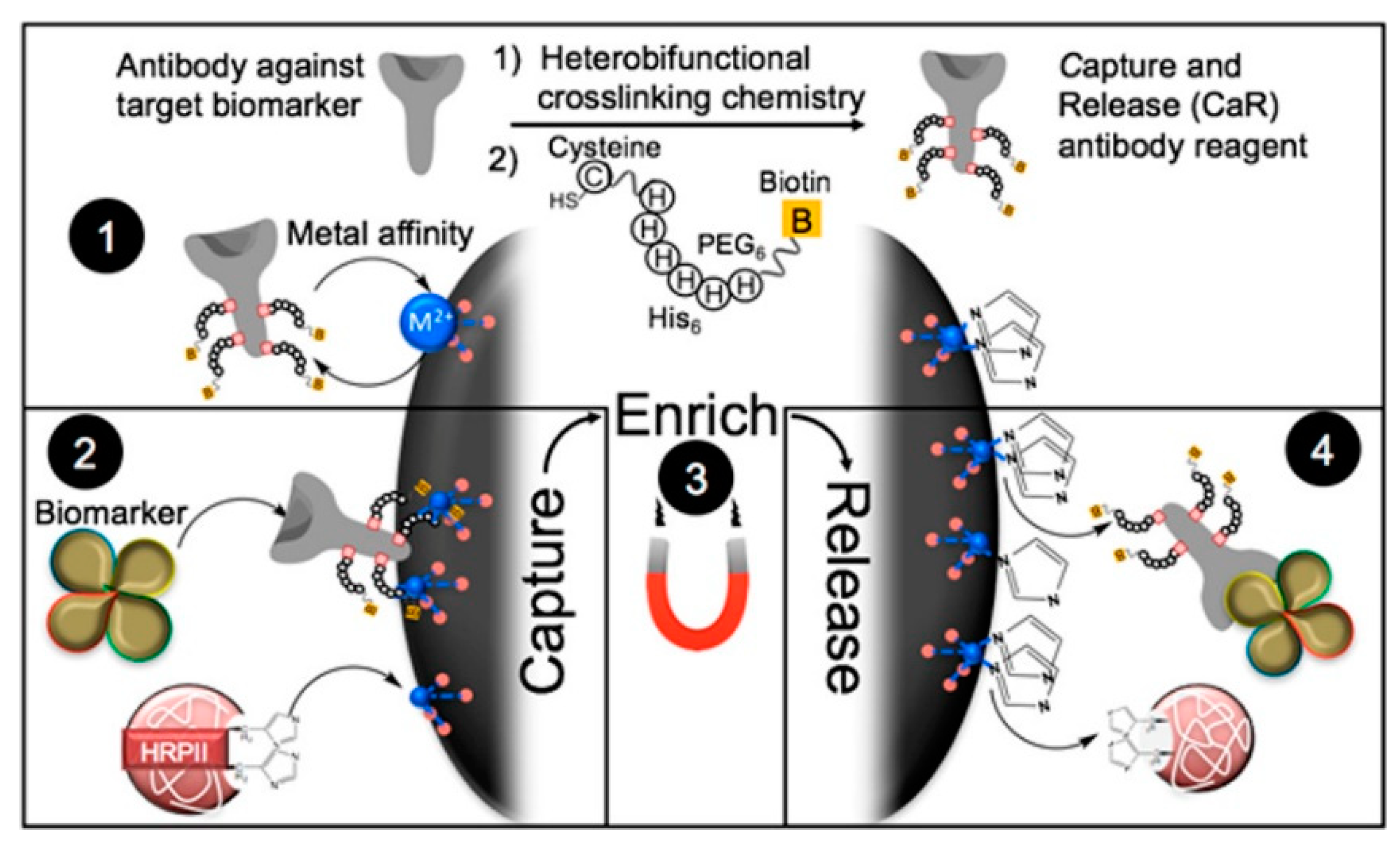
- Bauer, W.S.; Gulka, C.P.; Silva-Baucage, L.; Adams, N.M.; Haselton, F.R.; Wright, D.W. Metal affinity-enabled capture and release antibody reagents generate a multiplex biomarker enrichment system that improves detection limits of rapid diagnostic tests. Anal. Chem. 2017, 89, 10216–10223. [Google Scholar] [CrossRef]
- Yang, M.W.; Chen, D.J.; Hu, J.; Zheng, X.Y.; Lin, Z.J.; Zhu, H.M. The application of coffee-ring effect in analytical chemistry. TrAC-Trend. Anal. Chem. 2022, 157, 116752–116771. [Google Scholar] [CrossRef]
- Trantum, J.R.; Wright, D.W.; Haselton, F.R. Biomarker-mediated disruption of coffee-ring formation as a low resource diagnostic indicator. Langmuir 2012, 28, 2187–2193. [Google Scholar] [CrossRef]
- Gulka, C.P.; Swartz, J.D.; Trantum, J.R.; Davis, K.M.; Peak, C.M.; Denton, A.J.; Haselton, F.R.; Wright, D.W. Coffee rings as low-resource diagnostics: Detection of the malaria biomarker plasmodium falciparum histidine-rich protein-ii using a surface-coupled ring of Ni(II)NTA gold-plated polystyrene particles. ACS Appl. Mater. Interfaces 2014, 6, 6257–6263. [Google Scholar] [CrossRef]


Disclaimer/Publisher’s Note: The statements, opinions and data contained in all publications are solely those of the individual author(s) and contributor(s) and not of MDPI and/or the editor(s). MDPI and/or the editor(s) disclaim responsibility for any injury to people or property resulting from any ideas, methods, instructions or products referred to in the content. |
© 2023 by the authors. Licensee MDPI, Basel, Switzerland. This article is an open access article distributed under the terms and conditions of the Creative Commons Attribution (CC BY) license (https://creativecommons.org/licenses/by/4.0/).
Share and Cite
Zhu, L.; Chang, Y.; Li, Y.; Qiao, M.; Liu, L. Biosensors Based on the Binding Events of Nitrilotriacetic Acid–Metal Complexes. Biosensors 2023, 13, 507. https://doi.org/10.3390/bios13050507
Zhu L, Chang Y, Li Y, Qiao M, Liu L. Biosensors Based on the Binding Events of Nitrilotriacetic Acid–Metal Complexes. Biosensors. 2023; 13(5):507. https://doi.org/10.3390/bios13050507
Chicago/Turabian StyleZhu, Lin, Yong Chang, Yingying Li, Mingyi Qiao, and Lin Liu. 2023. "Biosensors Based on the Binding Events of Nitrilotriacetic Acid–Metal Complexes" Biosensors 13, no. 5: 507. https://doi.org/10.3390/bios13050507