A Review of Detection Methods for Vancomycin-Resistant Enterococci (VRE) Genes: From Conventional Approaches to Potentially Electrochemical DNA Biosensors

 , ,
, ,  and
and
Abstract
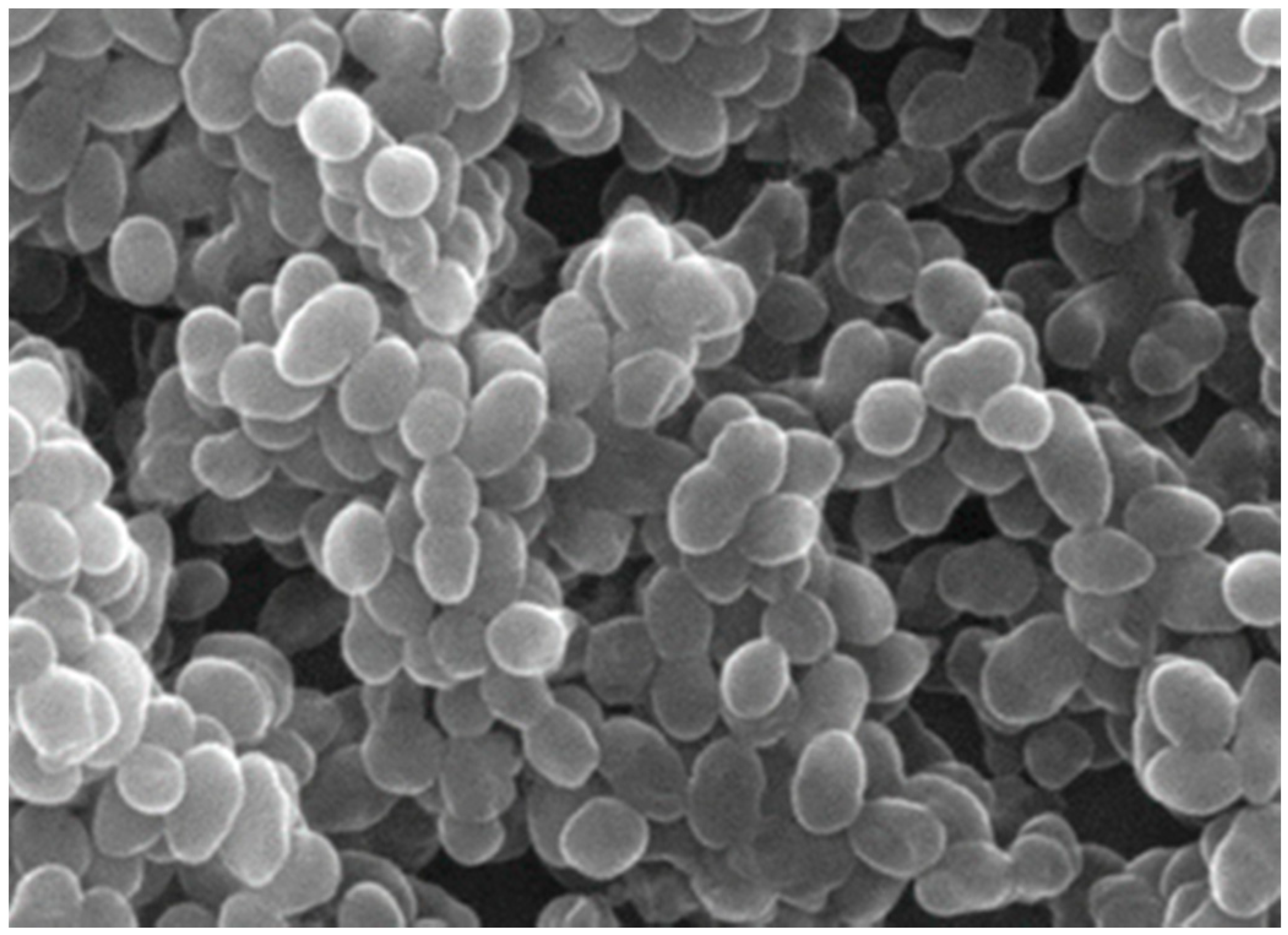
:1. Vancomycin-Resistant Enterococci (VRE)
Detection of VRE
2. Standard Clinical Diagnosis for VRE
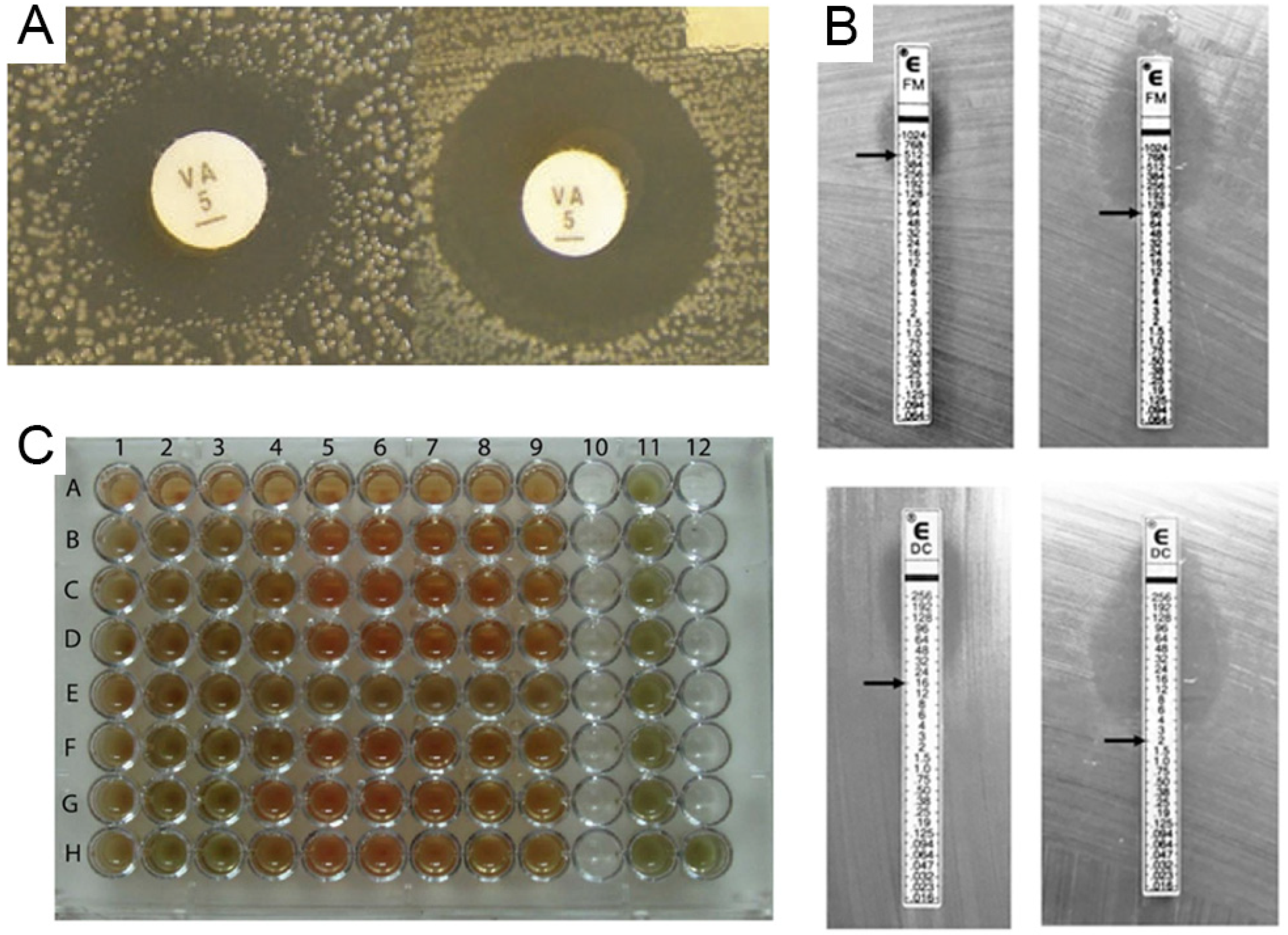
2.1. Traditional Methods
2.2. Immunoassay Methods
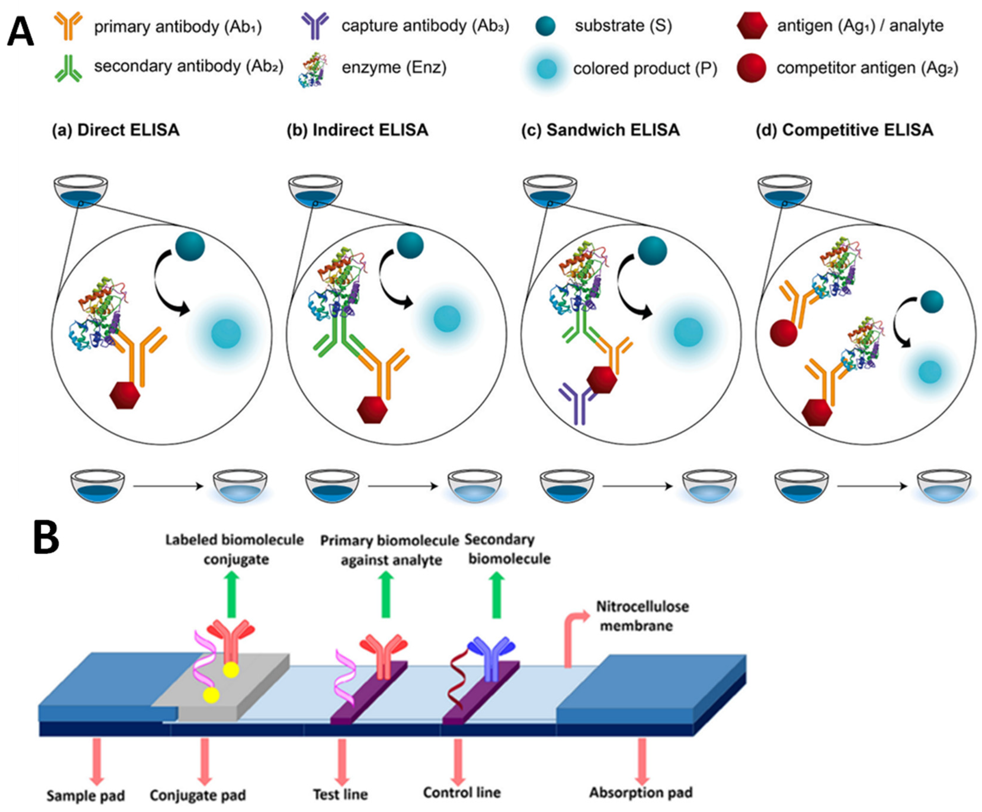
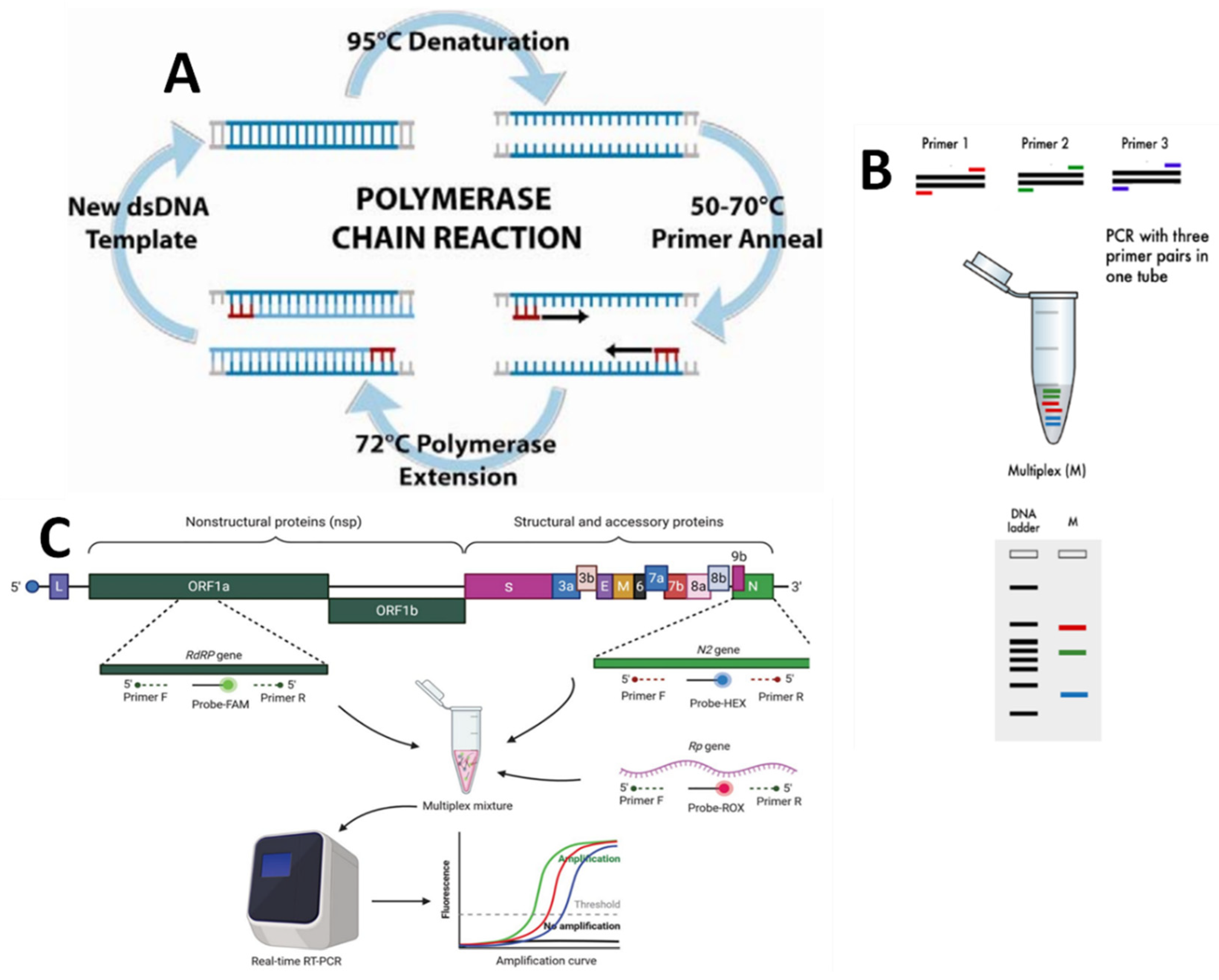
2.3. Molecular Methods

| Primer or Probe | Sequence (5′ > 3′) a | Size of Sequence (bp’s) | Amplified Gene or DNA Target Sequence | Ref |
|---|---|---|---|---|
| VanA (+) | GGGAAAACGACAATTGC | 732 | VanA VanA | [15] |
| VanA (−) | GTACAATGCGGCCGTTA | |||
| VanB (+) | ACGGAATGGGAAGCCGA | 647 | VanB | |
| VanB (−) | TGCACCCGATTTCGTTC | |||
| VanC | ATGGATTGGTAYTKGTATc | Van C1/2 | ||
| VanC | TAGCGGGAGTGMCYMGTAAc | |||
| VanD | TGTGGGATGCGATATTCAA | 500 | VanD | |
| VanD | TGCAGCCAAGTATCCGGTAA | |||
| VanE | TGTGGGATCGGAGCTGCAG | 430 | VanE | |
| VanE | ATAGTTTAGCTGGTAAC | |||
| VanG | CGGCATCCGCTGTTTTTGA | 941 | VanG | |
| VanG | GAACGATAGACCAATGCCTT | |||
| VanA | 5′-CATGAATAGAATAA AAGTTGCAATA-3′ | 1032 | VanA | [41] |
| 5′-CCCCTTTAACGCTA ATACGACGATCAA-3′ | ||||
| VanA1, VanA2 | 5′-GGGAAAACGACAATTGC 3′ and 5′-GTACAATGCGGCCGTTA 3′ | 732 bp | VanA | [42] |
| VanB1, VanB2 | 5′-ATGGGAAGCCGATAGTC-3′ and 5′-GATTTCGTTCCTCGACC-3′ | 635 bp | VanB | |
| VanA | GCT ATTCAG CTG TAC TC CAG CGG CCA TCA TAC GG | 783 bp | VanA | [18] |
| VanB | CAT CGC CGT CCC CGA ATT TCA AA GAT GCG GAA GAT ACC GTC GCT | 297 bp | VanB |
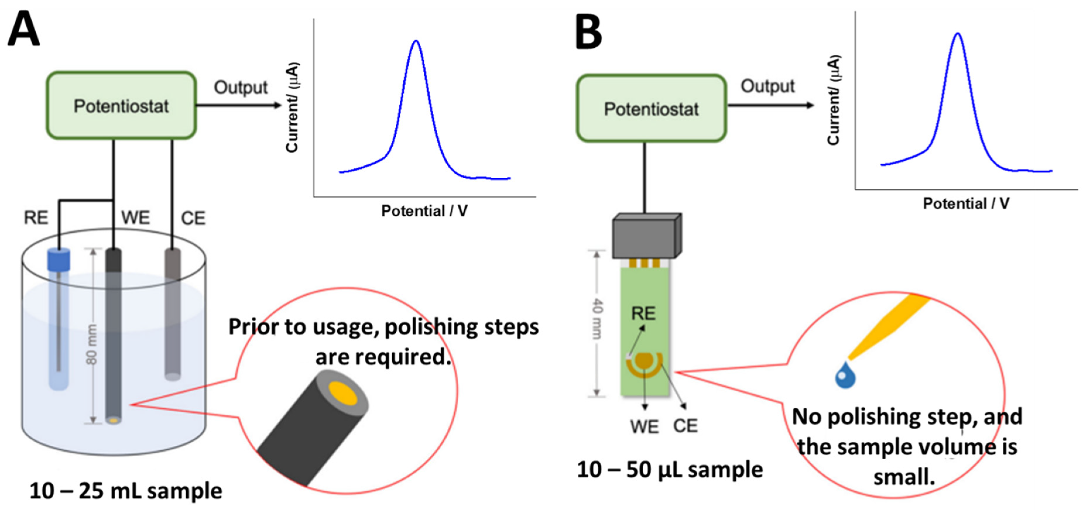
3. Potential and Strategies in Electrochemical DNA Biosensors of VRE Genes
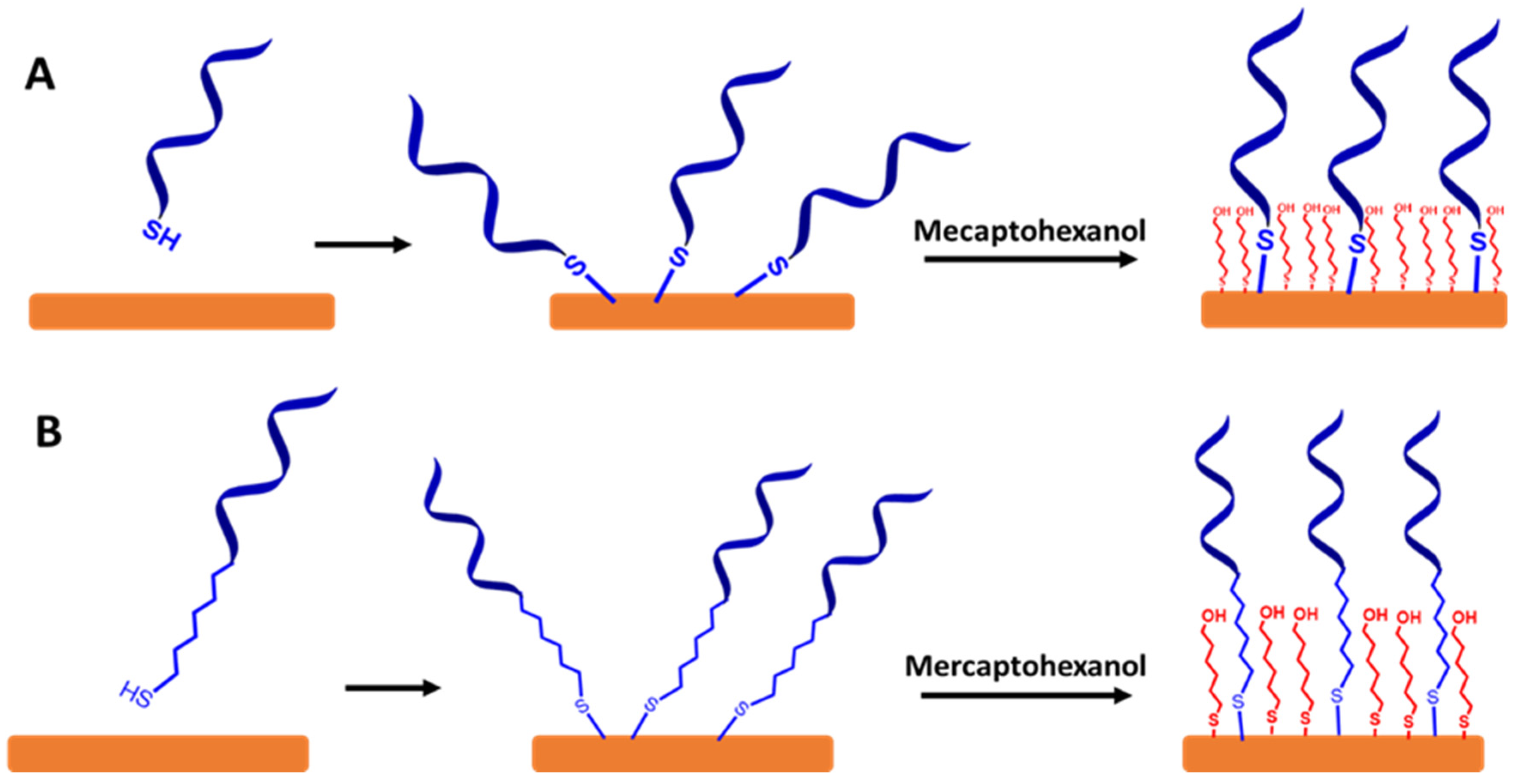
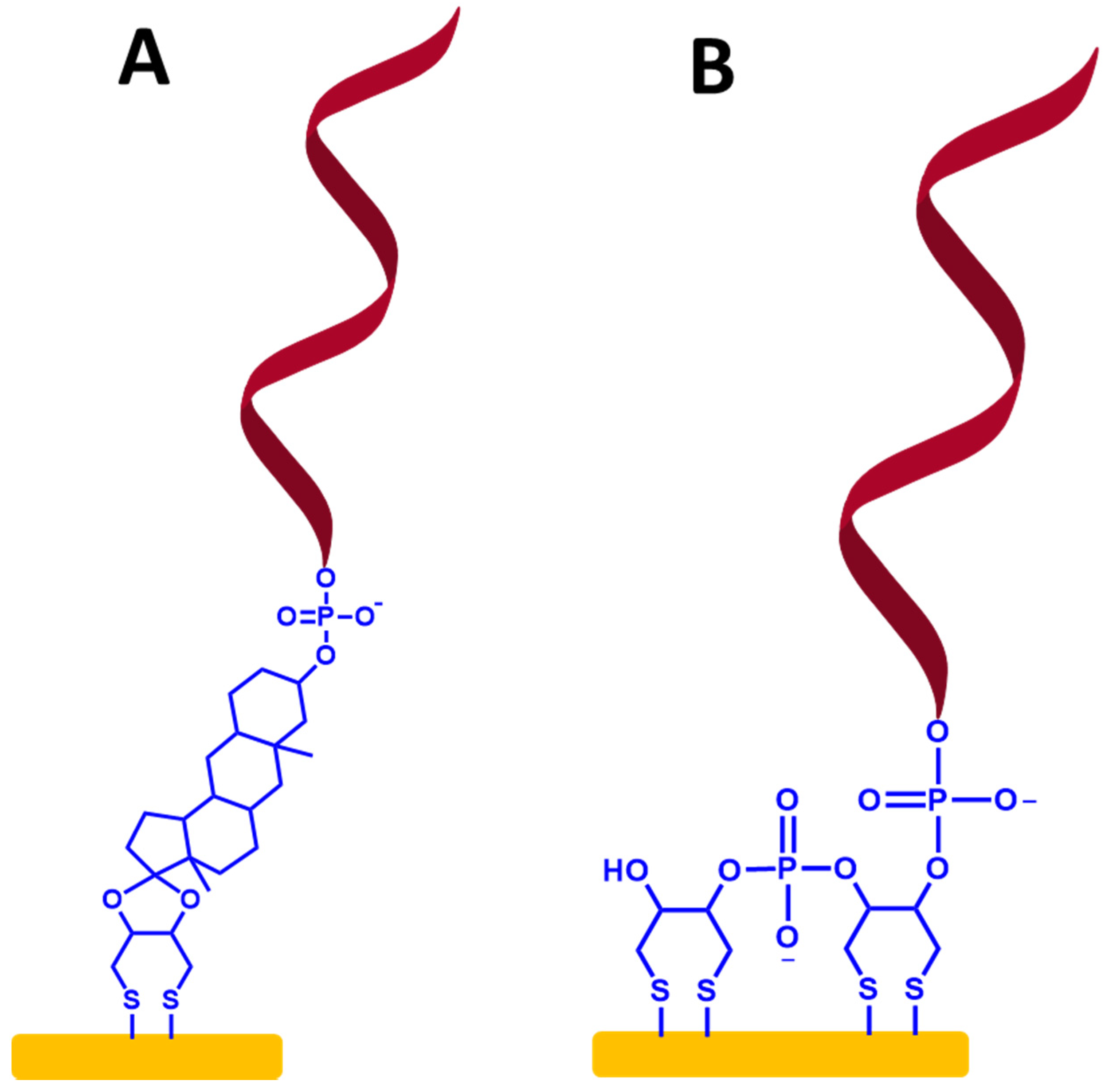
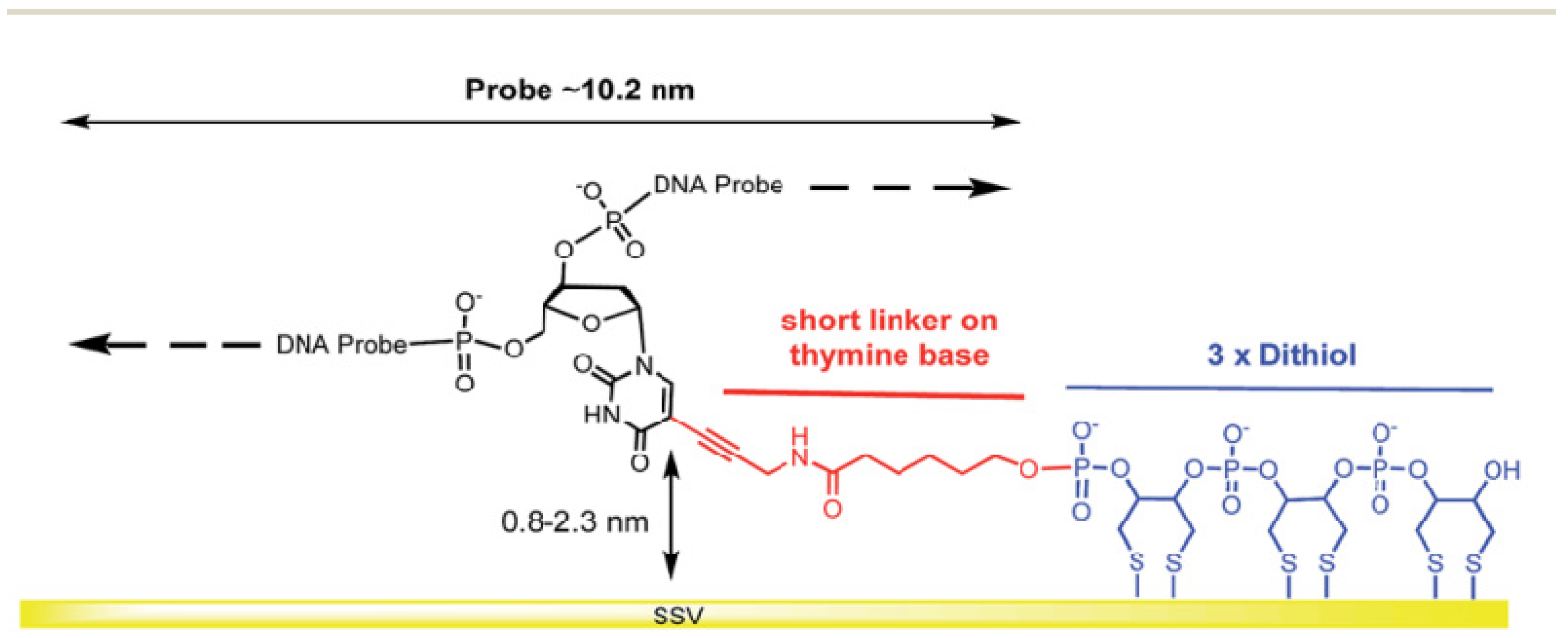
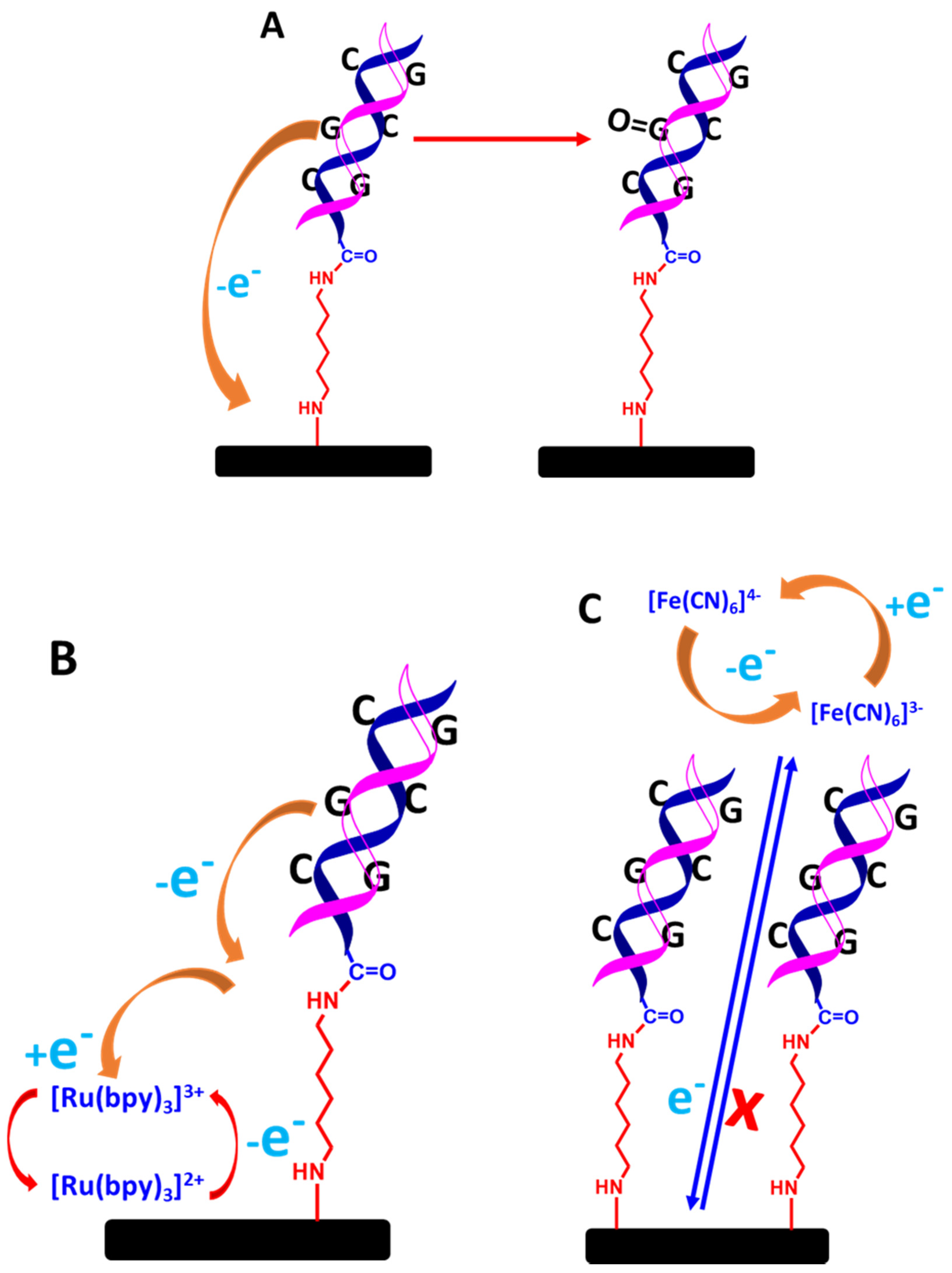
3.1. Immobilization ssDNA Probe-Based Self-Assembly Monolayer (SAM)
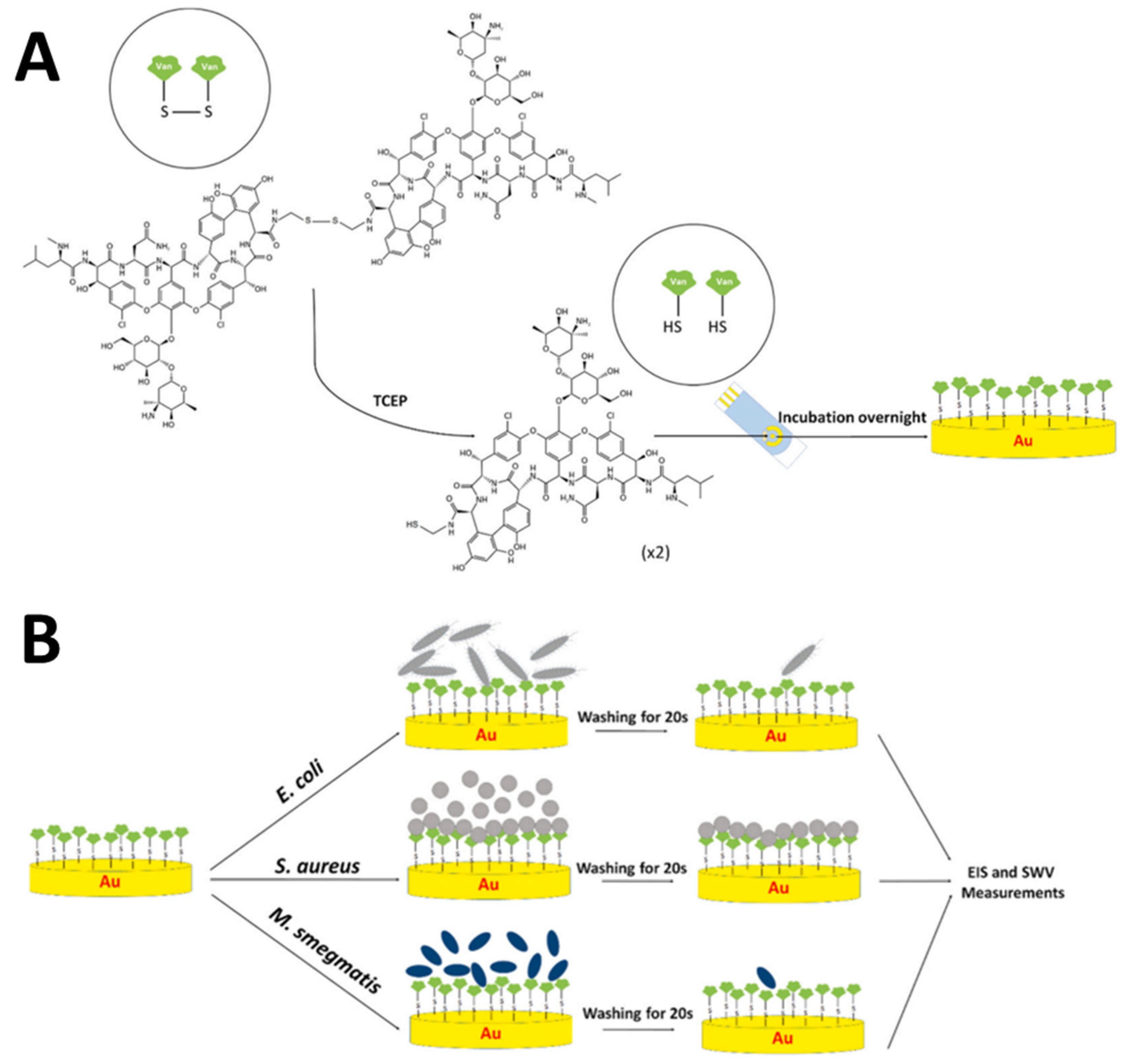
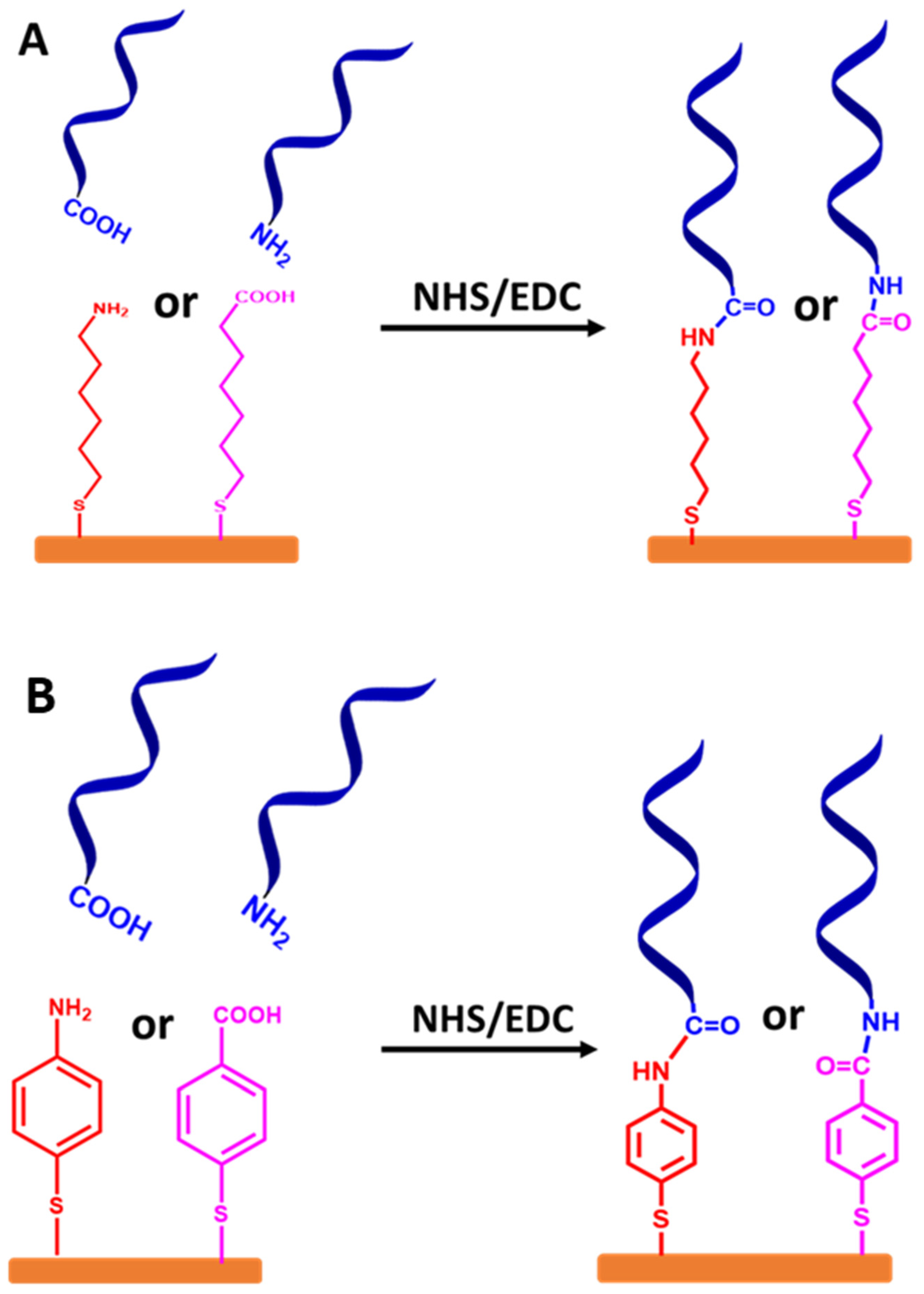
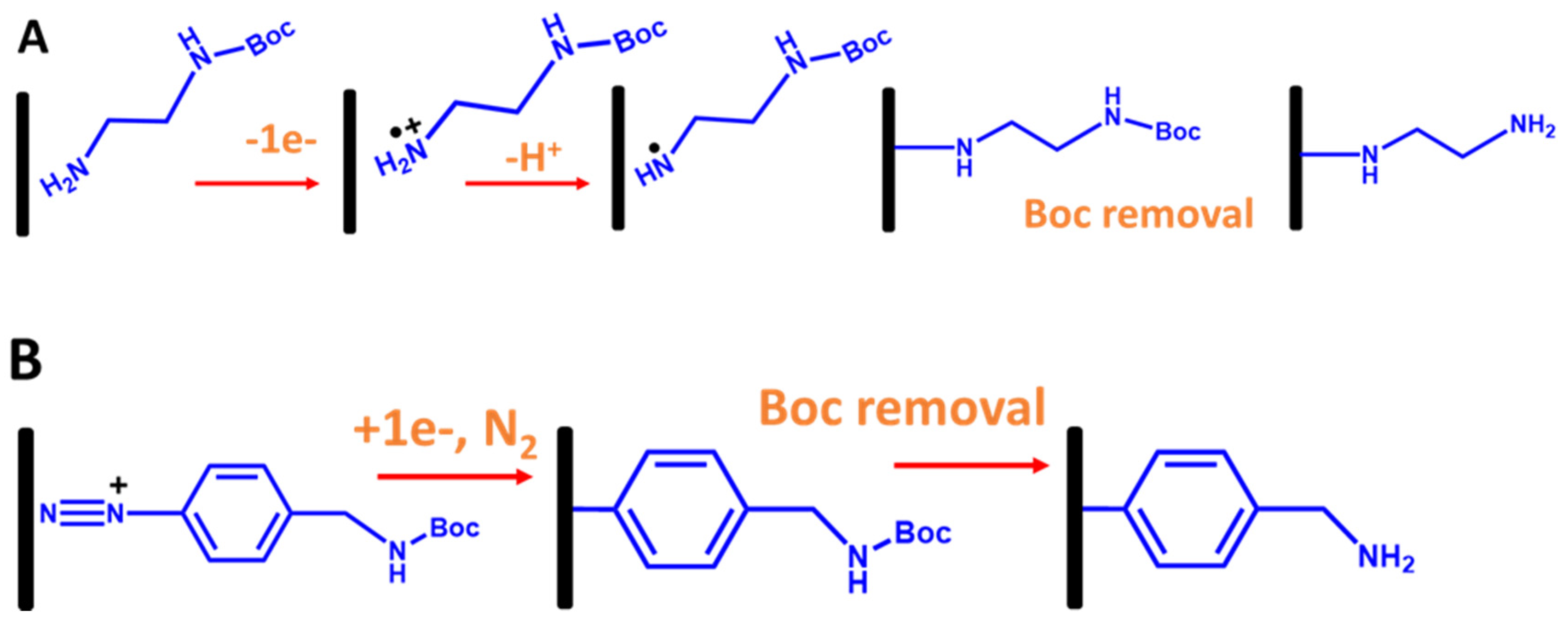
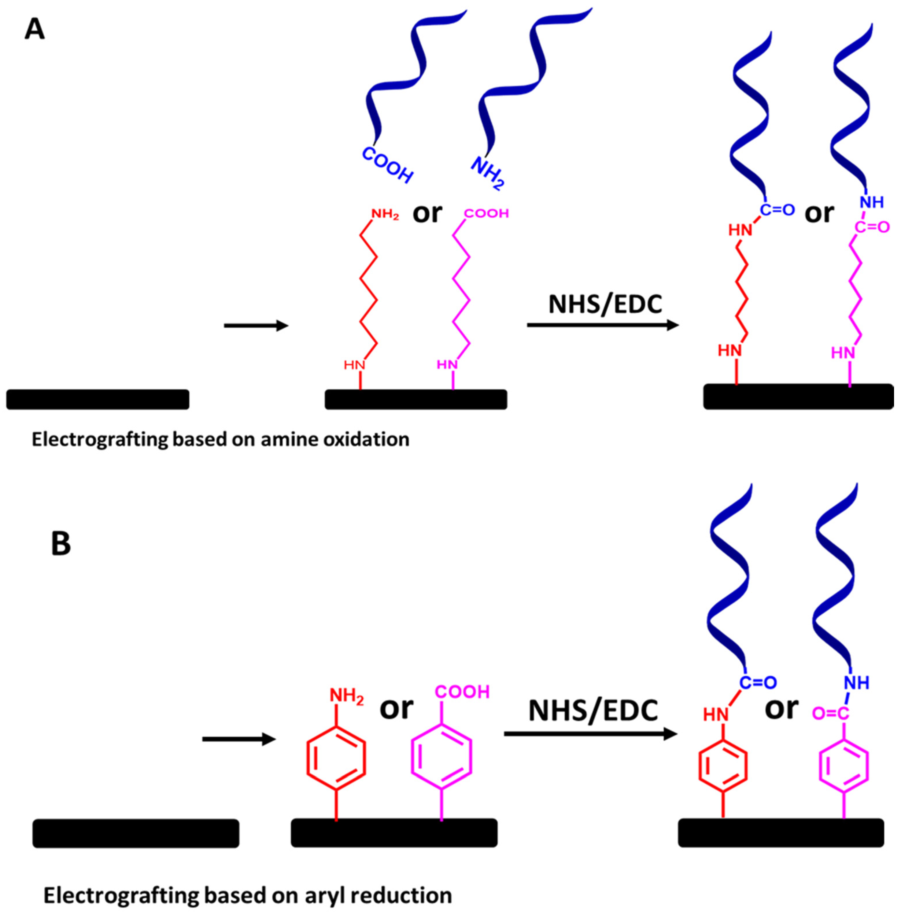
3.2. Immobilization ssDNA Probe-Based Covalent Attachment
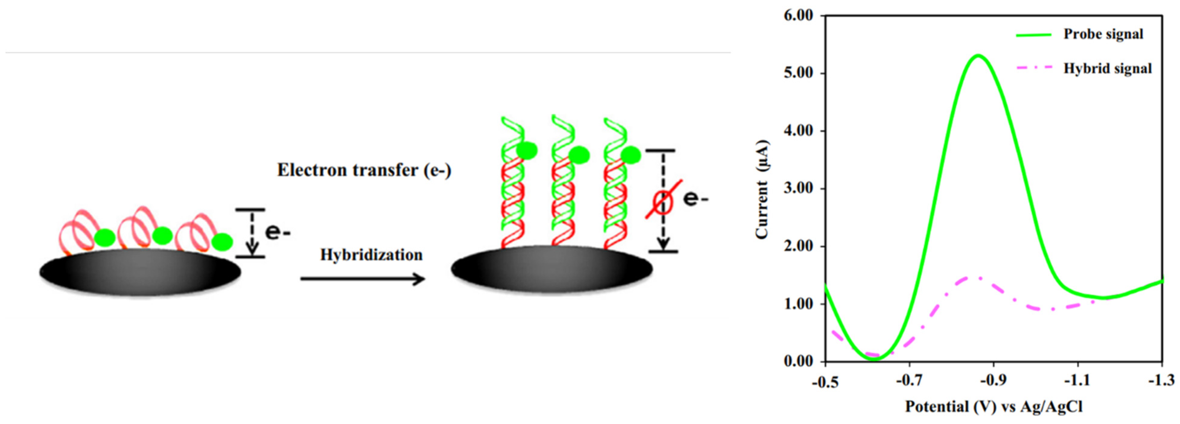
3.3. Label-Free and Labeled Detection Systems
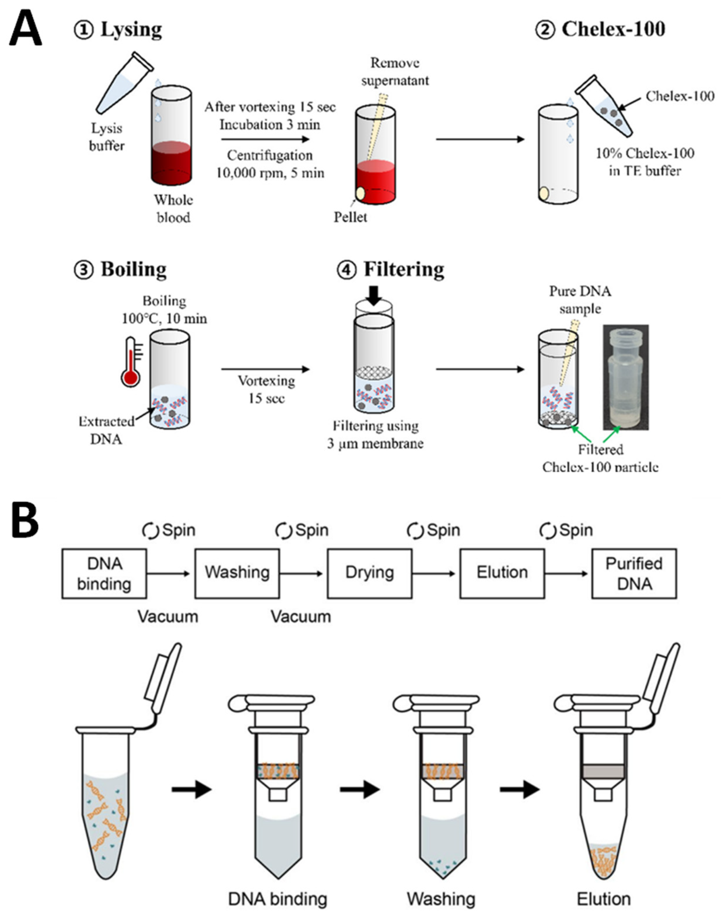
4. Method of DNA Extraction of Real Samples for Electrochemical DNA Biosensors
5. Future Outlook
Author Contributions
Funding
Conflicts of Interest
References
- O’Driscoll, T.; Crank, C.W. Vancomycin-resistant enterococcal infections: Epidemiology, clinical manifestations, and optimal management. Infect. Drug Resist. 2015, 8, 217–230. [Google Scholar] [CrossRef] [Green Version]
- Chang, W.H.; Yu, J.C.; Yang, S.Y.; Lin, Y.C.; Wang, C.H.; You, H.L.; Wu, J.J.; Lee, M.S.; Lee, G. Bin Vancomycin-resistant gene identification from live bacteria on an integrated microfluidic system by using low temperature lysis and loop-mediated isothermal amplification. Biomicrofluidics 2017, 11, 024101. [Google Scholar] [CrossRef] [Green Version]
- Bonten, M.J.; Willems, R.; Weinstein, R.A. Vancomycin-resistant enterococci: Why are they here, and where do they come from? Lancet Infect. Dis. 2001, 1, 314–325. [Google Scholar] [CrossRef]
- Lund, L.C.; Holzknecht, B.J.; Justesen, U.S. Treatment of vancomycin-resistant enterococcal infections. Ugeskr. Laeger 2018, 180, V07170530. [Google Scholar]
- Pérez-Hernández, X.; Méndez-Álvarez, S.; Claverie-Martín, F. A PCR assay for rapid detection of vancomycin-resistant enterococci. Diagn. Microbiol. Infect. Dis. 2002, 42, 273–277. [Google Scholar] [CrossRef]
- Ahmed, M.O.; Baptiste, K.E. Vancomycin-Resistant Enterococci: A Review of Antimicrobial Resistance Mechanisms and Perspectives of Human and Animal Health. Microb. Drug Resist. 2018, 24, 590–606. [Google Scholar] [CrossRef] [Green Version]
- Shlezinger, M.; Coppenhagen-Glazer, S.; Gelman, D.; Beyth, N.; Hazan, R. Eradication of vancomycin-resistant enterococci by combining phage and vancomycin. Viruses 2019, 11, 954. [Google Scholar] [CrossRef] [Green Version]
- Srinivasan, A.; Dick, J.D.; Perl, T.M. Vancomycin resistance in Staphylococci. Clin. Microbiol. Rev. 2002, 15, 430–438. [Google Scholar] [CrossRef] [Green Version]
- Gold, H.S. Vancomycin-resistant enterococci: Mechanisms and clinical observations. Clin. Infect. Dis. 2001, 33, 210–219. [Google Scholar] [CrossRef]
- Moosavian, M.; Ghadri, H.; Samli, Z. Molecular detection of vanA and vanB genes among vancomycin-resistant enterococci in ICU-hospitalized patients in Ahvaz in southwest of Iran. Infect. Drug Resist. 2018, 11, 2269–2275. [Google Scholar] [CrossRef] [Green Version]
- Gardete, S.; Tomasz, A. Mechanisms of vancomycin resistance in Staphylococcus aureus. J. Clin. Investig. 2014, 124, 2836–2840. [Google Scholar] [CrossRef]
- Purohit, G.; Gaind, R.; Dawar, R.; Verma, P.K.; Aggarwal, K.C.; Sardana, R.; Deb, M. Characterization of vancomycin resistant enterococci in hospitalized patients and role of gut colonization. J. Clin. Diagn. Res. 2017, 11, DC01–DC05. [Google Scholar] [CrossRef]
- Savini, V.; Marrollo, R.; Coclite, E.; Fusilli, P.; D’Incecco, C.; Fazii, P.; Gherardi, G. Liofilchem® Chromatic VRE and vancomycin MIC Test Strip detected glycopeptide resistance in a vanB neonatal Enterococcus faecium isolate showing alternate vancomycin susceptibility and resistance with bioMérieux Vitek2. Int. J. Clin. Exp. Pathol. 2014, 7, 6274–6277. [Google Scholar]
- Zerrouki, H.; Rebiahi, S.A.; Hadjadj, L.; Rolain, J.M.; Diene, S.M. Real-time PCR assay for rapid and simultaneous detection of vanA and vanB genes in clinical strains. Diagnostics 2021, 11, 2081. [Google Scholar] [CrossRef]
- Bhatt, P.; Sahni, A.K.; Praharaj, A.K.; Grover, N.; Kumar, M.; Chaudhari, C.N.; Khajuria, A. Detection of glycopeptide resistance genes in enterococci by multiplex PCR. Med. J. Armed Forces India 2015, 71, 43–47. [Google Scholar] [CrossRef] [Green Version]
- Velusamy, V.; Arshak, K.; Korostynska, O.; Oliwa, K.; Adley, C. An overview of foodborne pathogen detection: In the perspective of biosensors. Biotechnol. Adv. 2010, 28, 232–254. [Google Scholar] [CrossRef]
- Cesewski, E.; Johnson, B.N. Electrochemical biosensors for pathogen detection. Biosens. Bioelectron. 2020, 159, 112214. [Google Scholar] [CrossRef]
- Rengaraj, R. Detection of Vancomycin Resistance among Enterococcus faecalis and Staphylococcus aureus. J. Clin. Diagn. Res. 2016, 10, 8–10. [Google Scholar] [CrossRef]
- Jenkins, S.G.; Schuetz, A.N. Current concepts in laboratory testing to guide antimicrobial therapy. Mayo Clin. Proc. 2012, 87, 290–308. [Google Scholar] [CrossRef] [Green Version]
- Maurer, F.P.; Christner, M.; Hentschke, M.; Rohde, H. Advances in rapid identification and susceptibility testing of bacteria in the clinical microbiology laboratory: Implications for patient care and antimicrobial stewardship programs. Infect. Dis. Rep. 2017, 9, 18–27. [Google Scholar] [CrossRef] [Green Version]
- Rathe, M.; Kristensen, L.; Ellermann-Eriksen, S.; Thomsen, M.K.; Schumacher, H. Vancomycin-resistant Enterococcus spp.: Validation of susceptibility testing and in vitro activity of vancomycin, linezolid, tigecycline and daptomycin. APMIS 2010, 118, 66–73. [Google Scholar] [CrossRef]
- Davis, H.; Brown, R.; Ashcraft, D.; Pankey, G. In vitro synergy with fosfomycin plus doxycyclin against linezolid and vancomycin-resistant Enterococcus faecium. J. Glob. Antimicrob. Resist. 2020, 22, 78–83. [Google Scholar] [CrossRef]
- Ataee, A.A.; Habibian, S.; Mehrabi-Tavana, A.; Ahmadi, Z.; Jonaidi, N.; Salesi, M. Determination of vancomycin minimum inhibitory concentration for ceftazidime resistant Streptococcus pneumoniae in Iran. Ann. Clin. Microbiol. Antimicrob. 2014, 13, 53. [Google Scholar] [CrossRef] [Green Version]
- Balouiri, M.; Sadiki, M.; Ibnsouda, S.K. Methods for in Vitro Evaluating Antimicrobial Activity: A Review. J. Pharm. Anal. 2016, 6, 71–79. [Google Scholar] [CrossRef] [Green Version]
- Vila, M.M.D.C.; De Oliveira, R.M.; Gonçalves, M.M.; Tubino, M. Analytical methods for vancomycin determination in biological fluids and in pharmaceuticals. Quim. Nova 2007, 30, 395–399. [Google Scholar] [CrossRef]
- Sah, R.; Khadka, S.; Shrestha, N. Vancomycin Resistant Enterococcus Faecalis Causing Diarrhea Vancomycin Resistant Enterococcus Faecalis Causing. BMC Infect. Dis. 2017, 6, 133. [Google Scholar]
- Cenci-Goga, B.T.; Karama, M.; El-Ashram, S.; Saraiva, C.; García-Díez, J.; Chalias, A.; Grispoldi, L. Two Screening Assays to Detect Vancomycin-Resistant Enterococcus spp. Microbiol. Res. 2022, 13, 332–341. [Google Scholar] [CrossRef]
- Darwish, I.A. Immunoassay Methods and their Applications in Pharmaceutical Analysis: Basic Methodology and Recent Advances. Int. J. Biomed. Sci. 2006, 2, 217–235. [Google Scholar]
- Mello, L.D. Potential contribution of ELISA and LFI assays to assessment of the oxidative stress condition based on 8-oxodG biomarker. Anal. Biochem. 2021, 628, 114215. [Google Scholar] [CrossRef]
- Bahadır, E.B.; Sezgintürk, M.K. Lateral flow assays: Principles, designs and labels. TrAC Trends Anal. Chem. 2016, 82, 286–306. [Google Scholar] [CrossRef]
- Kong, D.; Xie, Z.; Liu, L.; Song, S.; Kuang, H.; Xu, C. Development of ic-ELISA and lateral-flow immunochromatographic assay strip for the detection of vancomycin in raw milk and animal feed. Food Agric. Immunol. 2017, 28, 414–426. [Google Scholar] [CrossRef] [Green Version]
- Oueslati, S.; Volland, H.; Cattoir, V.; Bernabeu, S.; Girlich, D.; Dulac, D.; Plaisance, M.; Laroche, M.; Dortet, L.; Simon, S.; et al. Development and Validation of a Lateral Flow Immunoassay for Rapid Detection of VanA-Producing Enterococci. J. Antimicrob. Chemother. 2021, 76, 146–151. [Google Scholar] [CrossRef]
- Bian, L.; Liang, J.; Zhao, H.; Ye, K.; Li, Z.; Liu, T.; Peng, J.; Wu, Y.; Lin, G. Rapid Monitoring of Vancomycin Concentration in Serum Using Europium (III) Chelate Nanoparticle-Based Lateral Flow Immunoassay. Front. Chem. 2021, 9, 763686. [Google Scholar] [CrossRef]
- Odekerken, J.C.E.; Logister, D.M.W.; Assabre, L.; Arts, J.J.C.; Walenkamp, G.H.I.M.; Welting, T.J.M. ELISA-Based Detection of Gentamicin and Vancomycin in Protein-Containing Samples. Springerplus 2015, 4, 614. [Google Scholar] [CrossRef] [Green Version]
- Lui, C.; Cady, N.C.; Batt, C.A. Nucleic Acid-Based Detection of Bacterial Pathogens Using Integrated Microfluidic Platform Systems. Sensors 2009, 9, 3713–3744. [Google Scholar] [CrossRef] [Green Version]
- Mohd Hanafiah, N.; Cheng, A.; Lim, P.E.; Sethuraman, G.; Mohd Zain, N.A.; Baisakh, N.; Mispan, M.S. Novel PCR-Based Multiplex Assays for Detecting Major Quality and Biotic Stress in Commercial and Weedy Rice. Life 2022, 12, 1542. [Google Scholar] [CrossRef]
- Tombuloglu, H.; Sabit, H.; Al-Khallaf, H.; Kabanja, J.H.; Alsaeed, M.; Al-Saleh, N.; Al-Suhaimi, E. Multiplex real-time RT-PCR method for the diagnosis of SARS-CoV-2 by targeting viral N, RdRP and human RP genes. Sci. Rep. 2022, 12, 2853. [Google Scholar] [CrossRef]
- Amberpet, R.; Sistla, S.; Parija, S.C.; Thabah, M.M. Screening for intestinal colonization with vancomycin resistant enterococci and associated risk factors among patients admitted to an adult intensive care unit of a large teaching hospital. J. Clin. Diagn. Res. 2016, 10, 6–9. [Google Scholar] [CrossRef]
- Lu, J.J.; Perng, C.L.; Chiueh, T.S.; Lee, S.Y.; Chen, C.H.; Chang, F.Y.; Wang, C.C.; Chi, W.M. Detection and typing of vancomycin-resistance genes of enterococci from clinical and nosocomial surveillance specimens by multiplex PCR. Epidemiol. Infect. 2001, 126, 357–363. [Google Scholar] [CrossRef] [Green Version]
- Nomura, T.; Hashimoto, Y.; Kurushima, J.; Hirakawa, H.; Tanimoto, K.; Zheng, B.; Ruan, G.; Xue, F.; Liu, J.; Hisatsune, J.; et al. New colony multiplex PCR assays for the detection and discrimination of vancomycin-resistant enterococcal species. J. Microbiol. Methods 2018, 145, 69–72. [Google Scholar] [CrossRef]
- Mashaly, M.; El-Mashad, N.; El-deeb, H. Detection of VanA type vancomycin resistance among MRSA isolates from an emergency hospital in Egypt. Comp. Clin. Pathol. 2019, 28, 971–976. [Google Scholar] [CrossRef]
- Özsoy, S.; İlki, A. Detection of vancomycin-resistant enterococci (VRE) in stool specimens submitted for Clostridium difficile toxin testing. Braz. J. Microbiol. 2017, 48, 489–492. [Google Scholar] [CrossRef]
- Tan, T.Y.; Jiang, B.; Ng, L.S.Y. Faster and economical screening for vancomycin-resistant enterococci by sequential use of chromogenic agar and real-time polymerase chain reaction. J. Microbiol. Immunol. Infect. 2017, 50, 448–453. [Google Scholar] [CrossRef] [Green Version]
- Awang, M.S.; Bustami, Y.; Hamzah, H.H.; Zambry, N.S.; Najib, M.A.; Khalid, M.F.; Aziah, I.; Manaf, A.A. Advancement in salmonella detection methods: From conventional to electrochemical-based sensing detection. Biosensors 2021, 11, 346. [Google Scholar] [CrossRef]
- Norouz Dizaji, A.; Ali, Z.; Ghorbanpoor, H.; Ozturk, Y.; Akcakoca, I.; Avci, H.; Dogan Guzel, F. Electrochemical-based “antibiotsensor” for the whole-cell detection of the vancomycin-susceptible bacteria. Talanta 2021, 234, 122695. [Google Scholar] [CrossRef]
- Kamal, N.N.A.; Anuar, N.S.; Noordin, R.; Rahumatullah, A.; Hamzah, H.H. Electrodeposited gold nanoparticle (AuNP)-film as a nanoplatform for a label-free electrochemical strongyloidiasis immunosensor. J. Electrochem. Soc. 2022, 169, 106514. [Google Scholar] [CrossRef]
- Love, J.C.; Estroff, L.A.; Kriebel, J.K.; Nuzzo, R.G.; Whitesides, G.M. Self-Assembled Monolayers of Thiolates on Methals as a Form of Nanotechnology. Chem. Rev. 2005, 105, 1103–1169. [Google Scholar] [CrossRef]
- Nuzzo, R.G.; Allara, D.L. Adsorption of bifunctional organic disulfides on gold surfaces. J. Am. Chem. Soc. 1983, 105, 4481–4483. [Google Scholar] [CrossRef]
- Singh, V.; Zharnikov, M.; Gulino, A.; Gupta, T. DNA immobilization, delivery and cleavage on solid supports. J. Mater. Chem. 2011, 21, 10602–10618. [Google Scholar] [CrossRef]
- Singh, Y.; Murat, P.; Defrancq, E. Recent developments in oligonucleotide conjugation. Chem. Soc. Rev. 2010, 39, 2054–2070. [Google Scholar] [CrossRef]
- Papadopoulou, E.; Gale, N.; Thompson, J.F.; Fleming, T.A.; Brown, T.; Bartlett, P.N. Specifically horizontally tethered DNA probes on Au surfaces allow labelled and label-free DNA detection using SERS and electrochemically driven melting. Chem. Sci. 2016, 7, 386–393. [Google Scholar] [CrossRef] [Green Version]
- Goodchild, S.A.; Gao, R.; Shenton, D.P.; McIntosh, A.J.S.; Brown, T.; Bartlett, P.N. Direct Detection and Discrimination of Nucleotide Polymorphisms Using Anthraquinone Labeled DNA Probes. Front. Chem. 2020, 8, 381. [Google Scholar] [CrossRef]
- Madaan, N.; Terry, A.; Harb, J.; Davis, R.C.; Schlaad, H.; Linford, M.R. Thiol–Ene–Thiol Photofunctionalization of Thiolated Monolayers with Polybutadiene and Functional Thiols, Including Thiolated DNA. J. Phys. Chem. C 2011, 115, 22931–22938. [Google Scholar] [CrossRef]
- Hamzah, H.H.; Ahmad Kamal, N.N.; Meneghello, M.; Shafiee, S.A.; Sönmez, T.; Mohamad Taib, M.N.A.; Mohd Samsuri, S.H.; Meor Zulkifli, M.F. Hexanediamine Monolayer Electrografted at Glassy Carbon Electrodes Enhances Oxygen Reduction Reaction in Aqueous Neutral Media. J. Electrochem. Soc. 2020, 167, 166508. [Google Scholar] [CrossRef]
- Al-Lolage, F.A.; Meneghello, M.; Ma, S.; Ludwig, R.; Bartlett, P.N. A Flexible Method for the Stable, Covalent Immobilization of Enzymes at Electrode Surfaces. ChemElectroChem 2017, 4, 1528–1534. [Google Scholar] [CrossRef] [Green Version]
- Ghanem, M.A.; Kocak, I.; Al-Mayouf, A.; Alhoshan, M.; Bartlett, P.N. Covalent modification of carbon nanotubes with anthraquinone by electrochemical grafting and solid phase synthesis. Electrochim. Acta 2012, 68, 74–80. [Google Scholar] [CrossRef]
- Hamzah, H.; Denuault, G.; Bartlett, P.; Pinczewska, A.; Kilburn, J. Electrografting of mono-N-Boc-ethylenediamine from an acetonitrile/aqueous NaHCO3 mixture. J. Electrochem. 2017, 23, 130–140. [Google Scholar] [CrossRef]
- Celiktas, A.; Ghanem, M.A.; Bartlett, P.N. Modification of nanostructured gold surfaces with organic functional groups using electrochemical and solid-phase synthesis methodologies. J. Electroanal. Chem. 2012, 670, 42–49. [Google Scholar] [CrossRef]
- Chrétien, J.-M.; Ghanem, M.A.; Bartlett, P.N.; Kilburn, J.D. Covalent tethering of organic functionality to the surface of glassy carbon electrodes by using electrochemical and solid-phase synthesis methodologies. Chem. A Eur. J. 2008, 14, 2548–2556. [Google Scholar] [CrossRef]
- Ghanem, M.A.; Chrétien, J.-M.; Kilburn, J.D.; Bartlett, P.N. Electrochemical and solid-phase synthetic modification of glassy carbon electrodes with dihydroxybenzene compounds and the electrocatalytic oxidation of NADH. Bioelectrochemistry 2009, 76, 115–125. [Google Scholar] [CrossRef]
- Gillan, L.; Teerinen, T.; Johansson, L.S.; Smolander, M. Controlled diazonium electrodeposition towards a biosensor for C-reactive protein. Sens. Int. 2021, 2, 100060. [Google Scholar] [CrossRef]
- Ferapontova, E.E. Electrochemistry of guanine and 8-oxoguanine at gold electrodes. Electrochim. Acta 2004, 49, 1751–1759. [Google Scholar] [CrossRef]
- Alves, L.M.; Rodovalho, V.R.; Castro, A.C.H.; Freitas, M.A.R.; Mota, C.M.; Mineo, T.W.P.; Mineo, J.R.; Madurro, J.M.; Brito-Madurro, A.G. Development of direct assays for Toxoplasma gondii and its use in genomic DNA sample. J. Pharm. Biomed. Anal. 2017, 145, 838–844. [Google Scholar] [CrossRef]
- Souza, E.; Nascimento, G.; Santana, N.; Ferreira, D.; Lima, M.; Natividade, E.; Martins, D.; José, L.F. Label-free electrochemical detection of the specific oligonucleotide sequence of dengue virus type I on pencil graphite electrodes. Sensors 2011, 11, 5616. [Google Scholar] [CrossRef] [Green Version]
- Drummond, T.G.; Hill, M.G.; Barton, J.K. Electrochemical DNA sensors. Nat. Biotechnol. 2003, 21, 1192–1199. [Google Scholar] [CrossRef] [Green Version]
- Anne, A.; Bouchardon, A.; Moiroux, J. 3′-Ferrocene-labeled oligonucleotide chains end-tethered to gold electrode surfaces: Novel model systems for exploring flexibility of short DNA using cyclic voltammetry. J. Am. Chem. Soc. 2003, 125, 1112–1113. [Google Scholar] [CrossRef]
- Hu, F.; Zhang, W.; Meng, W.; Ma, Y.; Zhang, X.; Xu, Y.; Wang, P.; Gu, Y. Ferrocene-labeled and purification-free electrochemical biosensor based on ligase chain reaction for ultrasensitive single nucleotide polymorphism detection. Anal. Chim. Acta 2020, 1109, 9–18. [Google Scholar] [CrossRef]
- García-González, R.; Costa-García, A.; Fernández-Abedul, M.T. Methylene blue covalently attached to single stranded DNA as electroactive label for potential bioassays. Sens. Actuators B Chem. 2014, 191, 784–790. [Google Scholar] [CrossRef]
- Jampasa, S.; Wonsawat, W.; Rodthongkum, N.; Siangproh, W.; Yanatatsaneejit, P.; Vilaivan, T.; Chailapakul, O. Electrochemical detection of human papillomavirus DNA type 16 using a pyrrolidinyl peptide nucleic acid probe immobilized on screen-printed carbon electrodes. Biosens. Bioelectron. 2014, 54, 428–434. [Google Scholar] [CrossRef]
- Pheeney, C.G.; Barton, J.K. DNA Electrochemistry with Tethered Methylene Blue. Langmuir 2012, 28, 7063–7070. [Google Scholar] [CrossRef] [Green Version]
- Bezinge, L.; Suea-Ngam, A.; Demello, A.J.; Shih, C.J. Nanomaterials for molecular signal amplification in electrochemical nucleic acid biosensing: Recent advances and future prospects for point-of-care diagnostics. Mol. Syst. Des. Eng. 2020, 5, 49–66. [Google Scholar] [CrossRef] [Green Version]
- Zakaria, N.D.; Omar, M.H.; Ahmad Kamal, N.N.; Abdul Razak, K.; Sönmez, T.; Balakrishnan, V.; Hamzah, H.H. Effect of Supporting Background Electrolytes on the Nanostructure Morphologies and Electrochemical Behaviors of Electrodeposited Gold Nanoparticles on Glassy Carbon Electrode Surfaces. ACS Omega 2021, 6, 24419–24431. [Google Scholar] [CrossRef]
- Lim, D.H.; Jee, H.; Moon, K.C.; Lim, C.S.; Jang, W.S. Development of a Simple DNA Extraction Method and Candida Pan Loop-Mediated Isothermal Amplification Assay for Diagnosis of Candidemia. Pathogens 2022, 11, 111. [Google Scholar] [CrossRef]
- Lee, H.; Na, W.; Park, C.; Park, K.H.; Shin, S. Centrifugation-free extraction of circulating nucleic acids using immiscible liquid under vacuum pressure. Sci. Rep. 2018, 8, 5467. [Google Scholar] [CrossRef] [Green Version]
- Sin, M.L.; Mach, K.E.; Wong, P.K.; Liao, J.C. Advances and challenges in biosensor-based diagnosis of infectious diseases. Expert Rev. Mol. Diagn. 2014, 14, 225–244. [Google Scholar] [CrossRef] [Green Version]
- Tan, S.C.; Yiap, B.C. DNA, RNA, and protein extraction: The past and the present. J. Biomed. Biotechnol. 2009, 2009, 574398. [Google Scholar] [CrossRef] [Green Version]
- Chacon-Cortes, D.; Haupt, L.M.; Lea, R.A.; Griffiths, L.R. Comparison of genomic DNA extraction techniques from whole blood samples: A time, cost and quality evaluation study. Mol. Biol. Rep. 2012, 39, 5961–5966. [Google Scholar] [CrossRef]
- Ghatak, S.; Muthukumaran, R.B.; Nachimuthu, S.K. A simple method of genomic DNA extraction from human samples for PCR-RFLP analysis. J. Biomol. Tech. 2013, 24, 224–231. [Google Scholar] [CrossRef] [Green Version]
- Javadi, A.; Shamaei, M.; Ziazi, L.M.; Pourabdollah, M.; Dorudinia, A.; Seyedmehdi, S.M.; Karimi, S. Qualification study of two genomic DNA extraction methods in different clinical samples. Tanaffos 2014, 13, 41–47. [Google Scholar]
- Barbier, F.F.; Chabikwa, T.G.; Ahsan, M.U.; Cook, S.E.; Powell, R.; Tanurdzic, M.; Beveridge, C.A. A phenol/chloroform-free method to extract nucleic acids from recalcitrant, woody tropical species for gene expression and sequencing. Plant Methods 2019, 15, 9–12. [Google Scholar] [CrossRef] [Green Version]
- Tagliaferro, S.S.; Zejnelagic, A.; Farrugia, R.; Wettinger, S.B. Comparison of DNA extraction methods for samples from old blood collections. Biotechniques 2021, 70, 243–250. [Google Scholar] [CrossRef]
- Podnecky, N.L.; Elrod, M.G.; Newton, B.R.; Dauphin, L.A.; Shi, J.; Chawalchitiporn, S.; Baggett, H.C.; Hoffmaster, A.R.; Gee, J.E. Comparison of DNA Extraction Kits for Detection of Burkholderia pseudomallei in Spiked Human Whole Blood Using Real-Time PCR. PLoS ONE 2013, 8, e58032. [Google Scholar] [CrossRef] [Green Version]
- Fidler, G.; Tolnai, E.; Stagel, A.; Remenyik, J.; Stundl, L.; Gal, F.; Biro, S.; Paholcsek, M. Tendentious effects of automated and manual metagenomic DNA purification protocols on broiler gut microbiome taxonomic profiling. Sci. Rep. 2020, 10, 3419. [Google Scholar] [CrossRef] [Green Version]
- Janabi, A.H.D.; Kerkhof, L.J.; McGuinness, L.R.; Biddle, A.S.; McKeever, K.H. Comparison of a modified phenol/chloroform and commercial-kit methods for extracting DNA from horse fecal material. J. Microbiol. Methods 2016, 129, 14–19. [Google Scholar] [CrossRef]
- Chen, Z.; Wu, Y.; Chen, H.; Mou, X.; Chen, Z.; Deng, Y.; Liu, B.; Wan, S. Design and Application of Automatic and Rapid Nucleic Acid Extractor Using Magnetic Nanoparticles. J. Nanosci. Nanotechnol. 2016, 16, 6998–7004. [Google Scholar] [CrossRef]
- Omar, M.H.; Razak, K.A.; Ab Wahab, M.N.; Hamzah, H.H. Recent progress of conductive 3D-printed electrodes based upon polymers/carbon nanomaterials using a fused deposition modelling (FDM) method as emerging electrochemical sensing devices. RSC Adv. 2021, 11, 16557–16571. [Google Scholar] [CrossRef]
- Paule, S.M.; Trick, W.E.; Tenover, F.C.; Lankford, M.; Cunningham, S.; Stosor, V.; Cordell, R.L.; Peterson, L.R. Comparison of PCR Assay to Culture for Surveillance Detection of Vancomycin-Resistant Enterococci. J. Clin. Microbiol. 2003, 41, 4805–4807. [Google Scholar] [CrossRef] [Green Version]
- Hadi, M.; Mollaei, T. Electroanalytical Determination of Vancomycin at a Graphene-modified Electrode: Comparison of Electrochemical Property between Graphene, Carbon Nanotube, and Carbon Black. Electroanalysis 2019, 31, 1224–1228. [Google Scholar] [CrossRef]
- Gill, A.A.S.; Singh, S.; Agrawal, N.; Nate, Z.; Chiwunze, T.E.; Thapliyal, N.B.; Chauhan, R.; Karpoormath, R. A poly(acrylic acid)-modified copper-organic framework for electrochemical determination of vancomycin. Microchim. Acta 2020, 187, 79. [Google Scholar] [CrossRef]
- Tan, F.; Zhai, M.; Meng, X.; Wang, Y.; Zhao, H.; Wang, X. Hybrid peptide-molecularly imprinted polymer interface for electrochemical detection of vancomycin in complex matrices. Biosens. Bioelectron. 2021, 184, 113220. [Google Scholar] [CrossRef]


| Diagnostic Method | Advantages | Disadvantages | Preparation and Detection Times | References |
|---|---|---|---|---|
| Conventional PCR | More cost effective than culture and staining | Lengthy analysis, sterile setting, no on-site testing | Less than 1 days | [42,87] |
| Multiplex PCR | Combined amplification of many gene types | Primer annealing temperatures | Less than 1 days | [39,40] |
| RT-PCR | Detection of living cells with high purity and specificity | Instability of the RNA molecule | Less than 1 days | [14,28,43] |
| ELISA | High specificity, user-friendliness, quantitative, and qualitative | The unstable, high false-positive rate | Less than 1 days | [31] |
| Immunoassay methods | Portable, disposable, and with a lower detection limit than conventional immunological methods | Batch-to-batch (or clone-to-clone) variability and antibody instability | 2–3 days (detection in 5–7 min) | [32] |
| Electrochemical DNA biosensors | Real-time detection, high sensitivity and specificity, and low cost and can be miniaturized. | Sample preparation is dependent on a bioreceptor. | 4–10 h (detection in 5 min) | [88,89,90] |
Disclaimer/Publisher’s Note: The statements, opinions and data contained in all publications are solely those of the individual author(s) and contributor(s) and not of MDPI and/or the editor(s). MDPI and/or the editor(s) disclaim responsibility for any injury to people or property resulting from any ideas, methods, instructions or products referred to in the content. |
© 2023 by the authors. Licensee MDPI, Basel, Switzerland. This article is an open access article distributed under the terms and conditions of the Creative Commons Attribution (CC BY) license (https://creativecommons.org/licenses/by/4.0/).
Share and Cite
Zakaria, N.D.; Hamzah, H.H.; Salih, I.L.; Balakrishnan, V.; Abdul Razak, K. A Review of Detection Methods for Vancomycin-Resistant Enterococci (VRE) Genes: From Conventional Approaches to Potentially Electrochemical DNA Biosensors. Biosensors 2023, 13, 294. https://doi.org/10.3390/bios13020294
Zakaria ND, Hamzah HH, Salih IL, Balakrishnan V, Abdul Razak K. A Review of Detection Methods for Vancomycin-Resistant Enterococci (VRE) Genes: From Conventional Approaches to Potentially Electrochemical DNA Biosensors. Biosensors. 2023; 13(2):294. https://doi.org/10.3390/bios13020294
Chicago/Turabian StyleZakaria, Nor Dyana, Hairul Hisham Hamzah, Ibrahim Luqman Salih, Venugopal Balakrishnan, and Khairunisak Abdul Razak. 2023. "A Review of Detection Methods for Vancomycin-Resistant Enterococci (VRE) Genes: From Conventional Approaches to Potentially Electrochemical DNA Biosensors" Biosensors 13, no. 2: 294. https://doi.org/10.3390/bios13020294