Cortisol Immunosensors: A Literature Review

 , , ,
, , ,
Abstract
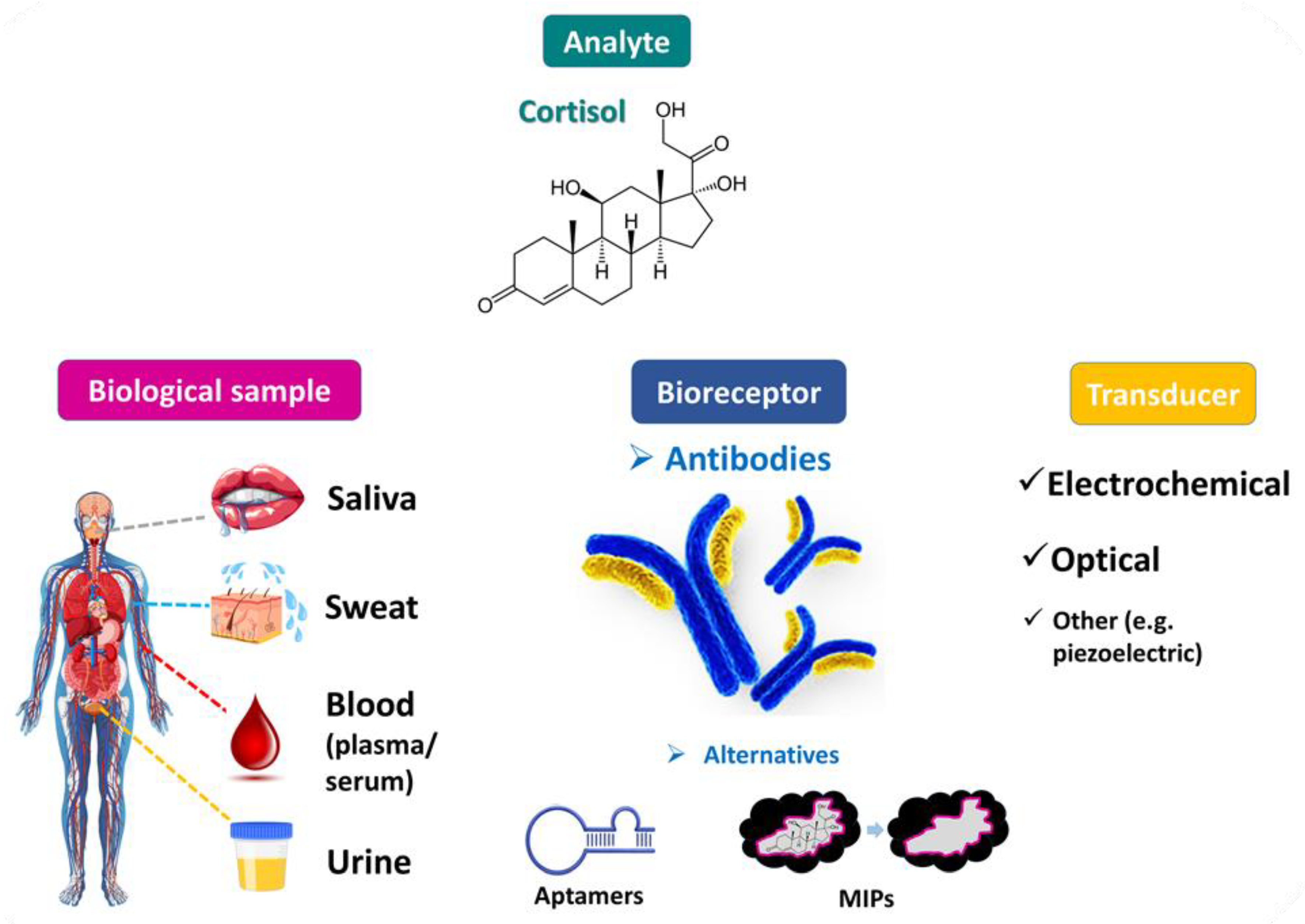
:1. Introduction
2. Immunosensors for Detecting Cortisol in Biological Samples
2.1. Electrochemical Cortisol Immunosensors
2.2. Optical Cortisol Immunosensors
2.3. Cortisol Aptasensors and MIP-Based Biosensors
3. Discussion—Future Perspectives
4. Conclusions
Author Contributions
Funding
Institutional Review Board Statement
Informed Consent Statement
Data Availability Statement
Acknowledgments
Conflicts of Interest
References
- Casals, G.; Hanzu, F.A. Cortisol Measurements in Cushing’s Syndrome: Immunoassay or Mass Spectrometry? Ann. Lab. Med. 2020, 40, 285–296. [Google Scholar] [CrossRef] [PubMed]
- Boolani, A.; Channaveerappa, D.; Dupree, E.J.; Jayathirtha, M.; Aslebagh, R.; Grobe, S.; Wilkinson, T.; Darie, C.C. Trends in Analysis of Cortisol and Its Derivatives. Adv. Exp. Med. Biol. 2019, 1140, 649–664. [Google Scholar] [CrossRef]
- Bougea, A.; Stefanis, L.; Chrousos, G. Stress system and related biomarkers in Parkinson’s disease. Adv. Clin. Chem. 2022, 111, 177–215. [Google Scholar] [CrossRef] [PubMed]
- Burke, H.M.; Davis, M.C.; Otte, C.; Mohr, D.C. Depression and cortisol responses to psychological stress: A meta-analysis. Psychoneuroendocrinology 2005, 30, 846–856. [Google Scholar] [CrossRef]
- Tsigos, C.; Chrousos, G.P. Hypothalamic-pituitary-adrenal axis, neuroendocrine factors and stress. J. Psychosom. Res. 2002, 53, 865–871. [Google Scholar] [CrossRef] [PubMed] [Green Version]
- Raff, H. Late Night Salivary Cortisol in the diagnosis of neoplastic hypercortisolism (including cyclic Cushing’s syndrome). Pituitary 2022, 25, 698–700. [Google Scholar] [CrossRef]
- Pappachan, J.M.; Hariman, C.; Edavalath, M.; Waldron, J.; Hanna, F.W. Cushing’s syndrome: A practical approach to diagnosis and differential diagnoses. J. Clin. Pathol. 2017, 70, 350–359. [Google Scholar] [CrossRef]
- Nieman, L.K.; Biller, B.M.; Findling, J.W.; Newell-Price, J.; Savage, M.O.; Stewart, P.M.; Montori, V.M. The diagnosis of Cushing’s syndrome: An Endocrine Society Clinical Practice Guideline. J. Clin. Endocrinol. Metab. 2008, 93, 1526–1540. [Google Scholar] [CrossRef]
- Lopez, A.G.; Fraissinet, F.; Lefebvre, H.; Brunel, V.; Ziegler, F. Pharmacological and analytical interference in hormone assays for diagnosis of adrenal incidentaloma. Ann. Endocrinol. 2019, 80, 250–258. [Google Scholar] [CrossRef]
- Zea, M.; Bellagambi, F.G.; Ben Halima, H.; Zine, N.; Jaffrezic-Renault, N.; Villa, R.; Gabriel, G.; Errachid, A. Electrochemical sensors for cortisol detections: Almost there. TrAC Trends Anal. Chem. 2020, 132, 116058. [Google Scholar] [CrossRef]
- Dorn, L.D.; Lucke, J.F.; Loucks, T.L.; Berga, S.L. Salivary cortisol reflects serum cortisol: Analysis of circadian profiles. Ann. Clin. Biochem. 2007, 44, 281–284. [Google Scholar] [CrossRef] [PubMed]
- Gozansky, W.S.; Lynn, J.S.; Laudenslager, M.L.; Kohrt, W.M. Salivary cortisol determined by enzyme immunoassay is preferable to serum total cortisol for assessment of dynamic hypothalamic--pituitary--adrenal axis activity. Clin. Endocrinol. 2005, 63, 336–341. [Google Scholar] [CrossRef] [PubMed]
- Umeda, T.; Hiramatsu, R.; Iwaoka, T.; Shimada, T.; Miura, F.; Sato, T. Use of saliva for monitoring unbound free cortisol levels in serum. Clin. Chim. Acta 1981, 110, 245–253. [Google Scholar] [CrossRef] [PubMed]
- Boroumand, M.; Olianas, A.; Cabras, T.; Manconi, B.; Fanni, D.; Faa, G.; Desiderio, C.; Messana, I.; Castagnola, M. Saliva, a bodily fluid with recognized and potential diagnostic applications. J. Sep. Sci. 2021, 44, 3677–3690. [Google Scholar] [CrossRef] [PubMed]
- Caruso, B.; Bovo, C.; Guidi, G.C. Causes of Preanalytical Interferences on Laboratory Immunoassays—A Critical Review. Ejifcc 2020, 31, 70–84. [Google Scholar]
- Kudielka, B.M.; Hellhammer, D.H.; Wüst, S. Why do we respond so differently? Reviewing determinants of human salivary cortisol responses to challenge. Psychoneuroendocrinology 2009, 34, 2–18. [Google Scholar] [CrossRef]
- Hellhammer, D.H.; Wüst, S.; Kudielka, B.M. Salivary cortisol as a biomarker in stress research. Psychoneuroendocrinology 2009, 34, 163–171. [Google Scholar] [CrossRef]
- Madhu, S.; Ramasamy, S.; Sekhar, P.; Bhansali, S.; Nagamony, P.; Manickam, P.; Viswanathan, C. Review—Towards Wearable Sensor Platforms for the Electrochemical Detection of Cortisol. J. Electrochem. Soc. 2020, 167, 067508. [Google Scholar] [CrossRef]
- Greff, M.J.E.; Levine, J.M.; Abuzgaia, A.M.; Elzagallaai, A.A.; Rieder, M.J.; van Uum, S.H.M. Hair cortisol analysis: An update on methodological considerations and clinical applications. Clin. Biochem. 2019, 63, 1–9. [Google Scholar] [CrossRef]
- Stalder, T.; Kirschbaum, C. Analysis of cortisol in hair--state of the art and future directions. Brain Behav. Immun. 2012, 26, 1019–1029. [Google Scholar] [CrossRef]
- Wennig, R. Potential problems with the interpretation of hair analysis results. Forensic Sci. Int. 2000, 107, 5–12. [Google Scholar] [CrossRef] [PubMed]
- Golub, Y.; Kuitunen-Paul, S.; Panaseth, K.; Stonawski, V.; Frey, S.; Steigleder, R.; Grimm, J.; Goecke, T.W.; Fasching, P.A.; Beckmann, M.W.; et al. Salivary and hair cortisol as biomarkers of emotional and behavioral symptoms in 6–9 year old children. Physiol. Behav. 2019, 209, 112584. [Google Scholar] [CrossRef] [PubMed]
- Pauli-Pott, U.; Schloß, S.; Skoluda, N.; Nater, U.M.; Becker, K. Low hair cortisol concentration predicts the development of attention deficit hyperactivity disorder. Psychoneuroendocrinology 2019, 110, 104442. [Google Scholar] [CrossRef] [PubMed]
- Fischer, S.; Schumacher, S.; Skoluda, N.; Strahler, J. Fingernail cortisol—State of research and future directions. Front. Neuroendocrinol. 2020, 58, 100855. [Google Scholar] [CrossRef]
- Izawa, S.; Sugaya, N.; Ogawa, N.; Shirotsuki, K.; Nomura, S. A validation study on fingernail cortisol: Correlations with one-month cortisol levels estimated by hair and saliva samples. Stress 2021, 24, 734–741. [Google Scholar] [CrossRef]
- Palme, R. Non-invasive measurement of glucocorticoids: Advances and problems. Physiol. Behav. 2019, 199, 229–243. [Google Scholar] [CrossRef] [PubMed]
- Kannankeril, J.; Carroll, T.; Findling, J.W.; Javorsky, B.; Gunsolus, I.L.; Phillips, J.; Raff, H. Prospective Evaluation of Late-Night Salivary Cortisol and Cortisone by EIA and LC-MS/MS in Suspected Cushing Syndrome. J. Endocr. Soc. 2020, 4, bvaa107. [Google Scholar] [CrossRef]
- Giacomello, G.; Scholten, A.; Parr, M.K. Current methods for stress marker detection in saliva. J. Pharm. Biomed. Anal. 2020, 191, 113604. [Google Scholar] [CrossRef]
- Russell, E.; Kirschbaum, C.; Laudenslager, M.L.; Stalder, T.; de Rijke, Y.; van Rossum, E.F.; Van Uum, S.; Koren, G. Toward standardization of hair cortisol measurement: Results of the first international interlaboratory round robin. Ther. Drug Monit. 2015, 37, 71–75. [Google Scholar] [CrossRef]
- Verbeeten, K.C.; Ahmet, A.H. The role of corticosteroid-binding globulin in the evaluation of adrenal insufficiency. J. Pediatr. Endocrinol. Metab. 2018, 31, 107–115. [Google Scholar] [CrossRef]
- Aydin, M.; Aydin, E.B.; Sezgintürk, M.K. Advances in immunosensor technology. Adv. Clin. Chem. 2021, 102, 1–62. [Google Scholar] [CrossRef] [PubMed]
- Mathew, M.; Radhakrishnan, S.; Vaidyanathan, A.; Chakraborty, B.; Rout, C.S. Flexible and wearable electrochemical biosensors based on two-dimensional materials: Recent developments. Anal Bioanal Chem 2021, 413, 727–762. [Google Scholar] [CrossRef]
- Xu, Y.; Suleiman, A.A. Reusable Amperometric Immunosensor for the Determination of Cortisol. Anal. Lett. 1997, 30, 2675–2689. [Google Scholar] [CrossRef]
- Cook, C.J. Real-time measurements of corticosteroids in conscious animals using an antibody-based electrode. Nat. Biotechnol. 1997, 15, 467–471. [Google Scholar] [CrossRef] [PubMed]
- Attili, B.S.; Suleiman, A.A. A piezoelectric immunosensor for the detection of cortisol. Anal. Lett. 1995, 28, 2149–2159. [Google Scholar] [CrossRef] [PubMed]
- Malon, R.S.; Sadir, S.; Balakrishnan, M.; Córcoles, E.P. Saliva-based biosensors: Noninvasive monitoring tool for clinical diagnostics. Biomed. Res. Int. 2014, 2014, 962903. [Google Scholar] [CrossRef] [PubMed] [Green Version]
- Perkins, H.; Higgins, M.; Marcato, M.; Galvin, P.; Teixeira, S.R. Immunosensor for Assessing the Welfare of Trainee Guide Dogs. Biosensors 2021, 11, 327. [Google Scholar] [CrossRef] [PubMed]
- Falk, M.; Psotta, C.; Cirovic, S.; Shleev, S. Non-Invasive Electrochemical Biosensors Operating in Human Physiological Fluids. Sensors 2020, 20, 6352. [Google Scholar] [CrossRef] [PubMed]
- Chung, M.; Fortunato, G.; Radacsi, N. Wearable flexible sweat sensors for healthcare monitoring: A review. J. R. Soc. Interface 2019, 16, 20190217. [Google Scholar] [CrossRef]
- Kaushik, A.; Vasudev, A.; Arya, S.K.; Pasha, S.K.; Bhansali, S. Recent advances in cortisol sensing technologies for point-of-care application. Biosens. Bioelectron. 2014, 53, 499–512. [Google Scholar] [CrossRef]
- Laochai, T.; Yukird, J.; Promphet, N.; Qin, J.; Chailapakul, O.; Rodthongkum, N. Non-invasive electrochemical immunosensor for sweat cortisol based on L-cys/AuNPs/ MXene modified thread electrode. Biosens. Bioelectron. 2022, 203, 114039. [Google Scholar] [CrossRef]
- Cheng, C.; Li, X.; Xu, G.; Lu, Y.; Low, S.S.; Liu, G.; Zhu, L.; Li, C.; Liu, Q. Battery-free, wireless, and flexible electrochemical patch for in situ analysis of sweat cortisol via near field communication. Biosens. Bioelectron. 2021, 172, 112782. [Google Scholar] [CrossRef] [PubMed]
- Liu, Q.; Shi, W.; Tian, L.; Su, M.; Jiang, M.; Li, J.; Gu, H.; Yu, C. Preparation of nanostructured PDMS film as flexible immunosensor for cortisol analysis in human sweat. Anal. Chim. Acta 2021, 1184, 339010. [Google Scholar] [CrossRef] [PubMed]
- Torrente-Rodríguez, R.M.; Tu, J.; Yang, Y.; Min, J.; Wang, M.; Song, Y.; Yu, Y.; Xu, C.; Ye, C.; IsHak, W.W.; et al. Investigation of cortisol dynamics in human sweat using a graphene-based wireless mHealth system. Matter 2020, 2, 921–937. [Google Scholar] [CrossRef] [PubMed]
- Bian, L.; Shao, W.; Liu, Z.; Zeng, Z.; Star, A. Detection of Stress Hormone with Semiconducting Single-Walled Carbon Nanotube-Based Field-Effect Transistors. J. Electrochem. Soc. 2022, 169, 057519. [Google Scholar] [CrossRef]
- Madhu, S.; Anthuuvan, A.J.; Ramasamy, S.; Manickam, P.; Bhansali, S.; Nagamony, P.; Chinnuswamy, V. ZnO Nanorod Integrated Flexible Carbon Fibers for Sweat Cortisol Detection. ACS Appl. Electron. Mater. 2020, 2, 499–509. [Google Scholar] [CrossRef]
- Cardinell, B.A.; Spano, M.L.; La Belle, J.T. Toward a Label-Free Electrochemical Impedance Immunosensor Design for Quantifying Cortisol in Tears. Crit. Rev. Biomed. Eng. 2019, 47, 207–215. [Google Scholar] [CrossRef]
- Cruz, A.F.; Norena, N.; Kaushik, A.; Bhansali, S. A low-cost miniaturized potentiostat for point-of-care diagnosis. Biosens. Bioelectron. 2014, 62, 249–254. [Google Scholar] [CrossRef]
- Kaushik, A.; Vasudev, A.; Arya, S.K.; Bhansali, S. Mediator and label free estimation of stress biomarker using electrophoretically deposited Ag@AgO-polyaniline hybrid nanocomposite. Biosens. Bioelectron. 2013, 50, 35–41. [Google Scholar] [CrossRef]
- Luo, D.; Fu, Q.; Gao, R.; Su, L.; Su, Y.; Liu, B. Signal-on photoelectrochemical immunoassay for salivary cortisol based on silver nanoclusters-triggered ion-exchange reaction with CdS quantum dots. Anal. Bioanal. Chem. 2022, 414, 3033–3042. [Google Scholar] [CrossRef]
- Abdulsattar, J.O.; Greenway, G.M.; Wadhawan, J.D. Electrochemical immunoassay for the detection of stress biomarkers. Heliyon 2020, 6, e03558. [Google Scholar] [CrossRef]
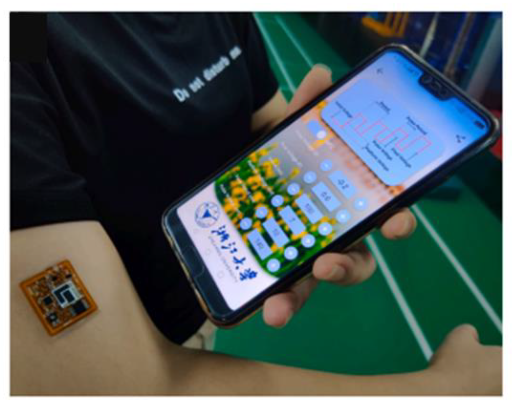
- Liu, J.; Xu, N.; Men, H.; Li, S.; Lu, Y.; Low, S.S.; Li, X.; Zhu, L.; Cheng, C.; Xu, G.; et al. Salivary Cortisol Determination on Smartphone-Based Differential Pulse Voltammetry System. Sensors 2020, 20, 1422. [Google Scholar] [CrossRef] [Green Version]
- Dhull, N.; Kaur, G.; Gupta, V.; Tomar, M. Highly sensitive and non-invasive electrochemical immunosensor for salivary cortisol detection. Sens. Actuators B Chem. 2019, 293, 281–288. [Google Scholar] [CrossRef]
- Khan, M.S.; Dighe, K.; Wang, Z.; Srivastava, I.; Schwartz-Duval, A.S.; Misra, S.K.; Pan, D. Electrochemical-digital immunosensor with enhanced sensitivity for detecting human salivary glucocorticoid hormone. Analyst 2019, 144, 1448–1457. [Google Scholar] [CrossRef] [PubMed]
- Liu, X.; Hsu, S.P.C.; Liu, W.C.; Wang, Y.M.; Liu, X.; Lo, C.S.; Lin, Y.C.; Nabilla, S.C.; Li, Z.; Hong, Y.; et al. Salivary Electrochemical Cortisol Biosensor Based on Tin Disulfide Nanoflakes. Nanoscale Res. Lett. 2019, 14, 189. [Google Scholar] [CrossRef] [PubMed] [Green Version]
- Tuteja, S.K.; Ormsby, C.; Neethirajan, S. Noninvasive Label-Free Detection of Cortisol and Lactate Using Graphene Embedded Screen-Printed Electrode. Nanomicro Lett. 2018, 10, 41. [Google Scholar] [CrossRef] [Green Version]
- Kämäräinen, S.; Mäki, M.; Tolonen, T.; Palleschi, G.; Virtanen, V.; Micheli, L.; Sesay, A.M. Disposable electrochemical immunosensor for cortisol determination in human saliva. Talanta 2018, 188, 50–57. [Google Scholar] [CrossRef]
- Kim, Y.H.; Lee, K.; Jung, H.; Kang, H.K.; Jo, J.; Park, I.K.; Lee, H.H. Direct immune-detection of cortisol by chemiresistor graphene oxide sensor. Biosens. Bioelectron. 2017, 98, 473–477. [Google Scholar] [CrossRef]
- Vabbina, P.K.; Kaushik, A.; Pokhrel, N.; Bhansali, S.; Pala, N. Electrochemical cortisol immunosensors based on sonochemically synthesized zinc oxide 1D nanorods and 2D nanoflakes. Biosens. Bioelectron. 2015, 63, 124–130. [Google Scholar] [CrossRef]
- Pasha, S.K.; Kaushik, A.; Vasudev, A.; Snipes, S.A.; Bhansali, S. Electrochemical Immunosensing of Saliva Cortisol. J. Electrochem. Soc. 2014, 161, B3077. [Google Scholar] [CrossRef]
- Yamaguchi, M.; Matsuda, Y.; Sasaki, S.; Sasaki, M.; Kadoma, Y.; Imai, Y.; Niwa, D.; Shetty, V. Immunosensor with fluid control mechanism for salivary cortisol analysis. Biosens. Bioelectron. 2013, 41, 186–191. [Google Scholar] [CrossRef] [Green Version]
- Tlili, C.; Myung, N.V.; Shetty, V.; Mulchandani, A. Label-free, chemiresistor immunosensor for stress biomarker cortisol in saliva. Biosens. Bioelectron. 2011, 26, 4382–4386. [Google Scholar] [CrossRef] [PubMed]
- Venugopal, M.; Arya, S.K.; Chornokur, G.; Bhansali, S. A Realtime and Continuous Assessment of Cortisol in ISF Using Electrochemical Impedance Spectroscopy. Sens. Actuators A Phys. 2011, 172, 154–160. [Google Scholar] [CrossRef] [Green Version]
- Vargas, E.; Povedano, E.; Krishnan, S.; Teymourian, H.; Tehrani, F.; Campuzano, S.; Dassau, E.; Wang, J. Simultaneous cortisol/insulin microchip detection using dual enzyme tagging. Biosens. Bioelectron. 2020, 167, 112512. [Google Scholar] [CrossRef] [PubMed]
- Sun, B.; Gou, Y.; Ma, Y.; Zheng, X.; Bai, R.; Ahmed Abdelmoaty, A.A.; Hu, F. Investigate electrochemical immunosensor of cortisol based on gold nanoparticles/magnetic functionalized reduced graphene oxide. Biosens. Bioelectron. 2017, 88, 55–62. [Google Scholar] [CrossRef] [PubMed]
- Wu, H.; Ohnuki, H.; Ota, S.; Murata, M.; Yoshiura, Y.; Endo, H. New approach for monitoring fish stress: A novel enzyme-functionalized label-free immunosensor system for detecting cortisol levels in fish. Biosens. Bioelectron. 2017, 93, 57–64. [Google Scholar] [CrossRef]
- Kaushik, A.; Yndart, A.; Jayant, R.D.; Sagar, V.; Atluri, V.; Bhansali, S.; Nair, M. Electrochemical sensing method for point-of-care cortisol detection in human immunodeficiency virus-infected patients. Int. J. Nanomed. 2015, 10, 677–685. [Google Scholar] [CrossRef] [Green Version]
- Liu, X.; Zhao, R.; Mao, W.; Feng, H.; Liu, X.; Wong, D.K.Y. Detection of cortisol at a gold nanoparticle|Protein G–DTBP-scaffold modified electrochemical immunosensor. Analyst 2011, 136, 5204–5210. [Google Scholar] [CrossRef]
- Moreno-Guzmán, M.; Eguílaz, M.; Campuzano, S.; González-Cortés, A.; Yáñez-Sedeño, P.; Pingarrón, J.M. Disposable immunosensor for cortisol using functionalized magnetic particles. Analyst 2010, 135, 1926–1933. [Google Scholar] [CrossRef]
- Kumar, A.; Aravamudhan, S.; Gordic, M.; Bhansali, S.; Mohapatra, S.S. Ultrasensitive detection of cortisol with enzyme fragment complementation technology using functionalized nanowire. Biosens. Bioelectron. 2007, 22, 2138–2144. [Google Scholar] [CrossRef]
- Arya, S.K.; Chornokur, G.; Venugopal, M.; Bhansali, S. Dithiobis(succinimidyl propionate) modified gold microarray electrode based electrochemical immunosensor for ultrasensitive detection of cortisol. Biosens. Bioelectron. 2010, 25, 2296–2301. [Google Scholar] [CrossRef] [Green Version]
- Cook, C.J. Measuring of extracellular cortisol and corticotropin-releasing hormone in the amygdala using immunosensor coupled microdialysis. J. Neurosci. Methods 2001, 110, 95–101. [Google Scholar] [CrossRef]
- Sekar, M.; Pandiaraj, M.; Bhansali, S.; Ponpandian, N.; Viswanathan, C. Carbon fiber based electrochemical sensor for sweat cortisol measurement. Sci. Rep. 2019, 9, 403. [Google Scholar] [CrossRef] [PubMed] [Green Version]
- Kinnamon, D.; Ghanta, R.; Lin, K.C.; Muthukumar, S.; Prasad, S. Portable biosensor for monitoring cortisol in low-volume perspired human sweat. Sci. Rep. 2017, 7, 13312. [Google Scholar] [CrossRef] [PubMed] [Green Version]
- Munje, R.D.; Muthukumar, S.; Jagannath, B.; Prasad, S. A new paradigm in sweat based wearable diagnostics biosensors using Room Temperature Ionic Liquids (RTILs). Sci. Rep. 2017, 7, 1950. [Google Scholar] [CrossRef] [PubMed] [Green Version]
- Munje, R.D.; Muthukumar, S.; Panneer Selvam, A.; Prasad, S. Flexible nanoporous tunable electrical double layer biosensors for sweat diagnostics. Sci. Rep. 2015, 5, 14586. [Google Scholar] [CrossRef] [PubMed] [Green Version]
- Stevens, R.C.; Soelberg, S.D.; Near, S.; Furlong, C.E. Detection of Cortisol in Saliva with a Flow-Filtered, Portable Surface Plasmon Resonance Biosensor System. Anal. Chem. 2008, 80, 6747–6751. [Google Scholar] [CrossRef] [Green Version]
- Frasconi, M.; Mazzarino, M.; Botrè, F.; Mazzei, F. Surface plasmon resonance immunosensor for cortisol and cortisone determination. Anal. Bioanal. Chem. 2009, 394, 2151–2159. [Google Scholar] [CrossRef]
- Mitchell, J.S.; Lowe, T.E.; Ingram, J.R. Rapid ultrasensitive measurement of salivary cortisol using nano-linker chemistry coupled with surface plasmon resonance detection. Analyst 2009, 134, 380–386. [Google Scholar] [CrossRef]
- Ilea, A.; Andrei, V.; Feurdean, C.N.; Băbțan, A.M.; Petrescu, N.B.; Câmpian, R.S.; Boșca, A.B.; Ciui, B.; Tertiș, M.; Săndulescu, R.; et al. Saliva, a Magic Biofluid Available for Multilevel Assessment and a Mirror of General Health-A Systematic Review. Biosensors 2019, 9, 27. [Google Scholar] [CrossRef] [PubMed] [Green Version]
- Soares, M.S.; Silva, L.C.B.; Vidal, M.; Loyez, M.; Facão, M.; Caucheteur, C.; Segatto, M.E.V.; Costa, F.M.; Leitão, C.; Pereira, S.O.; et al. Label-free plasmonic immunosensor for cortisol detection in a D-shaped optical fiber. Biomed. Opt. Express 2022, 13, 3259–3274. [Google Scholar] [CrossRef] [PubMed]
- Leitão, C.; Leal-Junior, A.; Almeida, A.R.; Pereira, S.O.; Costa, F.M.; Pinto, J.L.; Marques, C. Cortisol AuPd plasmonic unclad POF biosensor. Biotechnol. Rep. 2021, 29, e00587. [Google Scholar] [CrossRef]
- Safarian, S.M.; Kusov, P.A.; Kosolobov, S.S.; Borzenkova, O.V.; Khakimov, A.V.; Kotelevtsev, Y.V.; Drachev, V.P. Surface-specific washing-free immunosensor for time-resolved cortisol monitoring. Talanta 2021, 225, 122070. [Google Scholar] [CrossRef]
- Liu, Y.; Wu, B.; Tanyi, E.K.; Yeasmin, S.; Cheng, L.-J. Label-Free Sensitive Detection of Steroid Hormone Cortisol Based on Target-Induced Fluorescence Quenching of Quantum Dots. Langmuir 2020, 36, 7781–7788. [Google Scholar] [CrossRef]
- Shin, J.; Kim, S.; Yoon, T.; Joo, C.; Jung, H.-I. Smart Fatigue Phone: Real-time estimation of driver fatigue using smartphone-based cortisol detection. Biosens. Bioelectron. 2019, 136, 106–111. [Google Scholar] [CrossRef] [PubMed]
- Pinto, V.; Sousa, P.; Catarino, S.O.; Correia-Neves, M.; Minas, G. Microfluidic immunosensor for rapid and highly-sensitive salivary cortisol quantification. Biosens. Bioelectron. 2017, 90, 308–313. [Google Scholar] [CrossRef] [Green Version]
- Zangheri, M.; Cevenini, L.; Anfossi, L.; Baggiani, C.; Simoni, P.; Di Nardo, F.; Roda, A. A simple and compact smartphone accessory for quantitative chemiluminescence-based lateral flow immunoassay for salivary cortisol detection. Biosens. Bioelectron. 2015, 64, 63–68. [Google Scholar] [CrossRef]
- Shirtcliff, E.A.; Buck, R.L.; Laughlin, M.J.; Hart, T.; Cole, C.R.; Slowey, P.D. Salivary cortisol results obtainable within minutes of sample collection correspond with traditional immunoassays. Clin. Ther. 2015, 37, 505–514. [Google Scholar] [CrossRef] [PubMed] [Green Version]
- Tahara, Y.; Huang, Z.; Kiritoshi, T.; Onodera, T.; Toko, K. Development of Indirect Competitive Immuno-Assay Method Using SPR Detection for Rapid and Highly Sensitive Measurement of Salivary Cortisol Levels. Front. Bioeng. Biotechnol. 2014, 2, 15. [Google Scholar] [CrossRef]
- Yamaguchi, M.; Katagata, H.; Tezuka, Y.; Niwa, D.; Shetty, V. Automated-immunosensor with centrifugal fluid valves for salivary cortisol measurement. Sens. Biosensing Res. 2014, 1, 15–20. [Google Scholar] [CrossRef] [Green Version]
- van Smeden, L.; Saris, A.; Sergelen, K.; de Jong, A.M.; Yan, J.; Prins, M.W.J. Reversible Immunosensor for the Continuous Monitoring of Cortisol in Blood Plasma Sampled with Microdialysis. ACS Sens. 2022, 7, 3041–3048. [Google Scholar] [CrossRef] [PubMed]
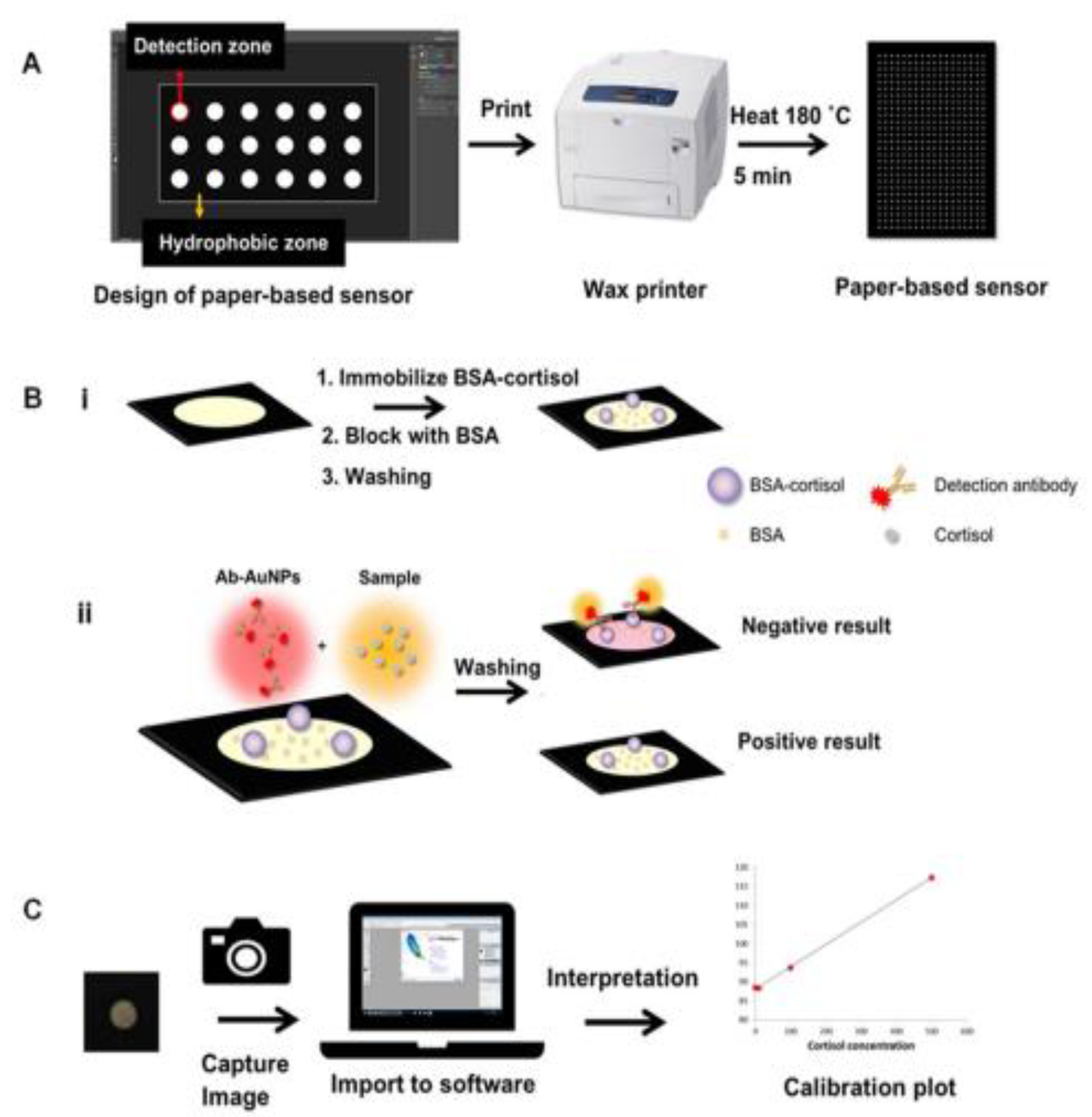
- Apilux, A.; Rengpipat, S.; Suwanjang, W.; Chailapakul, O. Paper-based immunosensor with competitive assay for cortisol detection. J. Pharm. Biomed. Anal. 2020, 178, 112925. [Google Scholar] [CrossRef] [PubMed]
- Li, Z.; Mohamed, M.A.; Vinu Mohan, A.M.; Zhu, Z.; Sharma, V.; Mishra, G.K.; Mishra, R.K. Application of Electrochemical Aptasensors toward Clinical Diagnostics, Food, and Environmental Monitoring: Review. Sensors 2019, 19, 5435. [Google Scholar] [CrossRef] [PubMed] [Green Version]
- Zhou, W.; Jimmy Huang, P.-J.; Ding, J.; Liu, J. Aptamer-based biosensors for biomedical diagnostics. Analyst 2014, 139, 2627–2640. [Google Scholar] [CrossRef] [Green Version]
- Martin, J.A.; Chávez, J.L.; Chushak, Y.; Chapleau, R.R.; Hagen, J.; Kelley-Loughnane, N. Tunable stringency aptamer selection and gold nanoparticle assay for detection of cortisol. Anal. Bioanal. Chem. 2014, 406, 4637–4647. [Google Scholar] [CrossRef] [PubMed]
- Ganguly, A.; Lin, K.C.; Muthukumar, S.; Prasad, S. Autonomous, Real-Time Monitoring Electrochemical Aptasensor for Circadian Tracking of Cortisol Hormone in Sub-microliter Volumes of Passively Eluted Human Sweat. ACS Sens. 2021, 6, 63–72. [Google Scholar] [CrossRef] [PubMed]
- Pali, M.; Jagannath, B.; Lin, K.-C.; Upasham, S.; Sankhalab, D.; Upashama, S.; Muthukumar, S.; Prasad, S. CATCH (Cortisol Apta WATCH): ‘Bio-mimic alarm’ to track Anxiety, Stress, Immunity in human sweat. Electrochim. Acta 2021, 390, 138834. [Google Scholar] [CrossRef]
- Pusomjit, P.; Teengam, P.; Thepsuparungsikul, N.; Sanongkiet, S.; Chailapakul, O. Impedimetric determination of cortisol using screen-printed electrode with aptamer-modified magnetic beads. Microchim. Acta 2021, 188, 41. [Google Scholar] [CrossRef]
- Sheibani, S.; Capua, L.; Kamaei, S.; Akbari, S.S.A.; Zhang, J.; Guerin, H.; Ionescu, A.M. Extended gate field-effect-transistor for sensing cortisol stress hormone. Commun. Mater. 2021, 2, 10. [Google Scholar] [CrossRef] [PubMed]
- Fernandez, R.E.; Umasankar, Y.; Manickam, P.; Nickel, J.C.; Iwasaki, L.R.; Kawamoto, B.K.; Todoki, K.C.; Scott, J.M.; Bhansali, S. Disposable aptamer-sensor aided by magnetic nanoparticle enrichment for detection of salivary cortisol variations in obstructive sleep apnea patients. Sci. Rep. 2017, 7, 17992. [Google Scholar] [CrossRef] [Green Version]
- Sanghavi, B.J.; Moore, J.A.; Chávez, J.L.; Hagen, J.A.; Kelley-Loughnane, N.; Chou, C.F.; Swami, N.S. Aptamer-functionalized nanoparticles for surface immobilization-free electrochemical detection of cortisol in a microfluidic device. Biosens. Bioelectron. 2016, 78, 244–252. [Google Scholar] [CrossRef] [PubMed]
- Mortazavi Moghadam, F.; Bigdeli, M.; Tamayol, A.; Shin, S.R. TISS nanobiosensor for salivary cortisol measurement by aptamer Ag nanocluster SAIE supraparticle structure. Sens. Actuators B Chem. 2021, 344, 130160. [Google Scholar] [CrossRef]
- Dalirirad, S.; Han, D.; Steckl, A.J. Aptamer-Based Lateral Flow Biosensor for Rapid Detection of Salivary Cortisol. ACS Omega 2020, 5, 32890–32898. [Google Scholar] [CrossRef] [PubMed]
- Dalirirad, S.; Steckl, A.J. Aptamer-based lateral flow assay for point of care cortisol detection in sweat. Sens. Actuators B Chem. 2019, 283, 79–86. [Google Scholar] [CrossRef]
- Ramanavičius, S.; Morkvėnaitė-Vilkončienė, I.; Samukaitė-Bubnienė, U.; Ratautaitė, V.; Plikusienė, I.; Viter, R.; Ramanavičius, A. Electrochemically Deposited Molecularly Imprinted Polymer-Based Sensors. Sensors 2022, 22, 1282. [Google Scholar] [CrossRef] [PubMed]
- Kadhem, A.J.; Gentile, G.J.; Fidalgo de Cortalezzi, M.M. Molecularly Imprinted Polymers (MIPs) in Sensors for Environmental and Biomedical Applications: A Review. Molecules 2021, 26, 6233. [Google Scholar] [CrossRef]
- Uygun, H.D.E.; Uygun, Z.O.; Canbay, E.; Sağın, F.G.; Sezer, E. Non-invasive cortisol detection in saliva by using molecularly cortisol imprinted fullerene-acrylamide modified screen printed electrodes. Talanta 2020, 206, 120225. [Google Scholar] [CrossRef] [PubMed]
- Manickam, P.; Pasha, S.; Snipes, S.; Bhansali, S. A Reusable Electrochemical Biosensor for Monitoring of Small Molecules (Cortisol) Using Molecularly Imprinted Polymers. J. Electrochem. Soc. 2017, 164, B54–B59. [Google Scholar] [CrossRef]
- Manickam, P.; Fernandez, R.E.; Umasankar, Y.; Gurusamy, M.; Arizaleta, F.; Urizar, G.; Bhansali, S. Salivary cortisol analysis using metalloporphyrins and multi-walled carbon nanotubes nanocomposite functionalized electrodes. Sens. Actuators B Chem. 2018, 274, 47–53. [Google Scholar] [CrossRef]
- Parlak, O.; Keene, S.T.; Marais, A.; Curto, V.F.; Salleo, A. Molecularly selective nanoporous membrane-based wearable organic electrochemical device for noninvasive cortisol sensing. Sci. Adv. 2018, 4, eaar2904. [Google Scholar] [CrossRef] [Green Version]
- Usha, S.P.; Shrivastav, A.M.; Gupta, B.D. A contemporary approach for design and characterization of fiber-optic-cortisol sensor tailoring LMR and ZnO/PPY molecularly imprinted film. Biosens. Bioelectron. 2017, 87, 178–186. [Google Scholar] [CrossRef] [PubMed]
- Visser, E.W.A.; Yan, J.; van, I.L.J.; Prins, M.W.J. Continuous biomarker monitoring by particle mobility sensing with single molecule resolution. Nat. Commun. 2018, 9, 2541. [Google Scholar] [CrossRef] [PubMed] [Green Version]
- Sonner, Z.; Wilder, E.; Heikenfeld, J.; Kasting, G.; Beyette, F.; Swaile, D.; Sherman, F.; Joyce, J.; Hagen, J.; Kelley-Loughnane, N.; et al. The microfluidics of the eccrine sweat gland, including biomarker partitioning, transport, and biosensing implications. Biomicrofluidics 2015, 9, 031301. [Google Scholar] [CrossRef] [PubMed] [Green Version]
- Hogenelst, K.; Soeter, M.; Kallen, V. Ambulatory measurement of cortisol: Where do we stand, and which way to follow? Sens. Bio-Sens. Res. 2019, 22, 100249. [Google Scholar] [CrossRef]
- Justino, C.I.L.; Duarte, A.C.; Rocha-Santos, T.A.P. Critical overview on the application of sensors and biosensors for clinical analysis. Trends Anal. Chem. 2016, 85, 36–60. [Google Scholar] [CrossRef]
- Yang, Y.; Gao, W. Wearable and flexible electronics for continuous molecular monitoring. Chem. Soc. Rev. 2019, 48, 1465–1491. [Google Scholar] [CrossRef]


| Immunoassay Principle | Signal Transduction Principle | Biological Sample | Range/LoD | Reference |
|---|---|---|---|---|
| Noncompetitive (Direct binding of cortisol to an anti-cortisol Ab 1, immobilized onto gold microelectrodes) | Cyclic voltammetry | Buffer | 10 pM–500 nM/ 1 pM | [48] |
| Noncompetitive (Direct binding of cortisol to an anti-cortisol Ab, immobilized onto silver/silver oxide (Ag/AgO)—polyaniline nanocomposites) | Cyclic voltammetry | Buffer | 1 pM–1 μM 0.64 pM | [49] |
| Noncompetitive (Direct binding of cortisol to an anti-cortisol Ab co-immobilized with HRP on the sensor electrode) | Amperometry | Buffer | 10−7–10−5 M | [33] |
| Competitive (Competition between free cortisol and a BSA-cortisol conjugate immobilized onto magnetic beads for binding to an anti-cortisol Ab labeled with silver nanoclusters (AgNCs) | Photoelectrochemistry | Saliva | 0.0001–100 ng/mL/ 0.06 pg/mL | [50] |
| Noncompetitive (Direct binding of cortisol to an anti-cortisol Ab immobilized on polyaniline-modified graphene electrodes) | Electrochemical impedance spectroscopy | Saliva (canine) | 0.0005–50 μg/mL/ 3.57 fg/mL | [37] |
| Noncompetitive (Direct binding of cortisol to an anti-cortisol Ab labeled with ferrocene-tags and immobilized on a modified tin-doped indium oxide electrode) | Cyclic voltammetry; square wave voltammetry | artificial saliva and zebrafish whole-body | 0.001–50 ng/mL/ 1.03 pg/mL | [51] |
| Noncompetitive (Direct binding of cortisol to an anti-cortisol Ab immobilized on a AuNP/MoS2/AuNP–modified screen-printed electrode) | Differential pulse voltammetry | Saliva | 0.5–200 nM/ 0.11 nM | [52] |
| Noncompetitive (Direct binding of cortisol to an anti-cortisol Ab covalently immobilized on NiO thin film/ITO 2 electrodes) | Cyclic voltammetry, Differential pulse voltammetry | Saliva | 1 pg/mL–10 μg/mL/ 0.32 pg/mL | [53] |
| Noncompetitive (Direct binding of cortisol to an anti-cortisol Ab covalently immobilized onto micro-Au electrodes) | Electrochemical impedance spectroscopy | Saliva | 1 pg/mL–10 ng/mL/ 0.87 ± 0.12 pg/mL | [54] |
| Noncompetitive (Direct binding of cortisol to an anti-cortisol Ab co-immobilized with BSA on glassy carbon electrodes that had been coated with tin disulfide nanoflakes) | Cyclic voltammetry, differential pulse voltammetry | Saliva | 100 pM–100 μM/ 100 pM | [55] |
| Competitive (Competition between an ALP-labeled cortisol conjugate and free cortisol for binding to an anti-cortisol Ab indirectly immobilized on disposable graphite screen-printed electrodes) | Square wave voltammetry | Saliva | 0.5–55.1 ng/mL/ 1.7 ng/mL | [57] |
| Noncompetitive (Direct binding of cortisol to an anti-cortisol Ab immobilized on electroreduced graphene oxide deposited on screen-printed electrodes) | Electrochemical chronoamperometry | Saliva, sweat | 0.1 ng/mL | [56] |
| Noncompetitive (Direct binding of cortisol to an anti-cortisol Ab covalently immobilized on reduced graphene oxide channels between two planar electrodes) | Resistance | Human saliva and buffer solution of rat adrenal gland acute slices | 10 pg/mL | [58] |
| Noncompetitive (Direct binding of cortisol to an anti-cortisol Ab immobilized on Au-substrates modified with ZnO nanostructures (1D nanorods, 2D nanoflakes)) | Cyclic voltammetry | Saliva | 1 pM | [59] |
| Noncompetitive (Direct binding of cortisol to an anti-cortisol Ab covalently immobilized on microfabricated interdigitated microelectrodes) | Cyclic voltammetry | Saliva | 10 pg/mL–100 ng/mL/ 10 pg/mL | [60] |
| Competitive (Competition between a GOD 3–cortisol conjugate and free cortisol for binding to an anti-cortisol Ab immobilized on platinum electrodes; lateral and vertical fluid control mechanisms were integrated in the sensor) | Amperometry | Saliva | 0.1–10 ng/mL | [61] |
| Noncompetitive (Direct binding of cortisol to an anti-cortisol Ab immobilized on gold microelectrode arrays) | Electrochemical impedance spectroscopy | Saliva and interstitial fluid | 1 pM–100 nM | [63] |
| Competitive (Competition between free cortisol and a cortisol analog covalently immobilized on single-walled carbon nanotubes and free cortisol for binding to an anti-cortisol Ab) | Resistance/conductance | Saliva | 1 pg/mL–10 ng/mL/ 1 pg/mL | [62] |
| Competitive (Competition between cortisol immobilized on naflon pretreated glassy carbon electrodes and free cortisol for binding to a biotinylated anti-cortisol Ab; detection was performed via reaction with HRP–streptavidin) | Electrochemical impedance spectroscopy, cyclic voltammetry | Plasma | 0.1–1000 ng/mL/ 0.05 ng/mL | [65] |
| Noncompetitive (Direct binding of cortisol to an anti-cortisol Ab co-immobilized with GOD on gold electrodes) | Amperometry | Plasma (fish) | 1.25–200 ng/mL | [66] |
| Noncompetitive (Direct binding of cortisol to an anti-cortisol Ab, immobilized on interdigitated gold microelectrodes) | Cyclic voltammetry | Plasma | 10 pg/mL–500 ng/mL/ 10 pg/mL | [67] |
| Competitive (Competition between an ALP-labeled cortisol conjugate and free cortisol for binding to an anti-cortisol Ab covalently immobilized on gold electrodes. | Amperometry | Serum | 0–250 ng/mL/ 13.4 ng/mL | [64] |
| Competitive (Competition between an ALP-labeled cortisol conjugate and free cortisol for binding to an anti-cortisol Ab immobilized through protein A on magnetic particles; the immunocomplexes formed were trapped on the surface of screen-printed electrodes with a small magnet and ALP activity was monitored) | Differential pulse voltammetry | Serum | 5 × 10⁻³–150 ng/mL/ 3.5 pg/mL | [69] |
| Competitive (Competition between an HRP-labeled cortisol conjugate and free cortisol for binding to an anti-cortisol Ab immobilized on gold electrodes functionalized with a AuNP–protein G–DTBP 4 scaffold) | Square wave voltammetry | Buffer, Serum | 50–2,500 pg/mL/ 16 pg/mL | [68] |
| Noncompetitive (Direct binding of cortisol and cortisone (transformed into cortisol via the enzyme 3α-hydroxysteroid dehydrogenase) to an anti-cortisol Ab immobilized on gold nanowires/working electrodes) | Square wave voltammetry | Buffer, Serum | 10–80 μM | [70] |
| Noncompetitive (Direct binding of cortisol to an anti-cortisol Ab immobilized on L-cys 5–AuNPs–MXene-modified electrodes) | Amperometry | Artificial Sweat | 5–180 ng/mL/ 0.54 ng/mL | [41] |
| Noncompetitive (Direct binding of cortisol to an anti-cortisol Ab immobilized on the surface of flexible screen-printed electrodes coated with AuNPs) | Differential pulse voltammetry | Sweat | 7.47 nM | [42] |
| Noncompetitive (Direct binding of cortisol to an anti-cortisol Ab, immobilized on a flexible electrode prepared on polydimethylsiloxane modified with multiwalled carbon nanotubes and AuNPs) | Cyclic voltammetry, differential pulse voltammetry | Sweat | 1 fg/mL–1 μg/mL/ 0.3 fg/mL | [43] |
| Competitive (Competition between an HRP–cortisol conjugate and free cortisol for binding to an anti-cortisol antibody immobilized on graphene-based electrode) | Amperometry | Sweat | 0.43–50.2 ng/mL | [44] |
| Noncompetitive (Direct binding of cortisol to an anti-cortisol antibody immobilized on a conductive carbon yarn functionalized with ellipsoidal Fe2O3 particles) | Cyclic voltammetry | Sweat | 1 fg/mL–1 μg/mL/ 0.005 fg/mL | [73] |
| Noncompetitive (Direct binding of cortisol to an anti-cortisol Ab immobilized on MoS2 sheets integrated into a nanoporous flexible electrode system) | Electrochemical impedance spectroscopy | Sweat | 1–500 ng/mL/ 1 ng/mL | [74] |
| Noncompetitive (Direct binding of cortisol to an anti-cortisol Ab immobilized on ZnO thin film deposited on a flexible nanoporous polyamide membrane; room temperature ionic liquids were employed to enhance sensor stability) | Electrochemical impedance spectroscopy | Sweat | 10–200 ng/mL/ 10 ng/mL | [75] |
| Noncompetitive (Direct binding of cortisol to an anti-cortisol Ab immobilized on a ZnO thin film deposited on a flexible nanoporous polyamide membrane) | Electrochemical impedance spectroscopy | Sweat | 10–200 ng/mL/ 1 ng/mL | [76] |
| Noncompetitive (Direct binding of cortisol to an anti-cortisol Ab, covalently immobilized on a gold microelectrode array) | Electrochemical impedance spectroscopy | Interstitial fluid | 1 pM–100 nM | [71] |
| Competitive (Competition between free cortisol and an HRP–cortisol conjugate for binding to an anti-cortisol antibody immobilized on a platinum electrode) | Amperometry | Dialysates of extracellular fluid of animal brain, amygdala region (sheep) | 0–100 ng/mL (in vitro measurement) | [72] |
| Competitive (Competition between HRP–cortisol conjugate and free cortisol for binding to an anti-cortisol antibody immobilized on platinum electrodes) | Potentiometry | Dialysates of animal circulating blood (sheep, cattle, rat) | 0.3 μg/100 mL | [34] |
| Immunoassay Principle | Signal Transduction Principle | Biological Sample | Range/LoD | Reference |
|---|---|---|---|---|
| Noncompetitive (Direct binding of cortisol to an anti-cortisol Ab immobilized on a D-shaped, gold-coated silica optical fiber) | Surface plasmon resonance (SPR) | Buffer | 0.01–100 ng/mL/ 1.46 ng/mL | [81] |
| Noncompetitive (Direct binding of cortisol to an anti-cortisol Ab immobilized on a plastic optical fiber coated with gold–palladium allοy) | SPR | Buffer | 1 pg/mL | [82] |
| Competitive (Competition between a fluorescently labeled BSA–cortisol conjugate and free cortisol for binding to an anti-cortisol Ab immobilized on glass substrate coated with gold) | Metal-enhanced fluorescence (MEF) | Buffer | 0.02 μg/mL | [83] |
| Noncompetitive (Direct binding between of cortisol to an anti-cortisol Ab or aptamer, immobilized on quantum dots) | Fluorescence quenching | Saliva | 1 nM (aptamer-based) 100 pM (Ab-based) | [84] |
| Lateral flow–type Competitive (Competition between a BSA–cortisol conjugate immobilized on the strip and free cortisol for binding to a Cy3-labeled anti-cortisol Ab) | Fluorescence (detected with a smartphone-linked reader) | Saliva | 0.1 ng/mL | [85] |
| Competitive (Competition between an HRP-labeled cortisol conjugate and free cortisol for binding to an anti-cortisol Ab indirectly immobilized on PDMS microfluidic channel) | Colorimetry | Saliva | 0.01–20 ng/mL/ 18 pg/mL | [86] |
| Lateral flow–type Competitive (Competition between an HRP-labeled cortisol conjugate and free cortisol for binding to an anti-cortisol Ab immobilized on the strip) | Chemiluminescence (detected through a smartphone camera) | Saliva | 0.3–60 ng/mL/ 0.3 ng/mL | [87] |
| Lateral flow–type Competitive (Based on europium fluorescent particle conjugates) | Fluorescence (Detected with a cassette reader transferring results through a Bluetooth device, manufactured by Oasis Diagnostics) | Saliva | 0.91 ng/mL | [88] |
| Competitive (Competition between a cortisol analogue, hydrocortisone 3-(O-carboxymethyl)oxime, covalently immobilized on gold surface and free cortisol for binding to an anti-cortisol Ab) | SPR | Saliva | 10 ppt–100 ppb/ 38 ppt | [89] |
| Competitive (Competition between a BSA–cortisol conjugate immobilized on a disposable disk chip and free cortisol for binding to an ALP-labeled anti-cortisol Ab) | Chemiluminescence | Saliva | 0.4–11.3 ng/mL | [90] |
| Competitive (Competition between an in-house prepared cortisol conjugate immobilized on a gold sensor surface and free cortisol for binding to the anti-cortisol Ab; a secondary Ab was used for signal increase) | SPR | Saliva | 91–934 pg/mL/ 49 pg/mL | [79] |
| Noncompetitive (Direct binding of cortisol to an anti-cortisol Ab covalently immobilized on the polycarboxylate hydrogel–coated sensing surface) | SPR | Saliva, urine | 3 μg/L | [78] |
| Competitive (Competition between a BSA–cortisol conjugate immobilized on the SPR-sensor surface and free cortisol for binding to a monoclonal anti-cortisol Ab) | SPR | Saliva, buffer | 1.0 ng/mL | [77] |
| Competitive (Competition between cortisol analogues, i.e., suitably prepared cortisol–ssDNA conjugates, and free cortisol for binding to a biotinylated anti-cortisol Ab immobilized on streptavidin-coated particles) | Particle mobility (detected through dark field microscopy) | Blood plasma (filtered or microdialysis-sampled) | High nM–low μM | [91] |
| Competitive (Competition between a BSA–cortisol conjugate immobilized on paper and free cortisol for binding to gold nanoparticles loaded with the anti-cortisol Ab) | Color | Blood serum | 21.5 μg/dL | [92] |
Disclaimer/Publisher’s Note: The statements, opinions and data contained in all publications are solely those of the individual author(s) and contributor(s) and not of MDPI and/or the editor(s). MDPI and/or the editor(s) disclaim responsibility for any injury to people or property resulting from any ideas, methods, instructions or products referred to in the content. |
© 2023 by the authors. Licensee MDPI, Basel, Switzerland. This article is an open access article distributed under the terms and conditions of the Creative Commons Attribution (CC BY) license (https://creativecommons.org/licenses/by/4.0/).
Share and Cite
Karachaliou, C.-E.; Koukouvinos, G.; Goustouridis, D.; Raptis, I.; Kakabakos, S.; Petrou, P.; Livaniou, E. Cortisol Immunosensors: A Literature Review. Biosensors 2023, 13, 285. https://doi.org/10.3390/bios13020285
Karachaliou C-E, Koukouvinos G, Goustouridis D, Raptis I, Kakabakos S, Petrou P, Livaniou E. Cortisol Immunosensors: A Literature Review. Biosensors. 2023; 13(2):285. https://doi.org/10.3390/bios13020285
Chicago/Turabian StyleKarachaliou, Chrysoula-Evangelia, Georgios Koukouvinos, Dimitrios Goustouridis, Ioannis Raptis, Sotirios Kakabakos, Panagiota Petrou, and Evangelia Livaniou. 2023. "Cortisol Immunosensors: A Literature Review" Biosensors 13, no. 2: 285. https://doi.org/10.3390/bios13020285