Application of Janus Particles in Point-of-Care Testing

 , and
, and
Abstract
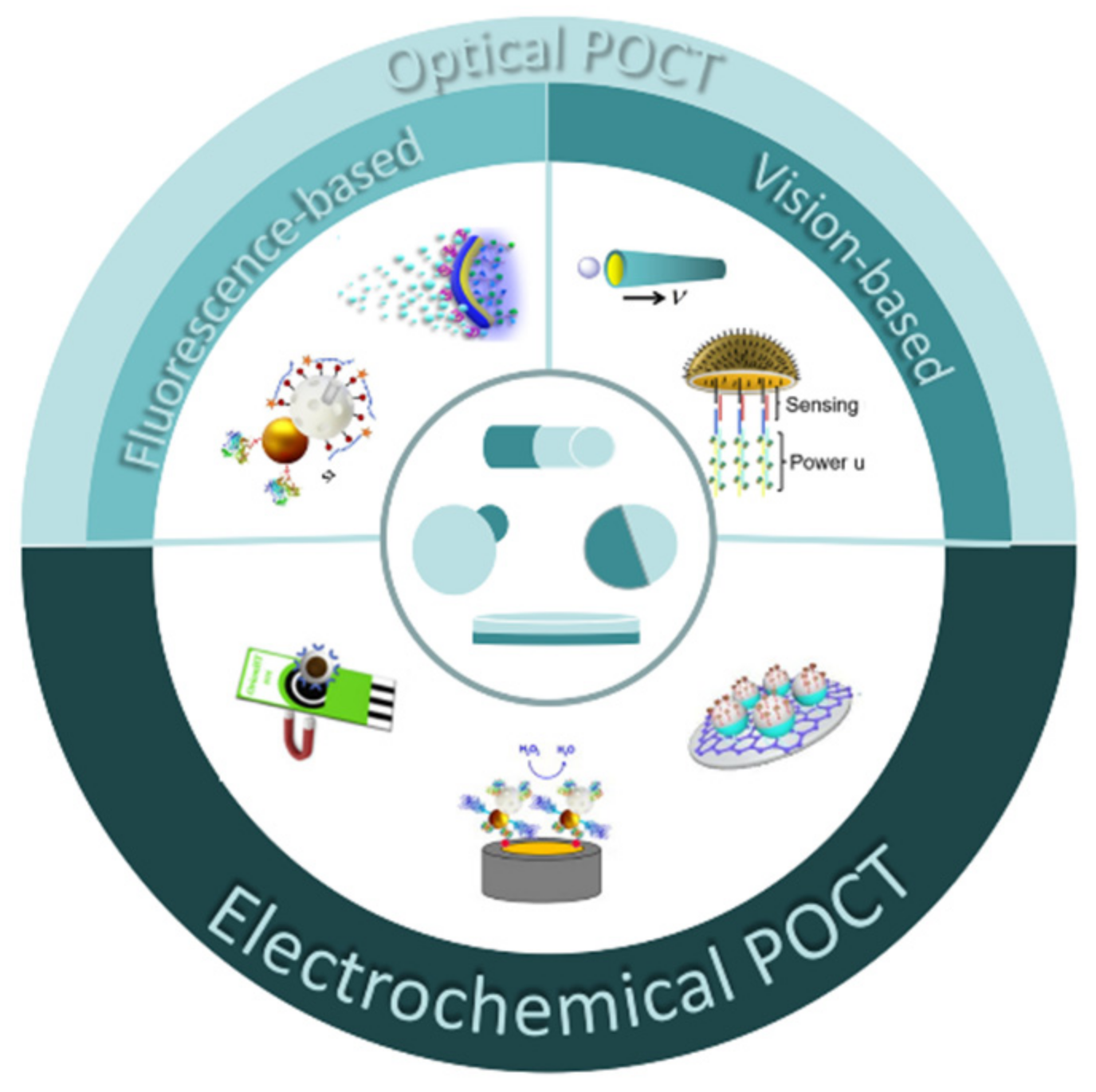
:1. Introduction
2. Preparation of Janus Particles
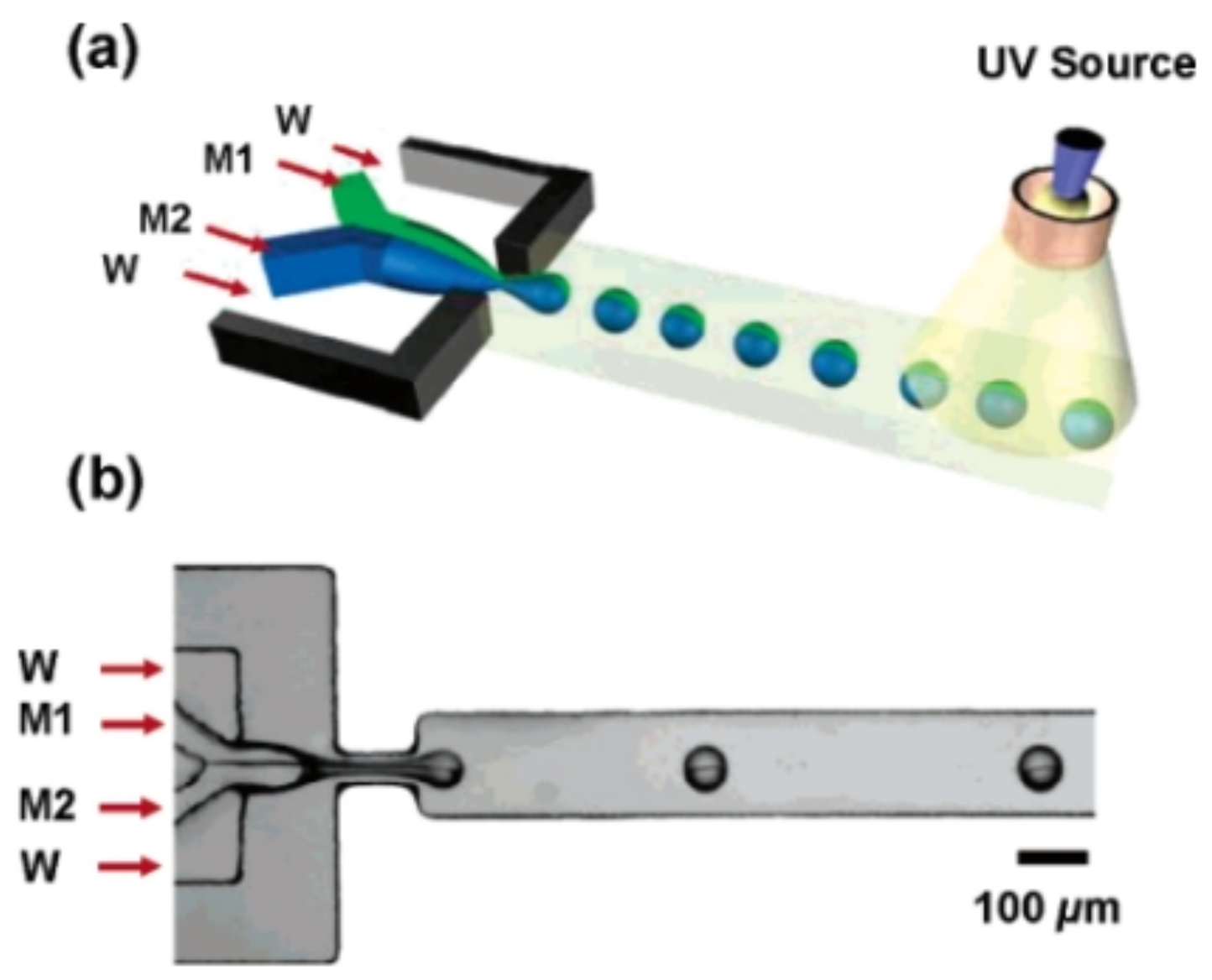
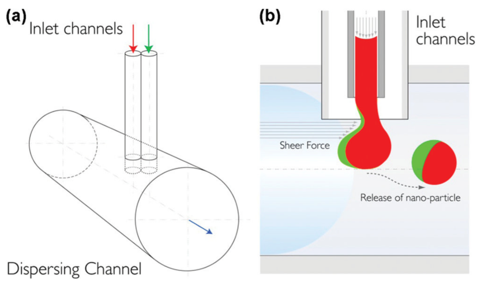
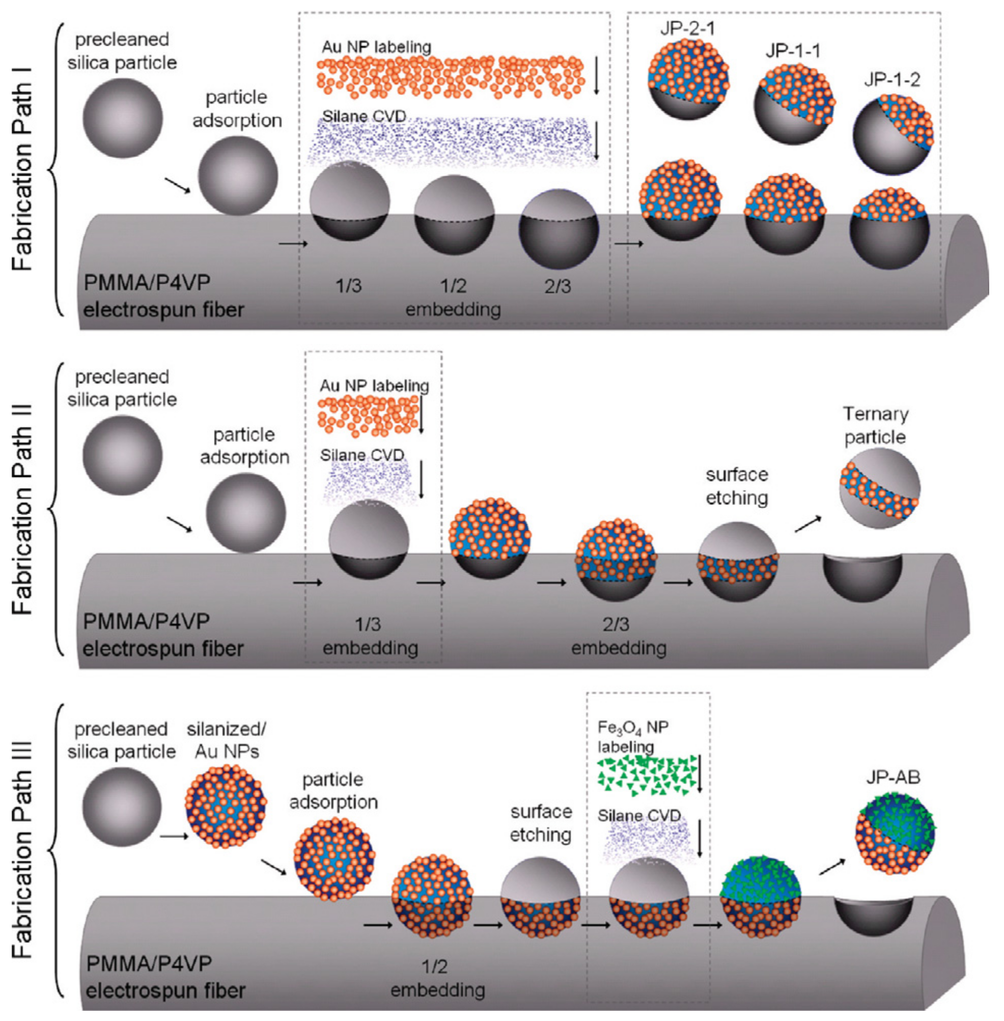
2.1. Microfluidic Method
2.2. Sputtering Method
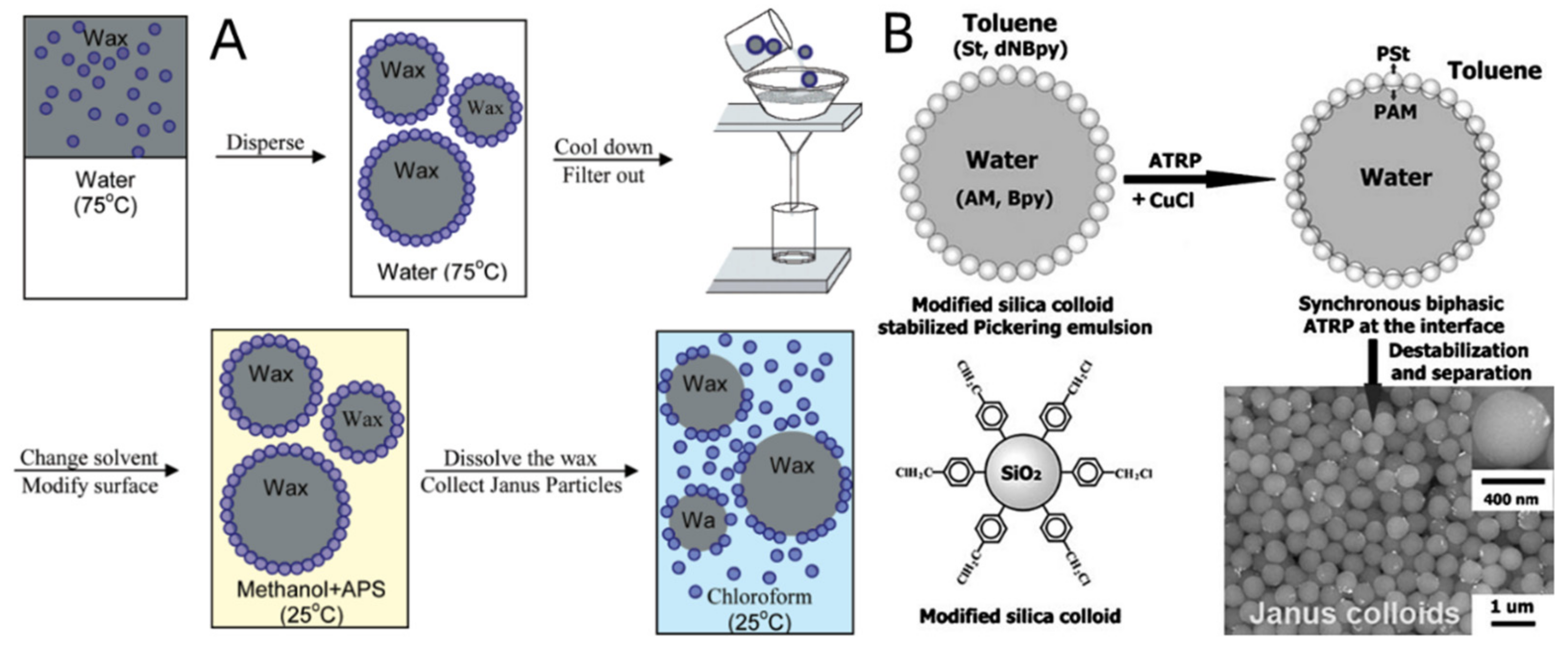
2.3. Phase-Separation Method
2.4. Pickering Emulsion Method
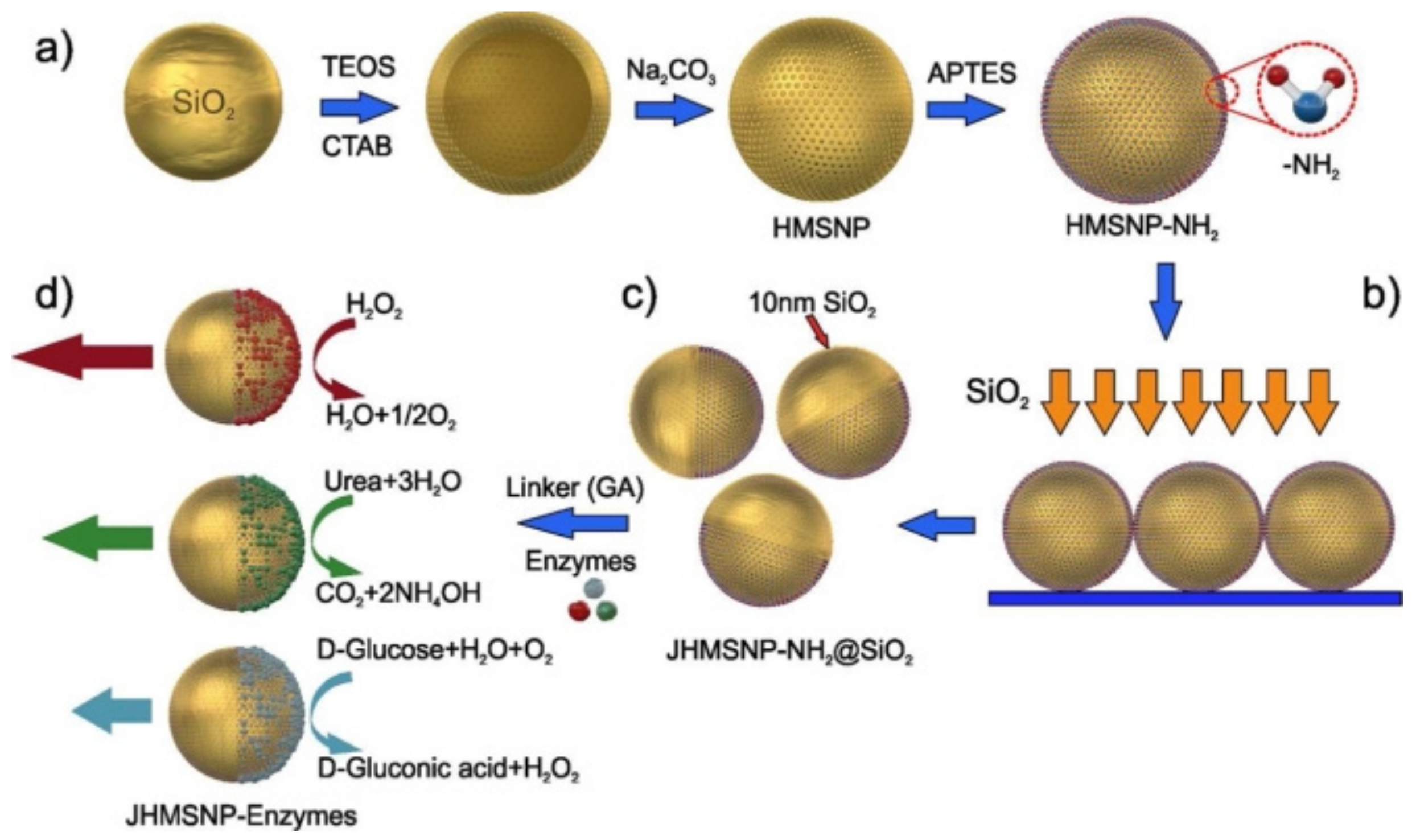
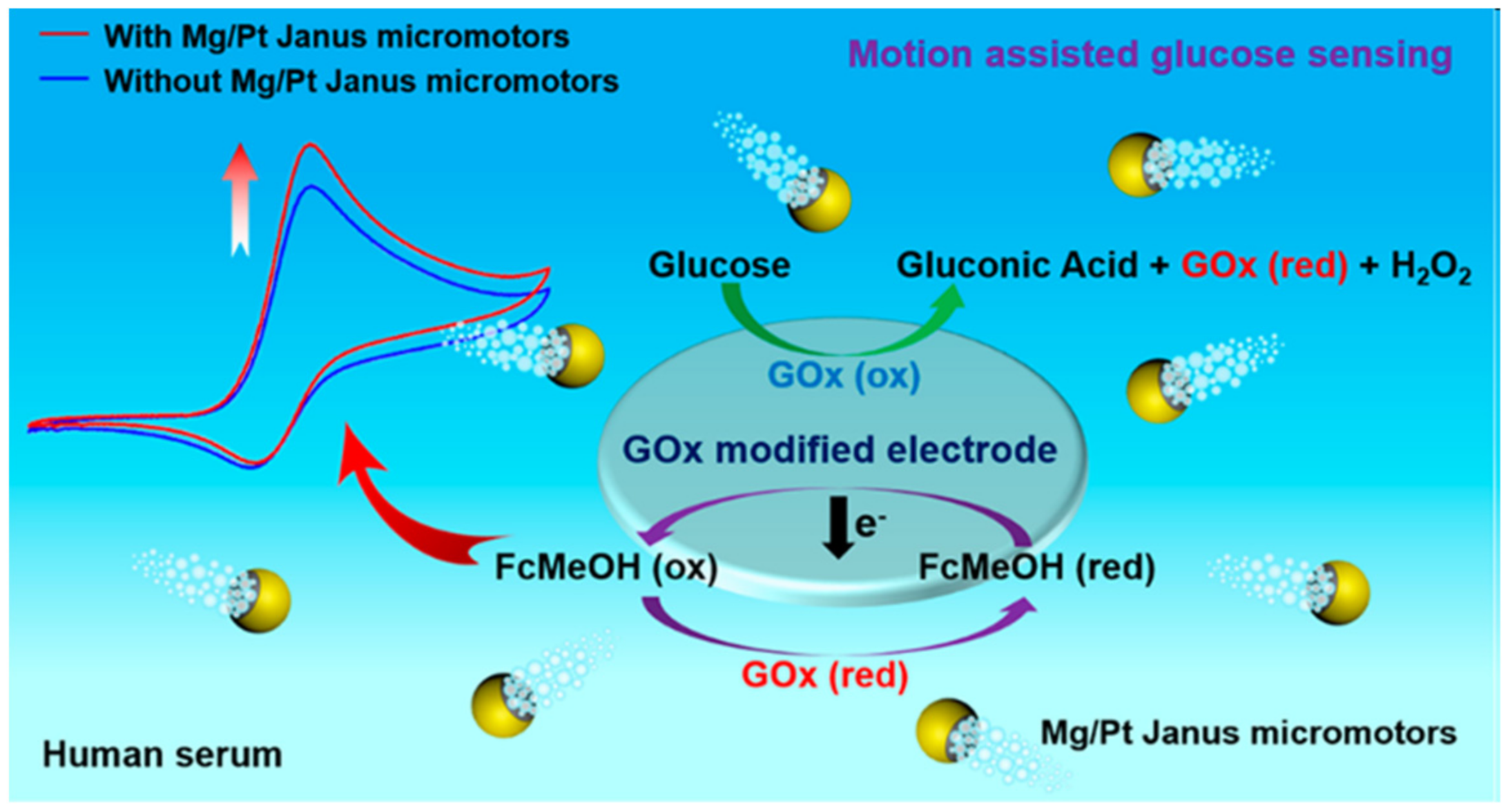
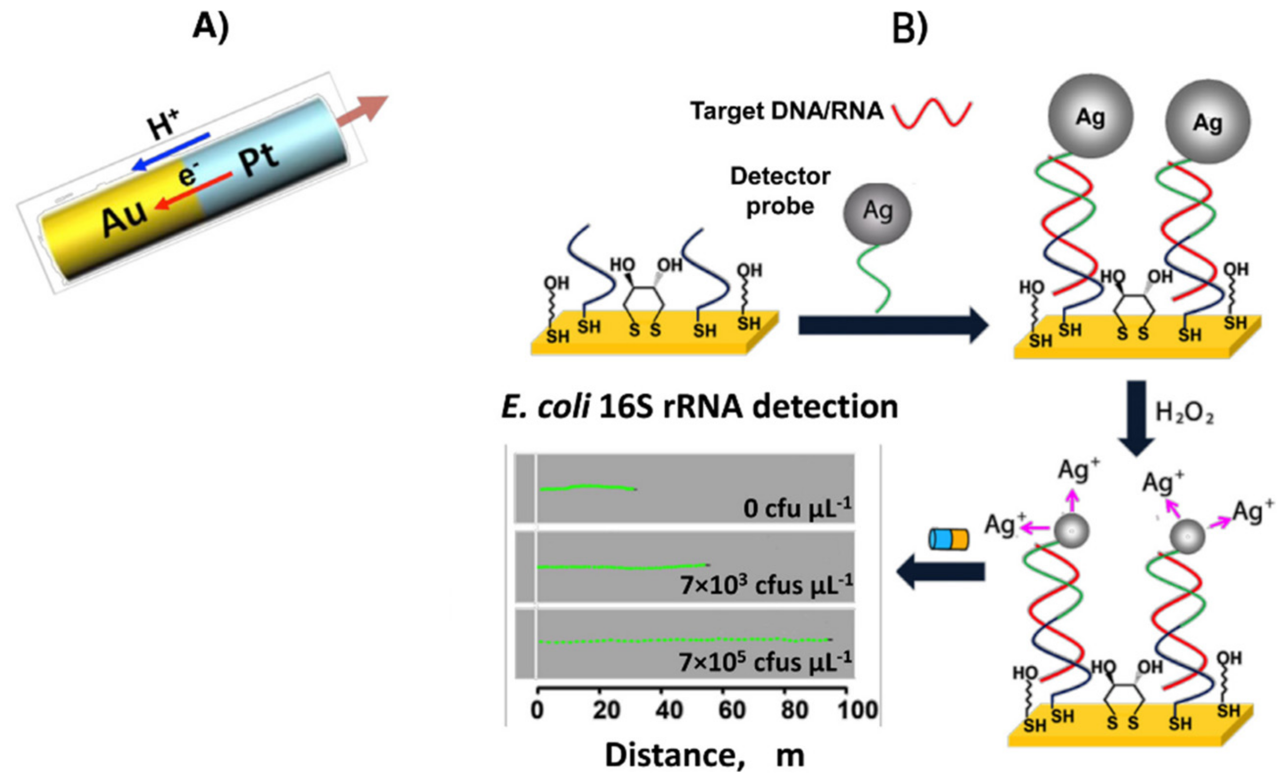
3. Janus Particles for Electrochemical POCT
4. Janus Particles for Optical POCT
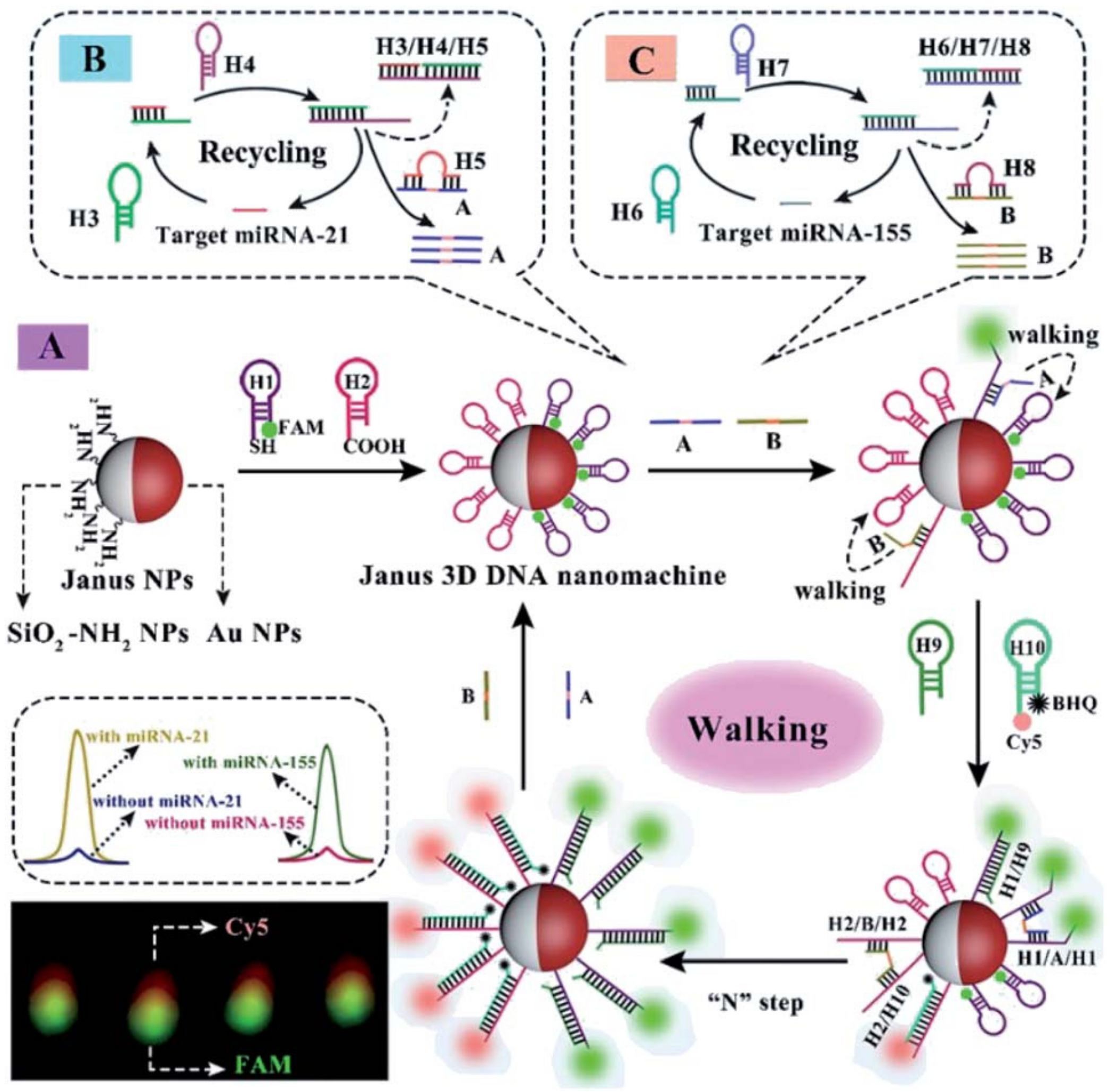
4.1. Janus Particles for Fluorescence-Based POCT
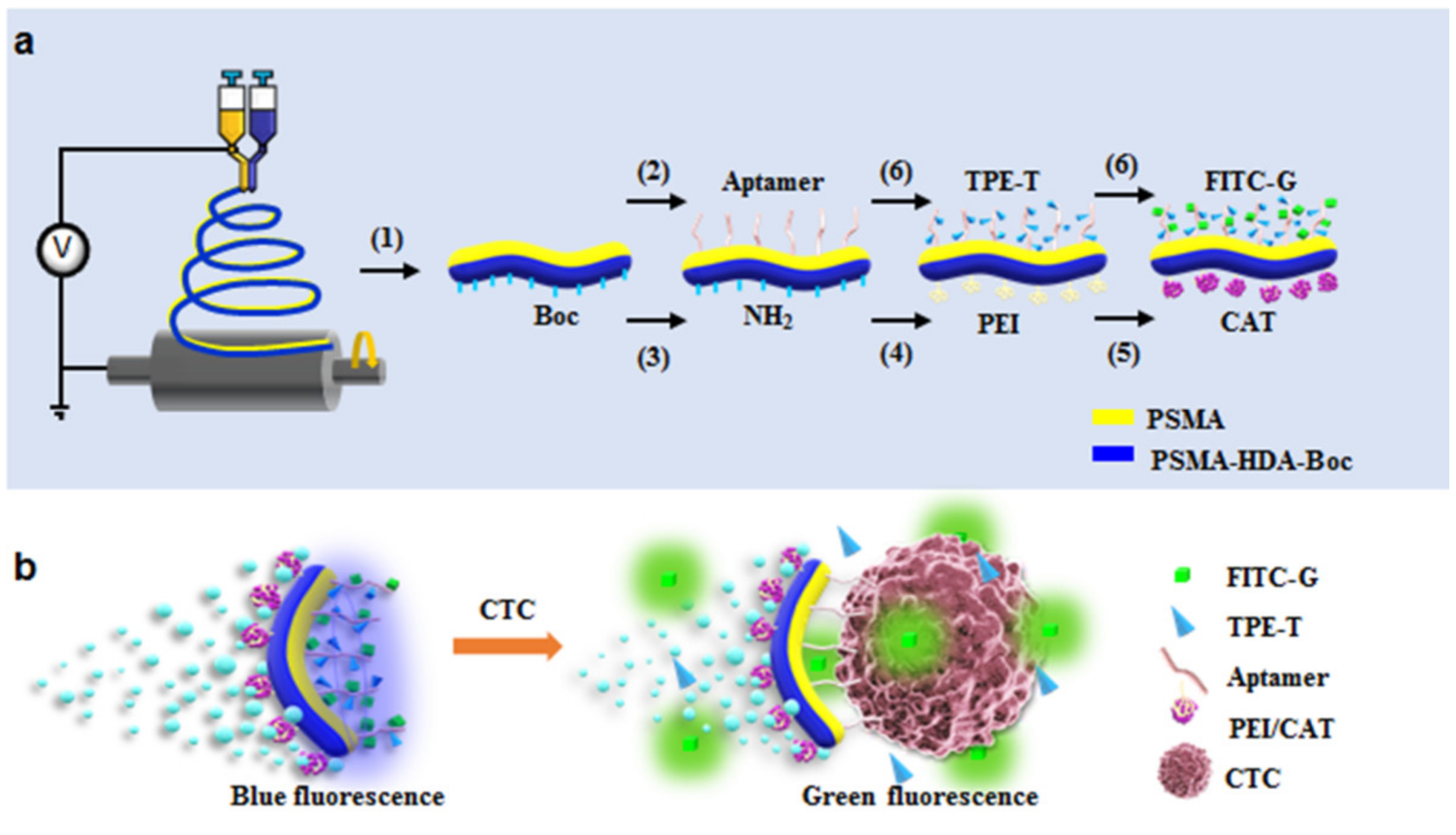
4.2. Janus Particles for Vision-Based POCT

| Detection Method | Analyte | Compositions | Type | Sensitivity | LoD | Ref. |
|---|---|---|---|---|---|---|
| Electrochemical biorecognition-signaling | Glucose | Au-SiO2 | Enzymesensor | 490 nM–600 mM | 360 nm | [154] |
| Mg/Pt Janus micromotors | Non-enzymesensor | 1–15 mM | 33.2 μM | [154] | ||
| Ochratoxin A | Au@SiO2 | Non-enzymesensor | 1 × 10⁻⁵–10 nM | 3.3 × 10⁻³ pM | [155] | |
| Carcinoembryonic antigen | Fe3O4@SiO2 | Non-enzymesensor | 5.5 pm to 28 nM | 1.2 pM | [157] | |
| C-reactive protein | Fe3O4@SiO2-Au | Enzymesensor | 10 pg/mL–1.0 ng/mL | 3.1 pg/mL | [158] | |
| IgG | Fe3O4@SiO2/Pt | Non-enzymesensor | 10 pg/mL to 100 ng/mL | 3.14 pg/mL | [159] | |
| Electrogenerated chemiluminescence | Glucose | glassy carbon | Enzymesensor | [160] | ||
| Flurorimetric detection | Urea | Au- SiO2 | Enzymesensor | 1.25–8.75 mM | 0.5 mM | [184] |
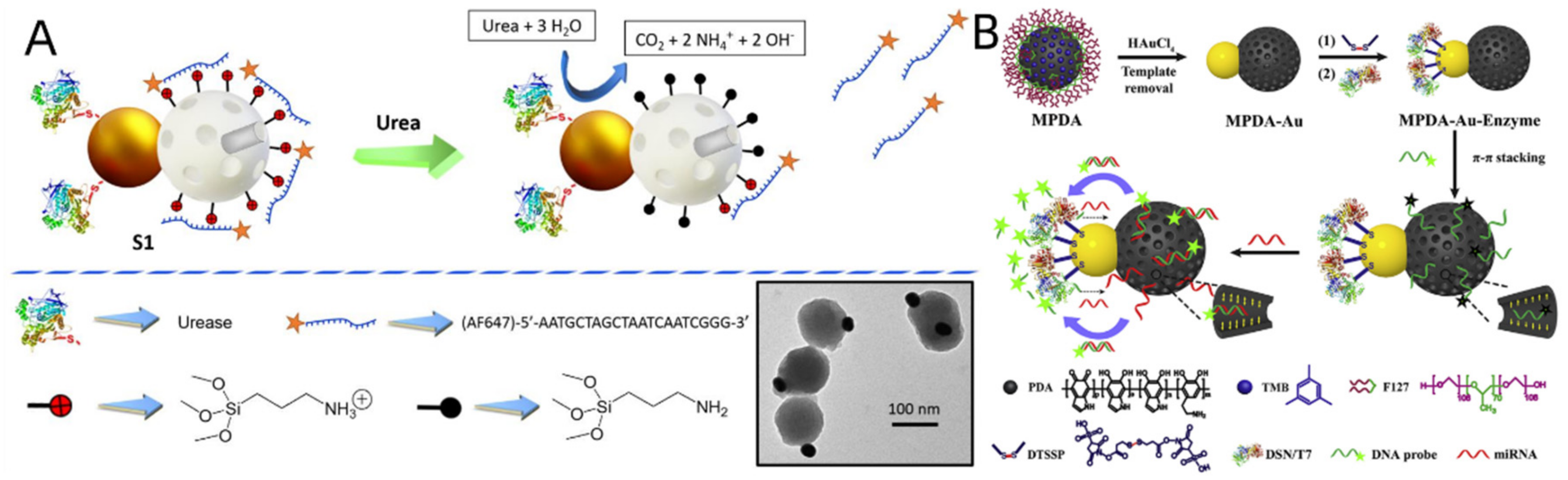
| MicroRNA | Au-MPDA | Enzymesensor | 20–500 fM | 32 fM | [185] | |
| Au@SiO2 | Non-enzymesensor | 1 pM–10 nM | 0.35 pM | [186] | ||
| circulating tumor cells | Janus fibers | Non-enzymesensor | 0–106 cells/mL | 25 cells/mL | [120] | |
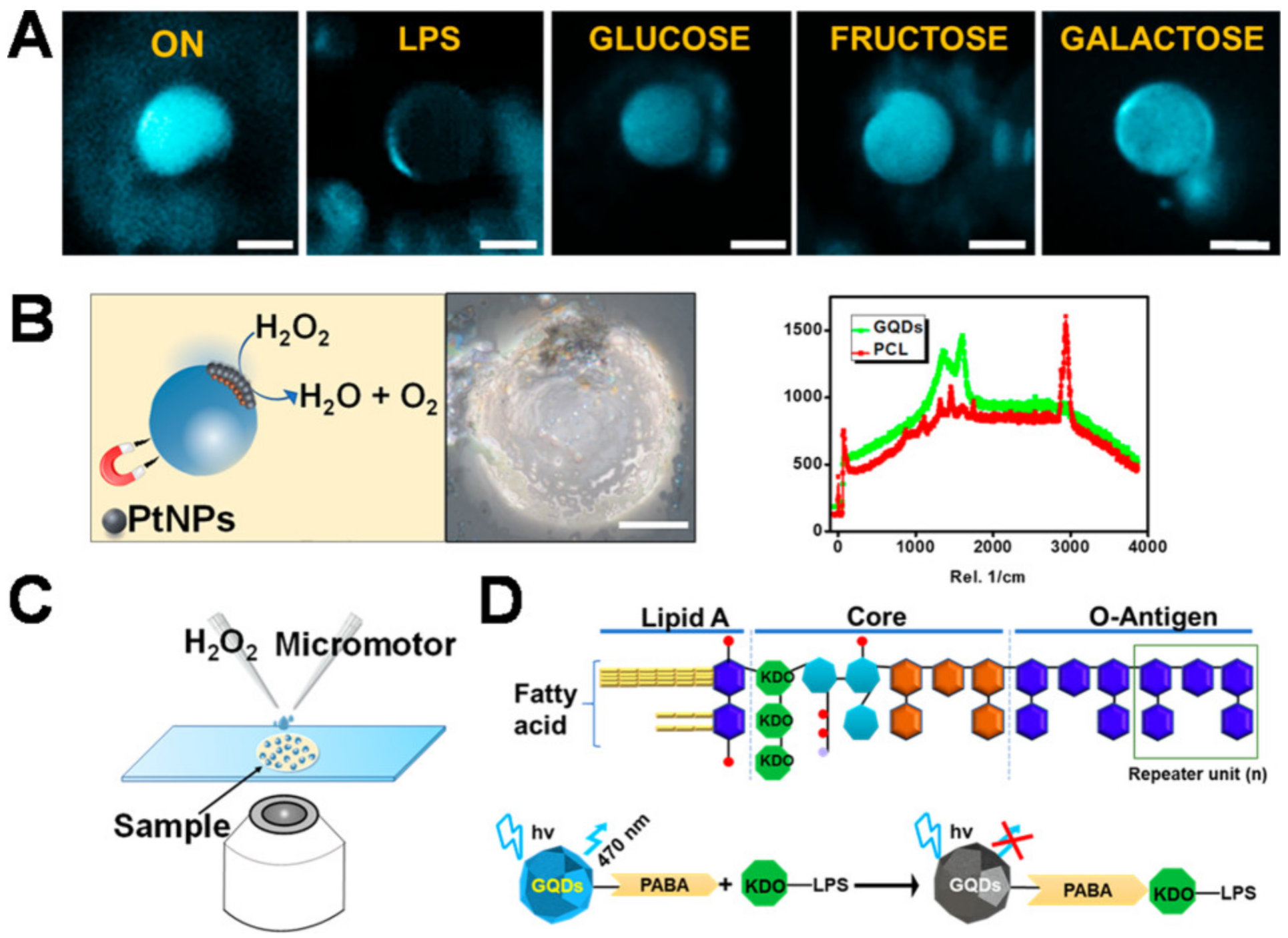
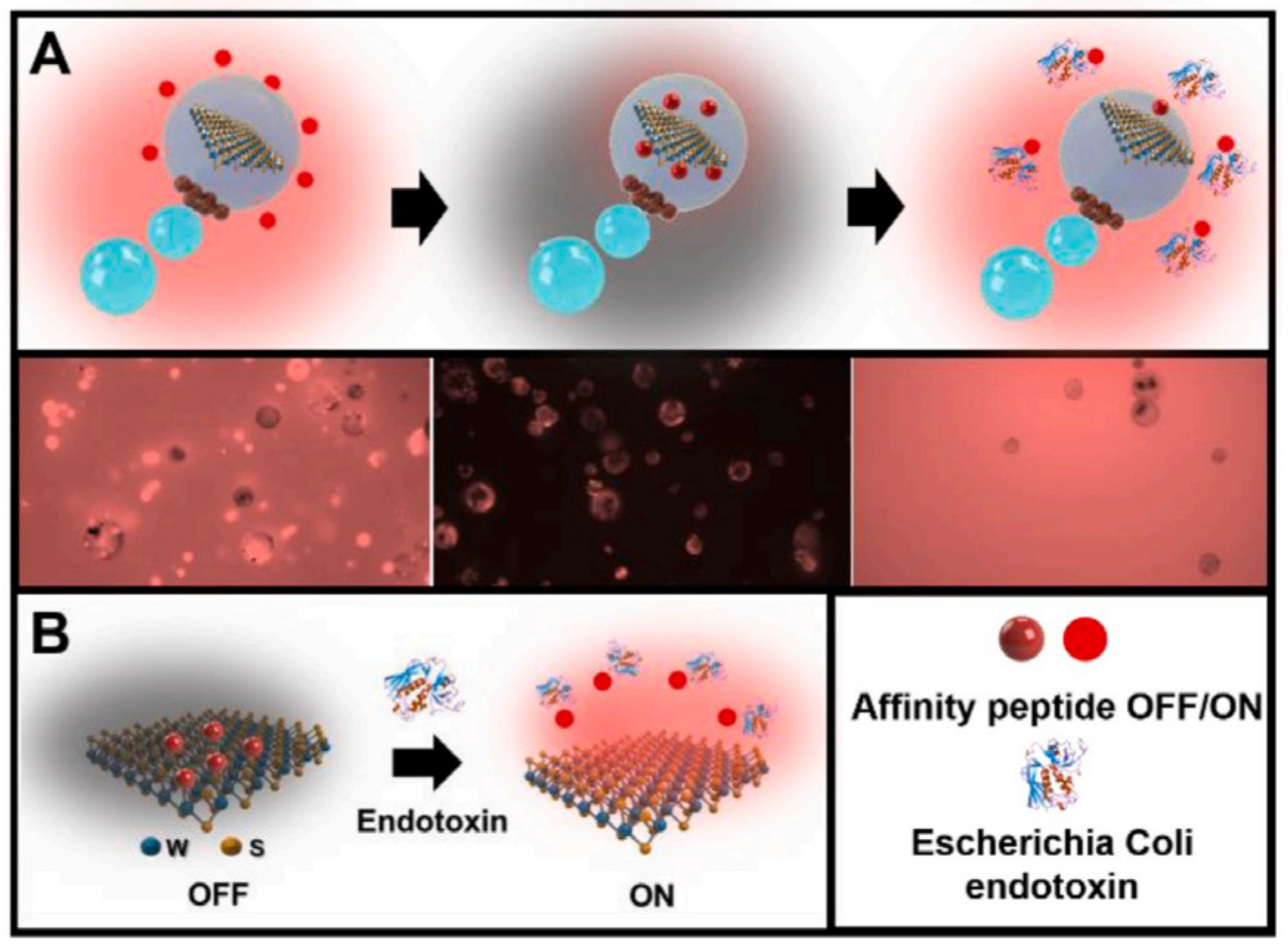
| Lipopolysaccharide | Graphene quantum dots | Non-enzymesensor | 0.2–1.0 ng/mL | 0.07 ng/mL | [126] | |
| WS2–Pt–Fe2O3 polycaprolactone | Non-enzymesensor | 4.0–1.0 × 10⁶ ng/mL | 120 pM | [188] | ||
| Salmonella enterica endotoxin | WS2–Pt–Fe2O3 | Non-enzymesensor | 4–333.3 µg/mL | 2.0 µg/mL | [189] | |
| MoS2–Pt–Fe2O3 | Non-enzymesensor | 9.8–333.3 µg/mL | 2.0 µg/mL | [189] | ||
| Emulsion agglutination assay | Tumor necrosis factors alpha | Au@SiO2 | Non-enzymesensor | 1 pg/mL–10 μg/mL | 1 pg/mL | [190] |
| DNA | Au@SiO2 | Non-enzymesensor | 0.1ng/μL | [191] | ||
| The anti-SARS-CoV-2 spike IgG antibody | Hydrocarbon and fluorocarbon oils | Non-enzymesensor | 0.2 μg/mL | [121] | ||
| Zika NS1 protein | Hydrocarbon and fluorocarbon phases | Non-enzymesensor | 100 nM | [195] | ||
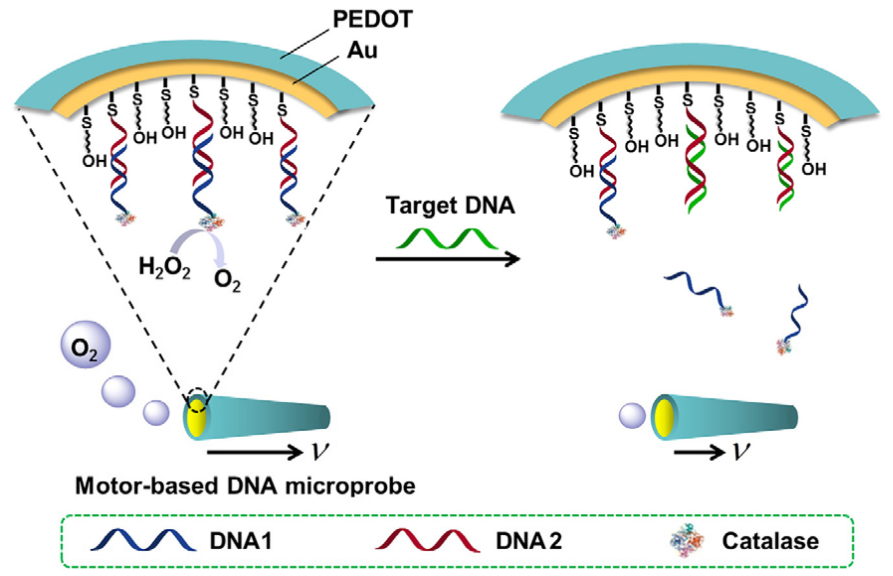
| Motion-based detection | DNA | Au–Ni–Au–Pt nanomotors | Enzymesensor | 100 pM–10 nM | 10 pM | [209] |
| PEDOT-PSS/Au | Enzymesensor | 10 nM–1 μM | [123] | |||
| PEDOT/Au | Enzymesensor | 0.5–10 mM | [210] | |||
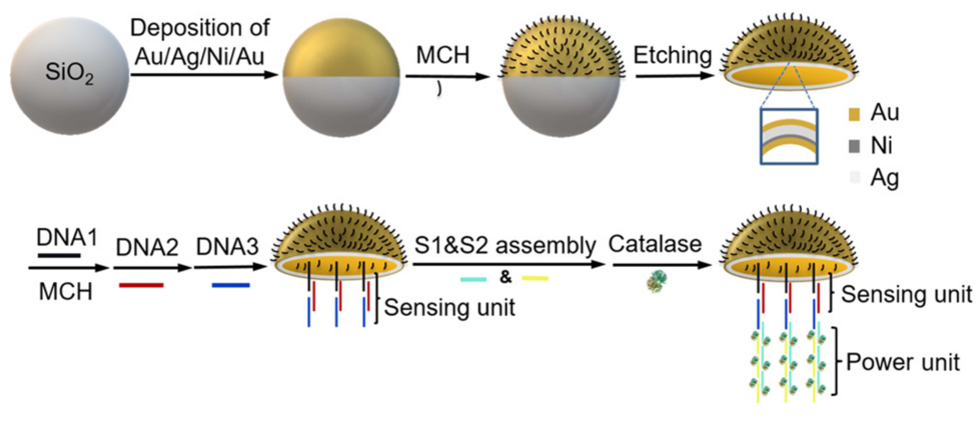
| Au/Ag/Ni/Au | Enzymesensor | 25–750 nM | [124] | |||
| HIV-1 RNA | Pt/Au@PS | Non-enzymesensor | [211] | |||
| Zika virus | Pt@PS | Non-enzymesensor | 100 particles/μL–106 particles/μL | 1 particle/μL | [212] | |
| Glutathione | Graphene-wrapped/PtNPs | Non-enzymesensor | 5–150 μM | 0.90 μM | [213] | |
| Colorimetric detection | Aspartic Acid | Janus AuNPs | Non-enzymesensor | 18 μM–1.8 nM | 33.9 μM | [55] |
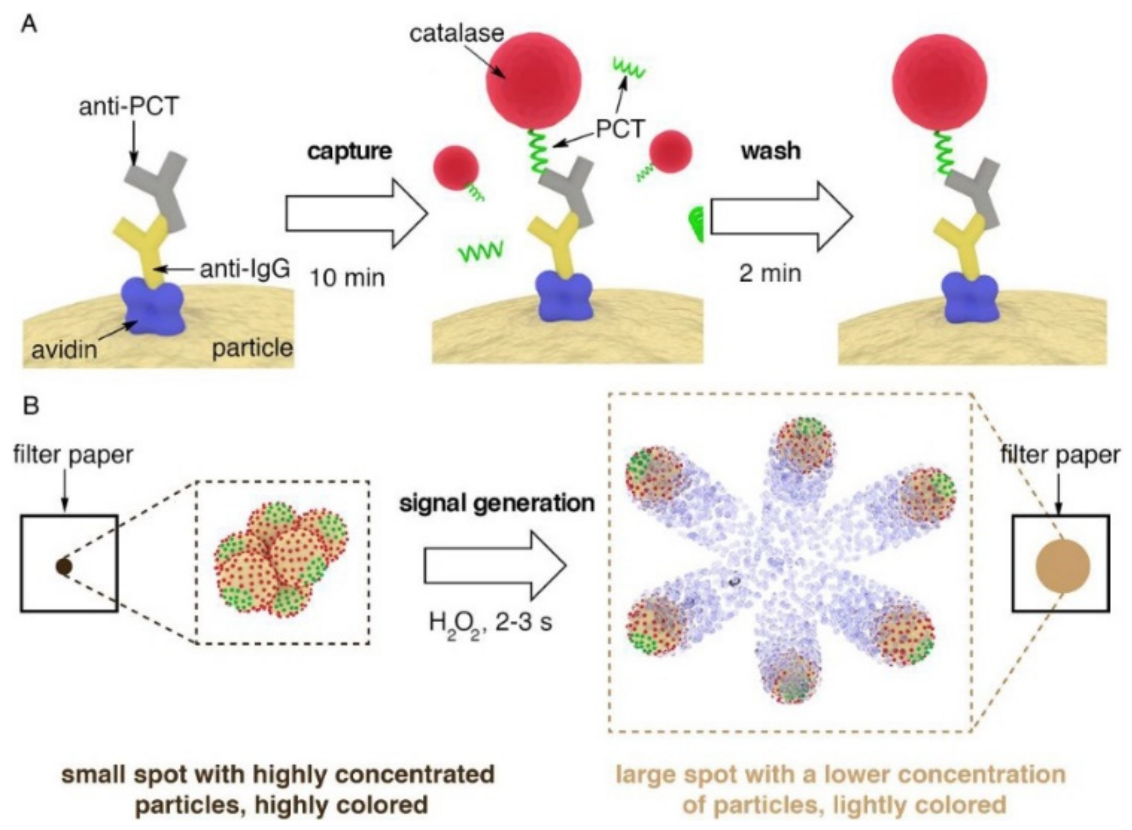
| Procalcitonin | Magnetic beads | Non-enzymesensor | 2 ng/mL | [214] | ||
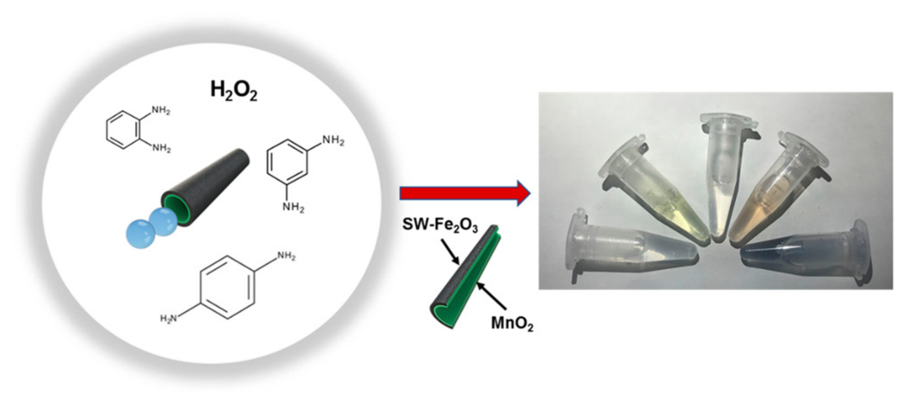
| Phenylenediamines Isomers | single-wall carbon nanotube (SW)-Fe2O3 | Non-enzymesensor | 20 μM | [79] | ||
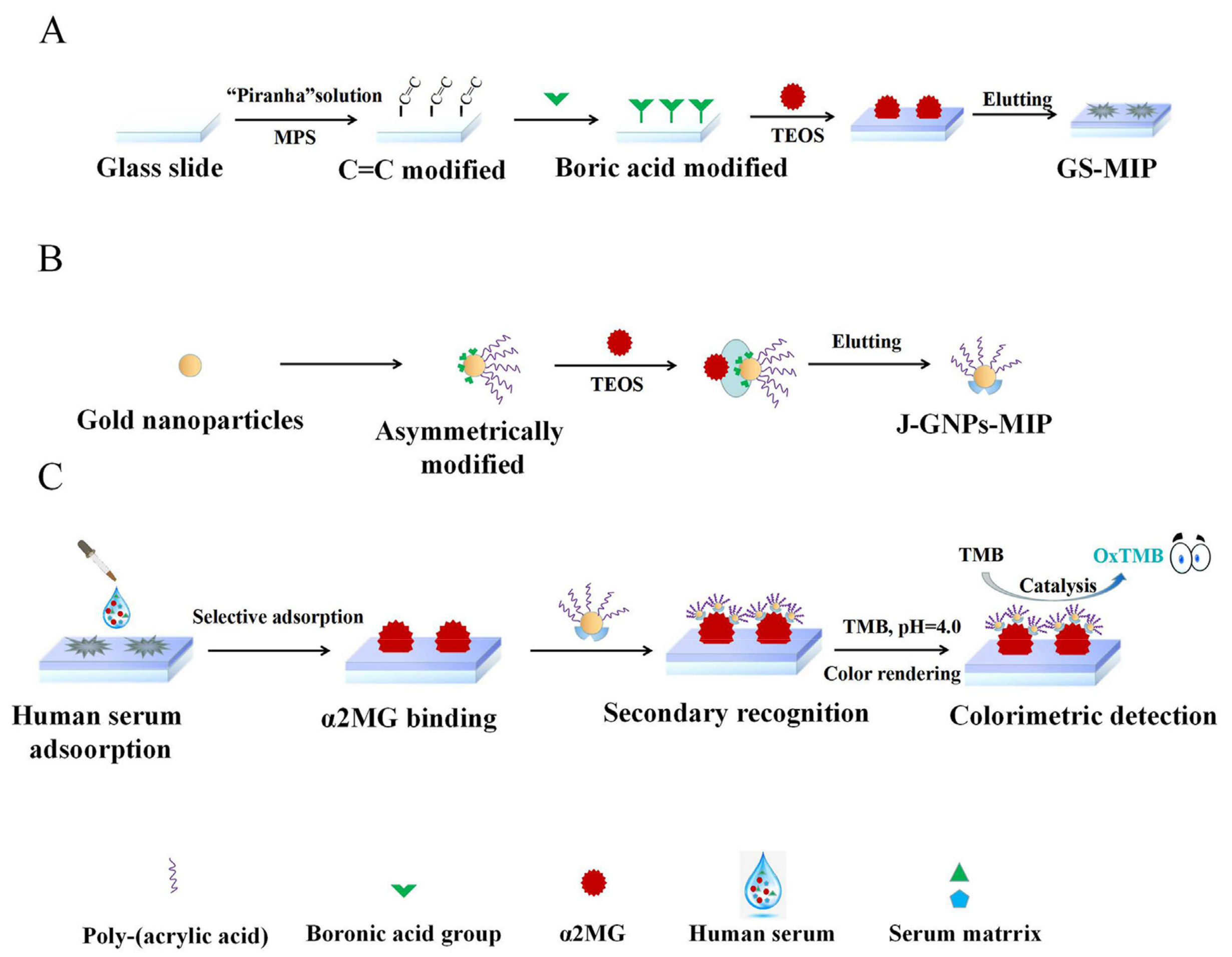
| Alpha-2-macroglobulin | Janus AuNPs | Non-enzymesensor | 0.297–130 mg/mL | 0.089 mg/mL | [215] |
5. General Conclusions, Challenges, and Perspectives
Author Contributions
Funding
Institutional Review Board Statement
Informed Consent Statement
Data Availability Statement
Conflicts of Interest
References
- Kirillova, A.; Marschelke, C.; Synytska, A. Hybrid Janus Particles: Challenges and Opportunities for the Design of Active Functional Interfaces and Surfaces. ACS Appl. Mater. Interfaces 2019, 11, 9643–9671. [Google Scholar] [CrossRef] [PubMed]
- Song, Y.; Chen, S. Janus Nanoparticles: Preparation, Characterization, and Applications. Chem. Asian J. 2013, 9, 418–430. [Google Scholar] [CrossRef]
- Zhang, Y.; Yang, F.; Wei, W.; Wang, Y.; Yang, S.; Li, J.; Xing, Y.; Zhou, L.; Dai, W.; Dong, H. Self-Propelled Janus Mesopo-rous Micromotor for Enhanced MicroRNA Capture and Amplified Detection in Complex Biological Samples. ACS Nano 2022, 16, 5587–5596. [Google Scholar] [CrossRef] [PubMed]
- Casagrande, C.; Fabre, P.; Raphael, E.; Veyssié, M. “Janus Beads”: Realization and Behaviour at Water/Oil Interfaces. EPL 1989, 9, 251. [Google Scholar] [CrossRef]
- Song, Y.; Zhou, J.; Fan, J.-B.; Zhai, W.; Meng, J.; Wang, S. Hydrophilic/Oleophilic Magnetic Janus Particles for the Rapid and Efficient Oil-Water Separation. Adv. Funct. Mater. 2018, 28, 1802493. [Google Scholar] [CrossRef]
- Schattling, P.; Thingholm, B.; Städler, B. Enhanced Diffusion of Glucose-Fueled Janus Particles. Chem. Mater. 2015, 27, 7412–7418. [Google Scholar] [CrossRef]
- Gröschel, A.H.; Walther, A.; Löbling, T.I.; Schmelz, J.; Hanisch, A.; Schmalz, H.; Müller, A.H.E. Facile, Solution-Based Synthesis of Soft, Nanoscale Janus Particles with Tunable Janus Balance. J. Am. Chem. Soc. 2012, 134, 13850–13860. [Google Scholar] [CrossRef]
- Jurado-Sánchez, B.; Escarpa, A. Janus Micromotors for Electrochemical Sensing and Biosensing Applications: A Review. Electroanalysis 2016, 29, 14–23. [Google Scholar] [CrossRef]
- Uspal, W.E. Theory of light-activated catalytic Janus particles. J. Chem. Phys. 2019, 150, 114903. [Google Scholar] [CrossRef]
- Huang, T.; Misko, V.R.; Gobeil, S.; Wang, X.; Nori, F.; Schütt, J.; Fassbender, J.; Cuniberti, G.; Makarov, D.; Baraban, L. Inverse Solidification Induced by Active Janus Particles. Adv. Funct. Mater. 2020, 30, 2003851. [Google Scholar] [CrossRef]
- Vilela, D.; Cossío, U.; Parmar, J.; Martinez-Villacorta, A.M.; Vallejo, V.G.; Llop, J.; Sánchez, S. Medical Imaging for the Tracking of Micromotors. ACS Nano 2018, 12, 1220–1227. [Google Scholar] [CrossRef]
- Yi, Y.; Sanchez, L.; Gao, Y.; Yu, Y. Janus particles for biological imaging and sensing. Analyst 2016, 141, 3526–3539. [Google Scholar] [CrossRef] [PubMed]
- Schick, I.; Lorenz, S.; Gehrig, D.; Tenzer, S.; Storck, W.; Fischer, K.; Strand, D.; Laquai, F.; Tremel, W. Inorganic Janus particles for biomedical applications. Beilstein J. Nanotechnol. 2014, 5, 2346–2362. [Google Scholar] [CrossRef] [PubMed]
- Yánez-Sedeño, P.; Campuzano, S.; Pingarrón, J. Janus particles for (bio)sensing. Appl. Mater. Today 2017, 9, 276–288. [Google Scholar] [CrossRef]
- Nosenko, V.; Luoni, F.; Kaouk, A.; Rubin-Zuzic, M.; Thomas, H. Active Janus particles in a complex plasma. Phys. Rev. Res. 2020, 2, 033226. [Google Scholar] [CrossRef]
- Nedev, S.; Carretero-Palacios, S.; Kühler, P.; Lohmueller, T.; Urban, A.S.; Anderson, L.J.E.; Feldmann, J. An Optically Controlled Microscale Elevator Using Plasmonic Janus Particles. ACS Photon 2015, 2, 491–496. [Google Scholar] [CrossRef]
- Huang, Z.; Chen, P.; Zhu, G.-L.; Yang, Y.; Xu, Z.; Yan, L.-T. Bacteria-Activated Janus Particles Driven by Chemotaxis. ACS Nano 2018, 12, 6725–6733. [Google Scholar] [CrossRef]
- Tang, Y.; Liu, H.; Chen, H.; Chen, Z.; Liu, Y.; Jin, L.; Deng, Y.; Li, S.; He, N. Advances in Aptamer Screening and Drug Delivery. J. Biomed. Nanotechnol. 2020, 16, 763–788. [Google Scholar] [CrossRef]
- Ji, X.; Yang, W.; Wang, T.; Mao, C.; Guo, L.; Xiao, J.; He, N. Coaxially electrospun core/shell structured poly(L-lactide) acid/chitosan nanofibers for potential drug carrier in tissue engineering. J. Biomed. Nanotechnol. 2013, 9, 1672–1678. [Google Scholar] [CrossRef]
- Xiao, X.; Yang, H.; Jiang, P.; Chen, Z.; Ji, C.; Nie, L. Multi-Functional Fe3O4@ MSiO2-AuNCs Composite Nanoparticles Used as Drug Delivery System. J. Biomed. Nanotechnol. 2017, 13, 1292–1299. [Google Scholar] [CrossRef]
- Liu, J.; Dong, S.; He, Q.; Yang, S.; Xie, M.; Deng, P.; Xia, Y.; Li, G. Facile Preparation of Fe3O4/C Nanocomposite and Its Ap-plication for Cost-Effective and Sensitive Detection of Tryptophan. Biomolecules 2019, 9, 245. [Google Scholar] [CrossRef] [PubMed]
- Li, X.; Chen, L.; Cui, D.; Jiang, W.; Han, L.; Niu, N. Preparation and application of Janus nanoparticles: Recent development and prospects. Co-Ord. Chem. Rev. 2021, 454, 214318. [Google Scholar] [CrossRef]
- Sharifi, M.; Hasan, A.; Attar, F.; Taghizadeh, A.; Falahati, M. Development of point-of-care nanobiosensors for breast cancers diagnosis. Talanta 2020, 217, 121091. [Google Scholar] [CrossRef] [PubMed]
- Guo, W.; Zhang, C.; Ma, T.; Liu, X.; Chen, Z.; Li, S.; Deng, Y. Advances in aptamer screening and aptasensors’ detection of heavy metal ions. J. Biomed. Nanobiotechnol. 2021, 19, 166. [Google Scholar] [CrossRef] [PubMed]
- Guo, Z.; Liu, Y.; He, N.; Deng, Y.; Jin, L. Discussion of the protein characterization techniques used in the identification of membrane protein targets corresponding to tumor cell aptamers. Chin. Chem. Lett. 2020, 32, 40–47. [Google Scholar] [CrossRef]
- Syedmoradi, L.; Daneshpour, M.; Alvandipour, M.; Gomez, F.A.; Hajghassem, H.; Omidfar, K. Point of care testing: The impact of nanotechnology. Biosens. Bioelectron. 2017, 87, 373–387. [Google Scholar] [CrossRef]
- Rusling, J.F.; Kumar, C.V.; Gutkind, J.S.; Patel, V. Measurement of biomarker proteins for point-of-care early detection and monitoring of cancer. Analyst 2010, 135, 2496–2511. [Google Scholar] [CrossRef]
- Ling, Y.; Zhu, Y.; Fan, H.; Zha, H.; Yang, M.; Wu, L.; Chen, H.; Li, W.; Wu, Y.; Chen, H. Rapid Method for Detection of Staphylococcus aureus in Feces. J. Biomed. Nanotechnol. 2019, 15, 1290–1298. [Google Scholar] [CrossRef]
- Xu, Y.; Wang, T.; Chen, Z.; Jin, L.; Wu, Z.; Yan, J.; Zhao, X.; Cai, L.; Deng, Y.; Guo, Y.; et al. The Point-of-Care-Testing of Nucleic Acids by Chip, Cartridge and Paper Sensors. Chin. Chem. Lett. 2021, 32, 3675–3686. [Google Scholar] [CrossRef]
- Ma, C.; Li, C.; He, N.; Wang, F.; Ma, N.; Zhang, L.; Lu, Z.; Ali, Z.; Xi, Z.; Li, X.; et al. Preparation and Characterization of Monodisperse Core–Shell Fe3O4 @SiO2 Microspheres and Its Application for Magnetic Separation of Nucleic Acids from E. coli BL21. J. Biomed. Nanotechnol. 2012, 8, 1000–1005. [Google Scholar] [CrossRef] [PubMed]
- Zhang, W.; Wang, R.; Luo, F.; Wang, P.; Lin, Z. Miniaturized electrochemical sensors and their point-of-care applications. Chin. Chem. Lett. 2019, 31, 589–600. [Google Scholar] [CrossRef]
- Wu, H.; Shi, C.; Zhu, Q.; Li, Y.; Xu, Z.; Wei, C.; Chen, D.; Huang, X. Capillary-driven blood separation and in-situ electrochemical detection based on 3D conductive gradient hollow fiber membrane. Biosens. Bioelectron. 2020, 171, 112722. [Google Scholar] [CrossRef] [PubMed]
- Gao, B.; Yang, Y.; Liao, J.; He, B.; Liu, H. Bioinspired multistructured paper microfluidics for POCT. Lab A Chip 2019, 19, 3602–3608. [Google Scholar] [CrossRef] [PubMed]
- Chen, Z.; Yang, T.; Yang, H.; Li, T.; Nie, L.; Mou, X.; Deng, Y.; He, N.; Li, Z.; Wang, L.; et al. A Portable Multi-Channel Turbidity System for Rapid Detection of Pathogens by Loop-Mediated Isothermal Amplification. J. Biomed. Nanotechnol. 2018, 14, 198–205. [Google Scholar] [CrossRef]
- Tu, F.; Lee, D. Shape-Changing and Amphiphilicity-Reversing Janus Particles with pH-Responsive Surfactant Properties. J. Am. Chem. Soc. 2014, 136, 9999–10006. [Google Scholar] [CrossRef]
- Fang, Y.; Liu, H.; Wang, Y.; Su, X.; Jin, L.; Wu, Y.; Deng, Y.; Li, S.; Chen, Z.; Chen, H.; et al. Fast and Accurate Control Strategy for Portable Nucleic Acid Detection (PNAD) System Based on Magnetic Nanoparticles. J. Biomed. Nanotechnol. 2021, 17, 407–415. [Google Scholar] [CrossRef]
- Chen, J.; Zhang, S.; Wang, Y.; Xie, R.; Liu, L.; Deng, Y. In Vivo Self-Assembly Based Cancer Therapy Strategy. J. Biomed. Nanotechnol. 2020, 16, 997–1017. [Google Scholar] [CrossRef]
- Deng, J.; Jiang, X. Advances in Reagents Storage and Release in Self-Contained Point-of-Care Devices. Adv. Mater. Technol. 2019, 4, 1800625. [Google Scholar] [CrossRef]
- Bunyarataphan, S.; Dharakul, T.; Fucharoen, S.; Paiboonsukwong, K.; Japrung, D. Glycated Albumin Measurement Using an Electrochemical Aptasensor for Screening and Monitoring of Diabetes Mellitus. Electroanalysis 2019, 31, 2254–2261. [Google Scholar] [CrossRef]
- Xu, Z.; Liu, Z.; Xiao, M.; Jiang, L.; Yi, C. A smartphone-based quantitative point-of-care testing (POCT) system for simultaneous detection of multiple heavy metal ions. Chem. Eng. J. 2020, 394, 124966. [Google Scholar] [CrossRef]
- Shao, D.; Zhang, X.; Liu, W.; Zhang, F.; Zheng, X.; Qiao, P.; Li, J.; Dong, W.-F.; Chen, L. Janus Silver-Mesoporous Silica Nanocarriers for SERS Traceable and pH-Sensitive Drug Delivery in Cancer Therapy. ACS Appl. Mater. Interfaces 2016, 8, 4303–4308. [Google Scholar] [CrossRef]
- Guo, L.; Chen, H.; He, N.; Deng, Y. Effects of Surface Modifications on the Physicochemical Properties of Iron Oxide Nano-particles and Their Performance as Anticancer Drug Carriers. Chin. Chem. Lett. 2018, 29, 1829–1833. [Google Scholar] [CrossRef]
- Yuet, K.P.; Hwang, D.K.; Haghgooie, R.; Doyle, P.S. Multifunctional Superparamagnetic Janus Particles. Langmuir 2009, 26, 4281–4287. [Google Scholar] [CrossRef] [PubMed]
- Li, S.; Zhang, L.; Chen, X.; Wang, T.; Zhao, Y.; Li, L.; Wang, C. Selective Growth Synthesis of Ternary Janus Nanoparticles for Imaging-Guided Synergistic Chemo- and Photothermal Therapy in the Second NIR Window. ACS Appl. Mater. Interfaces 2018, 10, 24137–24148. [Google Scholar] [CrossRef] [PubMed]
- He, Q.; Vijayamohanan, H.; Li, J.; Swager, T.M. Multifunctional Photonic Janus Particles. J. Am. Chem. Soc. 2022, 144, 5661–5667. [Google Scholar] [CrossRef] [PubMed]
- Zhang, Q.; Zhang, L.; Li, S.; Chen, X.; Zhang, M.; Wang, T.; Li, L.; Wang, C. Designed Synthesis of Au/Fe3O4 @C Janus Nanoparticles for Dual-Modal Imaging and Actively Targeted Chemo-Photothermal Synergistic Therapy of Cancer Cells. Chem. A Eur. J. 2017, 23, 17242–17248. [Google Scholar] [CrossRef]
- Yi, H.; Rehman, F.U.; Zhao, C.; Liu, B.; He, N. Recent advances in nano scaffolds for bone repair. Bone Res. 2016, 4, 16050. [Google Scholar] [CrossRef]
- Su, H.; Price, C.-A.H.; Jing, L.; Tian, Q.; Liu, J.; Qian, K. Janus particles: Design, preparation, and biomedical applications. Mater. Today Bio 2019, 4, 100033. [Google Scholar] [CrossRef]
- Yang, S.; Guo, F.; Kiraly, B.; Mao, X.; Lu, M.; Leong, K.; Huang, T.J. Microfluidic synthesis of multifunctional Janus particles for biomedical applications. Lab A Chip 2012, 12, 2097–2102. [Google Scholar] [CrossRef] [PubMed]
- Li, Q.; Hu, E.; Yu, K.; Xie, R.; Lu, F.; Lu, B.; Bao, R.; Zhao, T.; Dai, F.; Lan, G. Self-Propelling Janus Particles for Hemostasis in Perforating and Irregular Wounds with Massive Hemorrhage. Adv. Funct. Mater. 2020, 30, 2004153. [Google Scholar] [CrossRef]
- Shi, C.; Wu, Z.; Yang, F.; Tang, Y. Janus particles with pH switchable properties for high-efficiency adsorption of PPCPs in water. Solid State Sci. 2021, 119, 106702. [Google Scholar] [CrossRef]
- Le, T.; Zhai, J.; Chiu, W.-H.; A Tran, P.; Tran, N. Janus particles: Recent advances in the biomedical applications. Int. J. Nanomed. 2019, 14, 6749–6777. [Google Scholar] [CrossRef] [PubMed]
- Fujii, S.; Yokoyama, Y.; Miyanari, Y.; Shiono, T.; Ito, M.; Yusa, S.-I.; Nakamura, Y. Micrometer-Sized Gold–Silica Janus Particles as Particulate Emulsifiers. Langmuir 2013, 29, 5457–5465. [Google Scholar] [CrossRef] [PubMed]
- Sun, X.-T.; Zhang, Y.; Zheng, D.-H.; Yue, S.; Yang, C.-G.; Xu, Z.-R. Multitarget sensing of glucose and cholesterol based on Janus hydrogel microparticles. Biosens. Bioelectron. 2017, 92, 81–86. [Google Scholar] [CrossRef] [PubMed]
- Chen, X.-Y.; Ma, R.-T.; Ha, W.; Shi, Y.-P. Direct colorimetric detection of aspartic acid in rat brain based on oriented aggregation of Janus gold nanoparticle. Sens. Actuators B Chem. 2018, 274, 668–675. [Google Scholar] [CrossRef]
- Fan, J.-B.; Song, Y.; Liu, H.; Lu, Z.; Zhang, F.; Liu, H.; Meng, J.; Gu, L.; Wang, S.; Jiang, L. A general strategy to synthesize chemically and topologically anisotropic Janus particles. Sci. Adv. 2017, 3, e1603203. [Google Scholar] [CrossRef]
- Lan, Y.; Wu, J.; Han, S.H.; Yadavali, S.; Issadore, D.; Stebe, K.J.; Lee, D. Scalable Synthesis of Janus Particles with High Natu-rality. Chem. Eng. 2020, 7, 17680–17686. [Google Scholar]
- Zhang, W.; He, J.; Dong, X. Controlled fabrication of polymeric Janus nanoparticles and their solution behaviors. RSC Adv. 2016, 6, 105070–105075. [Google Scholar] [CrossRef]
- Zhang, J.; Grzybowski, B.A.; Granick, S. Janus Particle Synthesis, Assembly, and Application. Langmuir 2017, 33, 6964–6977. [Google Scholar] [CrossRef] [PubMed]
- Walther, A.; Müller, A.H.E. Janus Particles: Synthesis, Self-Assembly, Physical Properties, and Applications. Chem. Rev. 2013, 113, 5194–5261. [Google Scholar] [CrossRef]
- Park, B.J.; Brugarolas, T.; Lee, D. Janus particles at an oil–water interface. Soft Matter 2011, 7, 6413–6417. [Google Scholar] [CrossRef]
- Nie, Z.; Li, W.; Seo, M.; Xu, S.; Kumacheva, E. Janus and Ternary Particles Generated by Microfluidic Synthesis: Design, Synthesis, and Self-Assembly. J. Am. Chem. Soc. 2006, 128, 9408–9412. [Google Scholar] [CrossRef] [PubMed]
- Correia, E.L.; Brown, N.; Razavi, S. Janus Particles at Fluid Interfaces: Stability and Interfacial Rheology. Nanomaterials 2021, 11, 374. [Google Scholar] [CrossRef] [PubMed]
- Lan, Y.; Choi, J.; Li, H.; Jia, Y.; Huang, R.; Stebe, K.J.; Lee, D. Janus Particles with Varying Configurations for Emulsion Stabilization. Ind. Eng. Chem. Res. 2019, 58, 20961–20968. [Google Scholar] [CrossRef]
- Jiang, S.; Chen, Q.; Tripathy, M.; Luijten, E.; Schweizer, K.S.; Granick, S. Janus Particle Synthesis and Assembly. Adv. Mater. 2010, 22, 1060–1071. [Google Scholar] [CrossRef]
- Liu, B.; Zhang, C.; Liu, J.; Qu, X.; Yang, Z. Janus non-spherical colloids by asymmetric wet-etching. Chem. Commun. 2009, 26, 3871–3873. [Google Scholar] [CrossRef]
- Hu, J.; Zhou, S.; Sun, Y.; Fang, X.; Wu, L. Fabrication, properties and applications of Janus particles. Chem. Soc. Rev. 2012, 41, 4356–4378. [Google Scholar] [CrossRef]
- Poggi, E.; Gohy, J.-F. Janus particles: From synthesis to application. Colloid Polym. Sci. 2017, 295, 2083–2108. [Google Scholar] [CrossRef]
- Nie, L.; Liu, S.; Shen, W.; Chen, D.; Jiang, M. One-Pot Synthesis of Amphiphilic Polymeric Janus Particles and Their Self-Assembly into Supermicelles with a Narrow Size Distribution. Angew. Chem. Int. Ed. 2007, 46, 6321–6324. [Google Scholar] [CrossRef]
- Shang, L.; Cheng, Y.; Zhao, Y. Emerging Droplet Microfluidics. Chem. Rev. 2017, 117, 7964–8040. [Google Scholar] [CrossRef]
- Chen, C.H.; Shah, R.K.; Abate, A.R.; Weitz, D.A. Janus Particles Templated from Double Emulsion Droplets Generated Using Microfluidics. Langmuir 2009, 25, 4320–4323. [Google Scholar] [CrossRef] [PubMed]
- Chou, W.-L.; Lee, P.-Y.; Yang, C.-L.; Huang, W.-Y.; Lin, Y.-S. Recent Advances in Applications of Droplet Microfluidics. Micromachines 2015, 6, 1249–1271. [Google Scholar] [CrossRef]
- Yang, Y.-T.; Wei, J.; Li, X.; Wu, L.-J.; Chang, Z.-Q.; Serra, C.A. A side-by-side capillaries-based microfluidic system for synthesizing size- and morphology-controlled magnetic anisotropy janus beads. Adv. Powder Technol. 2014, 26, 156–162. [Google Scholar] [CrossRef]
- Baraban, L.; Harazim, S.M.; Sanchez, S.; Schmidt, O.G. Chemotactic Behavior of Catalytic Motors in Microfluidic Channels. Angew. Chem. Int. Ed. 2013, 52, 5552–5556. [Google Scholar] [CrossRef]
- Amirifar, L.; Besanjideh, M.; Nasiri, R.; Shamloo, A.; Nasrollahi, F.; de Barros, N.R.; Davoodi, E.; Erdem, A.; Mahmoodi, M.; Hosseini, V.; et al. Droplet-based microfluidics in biomedical applications. Biofabrication 2022, 14. [Google Scholar] [CrossRef]
- Khan, I.U.; Serra, C.A.; Anton, N.; Li, X.; Akasov, R.; Messaddeq, N.; Kraus, I.; Vandamme, T.F. Microfluidic conceived drug loaded Janus particles in side-by-side capillaries device. Int. J. Pharm. 2014, 473, 239–249. [Google Scholar] [CrossRef]
- Lone, S.; Cheong, I.W. Fabrication of polymeric Janus particles by droplet microfluidics. RSC Adv. 2014, 4, 13322–13333. [Google Scholar] [CrossRef]
- Nisisako, T. Recent advances in microfluidic production of Janus droplets and particles. Curr. Opin. Colloid Interface Sci. 2016, 25, 1–12. [Google Scholar] [CrossRef]
- María-Hormigos, R.; Jurado-Sánchez, B.; Escarpa, A. Self-Propelled Micromotors for Naked-Eye Detection of Phenylenediamines Isomers. Anal. Chem. 2018, 90, 9830–9837. [Google Scholar] [CrossRef]
- Xie, H.; She, Z.-G.; Wang, S.; Sharma, G.; Smith, J.W. One-Step Fabrication of Polymeric Janus Nanoparticles for Drug Delivery. Langmuir 2012, 28, 4459–4463. [Google Scholar] [CrossRef]
- Prasad, N.; Perumal, J.; Choi, C.-H.; Lee, C.-S.; Kim, D.-P. Generation of Monodisperse Inorganic-Organic Janus Microspheres in a Microfluidic Device. Adv. Funct. Mater. 2009, 19, 1656–1662. [Google Scholar] [CrossRef]
- Nisisako, T.; Torii, T.; Takahashi, T.; Takizawa, Y. Synthesis of Monodisperse Bicolored Janus Particles with Electrical Anisotropy Using a Microfluidic Co-Flow System. Adv. Mater. 2006, 18, 1152–1156. [Google Scholar] [CrossRef]
- Smentkowski, V.S. Trends in Sputtering. Prog. Surf. Sci. 2000, 64, 1–58. [Google Scholar] [CrossRef]
- Kelly, P.J.; Arnell, R.D. Magnetron sputtering: A review of recent developments and applications. Vacuum 2000, 56, 159–172. [Google Scholar] [CrossRef]
- Valbusa, U.; Boragno, C.; de Mongeot, F.B. Nanostructuring surfaces by ion sputtering. J. Phys. Condens. Matter 2002, 14, 8153–8175. [Google Scholar] [CrossRef]
- Cui, J.-Q.; Kretzschmar, I. Surface-Anisotropic Polystyrene Spheres by Electroless Deposition. Langmuir 2006, 22, 8281–8284. [Google Scholar] [CrossRef] [PubMed]
- Ma, X.; Jannasch, A.; Albrecht, U.-R.; Hahn, K.; Miguel-López, A.; Schaffer, E.; Sánchez, S. Enzyme-Powered Hollow Meso-porous Janus Nanomotors. Nano Lett. 2015, 15, 7043–7050. [Google Scholar] [CrossRef]
- Li, B.; Wang, M.; Chen, K.; Cheng, Z.; Chen, G.; Zhang, Z. Synthesis of Biofunctional Janus Particles. Macromol. Rapid Commun. 2015, 36, 1200–1204. [Google Scholar] [CrossRef]
- Shepard, K.B.; Christie, D.A.; Sosa, C.L.; Arnold, C.B.; Priestley, R.D. Patchy Janus particles with tunable roughness and composition via vapor-assisted deposition of macromolecules. Appl. Phys. Lett. 2015, 106, 093104. [Google Scholar] [CrossRef]
- Tian, L.; Zhang, B.; Li, W.; Li, X.; Fan, X.; Jia, X.; Zhang, H.; Zhang, Q. Facile fabrication of Fe3O4@PS/PGMA magnetic Janus particles via organic–inorganic dual phase separation. RSC Adv. 2014, 4, 27152–27158. [Google Scholar] [CrossRef]
- Yin, Y.; Lu, Y.; Gates, B.; Xia, Y. Template-Assisted Self-Assembly: A Practical Route to Complex Aggregates of Monodispersed Colloids with Well-Defined Sizes, Shapes, and Structures. J. Am. Chem. Soc. 2001, 123, 8718–8729. [Google Scholar] [CrossRef] [PubMed]
- Wang, Z.; Rutjes, F.P.J.T.; van Hest, J.C.M. pH responsive polymersome Pickering emulsion for simple and efficient Janus polymersome fabrication. Chem. Commun. 2014, 50, 14550–14553. [Google Scholar] [CrossRef] [PubMed]
- Sun, Z.; Wu, B.; Ren, Y.; Wang, Z.; Zhao, C.; Hai, M.; Weitz, D.A.; Chen, D. Diverse Particle Carriers Prepared by Co-Precipitation and Phase Separation: Formation and Applications. ChemPlusChem 2020, 86, 49–58. [Google Scholar] [CrossRef] [PubMed]
- Shah, R.K.; Kim, J.-W.; Weitz, D.A. Janus Supraparticles by Induced Phase Separation of Nanoparticles in Droplets. Adv. Mater. 2009, 21, 1949–1953. [Google Scholar] [CrossRef]
- Bahrami, R.; Löbling, T.I.; Schmalz, H.; Müller, A.H.; Altstädt, V. Synergistic effects of Janus particles and triblock terpolymers on toughness of immiscible polymer blends. Polymer 2017, 109, 229–237. [Google Scholar] [CrossRef]
- Deng, R.; Li, H.; Zhu, J.; Li, B.; Liang, F.; Jia, F.; Qu, X.; Yang, Z. Janus Nanoparticles of Block Copolymers by Emulsion Solvent Evaporation Induced Assembly. Macromolecules 2016, 49, 1362–1368. [Google Scholar] [CrossRef]
- Lone, S.; Kim, S.H.; Nam, S.W.; Park, S.; Joo, J.; Cheong, I.W. Microfluidic synthesis of Janus particles by UV-directed phase separation. Chem. Commun. 2011, 47, 2634–2636. [Google Scholar] [CrossRef]
- Jeong, J.; Um, E.; Park, J.-K.; Kim, M.W. One-Step Preparation of Magnetic Janus Particles Using Controlled Phase Separa-tion of Polymer Blends and Nanoparticles. RSC Adv. 2013, 3, 11801–11806. [Google Scholar] [CrossRef]
- Wang, Y.; Guo, B.-H.; Wan, X.; Xu, J.; Wang, X.; Zhang, Y.-P. Janus-like Polymer Particles Prepared via Internal Phase Sepa-ration from Emulsified Polymer/Oil Droplets. Polymer 2009, 50, 3361–3369. [Google Scholar] [CrossRef]
- Lin, C.-C.; Liao, C.-W.; Chao, Y.-C.; Kuo, C. Fabrication and Characterization of Asymmetric Janus and Ternary Particles. ACS Appl. Mater. Interfaces 2010, 2, 3185–3191. [Google Scholar] [CrossRef]
- Kaewsaneha, C.; Tangboriboonrat, P.; Polpanich, D.; Eissa, M.; Elaissari, A. Preparation of Janus colloidal particles via Pickering emulsion: An overview. Colloids Surfaces A Physicochem. Eng. Asp. 2013, 439, 35–42. [Google Scholar] [CrossRef]
- Walther, A.; Hoffmann, M.; Müller, A.H.E. Emulsion Polymerization Using Janus Particles as Stabilizers. Angew. Chem. 2008, 120, 723–726. [Google Scholar] [CrossRef]
- Hwang, Y.; Jeon, K.; A Ryu, S.; Kim, D.; Lee, H. Temperature-Responsive Janus Particles as Microsurfactants for On-Demand Coalescence of Emulsions. Small 2020, 16, e2005159. [Google Scholar] [CrossRef] [PubMed]
- Bärwinkel, S.; Bahrami, R.; Löbling, T.I.; Schmalz, H.; Müller, A.H.E.; Altstädt, V. Polymer Foams Made of Immiscible Polymer Blends Compatibilized by Janus Particles-Effect of Compatibilization on Foam Morphology: Polymer Foams Compatibilized by Janus Particles. Adv. Eng. Mater. 2015, 18, 814–825. [Google Scholar] [CrossRef]
- Zenerino, A.; Peyratout, C.; Aimable, A. Synthesis of fluorinated ceramic Janus particles via a Pickering emulsion method. J. Colloid Interface Sci. 2015, 450, 174–181. [Google Scholar] [CrossRef] [PubMed]
- Wu, H.; Yi, W.; Chen, Z.; Wang, H.; Du, Q. Janus graphene oxide nanosheets prepared via Pickering emulsion template. Carbon 2015, 93, 473–483. [Google Scholar] [CrossRef]
- Liang, F.; Liu, J.; Zhang, C.; Qu, X.; Li, J.; Yang, Z. Janus hollow spheres by emulsion interfacial self-assembled sol–gel process. Chem. Commun. 2010, 47, 1231–1233. [Google Scholar] [CrossRef]
- Hong, L.; Jiang, S.; Granick, S. Simple Method to Produce Janus Colloidal Particles in Large Quantity. Langmuir 2006, 22, 9495–9499. [Google Scholar] [CrossRef]
- Liu, B.; Wei, W.; Qu, X.; Yang, Z. Janus Colloids Formed by Biphasic Grafting at a Pickering Emulsion Interface. Angew. Chem. 2008, 120, 4037–4039. [Google Scholar] [CrossRef]
- Onishi, S.; Tokuda, M.; Suzuki, T.; Minami, H. Preparation of Janus Particles with Different Stabilizers and Formation of One-Dimensional Particle Arrays. Langmuir 2014, 31, 674–678. [Google Scholar] [CrossRef]
- Fernández-Rodriguez, M.A.; Rodriguez-Valverde, M.A.; Cabrerizo-Vilchez, M.A.; Hidalgo-Alvarez, R. Surface activity of Janus particles adsorbed at fluid–fluid interfaces: Theoretical and experimental aspects. Adv. Colloid Interface Sci. 2016, 233, 240–254. [Google Scholar] [CrossRef] [PubMed]
- Suzuki, D.; Tsuji, S.; Kawaguchi, H. Janus Microgels Prepared by Surfactant-Free Pickering Emulsion-Based Modification and Their Self-Assembly. J. Am. Chem. Soc. 2007, 129, 8088–8089. [Google Scholar] [CrossRef] [PubMed]
- Liu, Y.; Hu, J.; Yu, X.; Xu, X.; Gao, Y.; Li, H.; Liang, F. Preparation of Janus-type catalysts and their catalytic performance at emulsion interface. J. Colloid Interface Sci. 2017, 490, 357–364. [Google Scholar] [CrossRef] [PubMed]
- Perro, A.; Meunier, F.; Schmitt, V.; Ravaine, S. Production of large quantities of “Janus” nanoparticles using wax-in-water emulsions. Colloids Surfaces A Physicochem. Eng. Asp. 2009, 332, 57–62. [Google Scholar] [CrossRef]
- Li, Y.; Liu, F.; Chen, S.; Tsyrenova, A.; Miller, K.; Olson, E.; Mort, R.; Palm, D.; Xiang, C.; Yong, X.; et al. Self-stratification of amphiphilic Janus particles at coating surfaces. Mater. Horizons 2020, 7, 2047–2055. [Google Scholar] [CrossRef]
- Ma, X.; Sánchez, S. Bio-catalytic mesoporous Janus nano-motors powered by catalase enzyme. Tetrahedron 2017, 73, 4883–4886. [Google Scholar] [CrossRef]
- Zhang, X.; Ge, Y.; Liu, M.; Pei, Y.; He, P.; Song, W.; Zhang, S. DNA-Au Janus Nanoparticles for In Situ SERS Detection and Targeted Chemo-photodynamic Synergistic Therapy. Anal. Chem. 2022, 94, 7823–7832. [Google Scholar] [CrossRef]
- Li, Y.-H.; Zhou, S.; Jian, X.; Zhang, X.; Song, Y.-Y. Asymmetrically coating Pt nanoparticles on magnetic silica nanospheres for target cell capture and therapy. Mikrochim. Acta 2021, 188, 361. [Google Scholar] [CrossRef]
- Zhang, Q.; Xu, M.; Liu, X.; Zhao, W.; Zong, C.; Yu, Y.; Wang, Q.; Gai, H. Fabrication of Janus droplets by evaporation driven liquid–liquid phase separation. Chem. Commun. 2016, 52, 5015–5018. [Google Scholar] [CrossRef]
- Zhao, L.; Liu, Y.; Xie, S.; Ran, P.; Wei, J.; Liu, Q.; Li, X. Janus micromotors for motion-capture-ratiometric fluorescence detection of circulating tumor cells. Chem. Eng. J. 2019, 382, 123041. [Google Scholar] [CrossRef]
- Li, J.; Concellón, A.; Yoshinaga, K.; Nelson, Z.; He, Q.; Swager, T.M. Janus Emulsion Biosensors for Anti-SARS-CoV-2 Spike Antibody. ACS Central Sci. 2021, 7, 1166–1175. [Google Scholar] [CrossRef] [PubMed]
- Campanile, R.; Scardapane, E.; Forente, A.; Granata, C.; Germano, R.; Di Girolamo, R.; Minopoli, A.; Velotta, R.; Della Ventura, B.; Iannotti, V. Core-Shell Magnetic Nanoparticles for Highly Sensitive Magnetoelastic Immunosensor. Nanomaterials 2020, 10, 1526. [Google Scholar] [CrossRef] [PubMed]
- Fu, S.; Zhang, X.; Xie, Y.; Wu, J.; Ju, H. An efficient enzyme-powered micromotor device fabricated by cyclic alternate hybridization assembly for DNA detection. Nanoscale 2017, 9, 9026–9033. [Google Scholar] [CrossRef] [PubMed]
- Zhang, X.; Chen, C.; Wu, J.; Ju, H. Bubble-Propelled Jellyfish-like Micromotors for DNA Sensing. ACS Appl. Mater. Interfaces 2019, 11, 13581–13588. [Google Scholar] [CrossRef] [PubMed]
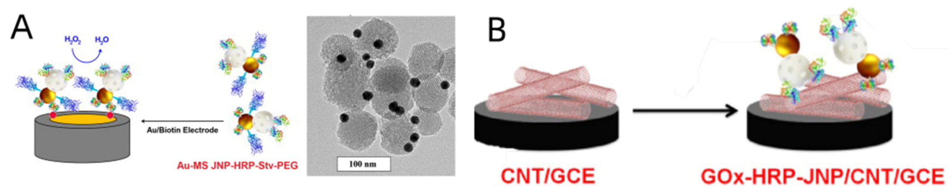
- Sánchez, A.; Díez, P.; Martínez-Ruíz, P.; Villalonga, R.; Pingarrón, J.M. Janus Au-mesoporous silica nanoparticles as electrochemical biorecognition-signaling system. Electrochem. Commun. 2013, 30, 51–54. [Google Scholar] [CrossRef]
- Pacheco, M.; Jurado-Sánchez, B.; Escarpa, A. Sensitive Monitoring of Enterobacterial Contamination of Food Using Self-Propelled Janus Microsensors. Anal. Chem. 2018, 90, 2912–2917. [Google Scholar] [CrossRef]
- Tian, Y.; Deng, P.; Wu, Y.; Ding, Z.; Li, G.; Liu, J.; He, Q. A Simple and Efficient Molecularly Imprinted Electrochemical Sensor for the Selective Determination of Tryptophan. Biomolecules 2019, 9, 294. [Google Scholar] [CrossRef]
- Magesa, F.; Wu, Y.; Dong, S.; Tian, Y.; Li, G.; Vianney, J.M.; Buza, J.; Liu, J.; He, Q. Electrochemical Sensing Fabricated with Ta2O5 Nanoparticle-Electrochemically Reduced Graphene Oxide Nanocomposite for the Detection of Oxytetracycline. Biomolecules 2020, 10, 110. [Google Scholar] [CrossRef]
- Deng, Y.; Wang, W.; Ma, C.; Li, Z.; Yan, D.; Wei, W.; Chao, M.; Zhiyang, L. Fabrication of an Electrochemical Biosensor Array for Simultaneous Detection of L-Glutamate and Acetylcholine. J. Biomed. Nanotechnol. 2013, 9, 1378–1382. [Google Scholar] [CrossRef]
- Deng, Y.; Wang, W.; Zhang, L.; Lu, Z.; Li, S.; Xu, L. Preparation and Electrochemical Behavior of L-Glutamate Electrochemi-cal Biosensor. J. Biomed. Nanotechnol. 2013, 9, 318–321. [Google Scholar] [CrossRef]
- Besis, A.; Gallou, D.; Avgenikou, A.; Serafeim, E.; Samara, C. Size-dependent in vitro inhalation bioaccessibility of PAHs and O/N PAHs—Implications to inhalation risk assessment. Environ. Pollut. 2022, 301, 119045. [Google Scholar] [CrossRef] [PubMed]
- He, Q.; Tian, Y.; Wu, Y.; Liu, J.; Li, G.; Deng, P.; Chen, D. Electrochemical Sensor for Rapid and Sensitive Detection of Tryp-tophan by a Cu2O Nanoparticles-Coated Reduced Graphene Oxide Nanocomposite. Biomolecules 2019, 9, 176. [Google Scholar] [CrossRef] [PubMed]
- Lai, Y.; Deng, Y.; Yang, G.; Li, S.; Zhang, C.; Liu, X. Molecular Imprinting Polymers Electrochemical Sensor Based on AuNPs/PTh Modified GCE for Highly Sensitive Detection of Carcinomaembryonic Antigen. J. Biomed. Nanotechnol. 2018, 14, 1688–1694. [Google Scholar] [CrossRef] [PubMed]
- He, L.; Huang, R.; Xiao, P.; Liu, Y.; Jin, L.; Liu, H.; Li, S.; Deng, Y.; Chen, Z.; Li, Z.; et al. Current Signal Amplification Strategies in Aptamer-Based Electrochemical Biosensor: A Review. Chin. Chem. Lett. 2021, 32, 1593–1602. [Google Scholar] [CrossRef]
- Li, Y.; He, R.; Niu, Y.; Li, F. Paper-Based Electrochemical Biosensors for Point-of-Care Testing of Neurotransmitters. J. Anal. Test. 2019, 3, 19–36. [Google Scholar] [CrossRef]
- Xu, L.; Du, J.; Deng, Y.; He, N. Electrochemical Detection of E. coli O157: H7 Using Porous Pseudo-Carbon Paste Electrode Modified with Carboxylic Multi-Walled Carbon Nanotubes, Glutaraldehyde and 3-Aminopropyltriethoxysilane. J. Biomed. Nanotechnol. 2012, 8, 1006–1011. [Google Scholar] [CrossRef]
- Xie, H.; Di, K.; Huang, R.; Khan, A.; Xia, Y.; Xu, H.; Liu, C.; Tan, T.; Tian, X.; Shen, H.; et al. Extracellular Vesicles Based Elec-trochemical Biosensors for Detection of Cancer Cells: A Review. Chin. Chem. Lett. 2020, 31, 1737–1745. [Google Scholar] [CrossRef]
- Wang, W.; Deng, Y.; Li, S.; Liu, H.; Lu, Z.; Zhang, L.; Lin, L.; Xu, L. A novel acetylcholine bioensor and its electrochemical behavior. J. Biomed. Nanotechnol. 2013, 9, 736–740. [Google Scholar] [CrossRef]
- Zhang, B.; Chen, M.; Cao, J.; Liang, Y.; Tu, T.; Hu, J.; Li, T.; Cai, Y.; Li, S.; Liu, B.; et al. An integrated electrochemical POCT platform for ultrasensitive circRNA detection towards hepatocellular carcinoma diagnosis. Biosens. Bioelectron. 2021, 192, 113500. [Google Scholar] [CrossRef]
- Liu, Y.; Lai, Y.; Yang, G.; Tang, C.; Deng, Y.; Li, S.; Wang, Z. Cd-Aptamer Electrochemical Biosensor Based on AuNPs/CS Modified Glass Carbon Electrode. J. Biomed. Nanotechnol. 2017, 13, 1253–1259. [Google Scholar] [CrossRef]
- Yang, G.; Lai, Y.; Xiao, Z.; Tang, C.; Deng, Y. Ultrasensitive electrochemical immunosensor of carcinoembryonic antigen based on gold-label silver-stain signal amplification. Chin. Chem. Lett. 2018, 29, 1857–1860. [Google Scholar] [CrossRef]
- Wu, Y.; Deng, P.; Tian, Y.; Feng, J.; Xiao, J.; Li, J.; Liu, J.; Li, G.; He, Q. Simultaneous and sensitive determination of ascorbic acid, dopamine and uric acid via an electrochemical sensor based on PVP-graphene composite. J. Nanobiotechnol. 2020, 18, 112. [Google Scholar] [CrossRef] [PubMed]
- Khanwalker, M.; Fujita, R.; Lee, J.; Wilson, E.; Ito, K.; Asano, R.; Ikebukuro, K.; LaBelle, J.; Sode, K. Development of a POCT type insulin sensor employing anti-insulin single chain variable fragment based on faradaic electrochemical impedance spectroscopy under single frequency measurement. Biosens. Bioelectron. 2021, 200, 113901. [Google Scholar] [CrossRef] [PubMed]
- Liu, M.; Yu, X.; Chen, Z.; Yang, T.; Yang, D.; Liu, Q.; Du, K.; Li, B.; Wang, Z.; Li, S.; et al. Aptamer selection and applications for breast cancer diagnostics and therapy. J. Nanobiotechnol. 2017, 15, 81. [Google Scholar] [CrossRef] [PubMed]
- Lai, Y.; Zhang, C.; Deng, Y.; Yang, G.; Li, S.; Tang, C.; He, N. A novel α-fetoprotein-MIP immunosensor based on AuNPs/PTh modified glass carbon electrode. Chin. Chem. Lett. 2018, 30, 160–162. [Google Scholar] [CrossRef]
- Liu, Y.; Li, T.; Ling, C.; Chen, Z.; Deng, Y.; He, N. Electrochemical sensor for Cd2+ and Pb2+ detection based on nano-porous pseudo carbon paste electrode. Chin. Chem. Lett. 2019, 30, 2211–2215. [Google Scholar] [CrossRef]
- Song, Y.; Wang, J.-Q.; Chen, X.-J.; Yu, S.-Z.; Ban, R.-L.; Yang, X.; Zhang, X.; Han, Y. Study the effects of dry-wet cycles and cadmium pollution on the mechanical properties and microstructure of red clay. Environ. Pollut. 2022, 302, 119037. [Google Scholar] [CrossRef]
- Akanda, R.; Joung, H.-A.; Tamilavan, V.; Park, S.; Kim, S.; Hyun, M.H.; Kim, M.-G.; Yang, H. An interference-free and rapid electrochemical lateral-flow immunoassay for one-step ultrasensitive detection with serum. Analyst 2014, 139, 1420–1425. [Google Scholar] [CrossRef]
- Liu, Y.; Deng, Y.; Li, T.; Chen, Z.; Chen, H.; Li, S.; Liu, H. Aptamer-Based Electrochemical Biosensor for Mercury Ions Detection Using AuNPs-Modified Glass Carbon Electrode. J. Biomed. Nanotechnol. 2018, 14, 2156–2161. [Google Scholar] [CrossRef]
- Liu, M.; Xi, L.; Tan, T.; Jin, L.; Wang, Z.; He, N. A novel aptamer-based histochemistry assay for specific diagnosis of clinical breast cancer tissues. Chin. Chem. Lett. 2020, 32, 1726–1730. [Google Scholar] [CrossRef]
- Kanno, Y.; Zhou, Y.; Fukuma, T.; Takahashi, Y. Alkaline Phosphatase-based Electrochemical Analysis for Point-of-Care Testing. Electroanalysis 2021, 34, 161–167. [Google Scholar] [CrossRef]
- Liu, L.-S.; Wu, C.; Zhang, S. Ultrasensitive Detection of DNA and Ramos Cell Using In Situ Selective Crystallization Based Quartz Crystal Microbalance. Anal. Chem. 2017, 89, 4309–4313. [Google Scholar] [CrossRef] [PubMed]
- Wu, C.; Sun, Z.; Liu, L.-S. Quantitative Control of CaCO3 Growth on Quartz Crystal Microbalance Sensors as a Signal Am-plification Method. Analyst 2017, 142, 2547–2551. [Google Scholar] [CrossRef] [PubMed]
- Boujakhrout, A.; Sánchez, E.; Díez, P.; Sánchez, A.; Martínez-Ruiz, P.; Parrado, C.; Pingarrón, J.M.; Villalonga, R. Single-Walled Carbon Nanotubes/Au-Mesoporous Silica Janus Nanoparticles as Building Blocks for the Preparation of a Bienzyme Biosensor. ChemElectroChem 2015, 2, 1735–1741. [Google Scholar] [CrossRef]
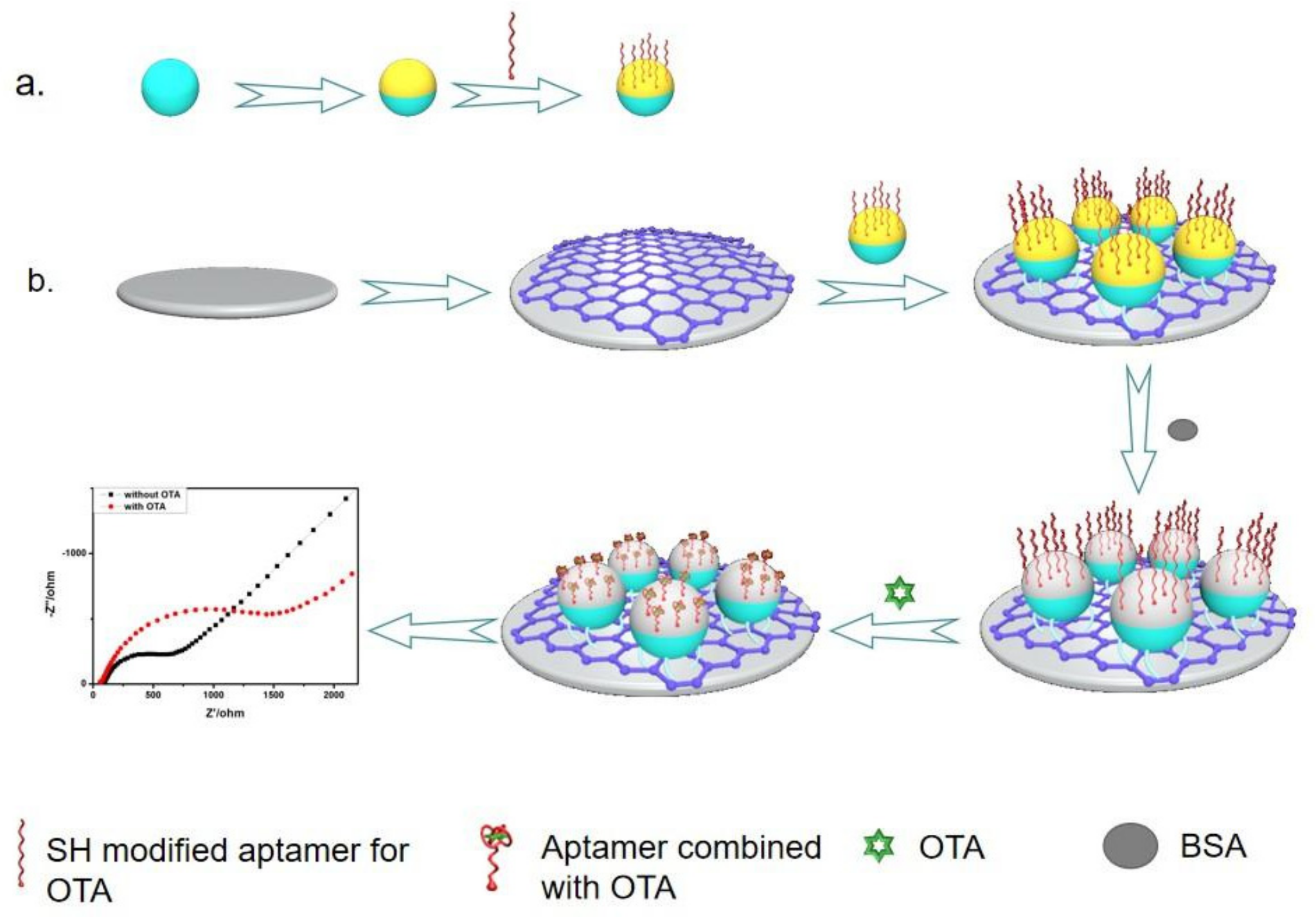
- Yang, Y.-J.; Zhou, Y.; Xing, Y.; Zhang, G.-M.; Zhang, Y.; Zhang, C.-H.; Lei, P.; Dong, C.; Deng, X.; He, Y.; et al. A Label-free aptasensor based on Aptamer/NH2 Janus particles for ultrasensitive electrochemical detection of Ochratoxin A. Talanta 2019, 199, 310–316. [Google Scholar] [CrossRef]
- Kong, L.; Rohaizad, N.; Nasir, M.Z.M.; Guan, J.; Pumera, M. Micromotor-Assisted Human Serum Glucose Biosensing. Anal. Chem. 2019, 91, 5660–5666. [Google Scholar] [CrossRef]
- Paniagua, G.; Villalonga, A.; Eguílaz, M.; Vegas, B.; Parrado, C.; Rivas, G.; Díez, P.; Villalonga, R. Amperometric aptasensor for carcinoembryonic antigen based on the use of bifunctionalized Janus nanoparticles as biorecognition-signaling element. Anal. Chim. Acta 2019, 1061, 84–91. [Google Scholar] [CrossRef]
- Villalonga, A.; Sánchez, A.; Vilela, D.; Mayol, B.; Martínez-Ruíz, P.; Villalonga, R. Electrochemical aptasensor based on anisotropically modified (Janus-type) gold nanoparticles for determination of C-reactive protein. Mikrochim. Acta 2022, 189, 309. [Google Scholar] [CrossRef]
- Ma, E.; Wang, K.; Wang, H. An immunoassay based on nanomotor-assisted electrochemical response for the detection of immunoglobulin. Mikrochim. Acta 2022, 189, 47. [Google Scholar] [CrossRef]
- Sentic, M.; Arbault, S.; Goudeau, B.; Manojlovic, D.; Kuhn, A.; Bouffier, L.; Sojic, N. Electrochemiluminescent swimmers for dynamic enzymatic sensing. Chem. Commun. 2014, 50, 10202–10205. [Google Scholar] [CrossRef]
- Nie, L.; Liu, F.; Ma, P.; Xiao, X. Applications of Gold Nanoparticles in Optical Biosensors. J. Biomed. Nanotechnol. 2014, 10, 2700–2721. [Google Scholar] [CrossRef] [PubMed]
- He, Z.; Tong, Z.; Tan, B.; He, X.; Zhang, T.; Guo, Y.; Jin, L.; He, N.; Li, S.; Chen, Z. Rapid Detection of DNA Methylation with a Novel Real-Time Fluorescence Recombinase-Aided Amplification Assay. J. Biomed. Nanotechnol. 2021, 17, 1364–1370. [Google Scholar] [CrossRef] [PubMed]
- Li, Z.; Wang, J.; Yang, H.; Chen, S.; Ma, G.; Zhang, X.; Zhu, M.; Yu, J.; Singh, R.; Zhang, Y.; et al. Ultrasensitive Detection of Gastric Cancer Plasma MicroRNAs via Magnetic Beads-Based Chemiluminescent Assay. J. Biomed. Nanotechnol. 2017, 13, 1272–1280. [Google Scholar] [CrossRef]
- Zhao, H.; Su, E.; Huang, L.; Zai, Y.; Liu, Y.; Chen, Z.; Li, S.; Jin, L.; Deng, Y.; He, N. Washing-Free Chemiluminescence Im-munoassay for Rapid Detection of Cardiac Troponin I in Whole Blood Samples. Chin. Chem. Lett. 2022, 33, 743–746. [Google Scholar] [CrossRef]
- Zhou, L.; Peng, Y.; Wang, Q.; Lin, Q. An ESIPT-based two-photon fluorescent probe detection of hydrogen peroxide in live cells and tissues. J. Photochem. Photobiol. B Biol. 2017, 167, 264–268. [Google Scholar] [CrossRef]
- Damborský, P.; Švitel, J.; Katrlík, J. Optical biosensors. Essays Biochem. 2016, 60, 91–100. [Google Scholar] [CrossRef]
- Lai, Y.; Wang, L.; Liu, Y.; Yang, G.; Tang, C.; Deng, Y.; Li, S. Immunosensors Based on Nanomaterials for Detection of Tumor Markers. J. Biomed. Nanotechnol. 2018, 14, 44–65. [Google Scholar] [CrossRef]
- Liu, Y.; Yang, G.; Li, T.; Deng, Y.; Chen, Z.; He, N. Selection of a DNA Aptamer for the Development of Fluorescent Ap-tasensor for Carbaryl Detection. Chin. Chem. Lett. 2021, 32, 1957–1962. [Google Scholar] [CrossRef]
- Mou, X.; Chen, Z.; Li, T.; Liu, M.; Liu, Y.; Ali, Z.; Li, S.; Zhu, Y.; Li, Z.; Deng, Y. A Highly Sensitive Strategy for Low-Abundance Hepatitis B Virus Detection via One-Step Nested Polymerase Chain Reaction, Chemiluminescence Technology and Magnetic Separation. J. Biomed. Nanotechnol. 2019, 15, 1832–1838. [Google Scholar] [CrossRef]
- Okumoto, S.; Jones, A.; Frommer, W.B. Quantitative Imaging with Fluorescent Biosensors. Annu. Rev. Plant Biol. 2012, 63, 663–706. [Google Scholar] [CrossRef]
- He, L.; Yang, H.; Xiao, P.; Singh, R.; He, N.; Liu, B.; Li, Z. Highly Selective, Sensitive and Rapid Detection of Escherichia coli O157:H7 Using Duplex PCR and Magnetic Nanoparticle-Based Chemiluminescence Assay. J. Biomed. Nanotechnol. 2017, 13, 1243–1252. [Google Scholar] [CrossRef]
- Tang, C.; He, Z.; Liu, H.; Xu, Y.; Huang, H.; Yang, G.; Xiao, Z.; Li, S.; Liu, H.; Deng, Y.; et al. Application of Magnetic Nano-particles in Nucleic Acid Detection. J. Biomed. Nanotechnol. 2020, 18, 62. [Google Scholar]
- Shi, Y.; Zhang, D.-D.; Liu, J.-B.; Yang, X.-L.; Xin, R.; Jia, C.-Y.; Wang, H.-M.; Lu, G.-X.; Wang, P.-Y.; Liu, Y.; et al. Comprehen-sive Analysis to Identify DLEU2L/TAOK1 Axis as a Prognostic Biomarker in Hepatocellular Carcinoma. Mol. Ther.-Nucleic Acids 2021, 23, 702–718. [Google Scholar] [CrossRef] [PubMed]
- Chen, Z.; Xiao, C.; Qiu, H.; Tan, X.; Jin, L.; He, Y.; Guo, Y.; He, N. Recent Advances of Artificial Intelligence in Cardiovascu-lar Disease. J. Biomed. Nanotechnol. 2020, 16, 1065–1081. [Google Scholar] [CrossRef]
- Pazos, E.; Vázquez, O.; Mascareñas, J.L.; Vázquez, M.E. Peptide-based fluorescent biosensors. Chem. Soc. Rev. 2009, 38, 3348–3359. [Google Scholar] [CrossRef]
- Yang, H.; Liang, W.; Si, J.; Li, Z.; He, N. Long spacer arm-functionalized magnetic nanoparticle platform for enhanced chemiluminescent detection of hepatitis B virus. J. Biomed. Nanotechnol. 2014, 10, 3610–3619. [Google Scholar] [CrossRef]
- Wang, R.; Zhang, Y.; Cai, J.; Cai, W.; Gao, T. Aptamer-Based Fluorescent Biosensors. Curr. Med. Chem. 2011, 18, 4175–4184. [Google Scholar] [CrossRef]
- Tang, Y.; Ali, Z.; Dai, J.; Liu, X.; Wu, Y.; Chen, Z.; He, N.; Li, S.; Wang, L. Single-Nucleotide Polymorphism Genotyping of exoS in Pseudomonas aeruginosa Using Dual-Color Fluorescence Hybridization and Magnetic Separation. J. Biomed. Nanotechnol. 2018, 14, 206–214. [Google Scholar] [CrossRef]
- Yang, H.; Liu, M.; Jiang, H.; Zeng, Y.; Jin, L.; Luan, T.; Deng, Y.; He, N.; Zhang, G.; Zeng, X. Copy Number Variation Analy-sis Based on Gold Magnetic Nanoparticles and Fluorescence Multiplex Ligation-Dependent Probe Amplification. J. Biomed. Nanotechnol. 2017, 13, 655–664. [Google Scholar] [CrossRef]
- Gong, L.; Zhao, L.; Tan, M.; Pan, T.; He, H.; Wang, Y.; He, X.; Li, W.; Tang, L.; Nie, L. Two-Photon Fluorescent Nanomateri-als and Their Applications in Biomedicine. J. Biomed. Nanotechnol. 2021, 17, 509–528. [Google Scholar] [CrossRef]
- Ma, F.; Li, Y.; Tang, B.; Zhang, C.-Y. Fluorescent Biosensors Based on Single-Molecule Counting. Accounts Chem. Res. 2016, 49, 1722–1730. [Google Scholar] [CrossRef] [PubMed]
- Jiang, H.; Zeng, X.; Xi, Z.; Liu, M.; Li, C.; Li, Z.; Jin, L.; Wang, Z.; Deng, Y.; He, N. Improvement on Controllable Fabrication of Streptavidin-Modified Three-Layer Core–Shell Fe3O4@SiO2@Au Magnetic Nanocomposites with Low Fluorescence Background. J. Biomed. Nanotechnol. 2013, 9, 674–684. [Google Scholar] [CrossRef] [PubMed]
- Li, T.; Yi, H.; Liu, Y.; Wang, Z.; Liu, S.; He, N.; Liu, H.; Deng, Y. One-Step Synthesis of DNA Templated Water-Soluble Au–Ag Bimetallic Nanoclusters for Ratiometric Fluorescence Detection of DNA. J. Biomed. Nanotechnol. 2018, 14, 150–160. [Google Scholar] [CrossRef] [PubMed]
- Llopis-Lorente, A.; Villalonga, R.; Marcos, M.D.; Martínez, M.D.M.; Sancenón, F. A Versatile New Paradigm for the Design of Optical Nanosensors Based on Enzyme-Mediated Detachment of Labeled Reporters: The Example of Urea Detection. Chem. A Eur. J. 2018, 25, 3575–3581. [Google Scholar] [CrossRef]
- Tang, J.; Xing, Y.; Wang, Z.; Yang, M.; Zhang, J.; Cai, K. Janus nanoparticles with asymmetrically subcompartmentalized sensing and amplification modules toward fluorescence detection of microRNA. Sensors Actuators B Chem. 2020, 320, 128438. [Google Scholar] [CrossRef]
- Yang, Z.; Peng, X.; Yang, P.; Zhuo, Y.; Chai, Y.-Q.; Liang, W.; Yuan, R. A Janus 3D DNA nanomachine for simultaneous and sensitive fluorescence detection and imaging of dual microRNAs in cancer cells. Chem. Sci. 2020, 11, 8482–8488. [Google Scholar] [CrossRef]
- Li, S.; Liu, H.; Deng, Y.; Lin, L.; He, N. Development of a magnetic nanoparticles microarray for simultaneous and simple detection of foodborne pathogens. J. Biomed. Nanotechnol. 2013, 9, 1254–1260. [Google Scholar] [CrossRef]
- Pacheco, M.; de la Asunción-Nadal, V.; Jurado-Sánchez, B.; Escarpa, A. Engineering Janus micromotors with WS2 and affinity peptides for turn-on fluorescent sensing of bacterial lipopolysaccharides. Biosens. Bioelectron. 2020, 165, 112286. [Google Scholar] [CrossRef]
- Pacheco, M.; Jurado-Sánchez, B.; Escarpa, A. Transition metal dichalcogenide-based Janus micromotors for on-the-fly Salmonella detection. Mikrochim. Acta 2022, 189, 194. [Google Scholar] [CrossRef]
- Chen, W.-L.; Chuang, H.-S. Trace Biomolecule Detection with Functionalized Janus Particles by Rotational Diffusion. Anal. Chem. 2020, 92, 12996–13003. [Google Scholar] [CrossRef]
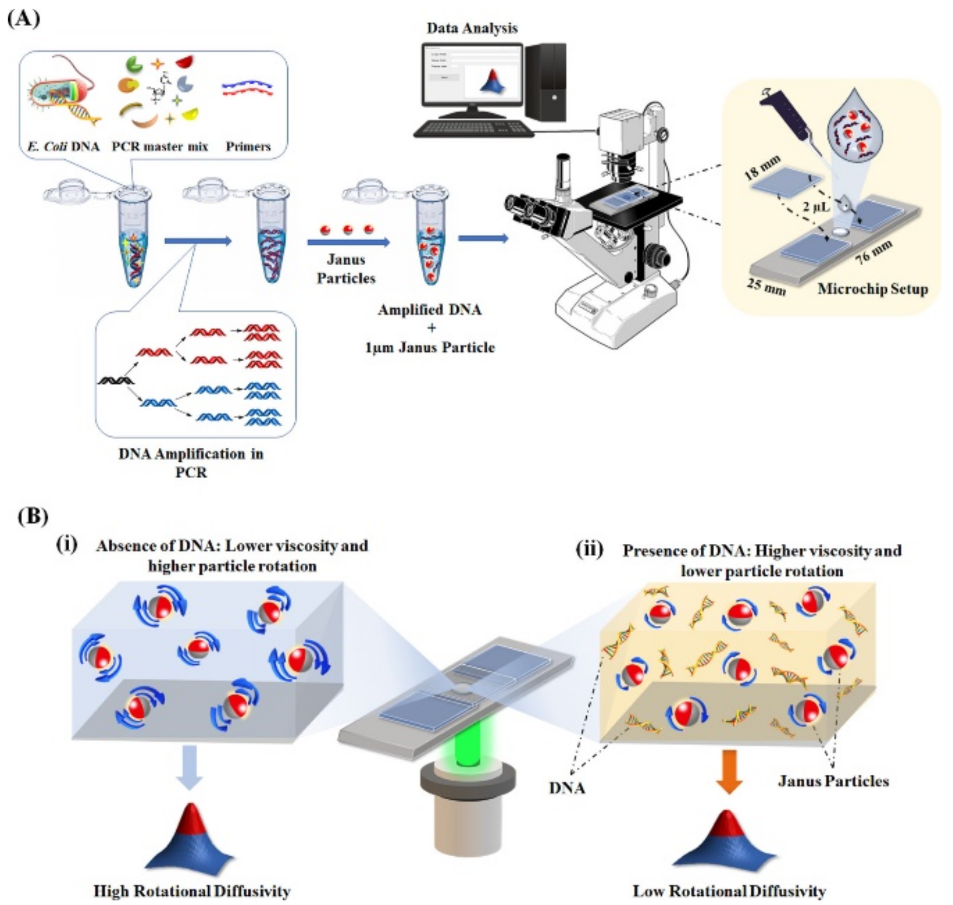
- Das, D.; Chen, W.-L.; Chuang, H.-S. Rapid and Sensitive Pathogen Detection by DNA Amplification Using Janus Particle-Enabled Rotational Diffusometry. Anal. Chem. 2021, 93, 13945–13951. [Google Scholar] [CrossRef]
- Zhang, Q.; Savagatrup, S.; Kaplonek, P.; Seeberger, P.H.; Swager, T.M. Janus Emulsions for the Detection of Bacteria. ACS Central Sci. 2017, 3, 309–313. [Google Scholar] [CrossRef] [PubMed]
- Li, J.; Savagatrup, S.; Nelson, Z.; Yoshinaga, K.; Swager, T.M. Fluorescent Janus Emulsions for Biosensing of Listeria Mono-cytogenes. Proc. Natl. Acad. Sci. USA 2020, 117, 11923–11930. [Google Scholar] [CrossRef] [PubMed]
- Chen, H.; Ma, X.; Zhang, X.; Hu, G.; Deng, Y.; Li, S.; Chen, Z.; He, N.; Wu, Y.; Jiang, Z. Novel Aerosol Detection Platform for SARS-CoV-2: Based on Specific Magnetic Nanoparticles Adsorption Sampling and Digital Droplet PCR Detection. Chin. Chem. Lett. 2022, in press. [Google Scholar] [CrossRef] [PubMed]
- Zhang, Q.; Zeininger, L.; Sung, K.-J.; Miller, E.A.; Yoshinaga, K.; Sikes, H.D.; Swager, T.M. Emulsion Agglutination Assay for the Detection of Protein–Protein Interactions: An Optical Sensor for Zika Virus. ACS Sens. 2019, 4, 180–184. [Google Scholar] [CrossRef]
- Liu, Y.; Li, T.; Yang, G.; Deng, Y.; Mou, X.; He, N. A Simple AuNPs-Based Colorimetric Aptasensor for Chlorpyrifos Detection. Chin. Chem. Lett. 2022, 33, 1913–1916. [Google Scholar] [CrossRef]
- Hridoy, R.H.; Akter, F.; Rakshit, A. Computer Vision Based Skin Disorder Recognition Using EfficientNet: A Transfer Learning Approach. In Proceedings of the 2021 International Conference on Information Technology (ICIT), Amman, Jordan, 14–15 July 2021; pp. 482–487. [Google Scholar]
- Xu, X.; He, N. Application of adaptive pressure-driven microfluidic chip in thyroid function measurement. Chin. Chem. Lett. 2021, 32, 1747–1750. [Google Scholar] [CrossRef]
- Morbioli, G.G.; Mazzu-Nascimento, T.; Stockton, A.M.; Carrilho, E. Technical aspects and challenges of colorimetric detection with microfluidic paper-based analytical devices (μPADs)—A review. Anal. Chim. Acta 2017, 970, 1–22. [Google Scholar] [CrossRef]
- Liu, S.; He, X.; Zhang, T.; Zhao, K.; Xiao, C.; Tong, Z.; Jin, L.; He, N.; Deng, Y.; Li, S.; et al. Highly sensitive smartphone-based detection of Listeria monocytogenes using SYTO9. Chin. Chem. Lett. 2021, 33, 1933–1935. [Google Scholar] [CrossRef]
- Zhu, Z. Smartphone-based apparatus for measuring upconversion luminescence lifetimes. Anal. Chim. Acta 2018, 1054, 122–127. [Google Scholar] [CrossRef]
- Liu, Y.; Li, T.; Ling, C.; Wang, Z.; Jin, L.; Zhao, Y.; Chen, Z.; Li, S.; Deng, Y.; He, N. A Simple Visual Method for DNA Detection Based on the Formation of Gold Nanoparticles. Chin. Chem. Lett. 2019, 30, 2359–2362. [Google Scholar] [CrossRef]
- Chen, Z.; Zhao, K.; He, Z.; Luo, X.; Qin, Z.; Tan, Y.; Zheng, X.; Wu, Z.; Deng, Y.; Chen, H.; et al. Development and Evalua-tion of a Thermostatic Nucleic Acid Testing Device Based on Magnesium Pyrophosphate Precipitation for Detecting Entero-cytozoon Hepatopenaei. Chin. Chem. Lett. 2022, 33, 4053–4056. [Google Scholar] [CrossRef]
- Yang, N.; Chen, C.; Wang, P.; Sun, J.; Mao, H. Structure optimization method of microfluidic paper chip based on image grey-level statistics for chromogenic reaction. Chem. Eng. Process. Process Intensif. 2019, 143, 107627. [Google Scholar] [CrossRef]
- Ou, J.; Liu, K.; Jiang, J.; Wilson, D.A.; Liu, L.; Wang, F.; Wang, S.; Tu, Y.; Peng, F. Micro-/Nanomotors toward Biomedical Applications: The Recent Progress in Biocompatibility. Small 2020, 16, e1906184. [Google Scholar] [CrossRef] [PubMed]
- Lin, R.; Yu, W.; Chen, X.; Gao, H. Self-Propelled Micro/Nanomotors for Tumor Targeting Delivery and Therapy. Adv. Health Mater. 2021, 10, 2001212. [Google Scholar] [CrossRef]
- Yang, R.; Cheng, W.; Chen, X.; Qian, Q.; Zhang, Q.; Pan, Y.; Duan, P.; Miao, P. Color Space Transformation-Based Smartphone Algorithm for Colorimetric Urinalysis. ACS Omega 2018, 3, 12141–12146. [Google Scholar] [CrossRef]
- Zou, J.; Zhang, Q. EyeSay: Make Eyes Speak for ALS Patients with Deep Transfer Learning-Empowered Wearable. In Proceedings of the 2021 43rd Annual International Conference of the IEEE Engineering in Medicine & Biology Society (EMBC), Virtual Conference, 1 November 2021; pp. 377–381. [Google Scholar]
- Wu, J.; Balasubramanian, S.; Kagan, D.; Manesh, K.M.; Campuzano, S.; Wang, J. Motion-based DNA detection using catalytic nanomotors. Nat. Commun. 2010, 1, 36. [Google Scholar] [CrossRef]
- Xie, Y.; Fu, S.; Wu, J.; Lei, J.; Ju, H. Motor-based microprobe powered by bio-assembled catalase for motion detection of DNA. Biosens. Bioelectron. 2017, 87, 31–37. [Google Scholar] [CrossRef]
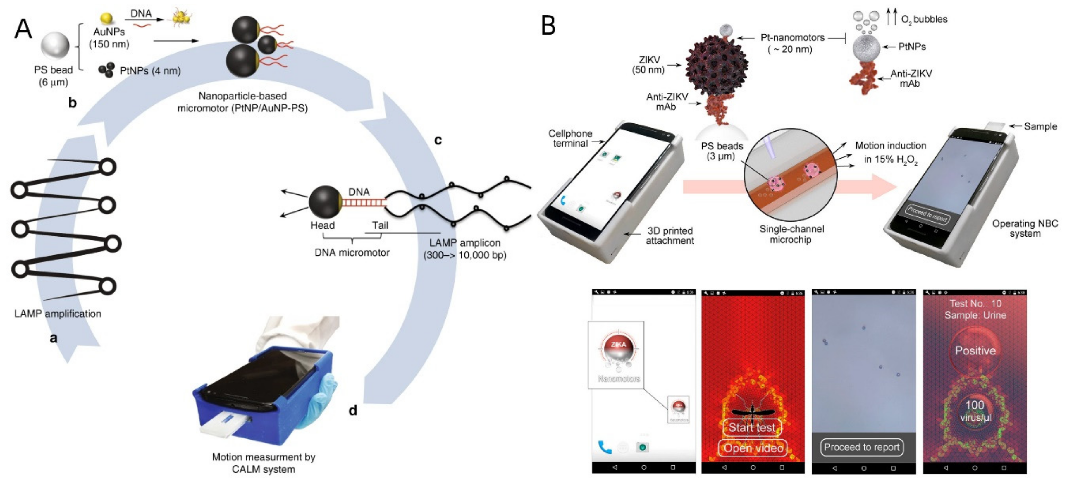
- Draz, M.S.; Kochehbyoki, K.M.; Vasan, A.; Battalapalli, D.; Sreeram, A.; Kanakasabapathy, M.K.; Kallakuri, S.; Tsibris, A.; Kuritzkes, D.R.; Shafiee, H. DNA engineered micromotors powered by metal nanoparticles for motion based cellphone diagnostics. Nat. Commun. 2018, 9, 4282. [Google Scholar] [CrossRef]
- Draz, M.S.; Lakshminaraasimulu, N.K.; Krishnakumar, S.; Battalapalli, D.; Vasan, A.; Kanakasabapathy, M.K.; Sreeram, A.; Kallakuri, S.; Thirumalaraju, P.; Li, Y.; et al. Motion-Based Immunological Detection of Zika Virus Using Pt-Nanomotors and a Cellphone. ACS Nano 2018, 12, 5709–5718. [Google Scholar] [CrossRef]
- Yuan, K.; Cuntín-Abal, C.; Jurado-Sánchez, B.; Escarpa, A. Smartphone-Based Janus Micromotors Strategy for Motion-Based Detection of Glutathione. Anal. Chem. 2021, 93, 16385–16392. [Google Scholar] [CrossRef] [PubMed]
- Russell, S.M.; Alba-Patiño, A.; Borges, M.; de la Rica, R. Multifunctional motion-to-color janus transducers for the rapid detection of sepsis biomarkers in whole blood. Biosens. Bioelectron. 2019, 140, 111346. [Google Scholar] [CrossRef] [PubMed]
- Zhang, Y.-D.; Shi, Y.-P. Colorimetric Detection of Human Alpha-2-Macroglobulin by Janus Imprinted Nanoparticles Con-structed Dual Molecular Imprinting Immunosandwich Strategy. Anal. Chim. Acta 2021, 1184, 339039. [Google Scholar] [CrossRef] [PubMed]
- Duan, W.; Qiu, Z.; Cao, S.; Guo, Q.; Huang, J.; Xing, J.; Lu, X.; Zeng, J. Pd–Fe3O4 Janus nanozyme with rational design for ultrasensitive colorimetric detection of biothiols. Biosens. Bioelectron. 2022, 196, 113724. [Google Scholar] [CrossRef] [PubMed]


| Synthetic Methods | Compositions | Morphology | Particle Size (µm) | Application | Ref. |
|---|---|---|---|---|---|
| Microfluidic method | Polyurethane | Spherical | 40–100 | [62] | |
| Polymer poly(lactic-co-glycolic acid) | Spherical | 0.3 | Drug delivery | [80] | |
| PSMA/PS | Fibriform | 1.9–20 | Biological detection | [120] | |
| Hydrocarbon and fluorocarbon oils | Droplet-shaped | 20 | Biological detection | [121] | |
| Sputtering method | Au@SiO2 | Spherical | 0.4 | [87] | |
| Pt@SiO2 | Spherical | 0.5 | Drug delivery | [122] | |
| SiO2 | Spherical | 0.1 | Drug delivery | [116] | |
| PEDOT-PSS/Au | Tubular | 13.5 | Biological detection | [123] | |
| Au/Ag/Ni/Au | Jellyfish-shaped | 20 | Biological detection | [124] | |
| Au/PEDOT/Pt | Tubular | 12 | Medical imaging | [11] | |
| Phase-separation method | PVP-Fe3O4 | Irregular spherical | 8.7 × 10³ | [98] | |
| Polystyrene(PS)/PMMA | Capped spherical | 10 | [99] | ||
| Au@SiO2 | Spherical | 0.45 | [100] | ||
| Au-SiO2 | Snowman-shaped | 0.1 | Biological detection | [125] | |
| PEG-CuS-Au-MnO2 | Snowman-shaped | 0.125 | Imaging and therapy | [44] | |
| AuNPs | Spherical | 1.3–4.5 × 10⁻² | Biological detection | [55] | |
| Fe3O4@PS/PGMA | Spherical | 18–30 | [90] | ||
| Au/Fe3O4@C | Snowman-shaped | 0.12 | Dual-modal imaging | [46] | |
| Pickering emulsion method | Molten paraffin | Spherical | 1.5 | [108] | |
| Toluene-SiO2 | Spherical | 0.45 | [109] | ||
| Graphene quantum dots | Spherical | 20 | Biological detection | [126] |
Publisher’s Note: MDPI stays neutral with regard to jurisdictional claims in published maps and institutional affiliations. |
© 2022 by the authors. Licensee MDPI, Basel, Switzerland. This article is an open access article distributed under the terms and conditions of the Creative Commons Attribution (CC BY) license (https://creativecommons.org/licenses/by/4.0/).
Share and Cite
Wang, Y.; Zhao, P.; Zhang, S.; Zhu, K.; Shangguan, X.; Liu, L.; Zhang, S. Application of Janus Particles in Point-of-Care Testing. Biosensors 2022, 12, 689. https://doi.org/10.3390/bios12090689
Wang Y, Zhao P, Zhang S, Zhu K, Shangguan X, Liu L, Zhang S. Application of Janus Particles in Point-of-Care Testing. Biosensors. 2022; 12(9):689. https://doi.org/10.3390/bios12090689
Chicago/Turabian StyleWang, Yuhan, Peixuan Zhao, Shihao Zhang, Kexiao Zhu, Xiaoya Shangguan, Lishang Liu, and Shusheng Zhang. 2022. "Application of Janus Particles in Point-of-Care Testing" Biosensors 12, no. 9: 689. https://doi.org/10.3390/bios12090689