Liquid Crystal Droplet-Based Biosensors: Promising for Point-of-Care Testing

 and
and 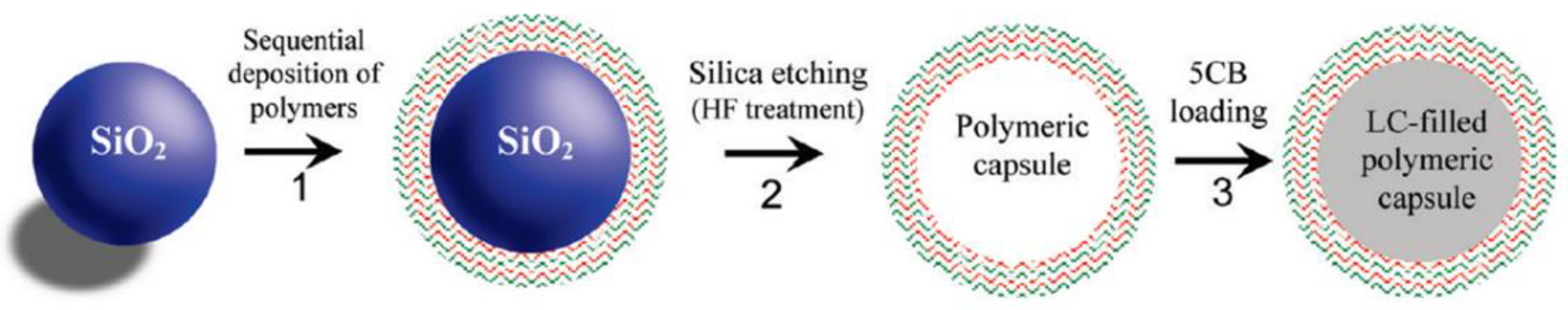
Abstract
:1. Introduction
2. Preparation of LC Droplets
2.1. Emulsion Method
2.2. LC Droplet Pattern Method
3. Overview of LC Droplet-Based Biosensors for POC Diagnosis of Diverse Targets
3.1. Bile Acid
3.2. Nucleic Acid
3.3. Protein and Peptides

3.4. Cell and Microorganism
3.4.1. Single-Cell Monitoring
3.4.2. Different Cells and Microorganisms’ Detection
3.5. Drug
3.6. Toxic Chemical
3.6.1. Toxin
3.6.2. Pesticide and Pollutant

3.7. Other Molecules
4. Conclusions and Future Directions
- 1.
- Spherical LC microstructures with one or more stable cores and multiple nesting may be created because of the rapid advancement of microfluidic technology. The designability and diversity of complex “core-shell microstructures,” as opposed to simple LC droplets and shells, will offer them new features, and raise new scientific challenges that call for more in-depth investigation.
- 2.
- The pointing vector configuration and defects in solvated LC droplets and shells and the related photonics applications are also fascinating research areas that need further exploration.
Author Contributions
Funding
Institutional Review Board Statement
Informed Consent Statement
Conflicts of Interest
References
- Kelker, H. Survey of the Early History of Liquid Crystals. Mol. Cryst. Liq. Cryst. 1988, 165, 1–43. [Google Scholar] [CrossRef]
- Sargazi, M.; Linford, M.R.; Kaykhaii, M. Liquid Crystals in Analytical Chemistry: A Review. Crit. Rev. Anal. Chem. 2019, 49, 243–255. [Google Scholar] [CrossRef] [PubMed]
- Price, A.D.; Schwartz, D.K. DNA Hybridization-Induced Reorientation of Liquid Crystal Anchoring at the Nematic Liquid Crystal/Aqueous Interface. J. Am. Chem. Soc. 2008, 130, 8188–8194. [Google Scholar] [CrossRef] [PubMed]
- Collings, P.J.; Hird, M.; Huang, C.C. Introduction to Liquid Crystals: Chemistry and Physics. Am. J. Phys. 1998, 66, 551. [Google Scholar] [CrossRef]
- McMillan, W.L. Simple Molecular Model for the Smectic A Phase of Liquid Crystals. Phys. Rev. A 1971, 4, 1238–1246. [Google Scholar] [CrossRef]
- Jenkins, J.T. Flows of Nematic Liquid Crystals. Annu. Rev. Fluid Mech. 1978, 10, 197–219. [Google Scholar] [CrossRef]
- Meier, G. Handbook of Liquid Crystals. Angew. Chem. Int. Ed. 1980, 92, 667–668. [Google Scholar] [CrossRef]
- Shvetsov, S.A.; Rudyak, V.Y.; Gruzdenko, A.A.; Emelyanenko, A.V. Axisymmetric Skyrmion-like Structures in Spherical-Cap Droplets of Chiral Nematic Liquid Crystal. J. Mol. Liq. 2020, 319, 114149. [Google Scholar] [CrossRef]
- Gupta, V.K. Optical Amplification of Ligand-Receptor Binding Using Liquid Crystals. Science 1998, 279, 2077–2080. [Google Scholar] [CrossRef]
- Brake, J.M.; Daschner, M.K.; Luk, Y.-Y.; Abbott, N.L. Biomolecular Interactions at Phospholipid-Decorated Surfaces of Liquid Crystals. Science 2003, 302, 2094–2097. [Google Scholar] [CrossRef]
- Lockwood, N.A.; Abbott, N.L. Self-Assembly of Surfactants and Phospholipids at Interfaces between Aqueous Phases and Thermotropic Liquid Crystals. Curr. Opin. Colloid Interface Sci. 2005, 10, 111–120. [Google Scholar] [CrossRef]
- Dong, Y.; Yang, Z. Beyond Displays: The Recent Progress of Liquid Crystals for Bio/Chemical Detections. Chin. Sci. Bull. 2013, 58, 2557–2562. [Google Scholar] [CrossRef]
- Woltman, S.J.; Jay, G.D.; Crawford, G.P. Liquid-Crystal Materials Find a New Order in Biomedical Applications. Nat. Mater. 2007, 6, 929–938. [Google Scholar] [CrossRef] [PubMed]
- Yang, H.; Liu, M.; Jiang, H.; Zeng, Y.; Jin, L.; Luan, T.; Deng, Y.; He, N.; Zhang, G.; Zeng, X. Copy Number Variation Analysis Based on Gold Magnetic Nanoparticles and Fluorescence Multiplex Ligation-Dependent Probe Amplification. J. Biomed. Nanotechnol. 2017, 13, 655–664. [Google Scholar] [CrossRef]
- Yan, J.; Lu, Y.; Xie, S.; Tan, H.; Tan, W.; Li, N.; Xu, L.; Xu, J. Highly Fluorescent N-Doped Carbon Quantum Dots Derived from Bamboo Stems for Selective Detection of Fe3+ Ions in Biological Systems. J. Biomed. Nanotechnol. 2021, 17, 312–321. [Google Scholar] [CrossRef]
- He, L.; Huang, R.; Xiao, P.; Liu, Y.; Jin, L.; Liu, H.; Li, S.; Deng, Y.; Chen, Z.; Li, Z.; et al. Current Signal Amplification Strategies in Aptamer-Based Electrochemical Biosensor: A Review. Chin. Chem. Lett. 2021, 32, 1593–1602. [Google Scholar] [CrossRef]
- Tang, Y.; Ali, Z.; Dai, J.; Liu, X.; Wu, Y.; Chen, Z.; He, N.; Li, S.; Wang, L. Single-Nucleotide Polymorphism Genotyping of ExoS in Pseudomonas Aeruginosa Using Dual-Color Fluorescence Hybridization and Magnetic Separation. J. Biomed. Nanotechnol. 2018, 14, 206–214. [Google Scholar] [CrossRef]
- Gong, L.; Zhao, L.; Tan, M.; Pan, T.; He, H.; Wang, Y.; He, X.; Li, W.; Tang, L.; Nie, L. Two-Photon Fluorescent Nanomaterials and Their Applications in Biomedicine. J. Biomed. Nanotechnol. 2021, 17, 509–528. [Google Scholar] [CrossRef]
- Sadati, M.; Apik, A.I.; Armas-Perez, J.C.; Martinez-Gonzalez, J.; Hernandez-Ortiz, J.P.; Abbott, N.L.; de Pablo, J.J. Liquid Crystal Enabled Early Stage Detection of Beta Amyloid Formation on Lipid Monolayers. Adv. Funct. Mater. 2015, 25, 6050–6060. [Google Scholar] [CrossRef]
- Popov, P.; Mann, E.K.; Jákli, A. Thermotropic Liquid Crystal Films for Biosensors and Beyond. J. Mater. Chem. B 2017, 5, 5061–5078. [Google Scholar] [CrossRef]
- Duong, T.D.S.; Jang, C.-H. A Label-Free Liquid Crystal Droplet-Based Sensor Used to Detect Lead Ions Using Single-Stranded DNAzyme. Colloids Surf. A 2020, 604, 125304. [Google Scholar] [CrossRef]
- Huang, J.-W.; Chang, J.-J.; Yang, R.-H.; Chen, C.-H. Agarose Dispersed Liquid Crystals as a Soft Sensing Platform for Detecting Mercuric Ions in Water. Res. Chem. Intermed. 2019, 45, 5409–5423. [Google Scholar] [CrossRef]
- Han, G.-R.; Jang, C.-H. Detection of Heavy-Metal Ions Using Liquid Crystal Droplet Patterns Modulated by Interaction between Negatively Charged Carboxylate and Heavy-Metal Cations. Talanta 2014, 128, 44–50. [Google Scholar] [CrossRef] [PubMed]
- Park, S.; Lee, S.S.; Kim, S. Photonic Multishells: Photonic Multishells Composed of Cholesteric Liquid Crystals Designed by Controlled Phase Separation in Emulsion Drops. Adv. Mater. 2020, 32, 2002166. [Google Scholar] [CrossRef] [PubMed]
- Kang, J.-H.; Kim, S.-H.; Fernandez-Nieves, A.; Reichmanis, E. Amplified Photon Upconversion by Photonic Shell of Cholesteric Liquid Crystals. J. Am. Chem. Soc. 2017, 139, 5708–5711. [Google Scholar] [CrossRef] [PubMed]
- Khan, M.; Park, S.-Y. General Liquid-Crystal Droplets Produced by Microfluidics for Urea Detection. Sens. Actuators B 2014, 202, 516–522. [Google Scholar] [CrossRef]
- Guo, Z.; Liu, Y.; He, N.; Deng, Y.; Jin, L. Discussion of the Protein Characterization Techniques Used in the Identification of Membrane Protein Targets Corresponding to Tumor Cell Aptamers. Chin. Chem. Lett. 2021, 32, 40–47. [Google Scholar] [CrossRef]
- Chen, Z.; Zhao, K.; He, Z.; Luo, X.; Qin, Z.; Tan, Y.; Zheng, X.; Wu, Z.; Deng, Y.; Chen, H.; et al. Development and Evaluation of a Thermostatic Nucleic Acid Testing Device Based on Magnesium Pyrophosphate Precipitation for Detecting Enterocytozoon Hepatopenaei. Chin. Chem. Lett. 2022, 33, 4053–4056. [Google Scholar] [CrossRef]
- He, Z.; Tong, Z.; Tan, B.; He, X.; Zhang, T.; Guo, Y.; Jin, L.; He, N.; Li, S.; Chen, Z. Rapid Detection of DNA Methylation with a Novel Real-Time Fluorescence Recombinase-Aided Amplification Assay. J. Biomed. Nanotechnol. 2021, 17, 1364–1370. [Google Scholar] [CrossRef]
- Lavrentovich, O.D. Topological Defects in Dispersed Words and Worlds around Liquid Crystals, or Liquid Crystal Drops. Liq. Cryst. 1998, 24, 117–126. [Google Scholar] [CrossRef]
- Xu, F.; Crooker, P.P. Chiral Nematic Droplets with Parallel Surface Anchoring. Phys. Rev. E 1997, 56, 6853–6860. [Google Scholar] [CrossRef]
- Seo, H.J.; Lee, S.S.; Noh, J.; Ka, J.-W.; Won, J.C.; Park, C.; Kim, S.-H.; Kim, Y.H. Robust Photonic Microparticles Comprising Cholesteric Liquid Crystals for Anti-Forgery Materials. J. Mater. Chem. C 2017, 5, 7567–7573. [Google Scholar] [CrossRef]
- Zhou, Y.; Bukusoglu, E.; Martínez-González, J.A.; Rahimi, M.; Roberts, T.F.; Zhang, R.; Wang, X.; Abbott, N.L.; de Pablo, J.J. Structural Transitions in Cholesteric Liquid Crystal Droplets. ACS Nano 2016, 10, 6484–6490. [Google Scholar] [CrossRef]
- Guo, J.-K.; Vij, J.K.; Song, J.-K. Tunable Transfer of Molecules between Liquid Crystal Microdroplets and Control of Photonic Crystallinity in Isolated Microdroplets. Adv. Opt. Mater. 2017, 5, 1700119. [Google Scholar] [CrossRef]
- Hartono, D.; Xue, C.-Y.; Yang, K.-L.; Yung, L.-Y.L. Decorating Liquid Crystal Surfaces with Proteins for Real-Time Detection of Specific Protein–Protein Binding. Adv. Funct. Mater. 2009, 19, 3574–3579. [Google Scholar] [CrossRef]
- Son, J.-H.; Baeck, S.-J.; Park, M.-H.; Lee, J.-B.; Yang, C.-W.; Song, J.-K.; Zin, W.-C.; Ahn, J.-H. Detection of Graphene Domains and Defects Using Liquid Crystals. Nat. Commun. 2014, 5, 3484. [Google Scholar] [CrossRef] [PubMed]
- Guo, L.; Wang, T.; Chen, Z.; He, N.; Chen, Y.; Yuan, T. Light Scattering Based Analyses of the Effects of Bovine Serum Proteins on Interactions of Magnetite Spherical Particles with Cells. Chin. Chem. Lett. 2018, 29, 1291–1295. [Google Scholar] [CrossRef]
- Chen, H.; Wu, Y.; Chen, Z.; Hu, Z.; Fang, Y.; Liao, P.; Deng, Y.; He, N. Performance Evaluation of a Novel Sample In–Answer Out (SIAO) System Based on Magnetic Nanoparticles. J. Biomed. Nanotechnol. 2017, 13, 1619–1630. [Google Scholar] [CrossRef] [PubMed]
- Liu, M.; Xi, L.; Tan, T.; Jin, L.; Wang, Z.; He, N. A Novel Aptamer-Based Histochemistry Assay for Specific Diagnosis of Clinical Breast Cancer Tissues. Chin. Chem. Lett. 2021, 32, 1726–1730. [Google Scholar] [CrossRef]
- Duan, R.; Li, Y.; Li, H.; Yang, J. Detection of Heavy Metal Ions Using Whispering Gallery Mode Lasing in Functionalized Liquid Crystal Microdroplets. Biomed. Opt. Express 2019, 10, 6073. [Google Scholar] [CrossRef]
- Duan, R.; Li, Y.; Shi, B.; Li, H.; Yang, J. Real-Time, Quantitative and Sensitive Detection of Urea by Whispering Gallery Mode Lasing in Liquid Crystal Microdroplet. Talanta 2020, 209, 120513. [Google Scholar] [CrossRef] [PubMed]
- Wang, Z.; Liu, Y.; Wang, H.; Wang, S.; Liu, K.; Xu, T.; Jiang, J.; Chen, Y.-C.; Liu, T. Ultra-Sensitive DNAzyme-Based Optofluidic Biosensor with Liquid Crystal-Au Nanoparticle Hybrid Amplification for Molecular Detection. Sens. Actuators B 2022, 359, 131608. [Google Scholar] [CrossRef]
- Vahala, K.J. Optical Microcavities. Nature 2003, 424, 839–846. [Google Scholar] [CrossRef]
- Lu, Y.; Yang, Y.; Wang, Y.; Wang, L.; Ma, J.; Zhang, L.; Sun, W.; Liu, Y. Tunable Liquid-Crystal Microshell-Laser Based on Whispering-Gallery Modes and Photonic Band-Gap Mode Lasing. Opt. Express 2018, 26, 3277. [Google Scholar] [CrossRef]
- Wang, Y.; Li, H.; Zhao, L.; Liu, Y.; Liu, S.; Yang, J. Tunable Whispering Gallery Modes Lasing in Dye-Doped Cholesteric Liquid Crystal Microdroplets. Appl. Phys. Lett. 2016, 109, 231906. [Google Scholar] [CrossRef]
- Zhao, L.; Wang, Y.; Yuan, Y.; Liu, Y.; Liu, S.; Sun, W.; Yang, J.; Li, H. Whispering Gallery Mode Laser Based on Cholesteric Liquid Crystal Microdroplets as Temperature Sensor. Opt. Commun. 2017, 402, 181–185. [Google Scholar] [CrossRef]
- Armani, A.M.; Kulkarni, R.P.; Fraser, S.E.; Flagan, R.C.; Vahala, K.J. Label-Free, Single-Molecule Detection with Optical Microcavities. Science 2007, 317, 783–787. [Google Scholar] [CrossRef] [PubMed]
- Duan, R.; Li, Y.; Li, H.; Yang, J. Real-Time Monitoring of the Enzymatic Reaction of Urease by Using Whispering Gallery Mode Lasing. Opt. Express 2019, 27, 35427. [Google Scholar] [CrossRef]
- Wang, W.; Deng, Y.; Li, S.; Liu, H.; Lu, Z.; Zhang, L.; Lin, L.; Xu, L. A Novel Acetylcholine Bioensor and Its Electrochemical Behavior. J. Biomed. Nanotechnol. 2013, 9, 736–740. [Google Scholar] [CrossRef]
- He, Q.; Liu, J.; Liu, X.; Li, G.; Deng, P.; Liang, J. Manganese Dioxide Nanorods/Electrochemically Reduced Graphene Oxide Nanocomposites Modified Electrodes for Cost-Effective and Ultrasensitive Detection of Amaranth. Colloids Surf. B 2018, 172, 565–572. [Google Scholar] [CrossRef]
- Deng, Y.; Wang, W.; Zhang, L.; Lu, Z.; Li, S.; Xu, L. Preparation and Electrochemical Behavior of L-Glutamate Electrochemical Biosensor. J. Biomed. Nanotechnol. 2013, 9, 318–321. [Google Scholar] [CrossRef] [PubMed]
- Ma, T.; Huang, H.; Guo, W.; Zhang, C.; Chen, Z.; Li, S.; Ma, L.; Deng, Y. Recent Progress in Black Phosphorus Sensors. J. Biomed. Nanotechnol. 2020, 16, 1045–1064. [Google Scholar] [CrossRef] [PubMed]
- Chen, H.; Ma, X.; Zhang, X.; Hu, G.; Deng, Y.; Li, S.; Chen, Z.; He, N.; Wu, Y.; Jiang, Z. Novel Aerosol Detection Platform for SARS-CoV-2: Based on Specific Magnetic Nanoparticles Adsorption Sampling and Digital Droplet PCR Detection. Chin. Chem. Lett. 2022, in press. [CrossRef] [PubMed]
- Dubtsov, A.V.; Pasechnik, S.V.; Shmeliova, D.V.; Saidgaziev, A.S.; Gongadze, E.; Iglič, A.; Kralj, S. Liquid Crystalline Droplets in Aqueous Environments: Electrostatic Effects. Soft Matter 2018, 14, 9619–9630. [Google Scholar] [CrossRef]
- Gupta, J.K.; Zimmerman, J.S.; de Pablo, J.J.; Caruso, F.; Abbott, N.L. Characterization of Adsorbate-Induced Ordering Transitions of Liquid Crystals within Monodisperse Droplets. Langmuir 2009, 25, 9016–9024. [Google Scholar] [CrossRef]
- Liu, M.; Yu, X.; Chen, Z.; Yang, T.; Yang, D.; Liu, Q.; Du, K.; Li, B.; Wang, Z.; Li, S.; et al. Aptamer Selection and Applications for Breast Cancer Diagnostics and Therapy. J. Nanobiotechnol. 2017, 15, 81. [Google Scholar] [CrossRef]
- Li, T.; He, N.; Wang, J.; Li, S.; Deng, Y.; Wang, Z. Effects of the I-Motif DNA Loop on the Fluorescence of Silver Nanoclusters. RSC Adv. 2016, 6, 22839–22844. [Google Scholar] [CrossRef]
- Xi, Z.; Huang, R.; Deng, Y.; He, N. Progress in Selection and Biomedical Applications of Aptamers. J. Biomed. Nanotechnol. 2014, 10, 3043–3062. [Google Scholar] [CrossRef]
- Nie, L.; Liu, F.; Ma, P.; Xiao, X. Applications of Gold Nanoparticles in Optical Biosensors. J. Biomed. Nanotechnol. 2014, 10, 2700–2721. [Google Scholar] [CrossRef]
- Lai, Y.; Wang, L.; Liu, Y.; Yang, G.; Tang, C.; Deng, Y.; Li, S. Immunosensors Based on Nanomaterials for Detection of Tumor Markers. J. Biomed. Nanotechnol. 2018, 14, 44–65. [Google Scholar] [CrossRef]
- Vennes, M.; Zentel, R. Liquid-Crystalline Colloidal Particles. Macromol. Chem. Phys. 2004, 205, 2303–2311. [Google Scholar] [CrossRef]
- Tixier, T.; Heppenstall-Butler, M.; Terentjev, E.M. Spontaneous Size Selection in Cholesteric and Nematic Emulsions. Langmuir 2006, 22, 2365–2370. [Google Scholar] [CrossRef] [PubMed]
- Hsu, P.; Poulin, P.; Weitz, D.A. Rotational Diffusion of Monodisperse Liquid Crystal Droplets. J. Colloid Interface Sci. 1998, 200, 182–184. [Google Scholar] [CrossRef]
- Vennes, M.; Martin, S.; Gisler, T.; Zentel, R. Anisotropic Particles from LC Polymers for Optical Manipulation. Macromolecules 2006, 39, 8326–8333. [Google Scholar] [CrossRef]
- Sivakumar, S.; Gupta, J.K.; Abbott, N.L.; Caruso, F. Monodisperse Emulsions through Templating Polyelectrolyte Multilayer Capsules. Chem. Mater. 2008, 20, 2063–2065. [Google Scholar] [CrossRef]
- Xu, X.; He, N. Application of Adaptive Pressure-Driven Microfluidic Chip in Thyroid Function Measurement. Chin. Chem. Lett. 2021, 32, 1747–1750. [Google Scholar] [CrossRef]
- Kwon, J.-Y.; Khan, M.; Park, S.-Y. PH-Responsive Liquid Crystal Double Emulsion Droplets Prepared Using Microfluidics. RSC Adv. 2016, 6, 55976–55983. [Google Scholar] [CrossRef]
- Deng, J.; Liang, W.; Fang, J. Liquid Crystal Droplet-Embedded Biopolymer Hydrogel Sheets for Biosensor Applications. ACS Appl. Mater. Interfaces 2016, 8, 3928–3932. [Google Scholar] [CrossRef]
- Lin, I.-H.; Miller, D.S.; Bertics, P.J.; Murphy, C.J.; de Pablo, J.J.; Abbott, N.L. Endotoxin-Induced Structural Transformations in Liquid Crystalline Droplets. Science 2011, 332, 1297–1300. [Google Scholar] [CrossRef]
- Sivakumar, S.; Wark, K.L.; Gupta, J.K.; Abbott, N.L.; Caruso, F. Liquid Crystal Emulsions as the Basis of Biological Sensors for the Optical Detection of Bacteria and Viruses. Adv. Funct. Mater. 2009, 19, 2260–2265. [Google Scholar] [CrossRef]
- Bera, T.; Fang, J. Polyelectrolyte-Coated Liquid Crystal Droplets for Detecting Charged Macromolecules. J. Mater. Chem. 2012, 22, 6807. [Google Scholar] [CrossRef]
- Hu, Q.-Z.; Jang, C.-H. Spontaneous Formation of Micrometer-Scale Liquid Crystal Droplet Patterns on Solid Surfaces and Their Sensing Applications. Soft Matter 2013, 9, 5779. [Google Scholar] [CrossRef]
- Zhang, M.; Jang, C.-H. Sensitive Detection of Trypsin Using Liquid-Crystal Droplet Patterns Modulated by Interactions between Poly-L-Lysine and a Phospholipid Monolayer. ChemPhysChem 2014, 15, 2569–2574. [Google Scholar] [CrossRef]
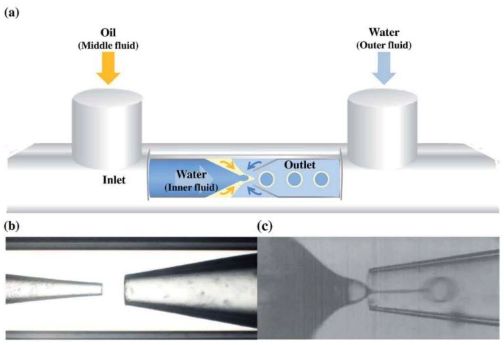
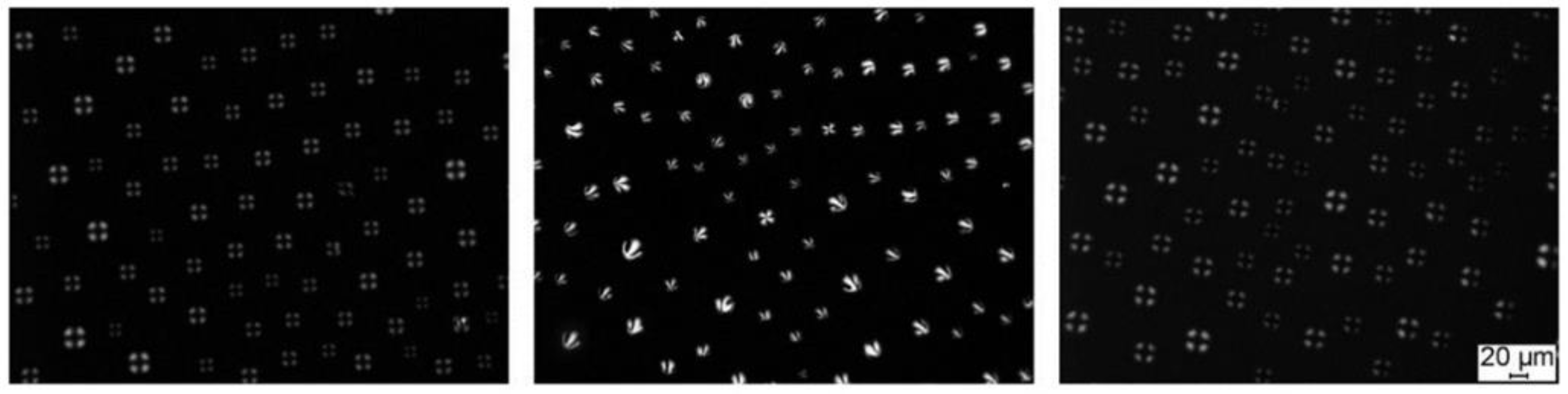
- Han, X.; Han, D.; Zeng, J.; Deng, J.; Hu, N.; Yang, J. Fabrication and Performance of Monodisperse Liquid Crystal Droplet-Based Microchips for the on-Chip Detection of Bile Acids. Microchem. J. 2020, 157, 105057. [Google Scholar] [CrossRef]
- Aliño, V.J.; Tay, K.X.; Khan, S.A.; Yang, K.-L. Inkjet Printing and Release of Monodisperse Liquid Crystal Droplets from Solid Surfaces. Langmuir 2012, 28, 14540–14546. [Google Scholar] [CrossRef]
- Degirolamo, C.; Modica, S.; Palasciano, G.; Moschetta, A. Bile Acids and Colon Cancer: Solving the Puzzle with Nuclear Receptors. Trends Mol. Med. 2011, 17, 564–572. [Google Scholar] [CrossRef]
- Kelly, P.N. Bile Acids and Liver Cancer. Science 2018, 360, 870–871. [Google Scholar] [CrossRef]
- Hegyi, P.; Maléth, J.; Walters, J.R.; Hofmann, A.F.; Keely, S.J. Guts and Gall: Bile Acids in Regulation of Intestinal Epithelial Function in Health and Disease. Physiol. Rev. 2018, 98, 1983–2023. [Google Scholar] [CrossRef]
- Griffiths, W.J.; Sjövall, J. Bile Acids: Analysis in Biological Fluids and Tissues. J. Lipid Res. 2010, 51, 23–41. [Google Scholar] [CrossRef]
- Deng, J.; Lu, X.; Constant, C.; Dogariu, A.; Fang, J. Design of β-CD–Surfactant Complex-Coated Liquid Crystal Droplets for the Detection of Cholic Acid via Competitive Host–Guest Recognition. Chem. Commun. 2015, 51, 8912–8915. [Google Scholar] [CrossRef]
- Hlaváček, A.; Farka, Z.; Hübner, M.; Horňáková, V.; Němeček, D.; Niessner, R.; Skládal, P.; Knopp, D.; Gorris, H.H. Competitive Upconversion-Linked Immunosorbent Assay for the Sensitive Detection of Diclofenac. Anal. Chem. 2016, 88, 6011–6017. [Google Scholar] [CrossRef] [PubMed]
- Xiong, Y.; Leng, Y.; Li, X.; Huang, X.; Xiong, Y. Emerging Strategies to Enhance the Sensitivity of Competitive ELISA for Detection of Chemical Contaminants in Food Samples. TrAC Trends Anal. Chem. 2020, 126, 115861. [Google Scholar] [CrossRef]
- Huang, X.; Chen, R.; Xu, H.; Lai, W.; Xiong, Y. Nanospherical Brush as Catalase Container for Enhancing the Detection Sensitivity of Competitive Plasmonic ELISA. Anal. Chem. 2016, 88, 1951–1958. [Google Scholar] [CrossRef] [PubMed]
- Bera, T.; Fang, J. Optical Detection of Lithocholic Acid with Liquid Crystal Emulsions. Langmuir 2013, 29, 387–392. [Google Scholar] [CrossRef]
- Niu, X.; Luo, D.; Chen, R.; Wang, F.; Sun, X.; Dai, H. Optical Biosensor Based on Liquid Crystal Droplets for Detection of Cholic Acid. Opt. Commun. 2016, 381, 286–291. [Google Scholar] [CrossRef]
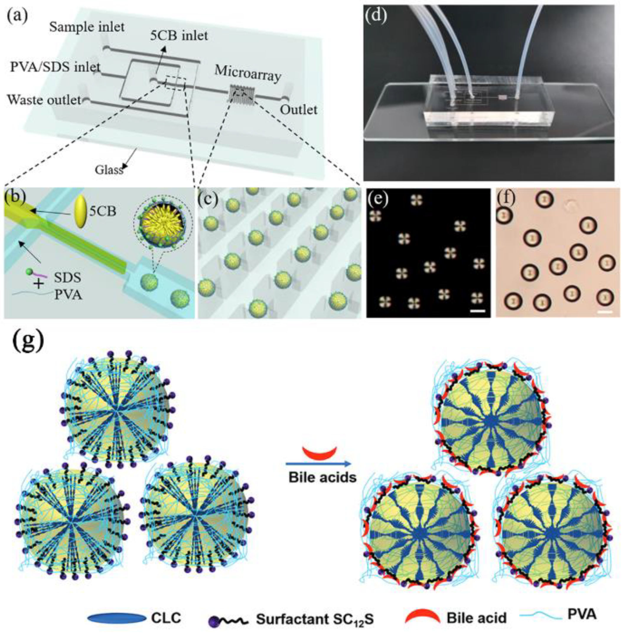
- Gollapelli, B.; Tatipamula, A.K.; Dewanjee, S.; Pathinti, R.S.; Vallamkondu, J. Detection of Bile Acids Using Optical Biosensors Based on Cholesteric Liquid Crystal Droplets. J. Mater. Chem. C 2021, 9, 13991–14002. [Google Scholar] [CrossRef]
- Margueron, R.; Reinberg, D. Chromatin Structure and the Inheritance of Epigenetic Information. Nat. Rev. Genet. 2010, 11, 285–296. [Google Scholar] [CrossRef]
- Annas, G.J. Privacy Rules for DNA Databanks: Protecting Coded “Future Diaries”. JAMA 1993, 270, 2346. [Google Scholar] [CrossRef]
- Li, S.; Liu, H.; Jia, Y.; Mou, X.; Deng, Y.; Lin, L.; Liu, B.; He, N. An Automatic High-Throughput Single Nucleotide Polymorphism Genotyping Approach Based on Universal Tagged Arrays and Magnetic Nanoparticles. J. Biomed. Nanotechnol. 2013, 9, 689–698. [Google Scholar] [CrossRef]
- Tang, C.; He, Z.; Liu, H.; Xu, Y.; Huang, H.; Yang, G.; Xiao, Z.; Li, S.; Liu, H.; Deng, Y.; et al. Application of Magnetic Nanoparticles in Nucleic Acid Detection. J. Nanobiotechnol. 2020, 18, 62. [Google Scholar] [CrossRef] [Green Version]
- Fang, Y.; Liu, H.; Wang, Y.; Su, X.; Jin, L.; Wu, Y.; Deng, Y.; Li, S.; Chen, Z.; Chen, H.; et al. Fast and Accurate Control Strategy for Portable Nucleic Acid Detection (PNAD) System Based on Magnetic Nanoparticles. J. Biomed. Nanotechnol. 2021, 17, 407–415. [Google Scholar] [CrossRef] [PubMed]
- Mou, X.; Li, T.; Wang, J.; Ali, Z.; Zhang, Y.; Chen, Z.; Deng, Y.; Li, S.; Su, E.; Jia, Q.; et al. Genetic Variation of BCL2 (Rs2279115), NEIL2 (Rs804270), LTA (Rs909253), PSCA (Rs2294008) and PLCE1 (Rs3765524, Rs10509670) Genes and Their Correlation to Gastric Cancer Risk Based on Universal Tagged Arrays and Fe3O4 Magnetic Nanoparticles. J. Biomed. Nanotechnol. 2015, 11, 2057–2066. [Google Scholar] [CrossRef]
- Liu, B.; Jia, Y.; Ma, M.; Li, Z.; Liu, H.; Li, S.; Deng, Y.; Zhang, L.; Lu, Z.; Wang, W.; et al. High Throughput SNP Detection System Based on Magnetic Nanoparticles Separation. J. Biomed. Nanotechnol. 2013, 9, 247–256. [Google Scholar] [CrossRef] [PubMed]
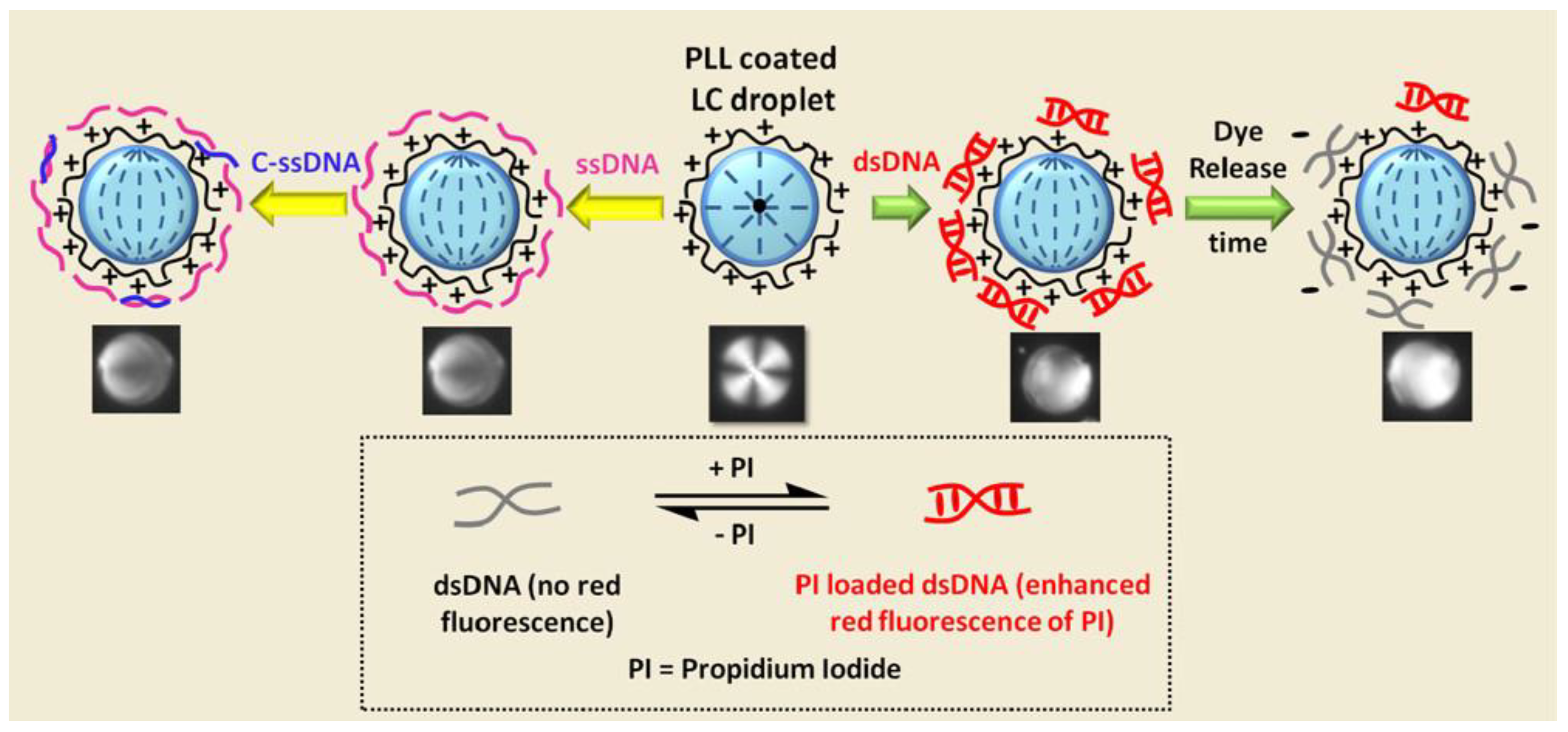
- Verma, I.; Sidiq, S.; Pal, S.K. Poly (L-Lysine)-Coated Liquid Crystal Droplets for Sensitive Detection of DNA and Their Applications in Controlled Release of Drug Molecules. ACS Omega 2017, 2, 7936–7945. [Google Scholar] [CrossRef]
- Mou, X.; Sheng, D.; Chen, Z.; Liu, M.; Liu, Y.; Deng, Y.; Xu, K.; Hou, R.; Zhao, J.; Zhu, Y.; et al. In-Situ Mutation Detection by Magnetic Beads-Probe Based on Single Base Extension and Its Application in Genotyping of Hepatitis B Virus Pre-C Region 1896nt Locus Single Nucleotide Polymorphisms. J. Biomed. Nanotechnol. 2019, 15, 2393–2400. [Google Scholar] [CrossRef] [PubMed]
- Ma, C.; Li, C.; Wang, F.; Ma, N.; Li, X.; Li, Z.; Deng, Y.; Wang, Z.; Xi, Z.; Tang, Y.; et al. Magnetic Nanoparticles-Based Extraction and Verification of Nucleic Acids from Different Sources. J. Biomed. Nanotechnol. 2013, 9, 703–709. [Google Scholar] [CrossRef] [PubMed]
- Xu, Y.; Wang, T.; Chen, Z.; Jin, L.; Wu, Z.; Yan, J.; Zhao, X.; Cai, L.; Deng, Y.; Guo, Y.; et al. The Point-of-Care-Testing of Nucleic Acids by Chip, Cartridge and Paper Sensors. Chin. Chem. Lett. 2021, 32, 3675–3686. [Google Scholar] [CrossRef]
- Li, T.; Yi, H.; Liu, Y.; Wang, Z.; Liu, S.; He, N.; Liu, H.; Deng, Y. One-Step Synthesis of DNA Templated Water-Soluble Au–Ag Bimetallic Nanoclusters for Ratiometric Fluorescence Detection of DNA. J. Biomed. Nanotechnol. 2018, 14, 150–160. [Google Scholar] [CrossRef]
- Ma, C.; Li, C.; He, N.; Wang, F.; Ma, N.; Zhang, L.; Lu, Z.; Ali, Z.; Xi, Z.; Li, X.; et al. Preparation and Characterization of Monodisperse Core–Shell Fe3O4@ SiO2 Microspheres and Its Application for Magnetic Separation of Nucleic Acids from E. Coli BL21. J. Biomed. Nanotechnol. 2012, 8, 1000–1005. [Google Scholar] [CrossRef]
- Wu, C.; Sun, Z.; Liu, L.-S. Quantitative Control of CaCO3 Growth on Quartz Crystal Microbalance Sensors as a Signal Amplification Method. Analyst 2017, 142, 2547–2551. [Google Scholar] [CrossRef]
- Liu, L.; Wu, C.; Zhang, S. Ultrasensitive Detection of DNA and Ramos Cell Using In Situ Selective Crystallization Based Quartz Crystal Microbalance. Anal. Chem. 2017, 89, 4309–4313. [Google Scholar] [CrossRef] [PubMed]
- Ma, Z.; Xu, M.; Zhou, S.; Shan, W.; Zhou, D.; Yan, Y.; Sun, W.; Liu, Y. Ultra-Low Sample Consumption Consecutive-Detection Method for Biochemical Molecules Based on a Whispering Gallery Mode with a Liquid Crystal Microdroplet. Opt. Lett. 2022, 47, 381. [Google Scholar] [CrossRef] [PubMed]
- Mustelin, T.; Vang, T.; Bottini, N. Protein Tyrosine Phosphatases and the Immune Response. Nat. Rev. Immunol. 2005, 5, 43–57. [Google Scholar] [CrossRef]
- Barry, W.S.; Pierce, N.F. Protein Deprivation Causes Reversible Impairment of Mucosal Immune Response to Cholera Toxoid/Toxin in Rat Gut. Nature 1979, 281, 64–65. [Google Scholar] [CrossRef]
- van den Heuvel, M.; Nusse, R.; Johnston, P.; Lawrence, P.A. Distribution of the Wingless Gene Product in Drosophila Embryos: A Protein Involved in Cell-Cell Communication. Cell 1989, 59, 739–749. [Google Scholar] [CrossRef]
- Lai, Y.; Deng, Y.; Yang, G.; Li, S.; Zhang, C.; Liu, X. Molecular Imprinting Polymers Electrochemical Sensor Based on AuNPs/PTh Modified GCE for Highly Sensitive Detection of Carcinomaembryonic Antigen. J. Biomed. Nanotechnol. 2018, 14, 1688–1694. [Google Scholar] [CrossRef] [PubMed]
- Zhao, H.; Su, E.; Huang, L.; Zai, Y.; Liu, Y.; Chen, Z.; Li, S.; Jin, L.; Deng, Y.; He, N. Washing-Free Chemiluminescence Immunoassay for Rapid Detection of Cardiac Troponin I in Whole Blood Samples. Chin. Chem. Lett. 2022, 33, 743–746. [Google Scholar] [CrossRef]
- Bao, P.; Paterson, D.A.; Harrison, P.L.; Miller, K.; Peyman, S.; Jones, J.C.; Sandoe, J.; Evans, S.D.; Bushby, R.J.; Gleeson, H.F. Lipid Coated Liquid Crystal Droplets for the On-Chip Detection of Antimicrobial Peptides. Lab Chip 2019, 19, 1082–1089. [Google Scholar] [CrossRef]
- Pani, I.; KM., F.N.; Sharma, M.; Pal, S.K. Probing Nanoscale Lipid–Protein Interactions at the Interface of Liquid Crystal Droplets. Nano Lett. 2021, 21, 4546–4553. [Google Scholar] [CrossRef]
- Bera, T.; Deng, J.; Fang, J. Protein-Induced Configuration Transitions of Polyelectrolyte-Modified Liquid Crystal Droplets. J. Phys. Chem. B 2014, 118, 4970–4975. [Google Scholar] [CrossRef]
- Verma, I.; Pani, I.; Sharma, D.; Maity, S.; Pal, S.K. Label-Free Imaging of Fibronectin Adsorption at Poly-(L-Lysine)-Decorated Liquid Crystal Droplets. J. Phys. Chem. C 2019, 123, 13642–13650. [Google Scholar] [CrossRef]
- Verma, I.; Sidiq, S.; Pal, S.K. Protein Triggered Ordering Transitions in Poly (L-Lysine)-Coated Liquid Crystal Emulsion Droplets. Liq. Cryst. 2019, 46, 1318–1326. [Google Scholar] [CrossRef]
- Schnurra, C.; Reiners, N.; Biemann, R.; Kaiser, T.; Trawinski, H.; Jassoy, C. Comparison of the Diagnostic Sensitivity of SARS-CoV-2 Nucleoprotein and Glycoprotein-Based Antibody Tests. J. Clin. Virol. 2020, 129, 104544. [Google Scholar] [CrossRef]
- Arts, R.; den Hartog, I.; Zijlema, S.E.; Thijssen, V.; van der Beelen, S.H.E.; Merkx, M. Detection of Antibodies in Blood Plasma Using Bioluminescent Sensor Proteins and a Smartphone. Anal. Chem. 2016, 88, 4525–4532. [Google Scholar] [CrossRef]
- Kim, H.; Hwang, S.G.; Guk, K.; Bae, Y.; Park, H.; Lim, E.-K.; Kang, T.; Jung, J. Development of Antibody against Drug-Resistant Respiratory Syncytial Virus: Rapid Detection of Mutant Virus Using Split Superfolder Green Fluorescent Protein-Antibody System. Biosens. Bioelectron. 2021, 194, 113593. [Google Scholar] [CrossRef]
- Arts, R.; Ludwig, S.K.J.; van Gerven, B.C.B.; Estirado, E.M.; Milroy, L.-G.; Merkx, M. Semisynthetic Bioluminescent Sensor Proteins for Direct Detection of Antibodies and Small Molecules in Solution. ACS Sens. 2017, 2, 1730–1736. [Google Scholar] [CrossRef] [PubMed]
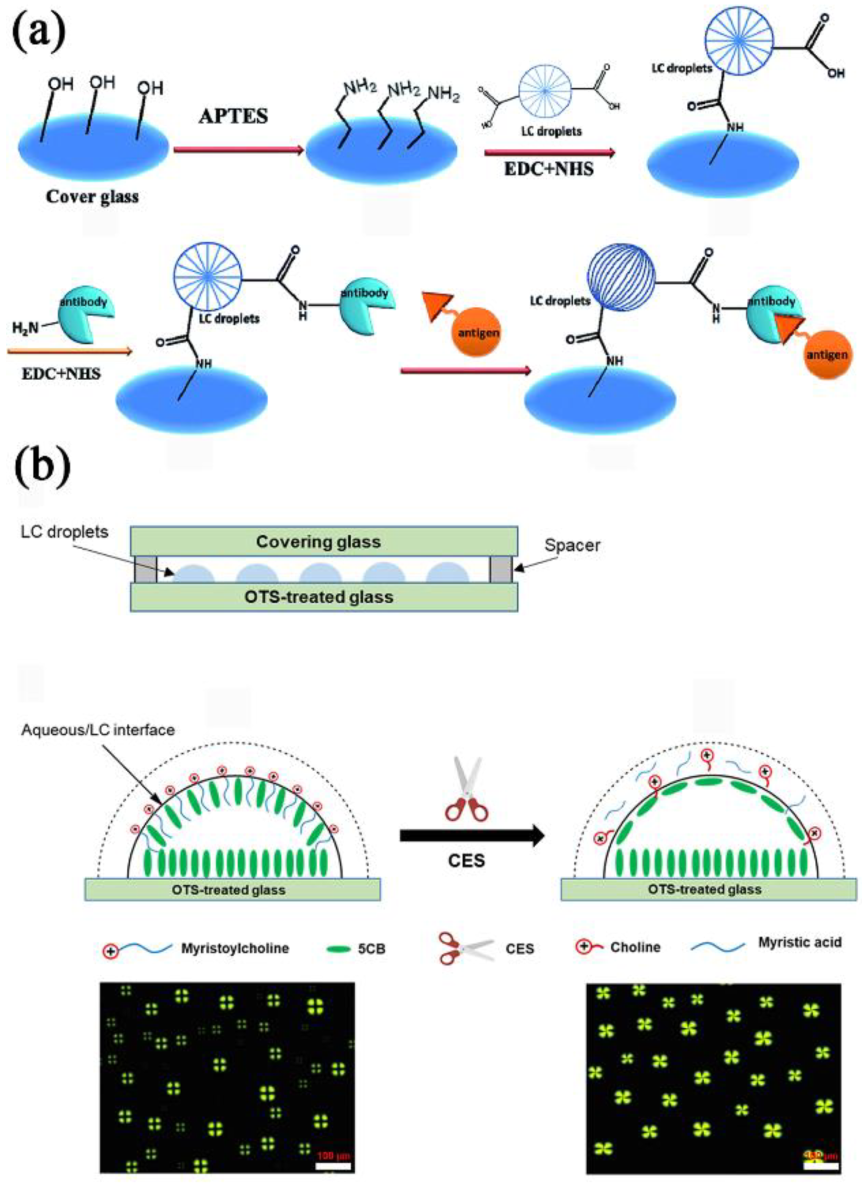
- Huan, Y.; Park, S.J.; Gupta, K.C.; Park, S.-Y.; Kang, I.-K. Slide Cover Glass Immobilized Liquid Crystal Microdroplets for Sensitive Detection of an IgG Antigen. RSC Adv. 2017, 7, 37675–37688. [Google Scholar] [CrossRef]
- Nguyen, D.-K.; Jang, C.-H. Simple and Label-Free Detection of Carboxylesterase and Its Inhibitors Using a Liquid Crystal Droplet Sensing Platform. Micromachines 2022, 13, 490. [Google Scholar] [CrossRef]
- Yang, L.; Khan, M.; Park, S.-Y. Liquid crystal droplets functionalized with charged surfactant and polyelectrolyte for non-specific protein detection. RSC Adv. 2015, 5, 97264–97271. [Google Scholar] [CrossRef]
- Khan, W.; Park, S.-Y. Configuration Change of Liquid Crystal Microdroplets Coated with a Novel Polyacrylic Acid Block Liquid Crystalline Polymer by Protein Adsorption. Lab Chip 2012, 12, 4553. [Google Scholar] [CrossRef]
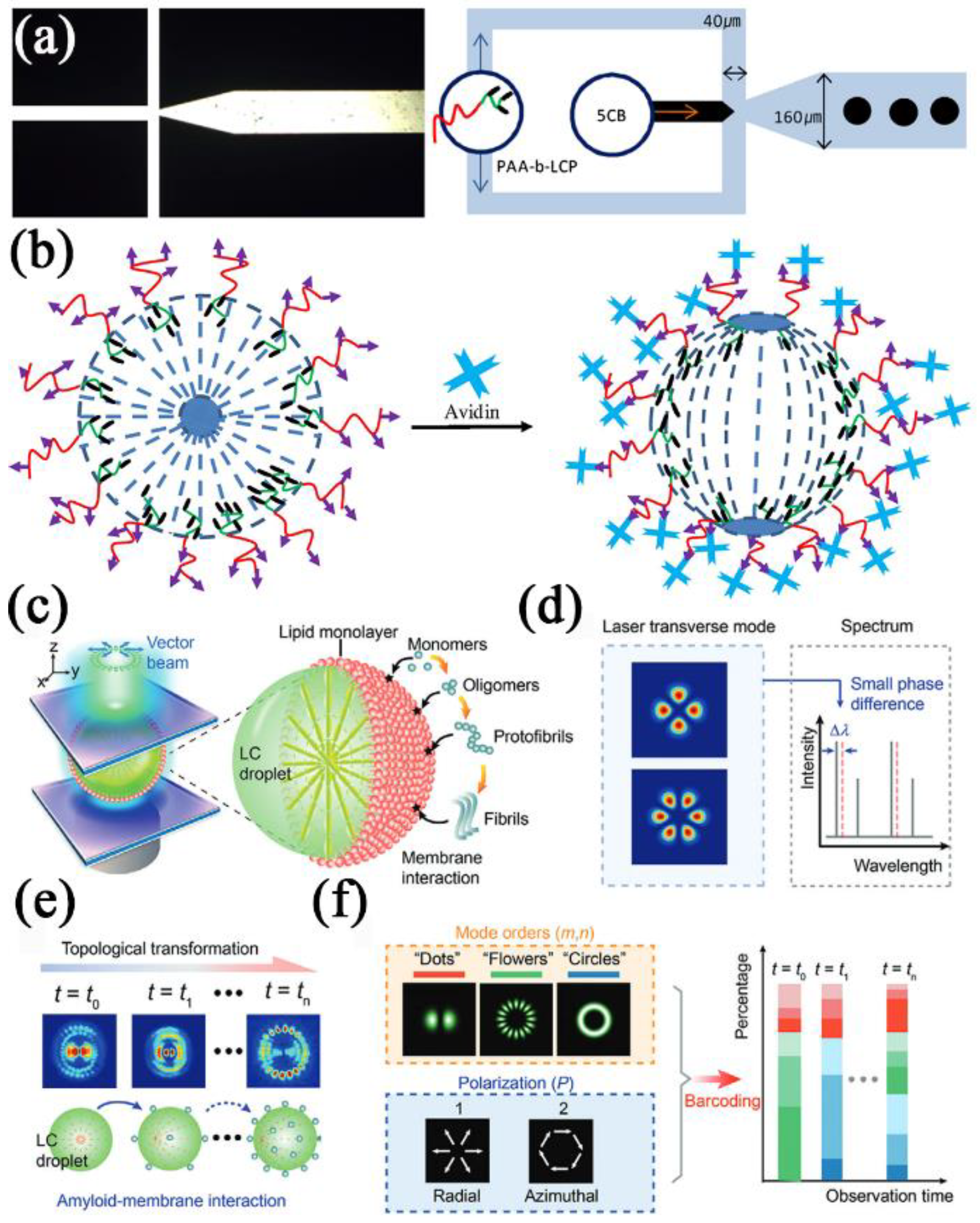
- Khan, M.; Park, S.-Y. Specific Detection of Avidin–Biotin Binding Using Liquid Crystal Droplets. Colloids Surf. B 2015, 127, 241–246. [Google Scholar] [CrossRef]
- Franklin, D.; Ueltschi, T.; Carlini, A.; Yao, S.; Reeder, J.; Richards, B.; Van Duyne, R.P.; Rogers, J.A. Bioresorbable Microdroplet Lasers as Injectable Systems for Transient Thermal Sensing and Modulation. ACS Nano 2021, 15, 2327–2339. [Google Scholar] [CrossRef] [PubMed]
- Rakić, A.D.; Taimre, T.; Bertling, K.; Lim, Y.L.; Dean, P.; Valavanis, A.; Indjin, D. Sensing and Imaging Using Laser Feedback Interferometry with Quantum Cascade Lasers. Appl. Phys. Rev. 2019, 6, 021320. [Google Scholar] [CrossRef]
- He, L.; Özdemir, Ş.K.; Zhu, J.; Kim, W.; Yang, L. Detecting Single Viruses and Nanoparticles Using Whispering Gallery Microlasers. Nat. Nanotechnol. 2011, 6, 428–432. [Google Scholar] [CrossRef] [PubMed]
- Wang, Z.; Zhang, Y.; Gong, X.; Yuan, Z.; Feng, S.; Xu, T.; Liu, T.; Chen, Y.-C. Bio-Electrostatic Sensitive Droplet Lasers for Molecular Detection. Nanoscale Adv. 2020, 2, 2713–2719. [Google Scholar] [CrossRef]
- Gong, C.; Qiao, Z.; Yuan, Z.; Huang, S.; Wang, W.; Wu, P.C.; Chen, Y. Topological Encoded Vector Beams for Monitoring Amyloid-Lipid Interactions in Microcavity. Adv. Sci. 2021, 8, 2100096. [Google Scholar] [CrossRef]
- Mou, X.; Chen, Z.; Li, T.; Liu, M.; Liu, Y.; Ali, Z.; Li, S.; Zhu, Y.; Li, Z.; Deng, Y. A Highly Sensitive Strategy for Low-Abundance Hepatitis B Virus Detection via One-Step Nested Polymerase Chain Reaction, Chemiluminescence Technology and Magnetic Separation. J. Biomed. Nanotechnol. 2019, 15, 1832–1838. [Google Scholar] [CrossRef]
- Chen, Z.; Yang, T.; Yang, H.; Li, T.; Nie, L.; Mou, X.; Deng, Y.; He, N.; Li, Z.; Wang, L.; et al. A Portable Multi-Channel Turbidity System for Rapid Detection of Pathogens by Loop-Mediated Isothermal Amplification. J. Biomed. Nanotechnol. 2018, 14, 198–205. [Google Scholar] [CrossRef]
- Liu, H.; Dong, H.; Chen, Z.; Lin, L.; Chen, H.; Li, S.; Deng, Y. Magnetic Nanoparticles Enhanced Microarray Detection of Multiple Foodborne Pathogens. J. Biomed. Nanotechnol. 2017, 13, 1333–1343. [Google Scholar] [CrossRef]
- Hussain, M.; Chen, Z.; Lv, M.; Xu, J.; Dong, X.; Zhao, J.; Li, S.; Deng, Y.; He, N.; Li, Z.; et al. Rapid and Label-Free Classification of Pathogens Based on Light Scattering, Reduced Power Spectral Features and Support Vector Machine. Chin. Chem. Lett. 2020, 31, 3163–3167. [Google Scholar] [CrossRef]
- Yang, H.; Liang, W.; Si, J.; Li, Z.; He, N. Long Spacer Arm-Functionalized Magnetic Nanoparticle Platform for Enhanced Chemiluminescent Detection of Hepatitis B Virus. J. Biomed. Nanotechnol. 2014, 10, 3610–3619. [Google Scholar] [CrossRef]
- Whiteley, M.; Diggle, S.P.; Greenberg, E.P. Progress in and Promise of Bacterial Quorum Sensing Research. Nature 2017, 551, 313–320. [Google Scholar] [CrossRef] [PubMed]
- Liu, S.; He, X.; Zhang, T.; Zhao, K.; Xiao, C.; Tong, Z.; Jin, L.; He, N.; Deng, Y.; Li, S.; et al. Highly Sensitive Smartphone-Based Detection of Listeria Monocytogenes Using SYTO9. Chin. Chem. Lett. 2022, 33, 1933–1935. [Google Scholar] [CrossRef]
- Zhou, L.; Peng, Y.; Wang, Q.; Lin, Q. An ESIPT-Based Two-Photon Fluorescent Probe Detection of Hydrogen Peroxide in Live Cells and Tissues. J. Photochem. Photobiol. B 2017, 167, 264–268. [Google Scholar] [CrossRef] [PubMed]
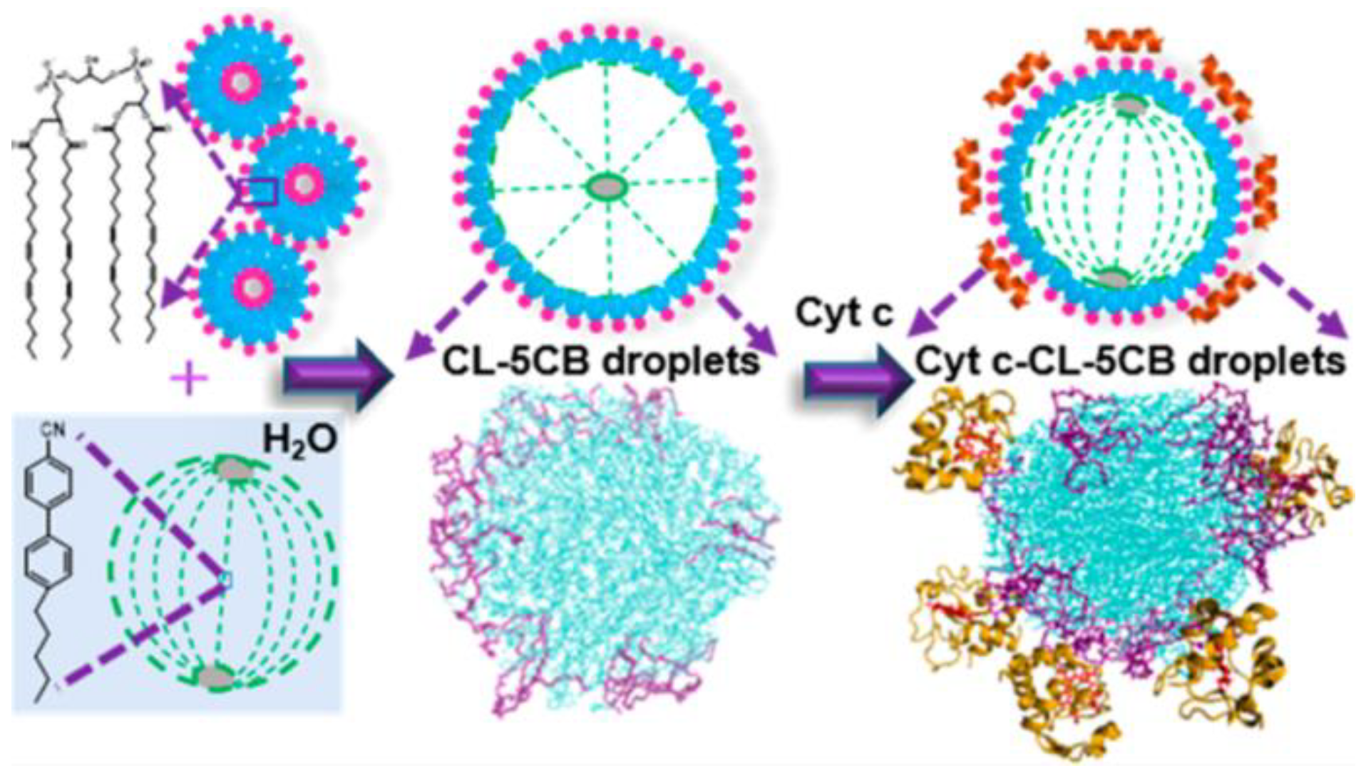
- Sidiq, S.; Prasad, G.V.R.K.; Mukhopadhaya, A.; Pal, S.K. Poly(L-Lysine)-Coated Liquid Crystal Droplets for Cell-Based Sensing Applications. J. Phys. Chem. B 2017, 121, 4247–4256. [Google Scholar] [CrossRef]
- Manna, U.; Zayas-Gonzalez, Y.M.; Carlton, R.J.; Caruso, F.; Abbott, N.L.; Lynn, D.M. Liquid Crystal Chemical Sensors That Cells Can Wear. Angew. Chem. Int. Ed. 2013, 52, 14011–14015. [Google Scholar] [CrossRef]
- Manna, U.; Zavala, Y.M.; Abbott, N.L.; Lynn, D.M. Structured Liquid Droplets as Chemical Sensors That Function Inside Living Cells. ACS Appl. Mater. Interfaces 2021, 13, 42502–42512. [Google Scholar] [CrossRef]
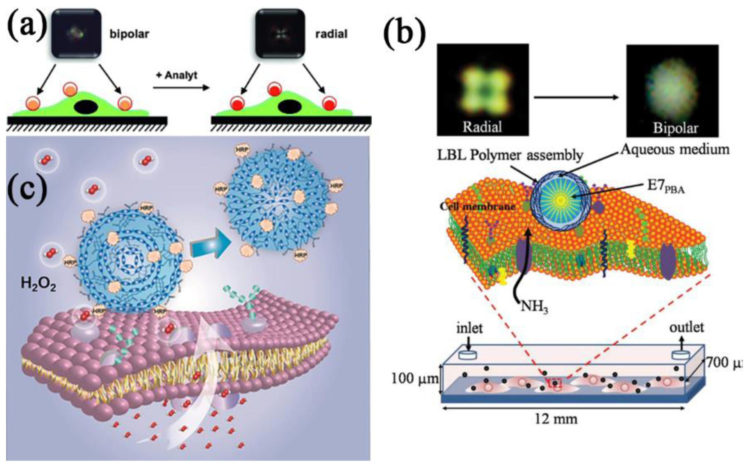
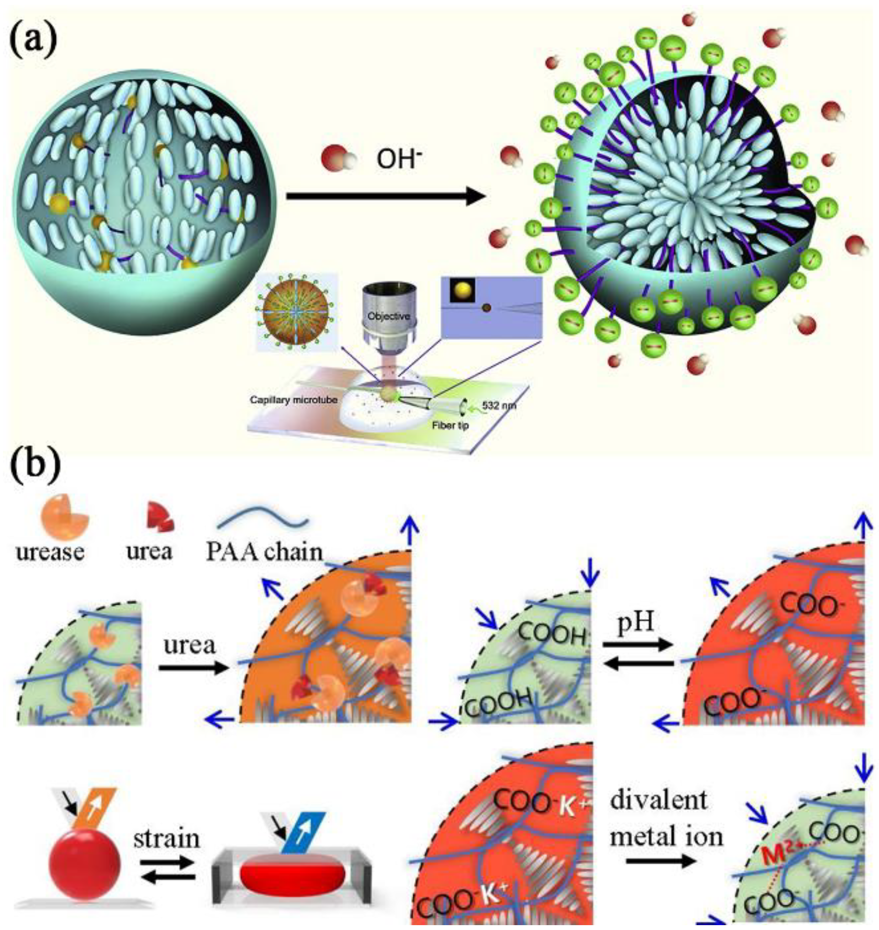
- Khan, M.; Li, W.; Mao, S.; Shah, S.N.A.; Lin, J. Real-Time Imaging of Ammonia Release from Single Live Cells via Liquid Crystal Droplets Immobilized on the Cell Membrane. Adv. Sci. 2019, 6, 1900778. [Google Scholar] [CrossRef]
- Li, W.; Khan, M.; Lin, L.; Zhang, Q.; Feng, S.; Wu, Z.; Lin, J. Monitoring H2O2 on the Surface of Single Cells with Liquid Crystal Elastomer Microspheres. Angew. Chem. Int. Ed. 2020, 59, 9282–9287. [Google Scholar] [CrossRef]
- Xie, H.; Di, K.; Huang, R.; Khan, A.; Xia, Y.; Xu, H.; Liu, C.; Tan, T.; Tian, X.; Shen, H.; et al. Extracellular Vesicles Based Electrochemical Biosensors for Detection of Cancer Cells: A Review. Chin. Chem. Lett. 2020, 31, 1737–1745. [Google Scholar] [CrossRef]
- Shen, Z.; Wu, A.; Chen, X. Current Detection Technologies for Circulating Tumor Cells. Chem. Soc. Rev. 2017, 46, 2038–2056. [Google Scholar] [CrossRef]
- Suzuki, T.; Kaji, N.; Yasaki, H.; Yasui, T.; Baba, Y. Mechanical Low-Pass Filtering of Cells for Detection of Circulating Tumor Cells in Whole Blood. Anal. Chem. 2020, 92, 2483–2491. [Google Scholar] [CrossRef] [PubMed]
- Jan, Y.J.; Chen, J.-F.; Zhu, Y.; Lu, Y.-T.; Chen, S.H.; Chung, H.; Smalley, M.; Huang, Y.-W.; Dong, J.; Chen, L.-C.; et al. NanoVelcro Rare-Cell Assays for Detection and Characterization of Circulating Tumor Cells. Adv. Drug Deliv. Rev. 2018, 125, 78–93. [Google Scholar] [CrossRef] [PubMed]
- Baird, Z.; Pirro, V.; Ayrton, S.; Hollerbach, A.; Hanau, C.; Marfurt, K.; Foltz, M.; Cooks, R.G.; Pugia, M. Tumor Cell Detection by Mass Spectrometry Using Signal Ion Emission Reactive Release Amplification. Anal. Chem. 2016, 88, 6971–6975. [Google Scholar] [CrossRef] [PubMed]
- Yoon, S.H.; Gupta, K.C.; Borah, J.S.; Park, S.-Y.; Kim, Y.-K.; Lee, J.-H.; Kang, I.-K. Folate Ligand Anchored Liquid Crystal Microdroplets Emulsion for In Vitro Detection of KB Cancer Cells. Langmuir 2014, 30, 10668–10677. [Google Scholar] [CrossRef]
- Choi, Y.; Lee, K.; Gupta, K.C.; Park, S.-Y.; Kang, I.-K. The Role of Ligand–Receptor Interactions in Visual Detection of HepG2 Cells Using a Liquid Crystal Microdroplet-Based Biosensor. J. Mater. Chem. B 2015, 3, 8659–8669. [Google Scholar] [CrossRef]
- Xiao, Z.; Chen, H.; Chen, H.; Wu, L.; Yang, G.; Wu, Y.; He, N. Advanced Diagnostic Strategies for Clostridium Difficile Infection (CDI). J. Biomed. Nanotechnol. 2019, 15, 1113–1134. [Google Scholar] [CrossRef]
- He, L.; Yang, H.; Xiao, P.; Singh, R.; He, N.; Liu, B.; Li, Z. Highly Selective, Sensitive and Rapid Detection of Escherichia coli O157:H7 Using Duplex PCR and Magnetic Nanoparticle-Based Chemiluminescence Assay. J. Biomed. Nanotechnol. 2017, 13, 1243–1252. [Google Scholar] [CrossRef]
- Dong, H.; Tang, C.; He, Z.; Liu, H.; Xu, Y.; Huang, H.; Yang, G.; Xiao, Z.; Li, S.; Deng, Y.; et al. Rapid Identification of Diarrheagenic Escherichia coli Based on Barcoded Magnetic Bead Hybridization. Chin. Chem. Lett. 2020, 31, 1812–1816. [Google Scholar] [CrossRef]
- Ling, Y.; Zhu, Y.; Fan, H.; Zha, H.; Yang, M.; Wu, L.; Chen, H.; Li, W.; Wu, Y.; Chen, H. Rapid Method for Detection of Staphylococcus aureus in Feces. J. Biomed. Nanotechnol. 2019, 15, 1290–1298. [Google Scholar] [CrossRef]
- Tang, Y.; Li, Z.; He, N.; Zhang, L.; Ma, C.; Li, X.; Li, C.; Wang, Z.; Deng, Y.; He, L. Preparation of Functional Magnetic Nanoparticles Mediated with PEG-4000 and Application in Pseudomonas aeruginosa Rapid Detection. J. Biomed. Nanotechnol. 2013, 9, 312–317. [Google Scholar] [CrossRef]
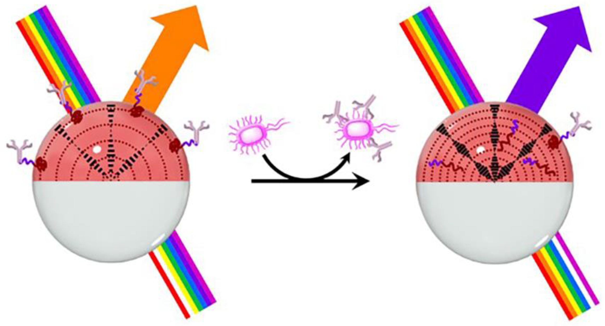
- Concellón, A.; Fong, D.; Swager, T.M. Complex Liquid Crystal Emulsions for Biosensing. J. Am. Chem. Soc. 2021, 143, 9177–9182. [Google Scholar] [CrossRef] [PubMed]
- Jones, C.M.; Baldwin, G.T.; Manocchio, T.; White, J.O.; Mack, K.A. Trends in Methadone Distribution for Pain Treatment, Methadone Diversion, and Overdose Deaths—United States, 2002–2014. MMWR Morb. Mortal. Wkly. Rep. 2016, 65, 667–671. [Google Scholar] [CrossRef] [PubMed]
- Dowell, D.; Noonan, R.K.; Houry, D. Underlying Factors in Drug Overdose Deaths. JAMA 2017, 318, 2295. [Google Scholar] [CrossRef] [PubMed]
- Magesa, F.; Wu, Y.; Dong, S.; Tian, Y.; Li, G.; Vianney, J.M.; Buza, J.; Liu, J.; He, Q. Electrochemical Sensing Fabricated with Ta2O5 Nanoparticle-Electrochemically Reduced Graphene Oxide Nanocomposite for the Detection of Oxytetracycline. Biomolecules 2020, 10, 110. [Google Scholar] [CrossRef] [PubMed]
- Bloomfield, M. A Sensitive and Rapid Assay for 4-Aminophenol in Paracetamol Drug and Tablet Formulation, by Flow Injection Analysis with Spectrophotometric Detection. Talanta 2002, 58, 1301–1310. [Google Scholar] [CrossRef]
- Chen, C.; Zhu, S.; Wang, S.; Zhang, W.; Cheng, Y.; Yan, X. Multiparameter Quantification of Liposomal Nanomedicines at the Single-Particle Level by High-Sensitivity Flow Cytometry. ACS Appl. Mater. Interfaces 2017, 9, 13913–13919. [Google Scholar] [CrossRef]
- Morrison, K.A.; Valenzuela, B.R.; Denis, E.H.; Nims, M.K.; Atkinson, D.A.; Clowers, B.H.; Ewing, R.G. Non-Contact Vapor Detection of Illicit Drugs via Atmospheric Flow Tube-Mass Spectrometry. Analyst 2020, 145, 6485–6492. [Google Scholar] [CrossRef]
- Peng, Y.; Gautam, L.; Hall, S.W. The Detection of Drugs of Abuse and Pharmaceuticals in Drinking Water Using Solid-Phase Extraction and Liquid Chromatography-Mass Spectrometry. Chemosphere 2019, 223, 438–447. [Google Scholar] [CrossRef]
- Jin, N.; Paraskevaidi, M.; Semple, K.T.; Martin, F.L.; Zhang, D. Infrared Spectroscopy Coupled with a Dispersion Model for Quantifying the Real-Time Dynamics of Kanamycin Resistance in Artificial Microbiota. Anal. Chem. 2017, 89, 9814–9821. [Google Scholar] [CrossRef] [Green Version]
- Yin, F.; Cheng, S.; Liu, S.; Ma, C.; Wang, L.; Zhao, R.; Lin, J.-M.; Hu, Q. A Portable Digital Optical Kanamycin Sensor Developed by Surface-Anchored Liquid Crystal Droplets. J. Hazard. Mater. 2021, 420, 126601. [Google Scholar] [CrossRef]
- Ong, K.C.; Khoo, H.-E. Biological Effects of Myricetin. Gen. Pharmacol. Vasc. Syst. 1997, 29, 121–126. [Google Scholar] [CrossRef]
- Jiang, M.; Zhu, M.; Wang, L.; Yu, S. Anti-Tumor Effects and Associated Molecular Mechanisms of Myricetin. Biomed. Pharmacother. 2019, 120, 109506. [Google Scholar] [CrossRef] [PubMed]
- Jung, S.K.; Lee, K.W.; Byun, S.; Kang, N.J.; Lim, S.H.; Heo, Y.-S.; Bode, A.M.; Bowden, G.T.; Lee, H.J.; Dong, Z. Myricetin Suppresses UVB-Induced Skin Cancer by Targeting Fyn. Cancer Res. 2008, 68, 6021–6029. [Google Scholar] [CrossRef] [PubMed]
- Xiong, Z.; Zhang, H.; Lu, Y.; Zhang, L.; Sun, W.; Liu, Y. Fast Detection of Myricetin with the Use of Dedicated Microdroplets. IEEE Sens. J. 2020, 20, 617–622. [Google Scholar] [CrossRef]
- Stephens, R.; Mythen, M. Endotoxin Immunization. Intensive Care Med. 2000, 26, S129–S136. [Google Scholar] [CrossRef]
- Roberts, R.S. Preparation of Endotoxin. Nature 1966, 209, 80. [Google Scholar] [CrossRef]
- Jiang, S.; Noh, J.; Park, C.; Smith, A.D.; Abbott, N.L.; Zavala, V.M. Using Machine Learning and Liquid Crystal Droplets to Identify and Quantify Endotoxins from Different Bacterial Species. Analyst 2021, 146, 1224–1233. [Google Scholar] [CrossRef] [PubMed]
- Berthiller, F.; Crews, C.; Dall’Asta, C.; Saeger, S.D.; Haesaert, G.; Karlovsky, P.; Oswald, I.P.; Seefelder, W.; Speijers, G.; Stroka, J. Masked Mycotoxins: A Review. Mol. Nutr. Food Res. 2013, 57, 165–186. [Google Scholar] [CrossRef]
- Dong, Y.; Wen, C.; She, Y.; Zhang, Y.; Chen, Y.; Zeng, J. Magnetic Relaxation Switching Immunoassay Based on Hydrogen Peroxide-Mediated Assembly of Ag@Au–Fe3O4 Nanoprobe for Detection of Aflatoxin B1. Small 2021, 17, 2104596. [Google Scholar] [CrossRef]
- Liu, D.; Li, W.; Zhu, C.; Li, Y.; Shen, X.; Li, L.; Yan, X.; You, T. Recent Progress on Electrochemical Biosensing of Aflatoxins: A Review. TrAC Trends Anal. Chem. 2020, 133, 115966. [Google Scholar] [CrossRef]
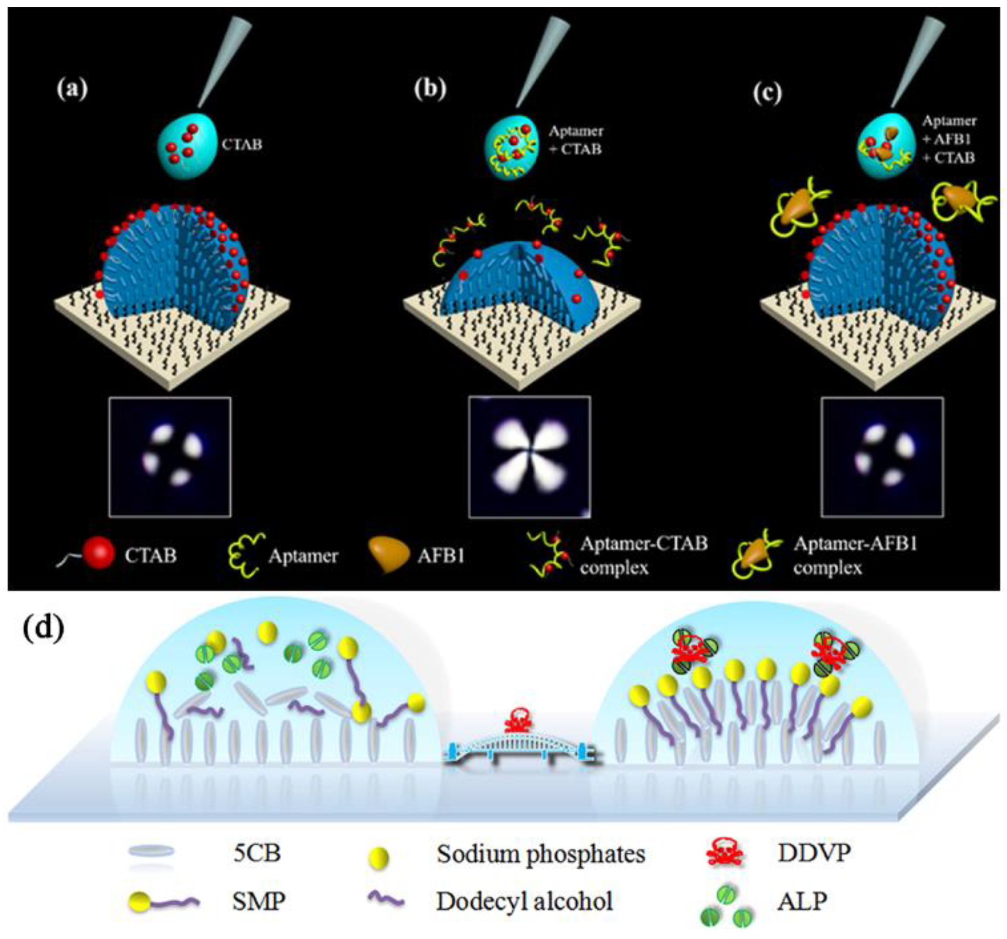
- Cheng, S.; Khan, M.; Yin, F.; Ma, C.; Yuan, J.; Jiang, T.; Liu, X.; Hu, Q. Surface-Anchored Liquid Crystal Droplets for the Semi-Quantitative Detection of Aflatoxin B1 in Food Samples. Food Chem. 2022, 390, 133202. [Google Scholar] [CrossRef] [PubMed]
- Guo, W.; Zhang, C.; Ma, T.; Liu, X.; Chen, Z.; Li, S.; Deng, Y. Advances in Aptamer Screening and Aptasensors’ Detection of Heavy Metal Ions. J. Nanobiotechnol. 2021, 19, 166. [Google Scholar] [CrossRef] [PubMed]
- Liu, Y.; Lai, Y.; Yang, G.; Tang, C.; Deng, Y.; Li, S.; Wang, Z. Cd-Aptamer Electrochemical Biosensor Based on AuNPs/CS Modified Glass Carbon Electrode. J. Biomed. Nanotechnol. 2017, 13, 1253–1259. [Google Scholar] [CrossRef]
- Liu, Y.; Yang, G.; Li, T.; Deng, Y.; Chen, Z.; He, N. Selection of a DNA Aptamer for the Development of Fluorescent Aptasensor for Carbaryl Detection. Chin. Chem. Lett. 2021, 32, 1957–1962. [Google Scholar] [CrossRef]
- Yang, G.; Liu, Y.; Deng, Y.; Chen, Z.; Chen, H.; Li, S.; He, N. Selection of a High-Affinity DNA Aptamer for the Recognition of Cadmium Ions. J. Biomed. Nanotechnol. 2021, 17, 2240–2246. [Google Scholar] [CrossRef]
- Liu, Y.; Li, T.; Yang, G.; Deng, Y.; Mou, X.; He, N. A Simple AuNPs-Based Colorimetric Aptasensor for Chlorpyrifos Detection. Chin. Chem. Lett. 2022, 33, 1913–1916. [Google Scholar] [CrossRef]
- Zhou, L.; Hu, Q.; Kang, Q.; Yu, L. Construction of Liquid Crystal Droplet-Based Sensing Platform for Sensitive Detection of Organophosphate Pesticide. Talanta 2018, 190, 375–381. [Google Scholar] [CrossRef]
- Kumar, P.; Deep, A.; Kim, K.-H.; Brown, R.J.C. Coordination Polymers: Opportunities and Challenges for Monitoring Volatile Organic Compounds. Prog. Polym. Sci. 2015, 45, 102–118. [Google Scholar] [CrossRef]
- Zhu, L.; Shen, D.; Luo, K.H. A Critical Review on VOCs Adsorption by Different Porous Materials: Species, Mechanisms and Modification Methods. J. Hazard. Mater. 2020, 389, 122102. [Google Scholar] [CrossRef]
- Salimi, M.; Hosseini, S.M.R.M. Smartphone-Based Detection of Lung Cancer-Related Volatile Organic Compounds (VOCs) Using Rapid Synthesized ZnO Nanosheet. Sens. Actuators B 2021, 344, 130127. [Google Scholar] [CrossRef]
- Jareño, J.; Munoz, M.A.; Wagner, C.; Civera, C.; Callol, L. Volatile Organic Compounds (VOC) in Exhaled Breath in Patients with Lung Cancer. Chest 2014, 145, 334A. [Google Scholar] [CrossRef]
- Phillips, M.; Gleeson, K.; Hughes, J.M.B.; Greenberg, J.; Cataneo, R.N.; Baker, L.; McVay, W.P. Volatile Organic Compounds in Breath as Markers of Lung Cancer: A Cross-Sectional Study. Lancet 1999, 353, 1930–1933. [Google Scholar] [CrossRef]
- An, Z.; Jang, C. Fabrication of Liquid Crystal Droplet Patterns for Monitoring Aldehyde Vapors. ChemPlusChem 2019, 84, 1554–1559. [Google Scholar] [CrossRef] [PubMed]
- Frazão, J.; Palma, S.I.C.J.; Costa, H.M.A.; Alves, C.; Roque, A.C.A.; Silveira, M. Optical Gas Sensing with Liquid Crystal Droplets and Convolutional Neural Networks. Sensors 2021, 21, 2854. [Google Scholar] [CrossRef]
- Yaling, T.; Deng, P.; Wu, Y.; Ding, Z.; Li, G.; Liu, J.; He, Q. A Simple and Efficient Molecularly Imprinted Electrochemical Sensor for the Selective Determination of Tryptophan. Biomolecules 2019, 9, 294. [Google Scholar] [CrossRef]
- He, Q.; Tian, Y.; Wu, Y.; Liu, J.; Li, G.; Deng, P.; Chen, D. Electrochemical Sensor for Rapid and Sensitive Detection of Tryptophan by a Cu2O Nanoparticles-Coated Reduced Graphene Oxide Nanocomposite. Biomolecules 2019, 9, 176. [Google Scholar] [CrossRef]
- Deng, Y.; Wang, W.; Ma, C.; Li, Z. Fabrication of an Electrochemical Biosensor Array for Simultaneous Detection of L-Glutamate and Acetylcholine. J. Biomed. Nanotechnol. 2013, 9, 1378–1382. [Google Scholar] [CrossRef]
- Liu, J.; Dong, S.; He, Q.; Yang, S.; Xie, M.; Deng, P.; Xia, Y.; Li, G. Facile Preparation of Fe3O4/C Nanocomposite and Its Application for Cost-Effective and Sensitive Detection of Tryptophan. Biomolecules 2019, 9, 245. [Google Scholar] [CrossRef]
- Wu, Y.; Deng, P.; Tian, Y.; Ding, Z.; Li, G.; Liu, J.; Zuberi, Z.; He, Q. Rapid Recognition and Determination of Tryptophan by Carbon Nanotubes and Molecularly Imprinted Polymer-Modified Glassy Carbon Electrode. Bioelectrochemistry 2020, 131, 107393. [Google Scholar] [CrossRef]
- Vanholder, R.; Gryp, T.; Glorieux, G. Urea and Chronic Kidney Disease: The Comeback of the Century? (In Uraemia Research). Nephrol. Dial. Transplant. 2018, 33, 4–12. [Google Scholar] [CrossRef]
- Laville, S.M.; Couturier, A.; Lambert, O.; Metzger, M.; Mansencal, N.; Jacquelinet, C.; Laville, M.; Frimat, L.; Fouque, D.; Combe, C.; et al. Urea Levels and Cardiovascular Disease in Patients with Chronic Kidney Disease. Nephrol. Dial. Transplant. 2022, gfac045. [Google Scholar] [CrossRef] [PubMed]
- Mascini, M.; Guilbault, G.G. Urease Coupled Ammonia Electrode for Urea Determination in Blood Serum. Anal. Chem. 1977, 49, 795–798. [Google Scholar] [CrossRef] [PubMed]
- Lim, J.-S.; Kim, Y.-J.; Park, S.-Y. Functional Solid-State Photonic Droplets with Interpenetrating Polymer Network and Their Applications to Biosensors. Sens. Actuators B 2021, 329, 129165. [Google Scholar] [CrossRef]
- Petersen, M.C.; Vatner, D.F.; Shulman, G.I. Regulation of Hepatic Glucose Metabolism in Health and Disease. Nat. Rev. Endocrinol. 2017, 13, 572–587. [Google Scholar] [CrossRef]
- Jiang, S.; Zhang, Y.; Yang, Y.; Huang, Y.; Ma, G.; Luo, Y.; Huang, P.; Lin, J. Glucose Oxidase-Instructed Fluorescence Amplification Strategy for Intracellular Glucose Detection. ACS Appl. Mater. Interfaces 2019, 11, 10554–10558. [Google Scholar] [CrossRef]
- Qu, F.; Guo, X.; Liu, D.; Chen, G.; You, J. Dual-Emission Carbon Nanodots as a Ratiometric Nanosensor for the Detection of Glucose and Glucose Oxidase. Sens. Actuators B 2016, 233, 320–327. [Google Scholar] [CrossRef]
- Fathollahzadeh, M.; Hosseini, M.; Haghighi, B.; Kolahdouz, M.; Fathipour, M. Fabrication of a Liquid-Gated Enzyme Field Effect Device for Sensitive Glucose Detection. Anal. Chim. Acta 2016, 924, 99–105. [Google Scholar] [CrossRef]
- Lee, H.-G.; Munir, S.; Park, S.-Y. Cholesteric Liquid Crystal Droplets for Biosensors. ACS Appl. Mater. Interfaces 2016, 8, 26407–26417. [Google Scholar] [CrossRef]
- Munir, S.; Park, S.-Y. Liquid-Crystal Droplets Functionalized with a Non-Enzymatic Moiety for Glucose Sensing. Sens. Actuators B 2018, 257, 579–585. [Google Scholar] [CrossRef]
- Dan, A.; Agnihotri, P.; Brugnoni, M.; Siemes, E.; Wöll, D.; Crassous, J.J.; Richtering, W. Microgel-Stabilized Liquid Crystal Emulsions Enable an Analyte-Induced Ordering Transition. Chem. Commun. 2019, 55, 7255–7258. [Google Scholar] [CrossRef]
- Norouzi, S.; Gonzalez, J.A.M.; Sadati, M. Chiral Liquid Crystal Microdroplets for Sensing Phospholipid Amphiphiles. Biosensors 2022, 12, 313. [Google Scholar] [CrossRef] [PubMed]
- Koklu, A.; Wustoni, S.; Guo, K.; Silva, R.; Salvigni, L.; Hama, A.; Diaz-Galicia, E.; Moser, M.; Marks, A.; McCulloch, I.; et al. Convection Driven Ultrarapid Protein Detection via Nanobody-Functionalized Organic Electrochemical Transistors. Adv. Mater. 2022, 34, 2202972. [Google Scholar] [CrossRef] [PubMed]
- Mirzajani, H.; Cheng, C.; Vafaie, R.H.; Wu, J.; Chen, J.; Eda, S.; Aghdam, E.N.; Ghavifekr, H.B. Optimization of ACEK-Enhanced, PCB-Based Biosensor for Highly Sensitive and Rapid Detection of Bisphenol a in Low Resource Settings. Biosens. Bioelectron. 2022, 196, 113745. [Google Scholar] [CrossRef] [PubMed]


Publisher’s Note: MDPI stays neutral with regard to jurisdictional claims in published maps and institutional affiliations. |
© 2022 by the authors. Licensee MDPI, Basel, Switzerland. This article is an open access article distributed under the terms and conditions of the Creative Commons Attribution (CC BY) license (https://creativecommons.org/licenses/by/4.0/).
Share and Cite
Xie, R.; Li, N.; Li, Z.; Chen, J.; Li, K.; He, Q.; Liu, L.; Zhang, S. Liquid Crystal Droplet-Based Biosensors: Promising for Point-of-Care Testing. Biosensors 2022, 12, 758. https://doi.org/10.3390/bios12090758
Xie R, Li N, Li Z, Chen J, Li K, He Q, Liu L, Zhang S. Liquid Crystal Droplet-Based Biosensors: Promising for Point-of-Care Testing. Biosensors. 2022; 12(9):758. https://doi.org/10.3390/bios12090758
Chicago/Turabian StyleXie, Ruwen, Na Li, Zunhua Li, Jinrong Chen, Kaixuan Li, Qiang He, Lishang Liu, and Shusheng Zhang. 2022. "Liquid Crystal Droplet-Based Biosensors: Promising for Point-of-Care Testing" Biosensors 12, no. 9: 758. https://doi.org/10.3390/bios12090758