Review

Multiple-Ion Releasing Bioactive Surface Pre-Reacted Glass-Ionomer (S-PRG) Filler: Innovative Technology for Dental Treatment and Care

1
Department of Dental Biomaterials, Osaka University Graduate School of Dentistry, Suita 565-0871, Osaka, Japan
2
Marketing Department, Shofu Inc., 11 Kamitakamatsu-cho, Fukuine, Higashiyama, Kyoto 605-0983, Kyoto, Japan
3
Division of Clinical Cariology and Endodontology, Department of Oral Rehabilitation, School of Dentistry, Health Sciences University of Hokkaido, Ishikari 061-0293, Hokkaido, Japan
4
Department of Preventive Dentistry, Osaka University Graduate School of Dentistry, Suita 565-0871, Osaka, Japan
5
Department of Pediatric Dentistry, Graduate School of Biomedical and Health Sciences, Hiroshima University, Hiroshima 734-8553, Hiroshima, Japan
6
Department of Pediatric Dentistry, Osaka University Graduate School of Dentistry, Suita 565-0871, Osaka, Japan
*
Author to whom correspondence should be addressed.
J. Funct. Biomater. 2023, 14(4), 236; https://doi.org/10.3390/jfb14040236
Submission received: 17 March 2023 / Revised: 12 April 2023 / Accepted: 19 April 2023 / Published: 21 April 2023
(This article belongs to the Special Issue Functional Materials for Dental Restorations)

Abstract

:
Surface Pre-Reacted Glass-ionomer (S-PRG) filler, which releases strontium (Sr2+), borate (BO33−), fluoride (F), sodium (Na+), silicate (SiO32−), and aluminum (Al3+) ions at high concentrations, is a unique glass filler that are utilized in dentistry. Because of its multiple-ion releasing characteristics, S-PRG filler exhibits several bioactivities such as tooth strengthening, acid neutralization, promotion of mineralization, inhibition of bacteria and fungi, inhibition of matrix metalloproteinases, and enhancement of cell activity. Therefore, S-PRG filler per se and S-PRG filler-containing materials have the potential to be beneficial for various dental treatments and care. Those include restorative treatment, caries prevention/management, vital pulp therapy, endodontic treatment, prevention/treatment of periodontal disease, prevention of denture stomatitis, and perforation repair/root end filling. This review summarizes bioactive functions exhibited by S-PRG filler and its possible contribution to oral health.

1. Introduction

Patient-oriented dental treatment and care are considered essential. The philosophy of restorative treatments has shifted from restoration with aggressive cutting to less intervention to preserve sound tooth structure as much as possible. Based on the findings from many clinical studies, various self/professional care approaches have been gaining support, and patients can receive more customized oral care. To develop dental treatment and care further, novel technologies are needed, and bioactive materials are proposed as one such solution [1,2,3,4,5].
The definition of bioactive materials in dentistry has not been well established. Some researchers focus on functions that promote (re)mineralization or hard tissue formation [6]. However, in the life science field, bioactivity refers generally to the functions that positively interact with living cells and tissues. Therefore, bioactivity useful for restorative/preventive treatments and care are not so simple just to induce mineralization but can be more diverse to contribute to oral health as listed in Table 1. The mechanism for those effects can be based on biological, chemical, or mixed actions, as described in the policy statement for bioactive restorative materials proposed by FDI World Dental Federation [7].
Usage of particles that can release specific components is an effective method for achieving bioactive functions in dental materials [2,4,5]. For example, Surface Pre-Reacted Glass-ionomer (S-PRG) filler, a fine glass powder which releases multiple ions, is one of promising technologies. To date, a large number of in vitro and in vivo studies have reported the ability of S-PRG filler and S-PRG filler-containing materials to provide bioactive effects. In this review, bioactive functions exhibited by S-PRG filler and its benefits for various dental treatments and care are summarized.

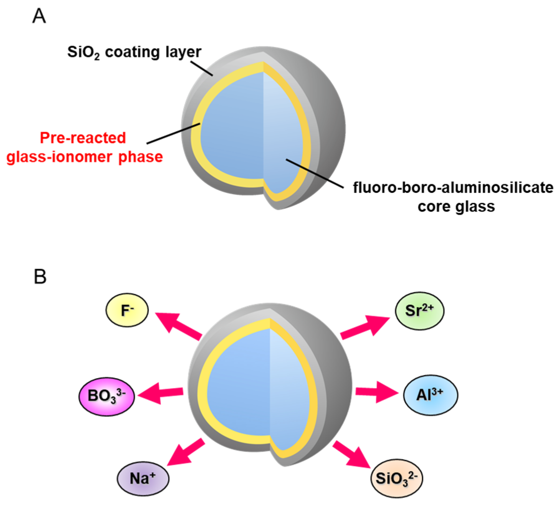
2. What Is S-PRG Filler

S-PRG filler technology was developed by Shofu Inc. and the resin composites incorporating S-PRG filler were first commercialized in 2000. S-PRG filler is a three-layered fine glass particle composed of a SiO2 coating in the outermost layer followed by a pre-reacted glass-ionomer phase and a glass core (Figure 1A). The pre-reacted glass-ionomer phase is prepared by spraying a polyacrylic acid that penetrates the SiO2 coating layer and causes an acid-base reaction with the fluoro-boro-aluminosilicate core glass. Many products containing S-PRG filler are already available on the market, and a series of those materials are called GIOMER. Three types of S-PRG filler with particle sizes of 3.0, 0.8, and 0.4 μm are utilized for different materials among the GIOMER series. In addition to commercial products, S-PRG filler has been experimentally incorporated into a variety of materials.
The pre-reacted glass-ionomer phase surrounding the glass core allows the S-PRG filler to release fluoride ions (F). Additionally, because specially fabricated fluoro-boro-aluminosilicate glass is used as the core material, S-PRG filler releases five other ions: strontium (Sr2+), borate (BO33−), sodium (Na+), silicate (SiO32−), and aluminum (Al3+) ions (Figure 1B). The concentrations of the six ions released into water from S-PRG filler, especially BO33−, Sr2+, and F, are relatively high [8], much greater than those liberated from unreacted filler of conventional glass-ionomer cement (Figure 2). Notably, the six ions do not form salts and are found separately in the eluate.

3. Bioactive Functions Exhibited by S-PRG Filler

Owing to the release of multiple ions, S-PRG filler exhibits various bioactive functions, which are described below.

3.1. Tooth Strengthening

Ions released from S-PRG filler contribute to the strengthening of enamel and dentin, improving their acid resistance. It is well known that fluoride ions strengthen the tooth substrate through the formation of fluorapatite. The release of F from S-PRG filler at high concentrations is effective in providing such effects.
Uo et al. [9] revealed that the strontium content in enamel and dentin became 100 times greater after immersion in the eluate of S-PRG filler compared with that before immersion. It was further demonstrated that Al3+, BO33−, and Sr2+ were taken up by enamel after 1 h of immersion in the eluate, and Sr showed remarkable incorporation of up to 7900 ppm after 28 days [10]. Strontium enhances the acid resistance of teeth by converting hydroxyapatite to strontium-apatite [11,12]. Uo et al. [9] investigated the local structure of Sr taken up by teeth using X-ray absorption fine structure analysis and determined that the structure of Sr in the enamel and dentin after immersion in the eluate of S-PRG filler was similar to that of synthetic Sr-incorporated hydroxyapatite, suggesting that Sr released from S-PRG filler can be incorporated into the Ca site of hydroxyapatite.
Hiraishi et al. [13] examined the interaction of borate ions released from S-PRG filler with the apatite of enamel and dentin and revealed that the borate ion adsorbed in the enamel and dentin was in a tetra-coordinated form, which possesses a buffer capacity, helps protect the tooth structure against acid attacks, and promotes remineralization.

3.2. Acid Neutralization

S-PRG filler modulates the pH of the surrounding medium and shifts it toward neutral and weak alkaline values [14]. Although further investigations are required to clarify the acid-buffering mechanism, the released Sr2+ and Na+ are considered to promote acid buffering.

3.3. Promotion of Mineralization

The S-PRG filler eluate containing Sr2+ at high concentrations can react with other ions in mineralizable solutions to induce chemical mineralization. The eluate obtained from S-PRG filler-containing materials promotes mineral deposit formation on the surface of phosvitin-immobilized agarose beads (model of demineralized dentin) placed in the mineralizing solution [8] (Figure 3). By micro-area X-ray diffraction analysis, the minerals that formed with a globular structure were determined to be apatite.
Nemoto et al. [15] reported that the eluate of S-PRG filler upregulated the mRNA expression level of the osteogenic differentiation marker (i.e., alkaline phosphatase) in human bone marrow-derived stromal cells. As a consequence, the S-PRG filler eluate promoted the mineralization of the extracellular matrix [15]. Ishigure et al. [16] reported that the eluate of S-PRG filler promoted the proliferation of dental pulp stem cells and enhanced their alkane phosphatase activity. These results suggest that S-PRG filler can biologically promote apatite formation and hard tissue formation, as well as chemically.

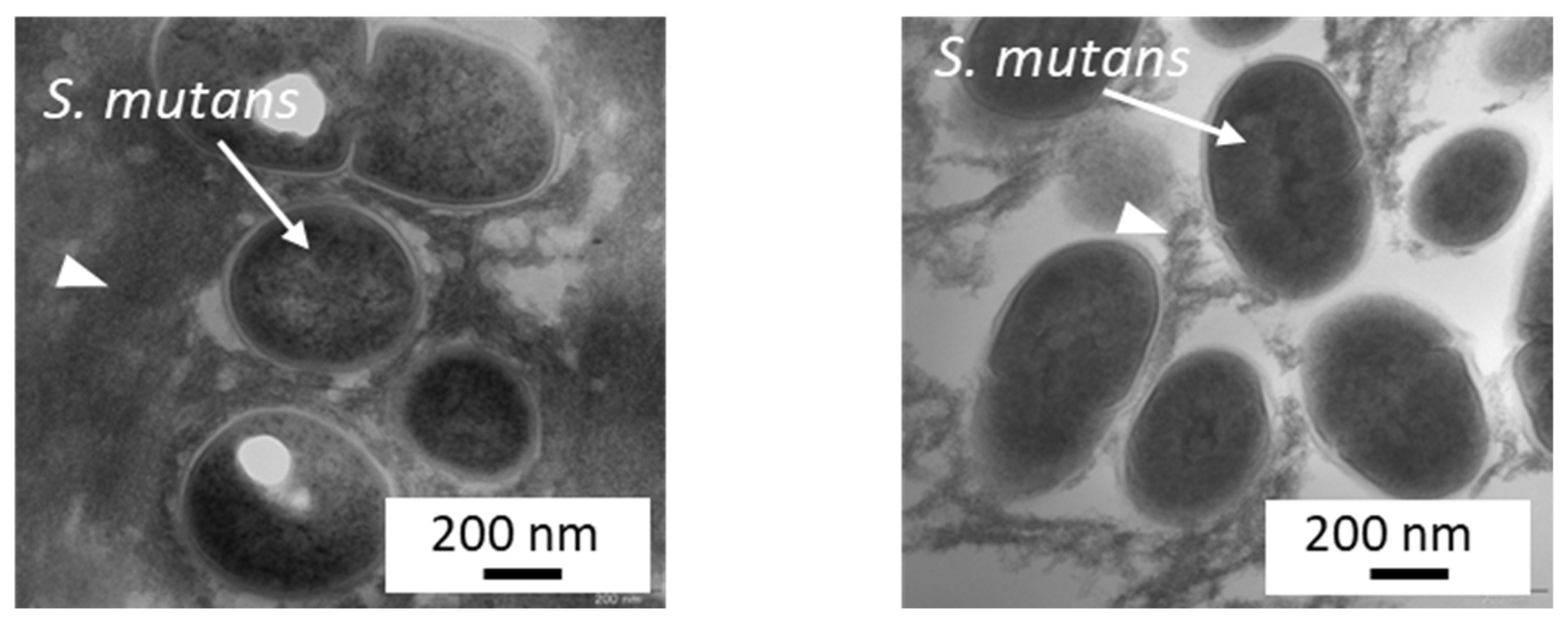
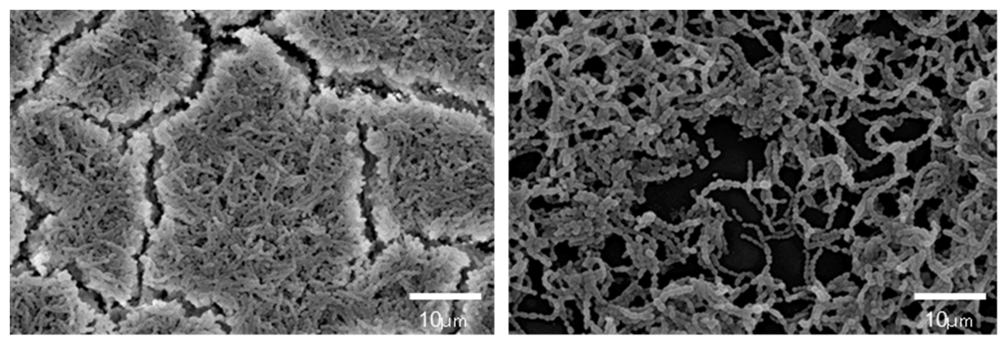
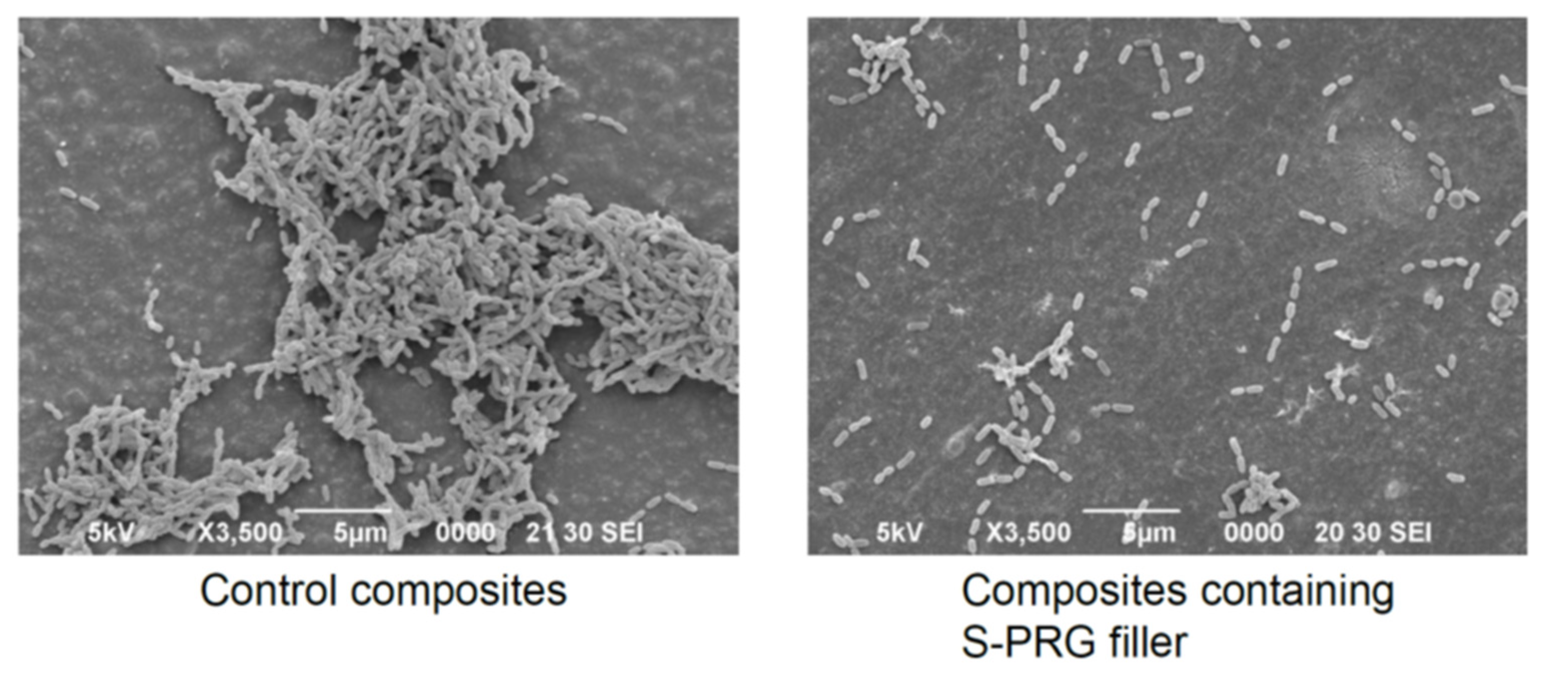
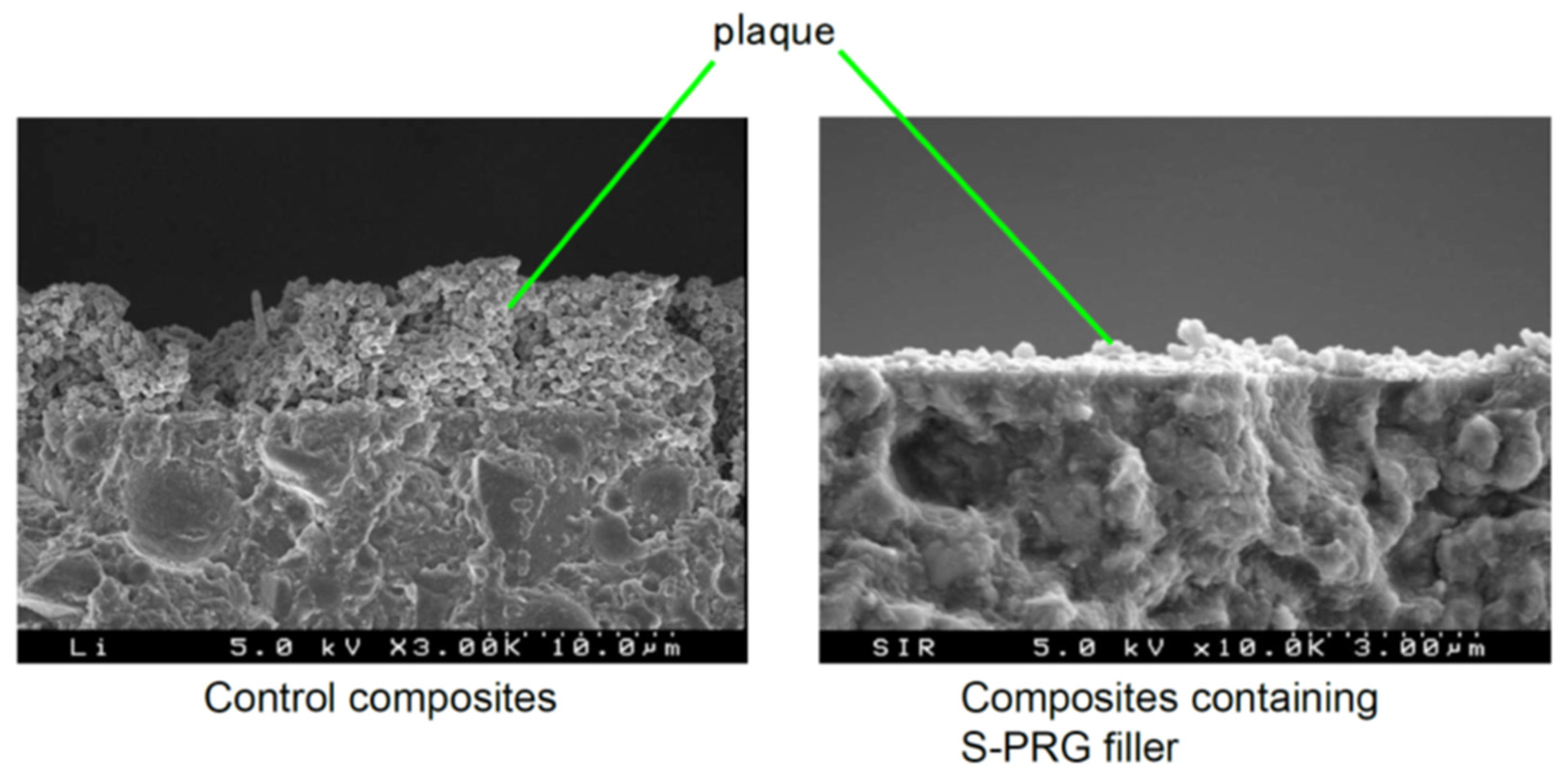
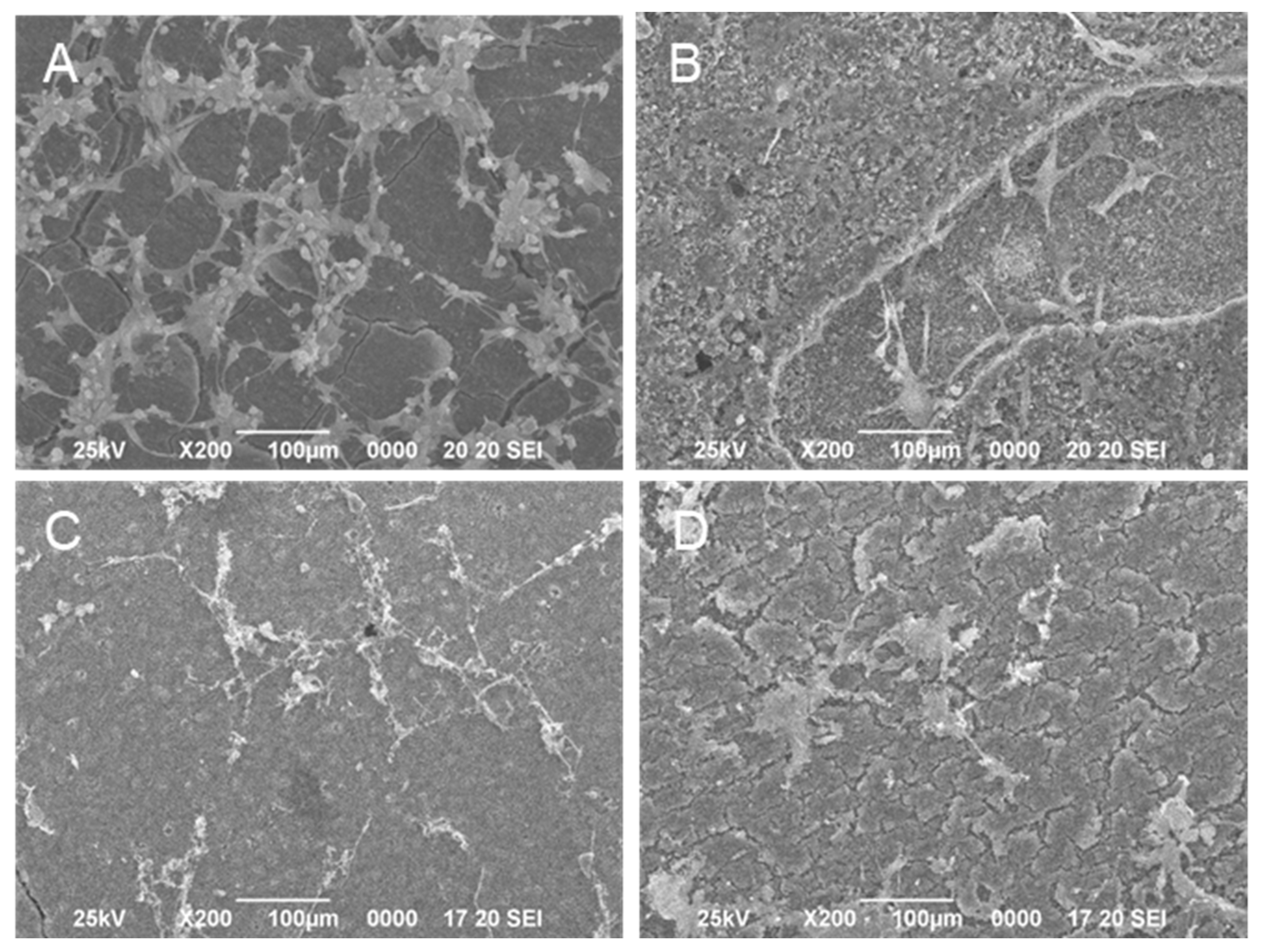
3.4. Inhibition of Bacteria and Fungi

Numerous studies have demonstrated antibacterial effects of the S-PRG filler eluate against bacteria in human saliva and dental plaque, such as streptococci, Enterococcus faecalis, Actinomyces israelii, Propionibacterium acnes, Porphyromonas gingivalis, and Fusobacterium nucleatum [17].
Nomura et al. [18] revealed that the S-PRG filler eluate inhibited Streptococcus mutans growth in a dose-dependent manner, especially before the logarithmic growth phase. The growth of S. mutans in the absence of S-PRG filler eluate reached a stationary phase 7 h after incubation, whereas the time for S. mutans growth was extended in the presence of S-PRG filler eluate. The ions mainly responsible for such effects are considered to be BO33− and F. Borate ions have been used as a preservative in the ophthalmic field, and fluoride ions at high concentrations are also known to inhibit bacteria. Miki et al. [19] investigated S. mutans growth in the presence of Na+, SiO32−, Sr2+, BO33−, Al3+, or F at the concentrations released from the experimental resin composites containing S-PRG filler at 55.9 vol% and determined that BO33− and F significantly inhibited the growth.
Kitagawa et al. [20] reported that even low concentrations of BO33− or F, at which bacterial growth was not affected, can inhibit S. mutans glucose metabolism and acid production. A detailed study conducted by Nomura et al. [18] clarified that the eluate of S-PRG filler prominently downregulated operons related to S. mutans sugar metabolism, such as the pdh operon encoding the pyruvate dehydrogenase complex and the glg operon encoding a putative glycogen synthase. According to such effects, when cultured with the 20–25% S-PRG filler eluate, glucan synthesis by S. mutans was inhibited (Figure 4), and the biofilm formed was determined to be sparse, with less extracellular matrix (Figure 5).
Ions released from S-PRG filler are effective in inhibiting bacteria related to periodontal disease and suppressing their pathogenic factors. It was reported that the eluate of S-PRG filler inhibited the growth of P. gingivalis, F. nucleatum, and Aggregatibacter actinomycetemcomitans [21], and suppressed the hemagglutination and gingipain activity of P. gingivalis [21,22]. It was also determined that the coaggregation of P. gingivalis and F. nucleatum, essential for subgingival biofilm formation, can be prevented in the presence of S-PRG filler eluate [22].
Tamura et al. [23] reported that the eluate of S-PRG filler decreased the amount of hydrogen peroxide and catalase activity in Candida albicans. Furthermore, the S-PRG filler eluate prevented the growth and biofilm formation of C. albicans and inhibited their dimorphism conversion. The eluate of S-PRG filler also suppressed the activity to produce secreted aspartyl proteinases, which are major pathogenic factors of C. albicans.

3.5. Inhibition of Matrix Metalloproteinases

Dentin matrix metalloproteinases (MMP) are a family of host-derived proteolytic enzymes that are trapped within the dentin matrix and can hydrolyze the organic matrix of dentin. MMPs break down collagen at the resin–dentin interface and compromise the durability of dentin bonding. Therefore, many reports are available on application of MMP inhibitors during bonding procedures to improve the long-term stability of dentin bonding. The S-PRG filler eluate inhibits the activity of collagen-bound MMPs and dentin matrix degradation, similar to 2% chlorhexidine digluconate, which is known as a MMP inhibitor [24]. Salim et al. [25] also reported that the eluate of S-PRG filler showed a reduction in MMP activity and revealed that F ions most effectively inhibited the enzymatic activity among the six ions released from S-PRG filler.

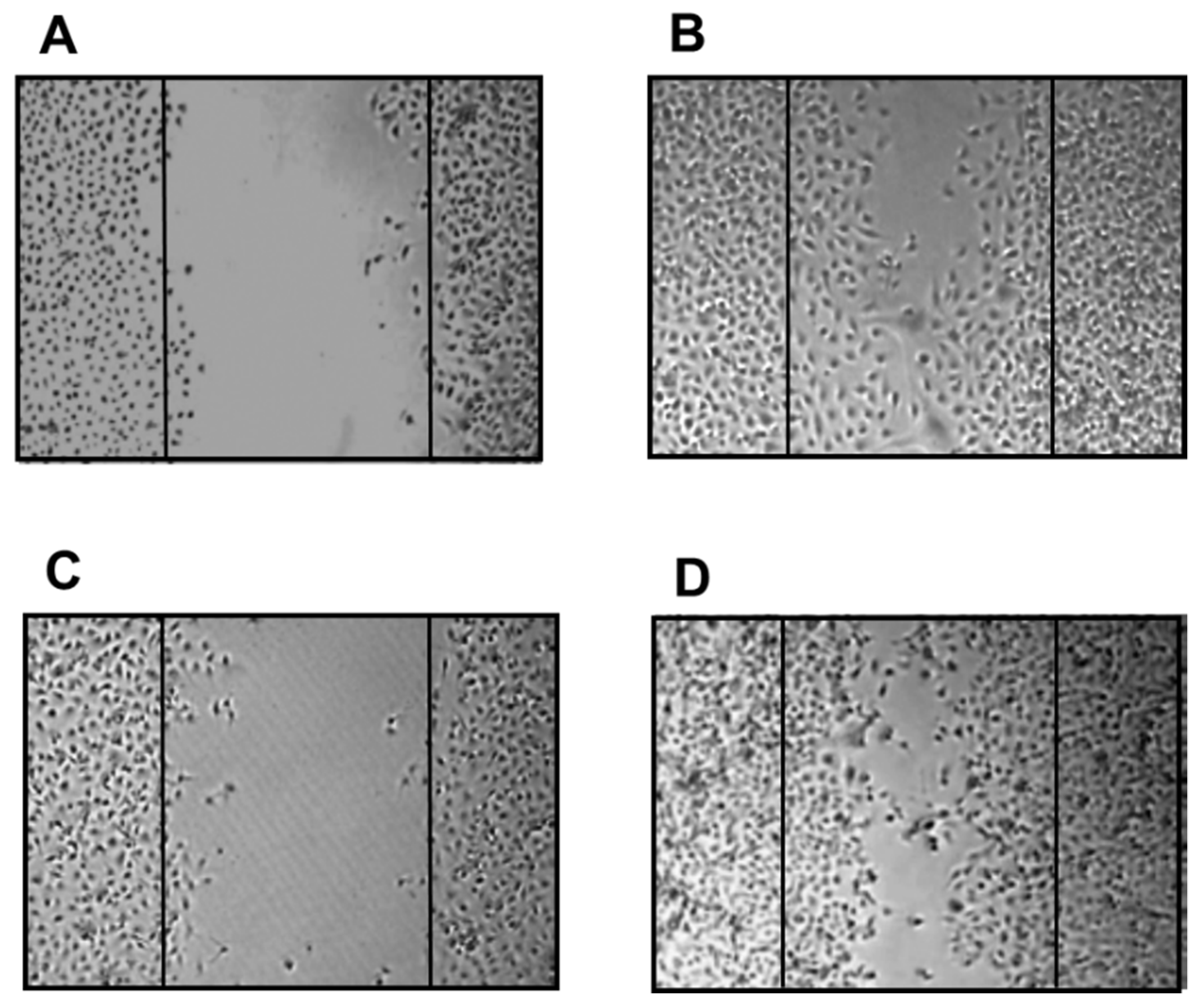
3.6. Enhancement of Cell Activity

Yamaguchi-Ueda et al. [26] examined the effects of various concentrations of S-PRG filler eluates on the growth and migration of gingival fibroblasts. They revealed that multiple ions present in the S-PRG filler eluate can stimulate migration of human gingival fibroblast cells via upregulation of the extracellular signal-regulated kinase 1/2 (ERK 1/2) signaling pathway. This finding indicates that the ions in the S-PRG filler eluate promoted cell activity, which may assist in oral wound healing.
Takeuchi et al. [27] reported that the eluate of S-PRG filler inhibited penetration of virulence factors of P. gingivalis, such as lipopolysaccharide and peptidoglycan, into gingival epithelial cells. The eluate of S-PRG filler induced the expression of coxsackievirus and adenovirus receptor (CXADR), a tight barrier of gingival epithelium, in immortalized human gingival epithelial cells, indicating that the S-PRG filler eluate enhanced the epithelial barrier function.

4. Benefits of S-PRG Filler for Dental Treatment and Care

Many products containing S-PRG filler are commercially available, such as resin composites, adhesives, resin cements, coating resins, fissure sealants, polishing pastes, and temporary fillings (Table 2). Additionally, S-PRG filler has been incorporated experimentally into inorganic cements, root canal sealers, denture bases, tissue conditioners, denture adhesives, toothpastes, varnishes, CAD/CAM composites, and toothbrush filaments. Even when S-PRG filler is incorporated into resin-based materials with silanization, multiple ions are released at high concentrations. The capacity for releasing six ions (i.e., Sr2+, BO33−, F, Na+, SiO32−, and Al3+) endows the S-PRG filler with various therapeutic effects, which are useful for restorative treatment, caries prevention/management, vital pulp therapy, endodontic treatment, prevention/treatment of periodontal disease, prevention of denture stomatitis, and perforation repair/root end filling. In this regard, S-PRG filler-containing materials differ from conventional ones that only release ions to promote remineralization, such as glass-ionomer cements or fluoride-releasing resin composites.

4.1. Restorative Treatment

To develop restorative treatments with preventive effects, S-PRG filler has been incorporated in resin composites, adhesives, and resin cements.
A ligand exchange mechanism within the glass-ionomer phase offers S-PRG filler the ability to release and recharge fluoride ions. The proprietary resin composites containing S-PRG filler (Beautifil II, Shofu Inc., Kyoto, Japan) release high concentrations of fluoride ions and can be recharged by exposure to 5000 ppm sodium fluoride solution for 5 min [28].
Several studies have demonstrated that resin composites containing S-PRG filler exhibit antibacterial effects against oral bacteria [4,5]. Miki et al. [19] examined the inhibitory effects of experimental resin composites containing different ratios of S-PRG filler on S. mutans growth and reported that the specimens containing S-PRG filler at 28.0 vol% or greater inhibited bacterial growth on their surfaces. They further demonstrated that the inhibitory effects were mainly attributed to the release of BO33− and F. It was also reported that the eluate of S-PRG filler suppressed the adherence of S. mutans [22], and thus, S. mutans biofilm formation was inhibited on the surface of resin composites containing S-PRG filler (Figure 6). An in situ study assessing plaque accumulation on resin composites placed in human mouths for 24 h showed significant suppression of dental plaque maturation on the surface of resin composites containing S-PRG filler [29] (Figure 7).
An investigation of demineralization in the tooth structure surrounding filling materials revealed that S-PRG filler-containing resin composites inhibited the demineralization of wall and outer lesions in enamel and dentin and increased the surrounding tooth hardness [30]. Lai et al. [31] investigated the anti-demineralization effects of restorations using S-PRG filler-containing resin composites (Beautifil II) and adhesives (FL-Bond II) on the root surfaces. Using an oral biofilm reactor and pH cycling artificial caries model, they demonstrated that the combination of Beautifil II and FL-Bond II exhibited a cumulative effect to inhibit demineralization of root dentin adjacent to restoration.
It has been reported that S. mutans contains esterases that can potentially degrade resin-based restorative materials [32]. Gautam et al. [33] revealed by in vitro test that degradation by S. mutans was reduced for resin composites containing S-PRG filler (Beautifil II) compared with other commercial resin composites. On the other hand, Yoshihara et al. [34] reported production of many holes on the surface of S-PRG filler-containing resin composites after immersion in lactic acid at pH 4.0 for 3 days, while no change in surface integrity was detected for other conventional resin composites. They described that such surface alteration was caused by dissolution of S-PRG filler. Although several clinical studies revealed their excellent performance [35,36], long-term surface durability of S-PRG filler-containing resin composites remains to be investigated in detail.
Recently, CAD/CAM resin composite blocks containing S-PRG filler have been developed for the restoration of primary molar teeth. Nakase et al. [37] reported that S-PRG filler-containing CAD/CAM resin composite blocks demonstrated acceptable physical properties and wear performance for clinical use. Moreover, an S-PRG filler-containing CAD/CAM resin composite crown is advantageous for primary tooth restoration because it can be cemented using a conventional glass-ionomer cement through the chelation reaction between Sr and polycarboxylic acid [38].

4.2. Caries Prevention/Management

The S-PRG filler modulates the pH of the surrounding medium and shifts it toward neutral and weak alkaline values [14]. Additionally, the release of F and Sr2+ from S-PRG filler strengthens the tooth substrate, as described above. These effects are beneficial for the prevention and management of caries. Many studies have demonstrated that ions released from S-PRG filler in various materials are taken up by adjacent enamel and dentin to prevent demineralization, including adhesives [39], endodontic sealers [40], orthodontic adhesives [41], pit/fissure sealants [42], coating materials [43,44], resinous vanishes [45], and denture base resins [46].
A resinous coating material containing S-PRG filler (PRG Barrier Coat, Shofu Inc.) protects the enamel surface from demineralization caused by acidic attack [44,47]. Ions released from S-PRG filler in PRG Barrier Coat exert acid-neutralizing effects near the coated surface. Uptake of F and Sr released from PRG Barrier Coat by the tooth substrate can be effective to inhibit demineralization [48]. Ma at al. [43] demonstrated that PRG Barrier Coat containing S-PRG filler can protect root surfaces from demineralization by producing a coating layer with a thickness of approximately 200 µm. It was also shown that the pH drop in S. mutans clumps caused by glucose on the surface of the PRG Barrier Coat was reduced, possibly because the glucose metabolism was hindered by the released ions as well as acid buffering [43].
The incorporation of S-PRG filler into a resin-based pit and fissure sealant provides acid neutralization capacity without deteriorating the sealing effectiveness [49,50] or the ability to release and recharge fluoride ions [51]. Some clinical studies demonstrated caries preventive effects of commercially available S-PRG filler-containing pit/fissure sealant (BeautiSealant, Shofu Inc.) [52,53]. Penha et al. [53] conducted a randomized clinical trial to compare the evolution of caries in newly erupted permanent molars and determined that the S-PRG sealant group presented higher sound teeth predominance compared with another fluoride-releasing sealant after 12 months.
A polishing paste containing S-PRG filler, including a commercial one containing 5% S-PRG filler (PRG Pro-Care Gel, Shofu Inc.), is useful for controlling caries incidence and remineralization. Iijima et al. [54] reported that human enamel polished with S-PRG filler-containing paste, following immersion in the demineralizing solution at pH 4.5, exhibited greater surface hardness and smoothness than those polished with fluoridated paste or nano-hydroxyapatite-containing paste. Furthermore, the surface hardness of demineralized dentin polished with the S-PRG filler-containing paste recovered after immersion in mineralizing solution for 1 month, indicating that the S-PRG filler-containing paste can promote dentin remineralization [55]. A recent study revealed that PRG Pro-Care Gel has a similar potential to remineralize the demineralized root dentin as a 38% silver diamine fluoride solution [56].
Amaechi et al. [57] examined the effectiveness of experimental toothpastes containing S-PRG filler in preventing tooth surface demineralization using Featherstone’s pH cycling model to develop early caries. It was determined that S-PRG filler-containing dentifrice was more effective in preventing tooth demineralization than 1100 ppm F-containing toothpaste, highlighting the potential of S-PRG filler to assist individuals at high risk of developing caries [58]. The potential of S-PRG filler-containing toothpaste to remineralize the demineralized enamel surface has also been shown in situ [59]. Experimental toothpaste containing 5% S-PRG filler showed remineralizing effects similar to those of toothpaste containing 1500 ppm F, even showing greater activity to recover mineral loss in the subsurface regions.
Moecke et al. [60] investigated the remineralizing effects of experimental varnishes containing S-PRG filler. They revealed that the varnish containing 40% S-PRG filler more effectively promoted the remineralization of demineralized bovine enamel compared with the varnish containing 5% sodium fluoride.

4.3. Vital Pulp Therapy

S-PRG filler can promote tertiary dentinogenesis. As pulp capping materials, several attempts have been made to incorporate S-PRG filler into adhesives [61,62], self-adhesive resins [63], and inorganic cements [64,65].
Experimental inorganic cement, prepared by mixing S-PRG filler with copolymers of acrylic acid and tricarboxylic acid, induces complete tertiary dentin formation at 2 and 4 weeks after pulp exposure in rat molars [64]. Strontium released from S-PRG cement was determined to transfer into the pulpal tissue and contribute to the induction of mineralization and dentinogenesis [65]. These functions are based on biological action because the S-PRG cement enhanced the expression of genes related to osteo/dentinogenic differentiation, such as CXCL-12 and TGF-β1, which contribute to tertiary dentin formation during the healing process in pulpal tissue [65].
It has been reported that lithium ions can activate the Wnt/β-catenin signaling pathway and induce dentin formation in pulpotomized teeth [66]. Therefore, trials to add LiCl to S-PRG cement were conducted, revealing that reparative dentin formation in rat teeth was enhanced through activation of the Wnt/β-catenin canonical signaling pathway without adversely affecting the sealing property of the cement [67,68].

4.4. Endodontic Treatment

Apical periodontitis is an infectious disease, and the treatment requires careful bacterial eradication inside filled root canals. However, the complete elimination of bacterial infection in root canals is difficult to achieve using only mechanical instrumentation, irrigation, and medication. Therefore, endodontic filling materials capable of eliminating residual bacteria inside root canals have been proposed as a possible solution to this problem. An experimental zinc oxide-based endodontic sealer containing S-PRG filler demonstrated the sustained release of multiple ions (i.e., Sr2+, BO33−, F, Na+, SiO32−, and Al3+) [40] and antibacterial effects against E. faecalis and P. gingivalis [69,70].
The eluate obtained from experimental root canal sealer containing S-PRG filler was reported to downregulate mRNA expression levels of proinflammatory cytokines, such as interleukin (IL)-1α, IL-6, and TNF-α, in LPS-stimulated RAW264.7 cells, suggesting its anti-inflammatory effects [71]. Moreover, an experimental sealer containing S-PRG filler can promote the healing of periapical lesions in vivo [70,72,73].

4.5. Prevention/Treatment of Periodontal Disease

The S-PRG filler eluate suppresses the activity of periodontitis-related bacteria [21,22] and inhibits penetration of P. gingivalis virulence factors into gingival epithelial cells [27]. It also promotes recovery of migration activity in epithelial cells infected with P. gingivalis (Figure 8). In recent years, several animal studies have verified the ability of S-PRG filler to help prevent the progression of periodontal disease. Iwamatsu-Kobayashi et al. [74] investigated the effects against tissue destruction induced in a mouse periodontal disease model and revealed that periodical dripping of the S-PRG filler eluate suppressed the bone loss and resulted in greater volume and density of alveolar bone. It has also been reported that ultrasonic irrigation with S-PRG filler dispersion can improve periodontal parameters, such as gingival index, bleeding on probing, probing pocket depth, and clinical attachment level when applied to a periodontal defect in a dog [75,76]. Irrigation with S-PRG filler dispersion reduced the ratio of red complex (i.e., Tannerella forsythia, P. gingivalis, and Treponema denticola) among the bacterial flora in the periodontal pocket [76].

4.6. Prevention of Denture Stomatitis

A denture base with antifungal effects may be useful for the prevention of denture stomatitis. The morphology of C. albicans cells remains in the yeast form in a normal resident flora, but it grows from the yeast form to the hyphal form by creating a longer germ tube when grown in an inflammatory environment in a human host [77]. The eluate of S-PRG filler prevents adherence of C. albicans to denture base resin and inhibits the dimorphism conversion of C. albicans [23]. The incorporation of S-PRG filler into polymethyl methacrylate-based resin effectively inhibited biofilm formation by C. albicans, with reduced length of the hyphae [78]. Similar results have been reported for the addition of S-PRG filler to other materials in the field of removable prosthodontics, such as tissue conditioners [79,80] and poly(methyl vinyl ether-alt-maleic anhydride)-based denture adhesives [81]. The multiple-ion release characteristics of S-PRG filler may contribute to the oral health of the elderly population by preventing the onset of oral candidiasis.

4.7. Perforation Repair/Root End Filling

Hirata-Tsuchiya et al. [72] reported that the application of commercially available resin composites containing S-PRG filler for sealing root perforation can facilitate periodontal tissue healing and support good clinical outcomes. Inorganic cement containing S-PRG filler exhibits excellent cytocompatibility for osteoblastic cells, similar to a calcium silicate-based cement (Figure 9). Considering the enhancement of cell activity by released ions, S-PRG filler-containing materials may be useful for perforation repair and root end filling, for which calcium silicate-based cements have been widely used.

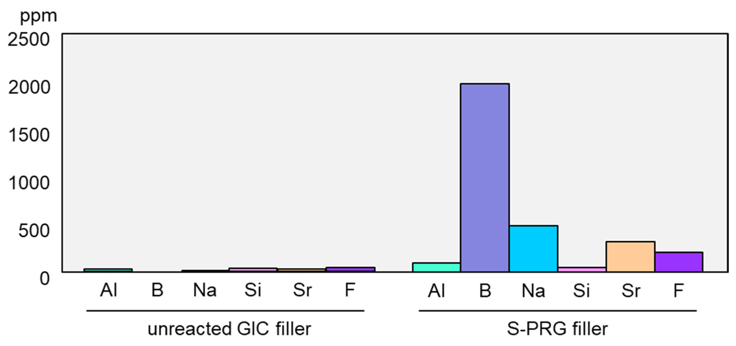
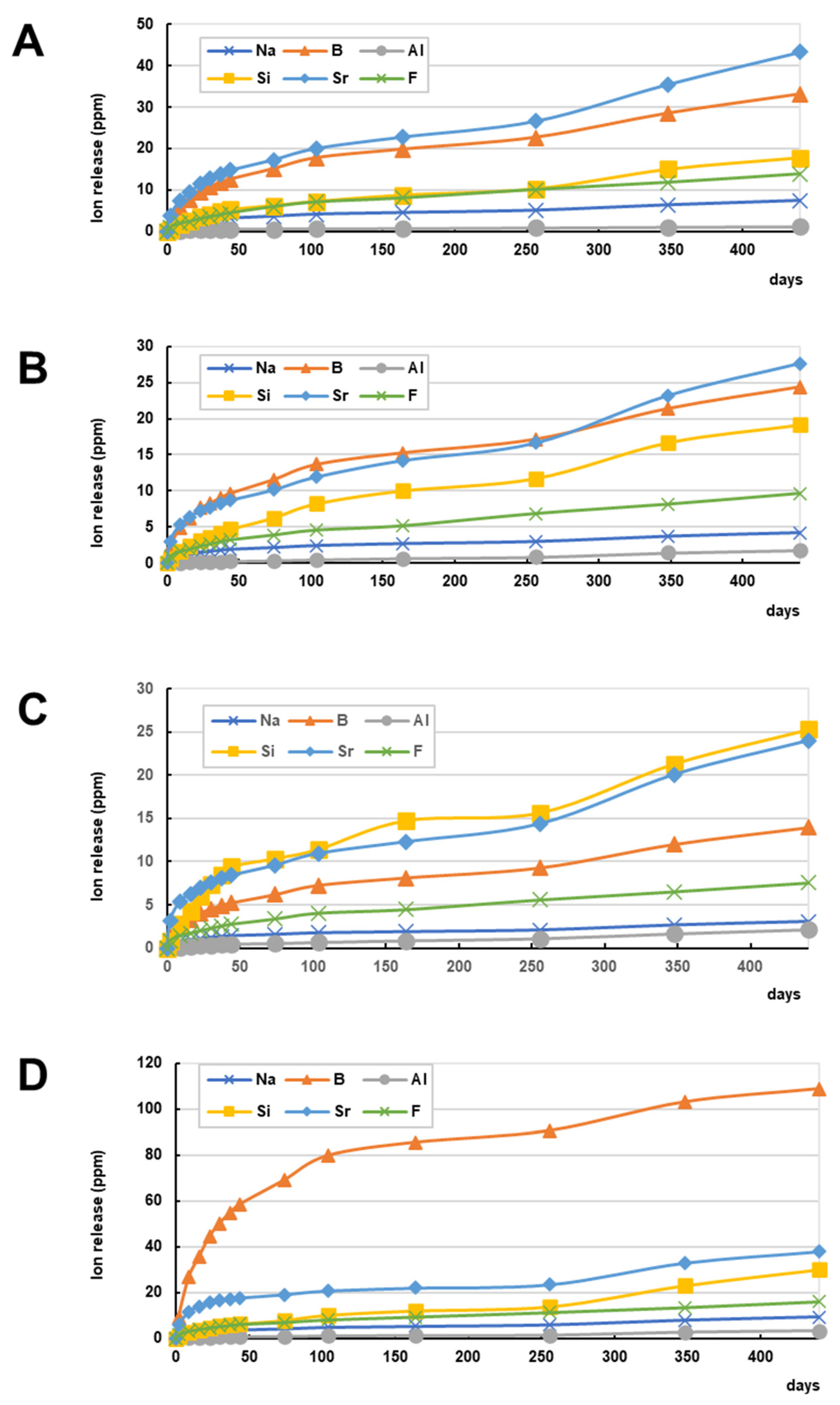
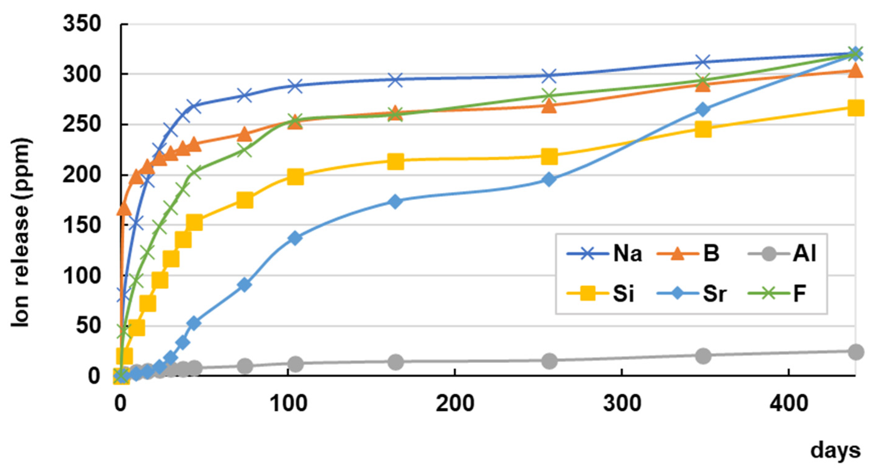
5. Ion Release Profile of S-PRG Filler-Containing Materials

To provide clinical benefits for restorative treatments, sustained or controlled release of active components is essential for agent-releasing bioactive materials. Figure 10 shows the release profile of six ions from commercially available S-PRG filler-containing resin composites, an S-PRG filler-containing resin cement for luting, and an S-PRG filler-containing bonding agent in a two-step self-etch adhesive. All materials demonstrated continuous release of six ions for up to 440 days, with the release of Sr2+ and BO33− at high concentrations for the resin composites and the resin cement. The bonding agent containing S-PRG filler exhibited a different release pattern and released a large amount of BO33−. Continuous release of fluoride for 440 days was confirmed for all restorative materials.
The ion release profile of a coating resin containing S-PRG filler is depicted in Figure 11. The concentrations of all ions released from the coating resin were much greater than those released from the restorative materials, and a large amount was released within 100 days, except for Sr2+, which showed relatively constant release pattern.

6. Conclusions

A large number of in vitro and in vivo (animal) studies have been conducted to assess the bioactive functions exhibited by S-PRG filler and S-PRG filler-containing materials. In addition to commercial products containing S-PRG filler, many reports on experimental materials are available. The S-PRG filler has the potential as a bioactive material to contribute to the success of various dental treatments and care. The main concern is that a limited number of clinical studies on the benefits of bioactive functions exhibited by S-PRG filler and S-PRG filler containing materials is available. Most of the evidence described in this review is based on in vitro or animal studies. Although several studies evaluated long-term action of ion-release from S-PRG filler, it is well known that the bench-to-clinic translation is not so simple. Future research in clinical settings and their accumulation are expected to elucidate further the practicality and efficacy of bioactive S-PRG filler for oral health promotion.

Author Contributions

Conceptualization, S.I. (Satoshi Imazato), T.N. and K.N.; investigation, S.I. (Satoshi Imazato), H.K., J.-I.S., H.T. and R.N.; methodology, H.K., S.Y., S.I. (Shuichi Ito) and R.N.; data curation, S.I. (Satoshi Imazato), T.N., H.K., H.T. and K.N.; writing—original draft preparation, S.I. (Satoshi Imazato), T.N., H.K. and J.-I.S.; writing—review and editing, S.I. (Satoshi Imazato), S.Y., R.N. and K.N.; supervision, S.I. (Satoshi Imazato), S.I. (Shuichi Ito) and K.N.; funding acquisition, S.I. (Satoshi Imazato) and K.N. All authors have read and agreed to the published version of the manuscript.

Funding

This work was supported by Grants-in-Aid for Scientific Research (21K19593 to S. Imazato) from the Japan Society for the Promotion of Science.

Data Availability Statement

The data that support the findings of this study are available from the corresponding author upon reasonable request.

Conflicts of Interest

The authors declare no conflict of interest.

References

  1. Imazato, S. Bio-active restorative materials with antibacterial effects: New dimension of innovation in restorative dentistry. Dent. Mater. J. 2009, 28, 11–19. [Google Scholar] [CrossRef] [PubMed]
  2. Imazato, S.; Ma, S.; Chen, J.H.; Xu, H.H. Therapeutic polymers for dental adhesives: Loading resins with bio-active components. Dent. Mater. 2014, 30, 97–104. [Google Scholar] [CrossRef] [PubMed]
  3. Imazato, S.; Kitagawa, H.; Tsuboi, R.; Kitagawa, R.; Thongthai, P.; Sasaki, J.I. Non-biodegradable polymer particles for drug delivery: A new technology for “bio-active” restorative materials. Dent. Mater. J. 2017, 36, 524–532. [Google Scholar] [CrossRef]
  4. Imazato, S.; Kohno, T.; Tsuboi, R.; Thongthai, P.; Xu, H.H.; Kitagawa, H. Cutting-edge filler technologies to release bio-active components for restorative and preventive dentistry. Dent. Mater. J. 2020, 39, 69–79. [Google Scholar] [CrossRef] [PubMed]
  5. Imazato, S.; Kitagawa, H. Dental resin-based materials with antibacterial properties: Contact inhibition and controlled release. In Oral Biofilms and Modern Dental Materials: Advances toward Bioactivity; Ionescu, A.C., Hahnel, S., Eds.; Springer: Cham, Switzerland, 2021; pp. 127–140. [Google Scholar]
  6. Spagnuolo, G. Bioactive dental materials: The current status. Materials 2022, 15, 2016. [Google Scholar] [CrossRef]
  7. Schmalz, G.; Hickel, R.; Price, R.B.; Platt, J.A. Bioactivity of dental restorative materials: FDI Policy Statement. Int. Dent. J. 2023, 73, 21–27. [Google Scholar] [CrossRef]
  8. Ito, S.; Iijima, M.; Hashimoto, M.; Tsukamoto, N.; Mizoguchi, I.; Saito, T. Effects of surface pre-reacted glass-ionomer fillers on mineral induction by phosphoprotein. J. Dent. 2011, 39, 72–79. [Google Scholar] [CrossRef]
  9. Uo, M.; Wada, T.; Asakura, K. Structural analysis of strontium in human teeth treated with surface pre-reacted glass-ionomer filler eluate by using extended X-ray absorption fine structure analysis. Dent. Mater. J. 2017, 36, 214–221. [Google Scholar] [CrossRef]
  10. Ogawa, A.; Wada, T.; Mori, Y.; Uo, M. Time dependence of multi-ion absorption into human enamel from surface prereacted glass-ionomer (S-PRG) filler eluate. Dent. Mater. J. 2019, 38, 707–712. [Google Scholar] [CrossRef]
  11. Dedhiya, M.G.; Young, F.; Higuchi, W.I. Mechanism for the retardation of the acid dissolution rate of hydroxyapatite by strontium. J. Dent. Res. 1973, 52, 1097–1109. [Google Scholar] [CrossRef]
  12. Featherstone, J.D.B.; Shields, C.P.; Khademazad, B.; Oldershaw, M.D. Acid reactivity of carbonated apatites with strontium and fluoride substitutions. J. Dent. Res. 1983, 62, 1049–1053. [Google Scholar] [CrossRef] [PubMed]
  13. Hiraishi, N.; Sayed, M.; Hill, R.; Tagami, J.; Hayashi, F. Interactions of boron released from surface pre-reacted glass ionomer with enamel/dentin and its effect on pH. Sci. Rep. 2021, 11, 15734. [Google Scholar] [CrossRef] [PubMed]
  14. Fujimoto, Y.; Iwasa, M.; Murayama, R.; Miyazaki, M.; Nagafuji, A.; Nakatsuka, T. Detection of ions released from S-PRG fillers and their modulation effect. Dent. Mater. J. 2010, 29, 392–397. [Google Scholar] [CrossRef] [PubMed]
  15. Nemoto, A.; Chosa, N.; Kyakumoto, S.; Yokota, S.; Kamo, M.; Noda, M.; Ishisaki, A. Water-soluble factors eluated from surface pre-reacted glass-ionomer filler promote osteoblastic differentiation of human mesenchymal stem cells. Mol. Med. Rep. 2018, 17, 3448–3454. [Google Scholar] [CrossRef]
  16. Ishigure, H.; Kawaki, H.; Shintani, K.; Ueno, K.; Mizuno-Kamiya, M.; Takayama, E.; Hotta, M.; Kondoh, N.; Nikaido, T. Effects of multi-components released from S-PRG filler on the activities of human dental pulp-derived stem cells. Dent. Mater. J. 2021, 40, 1329–1337. [Google Scholar] [CrossRef]
  17. Yoneda, M.; Suzuki, N.; Hirofuji, T. Antibacterial effect of surface pre-reacted glass ionomer filler and eluate–mini review. Pharm. Anal. Acta 2015, 6, 349. [Google Scholar]
  18. Nomura, R.; Morita, Y.; Matayoshi, S.; Nakano, K. Inhibitory effect of surface pre-reacted glass-ionomer (S-PRG) eluate against adhesion and colonization by Streptococcus mutans. Sci. Rep. 2018, 8, 5056. [Google Scholar] [CrossRef]
  19. Miki, S.; Kitagawa, H.; Kitagawa, R.; Kiba, W.; Hayashi, M.; Imazato, S. Antibacterial activity of resin composites containing surface pre-reacted glass-ionomer (S-PRG) filler. Dent. Mater. 2016, 32, 1095–1102. [Google Scholar] [CrossRef]
  20. Kitagawa, H.; Miki-Oka, S.; Mayanagi, G.; Abiko, Y.; Takahashi, N.; Imazato, S. Inhibitory effect of resin composite containing S-PRG filler on Streptococcus mutans glucose metabolism. J. Dent. 2018, 70, 92–96. [Google Scholar] [CrossRef]
  21. Kono, Y.; Tamura, M.; Cueno, M.E.; Tonogi, M.; Imai, K. S-PRG filler eluate induces oxidative stress in oral microorganism: Suppression of growth and pathogenicity, and possible clinical application. Antibiotics 2021, 10, 816. [Google Scholar] [CrossRef]
  22. Yoneda, M.; Suzuki, N.; Masuo, Y.; Fujimoto, A.; Iha, K.; Yamada, K.; Iwamoto, T.; Hirofuji, T. Effect of S-PRG eluate on biofilm formation and enzyme activity of oral bacteria. Int. J. Dent. 2012, 2012, 814913. [Google Scholar] [CrossRef] [PubMed]
  23. Tamura, M.; Cueno, M.E.; Abe, K.; Kamio, N.; Ochiai, K.; Imai, K. Ions released from a S-PRG filler induces oxidative stress in Candida albicans inhibiting its growth and pathogenicity. Cell Stress Chaperones 2018, 23, 1337–1343. [Google Scholar] [CrossRef] [PubMed]
  24. Mendes Soares, I.P.; Anselmi, C.; Guiné, I.; Fernandes, L.O.; Pires, M.L.B.A.; de Souza Costa, C.A.; Scheffel, D.L.S.; Hebling, J. Inhibitory activity of S-PRG filler on collagen-bound MMPs and dentin matrix degradation. J. Dent. 2022, 124, 104237. [Google Scholar] [CrossRef]
  25. Salim, I.; Seseogullari-Dirihan, R.; Imazato, S.; Tezvergil-Mutluay, A. The inhibitory effects of various ions released from S-PRG fillers on dentin protease activity. Dent. Mater. J. 2023, 42, 99–104. [Google Scholar] [CrossRef]
  26. Yamaguchi-Ueda, K.; Akazawa, Y.; Kawarabayashi, K.; Sugimoto, A.; Nakagawa, H.; Miyazaki, A.; Kurogoushi, R.; Iwata, K.; Kitamura, T.; Yamada, A.; et al. Combination of ions promotes cell migration via extracellular signal-regulated kinase 1/2 signaling pathway in human gingival fibroblasts. Mol. Med. Rep. 2019, 19, 5039–5045. [Google Scholar] [CrossRef] [PubMed]
  27. Takeuchi, H.; Kato, Y.; Sasaki, N.; Tanigaki, K.; Yamaga, S.; Mita, E.; Kuboniwa, M.; Matsusaki, M.; Amano, A. Surface pre-reacted glass-ionomer eluate protects gingival epithelium from penetration by lipopolysaccharides and peptidoglycans via transcription factor EB pathway. PLoS ONE 2022, 17, e0271192. [Google Scholar] [CrossRef]
  28. Naoum, S.; Ellakwa, A.; Martin, F.; Swain, M. Fluoride release, recharge and mechanical property stability of various fluoride-containing resin composites. Oper. Dent. 2011, 36, 422–432. [Google Scholar] [CrossRef]
  29. Saku, S.; Kotake, H.; Scougall-vilchis, R.J.; Ohashi, S.; Hotta, M.; Yamamoto, K. Antibacterial activity of composite resin with glass-ionomer filler particles. Dent. Mater. J. 2010, 29, 193–198. [Google Scholar] [CrossRef]
  30. Zhou, Y.; Hiraishi, N.; Shimada, Y.; Wang, G.; Tagami, J.; Feng, X. Evaluation of tooth demineralization and interfacial bacterial penetration around resin composites containing surface pre-reacted glass-ionomer (S-PRG) filler. Dent. Mater. 2021, 37, 849–862. [Google Scholar] [CrossRef]
  31. Lai, Y.J.; Takahashi, R.; Lin, P.Y.; Kuo, L.; Zhou, Y.; Matin, K.; Chiang, Y.C.; Shimada, Y.; Tagami, J. Anti-demineralization effects of dental adhesive-composites on enamel-root dentin junction. Polymers 2021, 13, 3327. [Google Scholar] [CrossRef]
  32. Bourbia, M.; Ma, D.; Cvitkovitch, D.G.; Santerre, J.P.; Finer, Y. Cariogenic bacteria degrade dental resin composites and adhesives. J. Dent. Res. 2013, 92, 989–994. [Google Scholar] [CrossRef]
  33. Gautam, A.K.; Thakur, R.; Shashikiran, N.D.; Shilpy, S.; Agarwal, N.; Tiwari, S. Degradation of resin restorative materials by Streptococcus mutans: A pilot study. J. Clin. Pediatr. Dent. 2017, 41, 225–227. [Google Scholar] [CrossRef]
  34. Yoshihara, K.; Nagaoka, N.; Maruo, Y.; Sano, H.; Yoshida, Y.; Van Meerbeek, B. Bacterial adhesion not inhibited by ion-releasing bioactive glass filler. Dent. Mater. 2017, 33, 723–734. [Google Scholar] [CrossRef]
  35. Ozer, F.; Patel, R.; Yip, J.; Yakymiv, O.; Saleh, N.; Blatz, M.B. Five-year clinical performance of two fluoride-releasing giomer resin materials in occlusal restorations. J. Esthet. Restor. Dent. 2022, 34, 1213–1220. [Google Scholar] [CrossRef] [PubMed]
  36. Toz-Akalin, T.; Öztürk-Bozkurt, F.; Kusdemir, M.; Özsoy, A.; Yüzbaşıoğlu, E.; Özcan, M. Clinical evaluation of low-shrinkage bioactive material giomer versus nanohybrid resin composite restorations: A two-year prospective controlled clinical trial. Oper. Dent. 2023, 48, 10–20. [Google Scholar] [CrossRef]
  37. Nakase, Y.; Yamaguchi, S.; Okawa, R.; Nakano, K.; Kitagawa, H.; Imazato, S. Physical properties and wear behavior of CAD/CAM resin composite blocks containing S-PRG filler for restoring primary molar teeth. Dent. Mater. 2022, 38, 158–168. [Google Scholar] [CrossRef]
  38. Akimoto, N.; Sakamoto, T.; Kubota, Y.; Kondo, Y.; Momoi, Y. A novel composite-to-composite adhesive bond mechanism. Dent. Mater. J. 2011, 30, 523–527. [Google Scholar] [CrossRef] [PubMed]
  39. Han, L.; Okamoto, A.; Fukushima, M.; Okiji, T. Evaluation of a new fluoride-releasing one-step adhesive. Dent. Mater. J. 2006, 25, 509–515. [Google Scholar] [CrossRef]
  40. Han, L.; Okiji, T. Evaluation of the ions release / incorporation of the prototype S-PRG filler-containing endodontic sealer. Dent. Mater. J. 2011, 30, 898–903. [Google Scholar] [CrossRef]
  41. Horiuchi, S.; Kaneko, K.; Mori, H.; Kawakami, E.; Tsukahara, T.; Yamamoto, K.; Hamada, K.; Asaoka, K.; Tanaka, E. Enamel bonding of self-etching and phosphoric acid-etching orthodontic adhesives in simulated clinical conditions: Debonding force and enamel surface. Dent. Mater. J. 2009, 28, 419–425. [Google Scholar] [CrossRef] [PubMed]
  42. Kaga, M.; Kakuda, S.; Ida, Y.; Toshima, H.; Hashimoto, M.; Endo, K.; Sano, H. Inhibition of enamel demineralization by buffering effect of S-PRG filler-containing dental sealant. Eur. J. Oral Sci. 2014, 122, 78–83. [Google Scholar] [CrossRef] [PubMed]
  43. Ma, S.; Imazato, S.; Chen, J.-H.; Mayanagi, G.; Takahashi, N.; Ishimoto, T.; Nakano, T. Effects of a coating resin containing S-PRG filler to prevent demineralization of root surfaces. Dent. Mater. J. 2012, 31, 909–915. [Google Scholar] [CrossRef]
  44. Kawasaki, K.; Kambara, M. Effects of ion-releasing tooth-coating material on demineralization of bovine tooth enamel. Int. J. Dent. 2014, 2014, 463149. [Google Scholar] [CrossRef]
  45. Shiiya, T.; Mukai, Y.; Tomiyama, K.; Teranaka, T. Anti-demineralization effect of a novel fluoride-releasing varnish on dentin. Am. J. Dent. 2012, 25, 347–350. [Google Scholar] [PubMed]
  46. Mukai, Y.; Kamijo, K.; Fujino, F.; Hirata, Y.; Teranaka, T.; Ten Cate, J.M. Effect of denture base-resin with prereacted glass-ionomer filler on dentin demineralization. Eur. J. Oral Sci. 2009, 117, 750–754. [Google Scholar] [CrossRef] [PubMed]
  47. Murayama, R.; Nagura, Y.; Yamauchi, K.; Moritake, N.; Iino, M.; Ishii, R.; Kurokawa, H.; Miyazaki, M.; Hosoya, Y. Effect of a coating material containing surface reaction-type pre-reacted glass-ionomer filler on prevention of primary enamel demineralization detected by optical coherence tomography. J. Oral Sci. 2018, 60, 367–373. [Google Scholar] [CrossRef]
  48. Funato, Y.; Matsuda, Y.; Okuyama, K.; Yamamoto, H.; Komatsu, H.; Sano, H. A new technique for analyzing trace element uptake by human enamel. Dent. Mater. J. 2015, 34, 240–245. [Google Scholar] [CrossRef]
  49. Hirayama, K.; Hanada, T.; Hino, R.; Saito, K.; Kobayashi, M.; Arakaki, M. Material properties on enamel and fissure of surface pre-reacted glass-ionomer filler-containing dental sealant. Pediatr. Dent. J. 2018, 28, 87–95. [Google Scholar] [CrossRef]
  50. Wang, Y.; Kaga, M.; Kajiwara, D.; Minamikawa, H.; Kakuda, S.; Hashimoto, M.; Yawaka, Y. Ion release and buffering capacity of S-PRG filler-containing pit and fissure sealant in lactic acid. Nano Biomed. 2011, 3, 275–281. [Google Scholar]
  51. Patil, S.S.; Kontham, U.R.; Kontham, R.K.; Patil, S.S.; Kamble, S.P. Fluoride release and fluoride-recharging ability of three different sealants. J. Indian Soc. Pedod. Prev. Dent. 2020, 38, 247–252. [Google Scholar]
  52. Hirayama, N.; Karaki, T.; Onaga, M.; Usuba, R.; Idaira, Y.; Asada, Y. Comparisons of retention rate and caries preventive effect between sealant containing S-PRG filler and resin-based sealant in children. Jpn. J. Pediatr. Dent. 2019, 57, 54–65. (In Japanese) [Google Scholar]
  53. Penha, K.J.S.; Roma, F.R.V.O.; Filho, E.M.M.; Ribeiro, C.C.C.; Firoozmand, L.M. Bioactive self-etching sealant on newly erupted molars: A split-mouth clinical trial. J. Dent. 2021, 115, 103857. [Google Scholar] [CrossRef]
  54. Iijima, M.; Kawaguchi, K.; Kawamura, N.; Ito, S.; Saito, T.; Mizoguchi, I. The effects of single application of pastes containing ion-releasing particles on enamel demineralization. Dent. Mater. J. 2017, 36, 461–468. [Google Scholar] [CrossRef] [PubMed]
  55. Iijima, M.; Ishikawa, R.; Kawaguchi, K.; Ito, S.; Saito, T.; Mizoguchi, I. Effects of pastes containing ion-releasing particles on dentin remineralization. Dent. Mater. J. 2019, 38, 271–277. [Google Scholar] [CrossRef]
  56. Okuwaki, T.; Sugimura, R.; Kurokawa, H.; Tsujimoto, A.; Takamizawa, T.; Miyazaki, M.; Garcia-Godoy, F. Effect of ion-releasing filler-containing gel application on dentin remineralization using optical coherent tomography. Am. J. Dent. 2021, 34, 286–292. [Google Scholar] [PubMed]
  57. Amaechi, B.T.; Kasundra, H.; Joshi, D.; Abdollahi, A.; Azees, P.A.A.; Okoye, L.O. Effectiveness of S-PRG filler-containing toothpaste in inhibiting demineralization of human tooth surface. Open Dent. J. 2018, 12, 811–819. [Google Scholar] [CrossRef] [PubMed]
  58. Amaechi, B.T.; Key, M.C.; Balu, S.; Okoye, L.O.; Gakunga, P.T. Evaluation of the caries-preventive effect of toothpaste containing surface prereacted glass-ionomer filler. J. Investig. Clin. Dent. 2017, 8, e12249. [Google Scholar] [CrossRef]
  59. Vertuan, M.; França da Silva, J.; Ferreira, A.M.; Braga, A.S.; Magalhães, A.C. Effect of a toothpaste containing surface pre-reacted glass-ionomer filler on the remineralization of artificial carious enamel lesions in situ. Caries Res. 2022, 56, 447–454. [Google Scholar] [CrossRef]
  60. Moecke, S.E.; Silva, A.G.C.S.; Andrade, A.C.M.; Borges, A.B.; Torres, C.R.G. Efficacy of S-PRG filler varnishes on enamel caries remineralization. J. Dent. 2022, 119, 104074. [Google Scholar] [CrossRef]
  61. Kawashima, S.; Shinkai, K.; Suzuki, M. The effect of multi-ion releasing filler contents on the dentin bond strength of an adhesive resin developed for direct pulp-capping. Dent. Mater. J. 2015, 34, 841–846. [Google Scholar] [CrossRef]
  62. Kawashima, S.; Shinkai, K.; Suzuki, M. Effect of an experimental adhesive resin containing multi-ion releasing fillers on direct pulp-capping. Dent. Mater. J. 2016, 35, 479–489. [Google Scholar] [CrossRef] [PubMed]
  63. Sato, F.; Suzuki, M.; Shinkai, K. Pulp tissue reaction to a self-adhesive, resin-based direct pulp capping material containing surface pre-reacted glass-ionomer filler. Dent. Mater. 2021, 37, 972–982. [Google Scholar] [CrossRef] [PubMed]
  64. Takahashi, Y.; Okamoto, M.; Komichi, S.; Imazato, S.; Nakatsuka, T.; Sakamoto, S.; Kimoto, K.; Hayashi, M. Application of a direct pulp capping cement containing S-PRG filler. Clin. Oral Investig. 2019, 23, 1723–1731. [Google Scholar] [CrossRef]
  65. Okamoto, M.; Ali, M.; Komichi, S.; Watanabe, M.; Huang, H.; Ito, Y.; Miura, J.; Hirose, Y.; Mizuhira, M.; Takahashi, Y.; et al. Surface pre-reacted glass filler contributes to tertiary dentin formation through a mechanism different than that of hydraulic calcium-silicate cement. J. Clin. Med. 2019, 8, 1440. [Google Scholar] [CrossRef]
  66. Li, L.; Peng, X.; Qin, Y.; Wang, R.; Tang, J.; Cui, X.; Wang, T.; Liu, W.; Pan, H.; Li, B. Acceleration of bone regeneration by activating Wnt/β-catenin signalling pathway via lithium released from lithium chloride/calcium phosphate cement in osteoporosis. Sci. Rep. 2017, 24, 45204. [Google Scholar] [CrossRef] [PubMed]
  67. Ali, M.; Okamoto, M.; Komichi, S.; Watanabe, M.; Huang, H.; Takahashi, Y.; Hayashi, M. Lithium-containing surface pre-reacted glass fillers enhance hDPSC functions and induce reparative dentin formation in a rat pulp capping model through activation of Wnt/β-catenin signaling. Acta Biomater. 2019, 96, 594–604. [Google Scholar] [CrossRef]
  68. Ali, M.; Okamoto, M.; Watanabe, M.; Huang, H.; Matsumoto, S.; Komichi, S.; Takahashi, Y.; Hayashi, M. Biological properties of lithium-containing surface pre-reacted glass fillers as direct pulp-capping cements. Dent. Mater. 2022, 38, 294–308. [Google Scholar] [CrossRef]
  69. Yassen, G.H.; Huang, R.; Al-Zain, A.; Yoshida, T.; Gregory, R.L.; Platt, J.A. Evaluation of selected properties of a new root repair cement containing surface pre-reacted glass ionomer fillers. Clin. Oral Investig. 2016, 20, 2139–2148. [Google Scholar] [CrossRef]
  70. Miyaji, H.; Mayumi, K.; Miyata, S.; Nishida, E.; Shitomi, K.; Hamamoto, A.; Tanaka, S.; Akasaka, T. Comparative biological assessments of endodontic root canal sealer containing surface pre-reacted glass-ionomer (S-PRG) filler or silica filler. Dent. Mater. J. 2020, 39, 287–294. [Google Scholar] [CrossRef]
  71. Thein, H.S.S.; Hashimoto, K.; Kawashima, N.; Noda, S.; Okiji, T. Evaluation of the anti-inflammatory effects of surface-reaction-type pre-reacted glass-ionomer filler containing root canal sealer in lipopolysaccharide-stimulated RAW264.7 macrophages. Dent. Mater. J. 2022, 41, 150–158. [Google Scholar] [CrossRef]
  72. Hirata-Tsuchiya, S.; Suzuki, S.; Nakamoto, T.; Kakimoto, N.; Yamada, S.; Shiba, H. Surgical sealing of laterally localized accessory root canal with resin containing S-PRG filler in combination with non-surgical endodontic treatment: A case report. Dent. J. 2020, 8, 131. [Google Scholar] [CrossRef] [PubMed]
  73. Xiong, B.; Shirai, K.; Matsumoto, K.; Abiko, Y.; Furuichi, Y. The potential of a surface pre-reacted glass root canal dressing for treating apical periodontitis in rats. Int. Endod. J. 2021, 54, 255–267. [Google Scholar] [CrossRef]
  74. Iwamatsu-Kobayashi, Y.; Abe, S.; Fujieda, Y.; Orimoto, A.; Kanehira, M.; Handa, K.; Venkataiah, V.S.; Zou, W.; Ishikawa, M.; Saito, M. Metal ions from S-PRG filler have the potential to prevent periodontal disease. Clin. Exp. Dent. Res. 2017, 3, 126–133. [Google Scholar] [CrossRef]
  75. Mayumi, K.; Miyaji, H.; Miyata, S.; Nishida, E.; Furihata, T.; Kanemoto, Y.; Sugaya, T.; Shitomi, K.; Akasaka, T. Antibacterial coating of tooth surface with ion-releasing pre-reacted glass-ionomer (S-PRG) nanofillers. Heliyon 2021, 7, e06147. [Google Scholar] [CrossRef] [PubMed]
  76. Miyaji, H.; Mayumi, K.; Kanemoto, Y.; Okamoto, I.; Hamamoto, A.; Kato, A.; Sugaya, T.; Akasaka, T.; Tanaka, S. Ultrasonic irrigation of periodontal pocket with surface pre-reacted glass-ionomer (S-PRG) nanofiller dispersion improves periodontal parameters in beagle dogs. J. Oral Biosci. 2022, 64, 222–228. [Google Scholar] [CrossRef]
  77. Sudbery, P.; Gow, N.; Berman, J. The distinct morphogenic states of Candida albicans. Trends Microbiol. 2004, 12, 317–324. [Google Scholar] [CrossRef]
  78. Tsutsumi, C.; Takakuda, K.; Wakabayashi, N. Reduction of Candida biofilm adhesion by incorporation of prereacted glass ionomer filler in denture base resin. J. Dent. 2016, 44, 37–43. [Google Scholar] [CrossRef] [PubMed]
  79. Takakusaki, K.; Fueki, K.; Tsutsumi, C.; Tsutsumi, Y.; Iwasaki, N.; Hanawa, T.; Takahashi, H.; Takakuda, K.; Wakabayashi, N. Effect of incorporation of surface pre-reacted glass ionomer filler in tissue conditioner on the inhibition of Candida albicans adhesion. Dent. Mater. J. 2018, 37, 453–459. [Google Scholar] [CrossRef]
  80. Tonprasong, W.; Inokoshi, M.; Tamura, M.; Uo, M.; Wada, T.; Takahashi, R.; Hatano, K.; Shimizubata, M.; Minakuchi, S. Tissue conditioner incorporating a nano-sized surface pre-reacted glass-ionomer (S-PRG) filler. Materials 2021, 14, 6648. [Google Scholar] [CrossRef]
  81. Hatano, K.; Inokoshi, M.; Tamura, M.; Uo, M.; Shimizubata, M.; Tonprasong, W.; Wada, T.; Takahashi, R.; Imai, K.; Minakuchi, S. Novel antimicrobial denture adhesive containing S-PRG filler. Dent. Mater. J. 2021, 40, 1365–1372. [Google Scholar] [CrossRef]
Figure 1. Surface Pre-Reacted Glass-ionomer (S-PRG) filler. (A) S-PRG filler is composed of three layers: outer SiO2 coating layer, pre-reacted glass-ionomer phase, and inner functional glass core. (B) S-PRG filler releases multiple ions: strontium (Sr2+), borate (BO33−), fluoride (F), sodium (Na+), silicate (SiO32−), and aluminum (Al3+) ions.
Jfb 14 00236 g001
Figure 2. Comparison of ion release from S-PRG filler and conventional glass-ionomer filler. Release of ions from S-PRG filler or unreacted filler of conventional glass-ionomer cement was determined after stirring for 24 h in distilled water (mixing ratio of 1:1). The concentration of fluoride ions in the eluate was measured using a fluoride ion electrode, and those of other ions were measured using an inductively coupled plasma atomic emission spectrometer.
Jfb 14 00236 g002
Figure 3. Mineral induction by eluate of S-PRG filler-containing adhesive. (A) Globular deposit formed in the presence of eluate of S-PRG filler-containing adhesive (Shake One). (B) Control. The eluate of S-PRG filler-containing adhesive promoted mineral formation on phosvitin-immobilized agarose beads placed in mineralizing solution consisting of 2.24 mM CaCl2, 1.34 mM KPO4, 150 mM KCl, and 10 mM Hepes at pH 7.4.
Jfb 14 00236 g003
Figure 4. Inhibition of S. mutans glucan synthesis by S-PRG filler eluate. S. mutans MT8148 (1.0 × 10⁷ CFU/mL) was incubated for 18 h at 37 °C with the addition of 1% sucrose without (left) or with (right) the 25% S-PRG filler eluate. Arrow: S. mutans cell, Arrowhead: glucan formed.
Jfb 14 00236 g004
Figure 5. Inhibition of biofilm formation by S-PRG filler eluate. S. mutans NCTC10449 suspension supplemented with 1% sucrose was incubated for 24 h without (left) or with the 20% S-PRG filler eluate (right). In the presence of the 20% S-PRG filler eluate, S. mutans biofilm formation was inhibited.
Jfb 14 00236 g005
Figure 6. Inhibitory effects of S. mutans biofilm formation on resin composites containing S-PRG filler. Reproduced with permission from Imazato et al. [4]. Copyright: Japanese Society for Dental Materials and Devices.
Jfb 14 00236 g006
Figure 7. Plaque accumulated on resin composites after placement in a human mouth for 24 h. Reproduced with permission from Imazato et al. [4]. Copyright: Japanese Society for Dental Materials and Devices.
Jfb 14 00236 g007
Figure 8. Recovery of migration activity in epithelial cells by S-PRG filler eluate. Human gingival epithelial cells were infected with P. gingivalis, and their migration was assessed after 24 h of culture. (A) Before culture, (B) after culture without P. gingivalis infection, (C) after culture with P. gingivalis infection, and (D) after culture with P. gingivalis infection and 1% S-PRG filler eluate.
Jfb 14 00236 g008
Figure 9. Proliferation of osteoblastic cells on the material surface. (A) Experimental S-PRG cement, (B) calcium silicate-based cement (ProRoot MTA), (C) reinforced zinc oxide cement, and (D) glass-ionomer cement. MC3T3-E1 cells were seeded on the set specimen and cultured for 24 h.
Jfb 14 00236 g009
Figure 10. Ion release profile of commercially available S-PRG filler-containing restorative materials. Cumulative concentrations of ions released from (A) resin composites (Beautifil II), (B) flowable-type resin composites (Beautifil Flow Plus), (C) resin cement for luting (ResiCem EX), and (D) bonding agent in a two-step self-etch adhesive (FL-Bond II). The disc-shaped cured specimen (15 mm diameter, 1 mm thickness) was immersed in 5 mL of distilled water, which was replaced periodically. The concentration of fluoride ions in the eluate was measured using a fluoride ion electrode, and those of other ions were measured using an inductively coupled plasma atomic emission spectrometer.
Jfb 14 00236 g010
Figure 11. Ion release profile of coating resin containing S-PRG filler. Cumulative concentrations of ions released from commercial coating resin containing S-PRG filler (PRG Barrier Coat). The disc-shaped cured specimen (15 mm diameter, 1 mm thickness) was immersed in 5 mL of distilled water, which was replaced periodically. The concentration of fluoride ions in the eluate was measured using a fluoride ion electrode, and those of other ions were measured using an inductively coupled plasma atomic emission spectrometer.
Jfb 14 00236 g011
Table 1. Bioactive functions for restorative/preventive treatments and care.
Promotion of mineralization/hard tissue formation
Control of bacterial infection
Prevention of inflammation
Promotion of tissue regeneration
Table 2. Commercially available materials containing S-PRG filler.
MaterialsTypeProduct Name
Resin compositesPackableBEAUTIFIL II
BEAUTIFIL II LS
BEAUTIFIL Next *
BEAUTIFIL Enamel
BEAUTIFIL Gingiva
BEAUTIFIL-Bulk Restorative
BEAUTIFIL e Posterior
BEAUTIFIL Unishade *
FlowableBEAUTIFIL Flow
BEAUTIFIL Flow Plus
BEAUTIFIL Flow Plus X
BEAUTIFIL-Bulk Flowable
BEAUTIFIL Injectable
BEAUTIFIL Injectable X
BEAUTIFIL Unishade Flow *
FIT SA
BEAUTIFIL Kids *
BEAUTIFIL Kids SA
Core build upBeautiCore Flow Paste *
BeautiCore LC *
Adhesives2-step self-etchFL-Bond II
1-step self-etchFluoro Bond Shake One *
Resin cementsWith adhesiveResiCem EX *
Self-adhesiveBeautiCem SA
Veneer cementBeautiCem Veneer
OrthodonitcsBeautiOrtho Bond II *
Teeth coating resinsPreventivePRG Barrier Coat
EstheticBeautiCoat
Fissure sealants BeautiSealant
Cleaning/polishing pastesFor tooth surfacePRG Pro-Care Gel α
For compositesPRG CompoGloss
Temporary fillingsFillingsPRG Protect Seal *
CementingIP Temp Cement *
* only available on Japanese market.
Disclaimer/Publisher’s Note: The statements, opinions and data contained in all publications are solely those of the individual author(s) and contributor(s) and not of MDPI and/or the editor(s). MDPI and/or the editor(s) disclaim responsibility for any injury to people or property resulting from any ideas, methods, instructions or products referred to in the content.

