
Biomaterials and Clinical Applications of Customized Healing Abutment—A Narrative Review

 and
and
Abstract
:1. Introduction
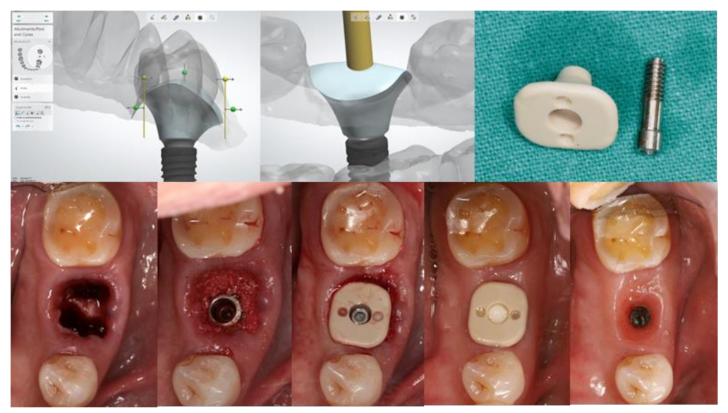
2. Customized Healing Abutment
3. Materials Used for Customized Healing Abutment and Their Properties
3.1. Materials Used for Customized Healing Abutment
3.1.1. Polyetheretherketone (PEEK)
3.1.2. Polymethyl methacrylate (PMMA)
3.1.3. Zirconia
3.1.4. Resin Composite
3.1.5. Titanium
3.2. Properties of Material Used for Customized Healing Abutments
3.2.1. Functional Properties
- Modulus of elasticity: it’s a numerical expression, indicating the measure of stiffness in a material. This allows the behavior of a material under a load to be calculated. When the material connected to the implant receives an occlusal load, one with a higher elastic modulus may deliver more stress to the cortical bone [66,67]. PEEK, CAD/CAM, and conventional PMMA and resin composites demonstrate an elastic modulus closer to human cortical bone than titanium and zirconia [68,69]. Zirconia presents the highest elastic modulus among these materials. This indicates that customized healing abutments made from titanium or zirconia can possibly cause more damage to bone than polymer and resin materials, especially when an implant is placed at the crestal level, which was shown to be significant to bone apposition at this level due to bone remodeling from the direct contact between the healing abutment and the crestal bone.
- Fracture resistance and flexural strength: Fracture resistance is a material property that describes the material’s capacity to resist fracture when experiencing a crack, while flexural strength is the ability of the material to withstand bending forces applied perpendicularly to its longitudinal axis. These properties are important because they contribute to resistance to occlusal loads that may cause a fracture or distortion of materials during the tissue maturation process. Zirconia and titanium present the highest strength values; PEEK has been reported to have lower fracture resistance than titanium but higher or comparable to zirconia and ceramic [60,70,71]. CAD/CAM PMMA was reported as having slightly lower fracture resistance compared to PEEK material [72]. A few minor mechanical complications were reported, such as the loosening of CAD/CAM PMMA customized healing abutments, which did not impact the outcome [22]. CAD/CAM resin composite has been reported with the lowest fracture resistance among the provisional materials in an in vitro study [73]. Resin composites usually present a low modulus of elasticity as well as low fracture toughness to protect the opposing tooth when used as a direct restoration [55,74,75], which may limit their use as a supporting material combines with a tougher material such as PEEK or titanium cylinder [30,35,57,76].
- Surface roughness is known to be related to the tissue response and cell adhesion [77,78] leading to soft tissue sealing [79]. Smooth abutment surfaces with a roughness value < 0.2 μm are recommended to ensure soft tissue sealing [80], and a roughness of <0.8 μm yields less bacterial colonization [81]. Polished zirconia was reported with lower surface roughness compared to polished titanium in an in vitro study [82]. Another study confirmed that the polishabilty of zirconia results in markedly low surface roughness and may contribute to its superior tissue adhesion [83]. An in vitro study showed PEEK presents lower surface roughness than a titanium abutment [84]. The study demonstrated the reduced surface roughness of PEEK after polishing [85]. CAD/CAM PMMA presents lower surface roughness compared to conventional heat-cured and light-cured PMMA [86]. Another in vitro study reported CAD/CAM PMMA demonstrated cellular behavior similar to that of lithium disilicate (current gold standard) and is, therefore, a material suitable for use as an implant provisional prosthesis. Since this material facilitates peri-implant soft tissue maturation [87] due to its low surface roughness. Resin composites also presented low surface roughness and a significantly lower value after proper polishing [88,89,90].
- Contact angle and wettability: Materials with a low contact angle and considered hydrophilic have demonstrated a positive correlation with plaque accumulation [91,92]. PEEK is inherently hydrophobic with a high contact angle, thus making it bioinert [93]. CAD/CAM PMMA presents more contact angle and hydrophobicity compared to conventional heat-cured PMMA [45,46]; both materials are considered hydrophobic. Resin composites have also demonstrated hydrophobicity due to their high contact angle [94]. Titanium and zirconia present a low contact angle, thus are classified as hydrophilic [82,95,96].
3.2.2. Biological Properties
- Tissue adhesion and tissue response: Fibroblasts and epithelial cells are known to acquire the major cellular composition of the peri-implant mucosa [65]. Therefore, the effect of fibroblast and epithelial adhesion could possibly lead to promoting the peri-implant seal and tissue maturation process. Tissue adhesion is known to be associated with the material surface roughness. A smooth surface is believed to provide the adhesion of tissue. PEEK showed biocompatibility with human fibroblast cells in an in vitro study by Peng et al. [103], which showed fibroblast adhesion effectiveness, metabolic activity, and pro-inflammatory responses similar to titanium alloy incubated fibroblasts. Moreover, PEEK promoted a more prominent soft-tissue response than a titanium healing cap in an animal study [104,105]. Clinical studies on the effects of PEEK material on human peri-implant tissue are scarce. Most of the studies reported optimum peri-implant soft tissue healing after sufficient healing periods [34,36,106]. CAD/CAM PMMA has been recommended to use around peri-implant tissue more than conventional self-cured material due to having more fibroblast attachment [98,107]. Studies have reported fibroblast and epithelial adhesion and proliferation on polished zirconia surfaces and better fibroblast adhesion on zirconia than the titanium surfaces [108,109]. It is well known that epithelial cells prefer a smoother surface, and titanium shows better epithelial adhesion than fibroblasts [110]. One study demonstrated a lower surface roughness on highly polished zirconia compared to titanium [111], which may lead to better epithelial proliferation. Resin composites demonstrated higher gingival epithelial attachment compared to partially cured composites [112]. Thus, when a resin composite has been used to fabricate a customized healing abutment intraorally, it should be removed be cured extraorally to ensure complete curing and enhance the tissue adhesion. Most materials seem to provide sufficient tissue healing and tissue maturation. Studies have reported the ability to preserve the papilla and facial mucosal—as well as the soft tissue—contour of the pre-existing teeth level with PEEK and CAD/CAM PMMA customized healing abutments [113,114]. Several other studies also mentioned papilla preservation in the short-term follow-up with the use of CAD/CAM PMMA customized healing abutments [24,77,114,115]. A 1-year randomized clinical trial reported a significantly higher Papilla Index [116] in customized healing abutments than in the standard group, which indicated more papillae present at the final outcome [115]. However, another short-term clinical study reported a significant mid-facial gingival height reduction at 1 and 3-month follow-ups with customized healing abutments made from a composite on a temporary cylinder [113]. The authors mentioned a significant reduction in the lingual soft tissue margin over 6 months due to a free gingival fiber collapse after tooth extraction. Another study reported similar facial mucosal reduction in the CAD/CAM PMMA customized healing abutment group compared to the standard group, where differences in gingival phenotypes between the two groups were reported as the possible confounding factors [115].
- Biocompatibility: refers to the ability of a biomaterial to perform its desired function without eliciting any undesirable local or systemic reactions but generating the most appropriate beneficial cellular or tissue response [117]. The properties related to biocompatibility include corrosion resistance, which is defined as the chemical or electro-chemical reaction between a material and its environment that produces a deterioration of the material itself and its properties [118]. The literatures support the high corrosion resistance of Cp-Ti and its alloys due to the stability of the Ti oxide (TiO2) layer [63,119]. However, some studies reported titanium and titanium alloy are not inert to corrosive attack if the stable oxide layer is disrupted and is unable to repair [120,121]. Titanium-wear was reported at the time of the implant placement and continued under the mastication forces. The TiO2 nanoparticles shed on peri-implant hard and soft tissue can further lead to local irritation [122]. Zirconia presents low corrosion and thus increases biocompatibility [123]. PEEK has also demonstrated high corrosion resistance in several in vitro studies [124,125] and no monomer release [126], which causes this material to have superior biocompatibility. Another factor related to biocompatibility is the release of the material substance to oral tissues. Self-cured PMMA contributes to the residual unpolymerized monomer during the polymerization reaction and has been reported to be associated with mucosal irritation [16,41,127], as well as tissue inflammation, and cytotoxicity [41,128]. Heat-cured and CAD/CAM PMMA showed a lower residual monomer release compared to self-cured PMMA. Therefore, they are likely to provide better biocompatibility [43,98,107]. There were several studies that reported good peri-implant tissue response to CAD/CAM PMMA customized healing abutments after 1–3 months of insertion, and an ability to preserve the soft tissue architecture with slight tissue inflammation [19,20,21,22,23,24,25,26,27,28]. There were some reports of resin composite leaching substances from dental composite resins and concerns about their biocompatibility, which can affect the growth and immune responsivity of gingival fibroblasts [129,130,131]. The leaching of inorganic ions was reported as dependent on the filler composition and filler treatment [132,133]. Resin composites might compromise soft tissue healing due to their release of substances and degradation in exposure to the oral environment. Stumpel and Wadhwani suggested a method to fabricate customized healing abutments from flowable composite extraorally to minimize the uncured composite contact with the peri-implant tissue [57].
- Bacterial formation: refers to material susceptible to bacterial deposition, which may cause inflammation of the peri-implant tissue, thus interfering in the tissue maturation process. PEEK demonstrated equal or lower biofilm formation compared to other materials, such as zirconia or titanium, in some in vitro studies [64,134]. Moreover, in vivo studies have reported significantly less plaque accumulation on zirconia compared to titanium in the oral cavity [135,136]. Studies have concluded that bacterial adhesion was influenced by the low surface energy of zirconia. Although conventional PMMA promotes bacterial formation due to its porosity, CAD/CAM PMMA provides less bacterial accumulation due to its enhanced hydrophobicity [91,107]. Resin composites have demonstrated marked plaque accumulation, which may lead to mucosal inflammation, compared to titanium [137].
4. Clinical Applications and Clinical Importance of Customized Healing Abutments
4.1. Clinical Applications
4.1.1. Immediate Implant Placement (IIP)
4.1.2. Delayed Placement
4.2. Clinical Importance
5. Conclusions
Author Contributions
Funding
Institutional Review Board Statement
Informed Consent Statement
Data Availability Statement
Acknowledgments
Conflicts of Interest
References
- Linkevicius, T.; Puisys, A.; Linkeviciene, L.; Peciuliene, V.; Schlee, M. Crestal Bone Stability around Implants with Horizontally Matching Connection after Soft Tissue Thickening: A Prospective Clinical Trial. Clin. Implant Dent. Relat. Res. 2015, 17, 497–508. [Google Scholar] [CrossRef] [PubMed]
- Prati, C.; Zamparini, F.; Canullo, L.; Pirani, C.; Botticelli, D.; Gandolfi, M. Factors Affecting Soft and Hard Tissues Around Two-Piece Transmucosal Implants: A 3-Year Prospective Cohort Study. Int. J. Oral Maxillofac. Implant. 2020, 35, 1022–1036. [Google Scholar] [CrossRef] [PubMed]
- Janakievski, J. Case report: Maintenance of gingival form following immediate implant placement—The custom-healing abutment. Adv. Esthet. Interdiscip. Dent. 2007, 3, 24–28. [Google Scholar]
- Elian, N.; Tabourian, G.; Jalbout, Z.N.; Classi, A.; Cho, S.-C.; Froum, S.; Tarnow, D.P. Accurate transfer of peri-implant soft tissue emergence profile from the provisional crown to the final prosthesis using an emergence profile cast. J. Esthet. Restor. Dent. 2007, 19, 306–314; discussion 315. [Google Scholar] [CrossRef] [PubMed]
- Oh, K.C.; Kim, J.-H.; Woo, C.-W.; Moon, H.S. Accuracy of Customized Prefabricated Screw-Type Immediate Provisional Restorations after Single-Implant Placement. J. Clin. Med. 2019, 8, 490. [Google Scholar] [CrossRef] [PubMed] [Green Version]
- Misch, C.E. Contemporary Implant Dentistry; Mosby: St. Louis, MO, USA, 1993. [Google Scholar]
- Weigl, P.; Trimpou, G.; Grizas, E.; Hess, P.; Nentwig, G.-H.; Lauer, H.-C.; Lorenz, J. All-ceramic versus titanium-based implant supported restorations: Preliminary 12-months results from a randomized controlled trial. J. Adv. Prosthodont. 2019, 11, 48–54. [Google Scholar] [CrossRef] [PubMed] [Green Version]
- Pow, E.H.N.; McMillan, A.S. A modified implant healing abutment to optimize soft tissue contours: A case report. Implant Dent. 2004, 13, 297–300. [Google Scholar] [CrossRef]
- Teślak, M.; Ziemlewski, A.; Foltyn, I.; Ordyniec-Kwaśnica, I.; Drogoszewska, B. Development of Custom Anatomic Healing Abutment Based on Cone-Beam Computer Tomography Measurement on Human Teeth Cross-Section. Materials 2021, 14, 4570. [Google Scholar] [CrossRef]
- Lemongello, G.J. Customized provisional abutment and provisional restoration for an immediately-placed implant. Pract. Proced. Aesthet. Dent. 2007, 19, 419–424; quiz 426. [Google Scholar]
- Wittneben, J.-G.; Buser, D.; Brägger, U.; Belser, U.C. Peri-implant soft tissue conditioning with provisional restorations in the esthetic zone: The dynamic compression technique. Int. J. Periodontics Restor. Dent. 2013, 33, 447–455. [Google Scholar] [CrossRef] [Green Version]
- Kan, J.Y.K.; Rungcharassaeng, K.; Lozada, J.L. Immediate placement and provisionalization of maxillary anterior single implants: 1-year prospective study. Int. J. Oral Maxillofac. Implant. 2003, 18, 31–39. [Google Scholar]
- Lorenzoni, M.; Pertl, C.; Zhang, K.; Wimmer, G.; Wegscheider, W.A. Immediate loading of single-tooth implants in the anterior maxilla. Preliminary results after one year. Clin. Oral Implant. Res. 2003, 14, 180–187. [Google Scholar] [CrossRef]
- Meng, H.-W.; Chien, E.Y.; Chien, H.-H. Immediate Implant Placement and Provisionalization in the Esthetic Zone: A 6.5-Year Follow-Up and Literature Review. Case Rep. Dent. 2021, 2021, 4290193. [Google Scholar] [CrossRef] [PubMed]
- Block, M.S.; Mercante, D.E.; Lirette, D.; Mohamed, W.; Ryser, M.; Castellon, P. Prospective evaluation of immediate and delayed provisional single tooth restorations. J. Oral Maxillofac. Surg. 2009, 67 (Suppl. 11), 89–107. [Google Scholar] [CrossRef] [PubMed]
- Ionescu, R.N.; Totan, A.R.; Imre, M.M.; Țâncu, A.M.C.; Pantea, M.; Butucescu, M.; Farcașiu, A.T. Prosthetic Materials Used for Implant-Supported Restorations and Their Biochemical Oral Interactions: A Narrative Review. Materials 2022, 15, 1016. [Google Scholar] [CrossRef]
- Lepidi, L.; Galli, M.; Suriano, C.; Ruggiero, G.; Calabrese, L.; Li, J.; Venezia, P. Digital planning of a customized CAD-CAM healing abutment for soft tissue conditioning at the time of implant placement: A different digital and clinical perspective in molar implant rehabilitation. J. Osseointegr. 2022, 13 (Suppl. 4), S305–S310. [Google Scholar]
- Lilet, R.; Desiron, M.; Finelle, G.; Lecloux, G.; Seidel, L.; Lambert, F. Immediate implant placement combining socket seal abutment and peri-implant socket filling: A prospective case series. Clin. Oral Implant. Res. 2022, 33, 33–44. [Google Scholar] [CrossRef]
- Wang, L.; Wang, T.; Lu, Y.; Fan, Z. Comparing the Clinical Outcome of Peri-implant Hard and Soft Tissue Treated with Immediate Individualized CAD/CAM Healing Abutments and Conventional Healing Abutments for Single-Tooth Implants in Esthetic Areas Over 12 Months: A Randomized Clinical Trial. Int. J. Oral Maxillofac. Implant. 2021, 36, 977–984. [Google Scholar] [CrossRef]
- Raheem, I.M.A.; Hammad, I.A.; Kader, S.H.A.; Fahmy, R.A. Fabrication of a CAD-CAM custom healing abutment guided by a conventional dental radiograph for delayed loaded dental implants: A dental technique. J. Prosthet. Dent. 2022, 127, 49–54. [Google Scholar] [CrossRef]
- Proussaefs, P. Use of CAD/CAM Healing Abutment Immediately After Dental Implant Placement for the Non-Esthetic Zone: A Guided Soft Tissue Healing Technique. J. Oral Implant. 2016, 42, 189–193. [Google Scholar] [CrossRef] [Green Version]
- Finelle, G.; Sanz-Martín, I.; Knafo, B.; Figué, M.; Popelut, A. Digitalized CAD/CAM protocol for the fabrication of customized sealing socket healing abutments in immediate implants in molar sites. Int. J. Comput. Dent. 2019, 22, 187–204. [Google Scholar] [PubMed]
- Finelle, G.; Lee, S. Guided Immediate Implant Placement with Wound Closure by Computer-Aided Design/Computer-Assisted Manufacture Sealing Socket Abutment: Case Report. Int. J. Oral Maxillofac. Implant. 2017, 32, e63–e67. [Google Scholar] [CrossRef] [PubMed] [Green Version]
- Finelle, G.; Popelut, A.; Knafo, B.; Martín, I. Sealing Socket Abutments (SSAs) in Molar Immediate Implants with a Digitalized CAD/CAM Protocol: Soft Tissue Contour Changes and Radiographic Outcomes After 2 Years. Int. J. Periodontics Restor. Dent. 2021, 41, 235–244. [Google Scholar] [CrossRef] [PubMed]
- Hartman, M.J. A Workflow to Design and Fabricate a Customized Healing Abutment From a Dynamic Navigation Virtual Treatment Plan. Compend. Contin. Educ. Dent. 2021, 42, 86–92. [Google Scholar] [PubMed]
- Joda, T.; Ferrari, M.; Braegger, U. A digital approach for one-step formation of the supra-implant emergence profile with an individualized CAD/CAM healing abutment. J. Prosthodont. Res. 2016, 60, 220–223. [Google Scholar] [CrossRef] [Green Version]
- Proussaefs, P. Custom CAD-CAM healing abutment and impression coping milled from a poly(methyl methacrylate) block and bonded to a titanium insert. J. Prosthet. Dent. 2016, 116, 657–662. [Google Scholar] [CrossRef]
- Junior, D.; Machado, A. Rehabilitation of Anterior Tooth Loss with Immediate Implant and Preservation of Gingival Aesthetics with Use of Vertical Extraction System and Customized Healing Abutment made by CAD-CAM Technology: Case Report. SVOA Dent. 2021, 2, 232–237. [Google Scholar]
- Alshhrani, W.M.; Al Amri, M.D. Customized CAD-CAM healing abutment for delayed loaded implants. J. Prosthet. Dent. 2016, 116, 176–179. [Google Scholar] [CrossRef]
- Akin, R. A New Concept in Maintaining the Emergence Profile in Immediate Posterior Implant Placement: The Anatomic Harmony Abutment. J. Oral Maxillofac. Surg. 2016, 74, 2385–2392. [Google Scholar] [CrossRef] [Green Version]
- Suphangul, S.; Rokaya, D.; Kanchanasobhana, C.; Rungsiyakull, P.; Chaijareenont, P. PEEK Biomaterial in Long-Term Provisional Implant Restorations: A Review. J. Funct. Biomater. 2022, 13, 33. [Google Scholar] [CrossRef]
- Kurtz, S.M.; Devine, J.N. PEEK biomaterials in trauma, orthopedic, and spinal implants. Biomaterials 2007, 28, 4845–4869. [Google Scholar] [CrossRef] [Green Version]
- Papathanasiou, I.; Kamposiora, P.; Papavasiliou, G.; Ferrari, M. The use of PEEK in digital prosthodontics: A narrative review. BMC Oral Health 2020, 20, 217. [Google Scholar] [CrossRef] [PubMed]
- Beretta, M.; Poli, P.P.; Pieriboni, S.; Tansella, S.; Manfredini, M.; Cicciù, M.; Maiorana, C. Peri-Implant Soft Tissue Conditioning by Means of Customized Healing Abutment: A Randomized Controlled Clinical Trial. Materials 2019, 12, 3041. [Google Scholar] [CrossRef] [PubMed] [Green Version]
- Ruales-Carrera, E.; Pauletto, P.; Apaza-Bedoya, K.; Volpato, C.A.M.; Özcan, M.; Benfatti, C.A.M. Peri-implant tissue management after immediate implant placement using a customized healing abutment. J. Esthet. Restor. Dent. 2019, 31, 533–541. [Google Scholar] [CrossRef]
- Bezerra, F.J.B.; Araujo, F.M.; De Oliveira, G.J.P.L.; Ghiraldini, B. Clinical application of the customizable PEEK healing abutment. A case report. J. Multidiscip. Dent. 2020, 10, 93–96. [Google Scholar] [CrossRef]
- Sarfaraz, H.; Rasheed, M.N.; Shetty, S.S.; Prabhu, U.M.; Fernandes, K.; Mohandas, S. Comparison of the Bond Strength of Composite Resin to Zirconia and Composite Resin to Polyether Ether Ketone: An In Vitro Study. J. Pharm. Bioallied Sci. 2020, 12 (Suppl. 1), S504–S509. [Google Scholar]
- Hassan, M.; Asghar, M.; UdDin, S.; Zafar, M.S. Chapter 8—Thermoset Polymethacrylate-Based Materials for Dental Applications; Grumezescu, V., Grumezescu, A.M., Eds.; Elsevier: Amsterdam, The Netherlands, 2019; pp. 273–308. [Google Scholar]
- Ronald, L.; Sakaguchi, J.L.F.; John, M. Powers, Craig’s Restorative Dental Materials, 14th ed.; Elsevier: St. Louis, MO, USA, 2019. [Google Scholar]
- Zafar, M.S. Prosthodontic Applications of Polymethyl Methacrylate (PMMA): An Update. Polymers 2020, 12, 2299. [Google Scholar] [CrossRef]
- Jorge, J.H.; Giampaolo, E.T.; Machado, A.L.; Vergani, C.E. Cytotoxicity of denture base acrylic resins: A literature review. J. Prosthet. Dent. 2003, 90, 190–193. [Google Scholar] [CrossRef]
- de Sá, J.; Vieira, F.; Aroso, C.M.; Cardoso, M.; Mendes, J.M.; Silva, A.S. The Influence of Saliva pH on the Fracture Resistance of Three Complete Denture Base Acrylic Resins. Int. J. Dent. 2020, 2020, 8941876. [Google Scholar]
- Srinivasan, M.; Gjengedal, H.; Cattani-Lorente, M.; Moussa, M.; Durual, S.; Schimmel, M.; Müller, F. CAD/CAM milled complete removable dental prostheses: An in vitro evaluation of biocompatibility, mechanical properties, and surface roughness. Dent. Mater. J. 2018, 37, 526–533. [Google Scholar] [CrossRef] [Green Version]
- Kalberer, N.; Mehl, A.; Schimmel, M.; Müller, F.; Srinivasan, M. CAD-CAM milled versus rapidly prototyped (3D-printed) complete dentures: An in vitro evaluation of trueness. J. Prosthet. Dent. 2019, 121, 637–643. [Google Scholar] [CrossRef] [PubMed]
- Alp, G.; Murat, S.; Yilmaz, B. Comparison of Flexural Strength of Different CAD/CAM PMMA-Based Polymers. J. Prosthodont. 2018, 28, e491–e495. [Google Scholar] [CrossRef] [PubMed]
- Arslan, M.; Murat, S.; Alp, G.; Zaimoglu, A. Evaluation of flexural strength and surface properties of prepolymerized CAD/CAM PMMA-based polymers used for digital 3D complete dentures. Int. J. Comput. Dent. 2018, 21, 31–40. [Google Scholar] [PubMed]
- Al-Dwairi, Z.N.; Tahboub, K.Y.; Baba, N.Z.; Goodacre, C.J.; Özcan, M. A Comparison of the Surface Properties of CAD/CAM and Conventional Polymethylmethacrylate (PMMA). J. Prosthodont. 2019, 28, 452–457. [Google Scholar] [CrossRef] [Green Version]
- Della Bona, A.; Pecho, O.E.; Alessandretti, R. Zirconia as a Dental Biomaterial. Materials 2015, 8, 4978–4991. [Google Scholar] [CrossRef] [Green Version]
- Jitwirachot, K.; Rungsiyakull, P.; Holloway, J.A.; Jia-Mahasap, W. Wear Behavior of Different Generations of Zirconia: Present Literature. Int. J. Dent. 2022, 2022, 9341616. [Google Scholar] [CrossRef]
- Jia-Mahasap, W.; Jitwirachot, K.; Holloway, J.A.; Rangsri, W.; Rungsiyakull, P. Wear of various restorative materials against 5Y-ZP zirconia. J. Prosthet. Dent. 2022, 128, e1–e814. [Google Scholar] [CrossRef]
- Bona, A.D. Bonding to Ceramics: Scientific Evidences for Clinical Dentistry; Editoria Artes Medicas Ltda: São Paulo, Brazil, 2009. [Google Scholar]
- Nistor, L.; Grădinaru, M.; Rîcă, R.; Mărășescu, P.; Stan, M.; Manolea, H.; Ionescu, A.; Moraru, I. Zirconia Use in Dentistry-Manufacturing and Properties. Curr. Health Sci. J. 2019, 45, 28–35. [Google Scholar]
- Sundh, A.; Molin, M.; Sjögren, G. Fracture resistance of yttrium oxide partially-stabilized zirconia all-ceramic bridges after veneering and mechanical fatigue testing. Dent. Mater. 2005, 21, 476–482. [Google Scholar] [CrossRef]
- Zhou, X.; Huang, X.; Li, M.; Peng, X.; Wang, S.; Zhou, X.; Cheng, L. Development and status of resin composite as dental restorative materials. J. Appl. Polym. Sci. 2019, 136, 48180. [Google Scholar] [CrossRef] [Green Version]
- Cho, K.; Rajan, G.; Farrar, P.; Prentice, L.; Prusty, B.G. Dental resin composites: A review on materials to product realizations. Compos. Part B Eng. 2021, 230, 109495. [Google Scholar] [CrossRef]
- Stumpel, L.J.; Wadhwani, C. Development and capture of soft tissue contours at time of implant placement. J. Prosthet. Dent. 2017, 117, 709–713. [Google Scholar] [CrossRef] [PubMed]
- Stumpel, L.J.; Wadhwani, C. A Customized Healing Abutment for Immediate and Delayed Implant Cases. Compend. Contin. Educ. Dent. 2017, 38, 672–678. [Google Scholar]
- Magne, P.; Oderich, E.; Boff, L.L.; Cardoso, A.C.; Belser, U.C. Fatigue resistance and failure mode of CAD/CAM composite resin implant abutments restored with type III composite resin and porcelain veneers. Clin. Oral Implant. Res. 2011, 22, 1275–1281. [Google Scholar] [CrossRef] [PubMed]
- Magne, P.; Paranhos, M.P.G.; Burnett, L.H., Jr.; Magne, M.; Belser, U.C. Fatigue resistance and failure mode of novel-design anterior single-tooth implant restorations: Influence of material selection for type III veneers bonded to zirconia abutments. Clin. Oral Implant. Res. 2010, 22, 195–200. [Google Scholar] [CrossRef] [PubMed]
- Neumann, E.A.F.; Villar, C.C.; França, F.M.G. Fracture resistance of abutment screws made of titanium, polyetheretherketone, and carbon fiber-reinforced polyetheretherketone. Braz. Oral Res. 2014, 28, 1–5. [Google Scholar] [CrossRef] [PubMed] [Green Version]
- Liu, X.; Chen, S.; Tsoi, J.K.; Matinlinna, J.P. Binary titanium alloys as dental implant materials-a review. Regen. Biomater. 2017, 4, 315–323. [Google Scholar] [CrossRef] [Green Version]
- Nicholson, J.W. Titanium Alloys for Dental Implants: A Review. Prosthesis 2020, 2, 100–116. [Google Scholar] [CrossRef]
- Özcan, M.; Hammerle, C. Titanium as a Reconstruction and Implant Material in Dentistry: Advantages and Pitfalls. Materials 2012, 5, 1528–1545. [Google Scholar] [CrossRef]
- Hahnel, S.; Wieser, A.; Lang, R.; Rosentritt, M. Biofilm formation on the surface of modern implant abutment materials. Clin. Oral Implant. Res. 2015, 26, 1297–1301. [Google Scholar] [CrossRef]
- Berglundh, T.; Abrahamsson, I.; Welander, M.; Lang, N.P.; Lindhe, J. Morphogenesis of the peri-implant mucosa: An experimental study in dogs. Clin. Oral Implant. Res. 2007, 18, 1–8. [Google Scholar] [CrossRef] [PubMed]
- Datte, C.; Tribst, J.P.; Piva, A.D.; Nishioka, R.; Bottino, M.A.; Evangelhista, A.; Monteiro, F.; Borges, A. Influence of different restorative materials on the stress distribution in dental implants. J. Clin. Exp. Dent. 2018, 10, e439–e444. [Google Scholar] [CrossRef] [PubMed]
- Pumnil, S.; Rungsiyakull, P.; Rungsiyakull, C.; Elsaka, S. Effect of Different Customized Abutment Types on Stress Distribution in Implant-Supported Single Crown: A 3D Finite Element Analysis. J. Prosthodont. 2022, 31, e2–e11. [Google Scholar] [CrossRef] [PubMed]
- Moon, S.-M.; Ingalhalikar, A.; Highsmith, J.M.; Vaccaro, A.R. Biomechanical rigidity of an all-polyetheretherketone anterior thoracolumbar spinal reconstruction construct: An in vitro corpectomy model. Spine J. 2009, 9, 330–335. [Google Scholar] [CrossRef]
- Hada, T.; Kanazawa, M.; Iwaki, M.; Katheng, A.; Minakuchi, S. Comparison of Mechanical Properties of PMMA Disks for Digitally Designed Dentures. Polymers 2021, 13, 1745. [Google Scholar] [CrossRef]
- Beuer, F.; Steff, B.; Naumann, M.; Sorensen, J.A. Load-bearing capacity of all-ceramic three-unit fixed partial dentures with different computer-aided design (CAD)/computer-aided manufacturing (CAM) fabricated framework materials. Eur. J. Oral Sci. 2008, 116, 381–386. [Google Scholar] [CrossRef]
- Atsü, S.; Aksan, M.; Bulut, A. Fracture Resistance of Titanium, Zirconia, and Ceramic-Reinforced Polyetheretherketone Implant Abutments Supporting CAD/CAM Monolithic Lithium Disilicate Ceramic Crowns After Aging. Int. J. Oral Maxillofac. Implant. 2019, 34, 622–630. [Google Scholar] [CrossRef]
- Benli, M.; Eker-Gumus, B.; Kahraman, Y.; Huck, O.; Ozcan, M. Can polylactic acid be a CAD/CAM material for provisional crown restorations in terms of fit and fracture strength? Dent. Mater. J. 2021, 40, 772–780. [Google Scholar] [CrossRef]
- Abdullah, A.O.; Tsitrou, E.A.; Pollington, S. Comparative in vitro evaluation of CAD/CAM vs conventional provisional crowns. J. Appl. Oral Sci. 2016, 24, 258–263. [Google Scholar] [CrossRef]
- Iftekhar, H. 9-Nanocomposite Restorative Materials for Dental Caries Management. In Applications of Nanocomposite Materials in Dentistry; Asiri, A.M., Inamuddin, M.A., Eds.; Woodhead Publishing: Philadelphia, PA, USA, 2019; pp. 161–169. [Google Scholar]
- Lucsanszky, I.J.; Ruse, N.D. Fracture Toughness, Flexural Strength, and Flexural Modulus of New CAD/CAM Resin Composite Blocks. J. Prosthodont. 2019, 29, 34–41. [Google Scholar] [CrossRef]
- Hu, C.; Lin, W.; Gong, T.; Zuo, Y.; Qu, Y.; Man, Y. Early Healing of Immediate Implants Connected With Two Types of Healing Abutments: A Prospective Cohort Study. Implant. Dent. 2018, 27, 646–652. [Google Scholar] [CrossRef] [PubMed]
- Ponsonnet, L.; Reybier, K.; Jaffrezic, N.; Comte, V.; Lagneau, C.; Lissac, M.; Martelet, C. Relationship between surface properties (roughness, wettability) of titanium and titanium alloys and cell behaviour. Mater. Sci. Eng. C 2003, 23, 551–560. [Google Scholar] [CrossRef]
- Kearns, V.R.; Williams, R.L.; Mirvakily, F.; Doherty, P.J.; Martin, N. Guided gingival fibroblast attachment to titanium surfaces: An in vitro study. J. Clin. Periodontol. 2012, 40, 99–108. [Google Scholar] [CrossRef] [PubMed]
- Bollenl, C.M.; Lambrechts, P.; Quirynen, M. Comparison of surface roughness of oral hard materials to the threshold surface roughness for bacterial plaque retention: A review of the literature. Dent. Mater. 1997, 13, 258–269. [Google Scholar] [CrossRef]
- Mustafa, K.; Odén, A.; Wennerberg, A.; Hultenby, K.; Arvidson, K. The influence of surface topography of ceramic abutments on the attachment and proliferation of human oral fibroblasts. Biomaterials 2005, 26, 373–381. [Google Scholar] [CrossRef]
- Verran, J.; Airey, P.; Packer, A.; Whitehead, K.A. Microbial retention on open food contact surfaces and implications for food contamination. Adv. Appl. Microbiol. 2008, 64, 223–246. [Google Scholar]
- Al-Radha, A.S.D.; Dymock, D.; Younes, C.; O’Sullivan, D. Surface properties of titanium and zirconia dental implant materials and their effect on bacterial adhesion. J. Dent. 2012, 40, 146–153. [Google Scholar] [CrossRef]
- Linkevicius, T.; Valantiejiene, V.; Alkimavicius, J.; Gineviciute, E.; Andrijauskas, R.; Linkeviciene, L. The Effect of a Polishing Protocol on the Surface Roughness of Zirconium Oxide. Int. J. Prosthodont. 2020, 33, 217–223. [Google Scholar] [CrossRef]
- Ramakrishnan, H.; Ragupathi, M.; Mahadevan, V.; Azhagarasan, N.; Jayakrishnakumar, S. Comparative evaluation of the wear resistance of two different implant abutment materials after cyclic loading—An in vitro study. Contemp. Clin. Dent. 2020, 11, 229–236. [Google Scholar] [CrossRef]
- Batak, B.; Çakmak, G.; Johnston, W.M.; Yilmaz, B. Surface roughness of high-performance polymers used for fixed implant-supported prostheses. J. Prosthet. Dent. 2021, 126, 254.e1–254.e6. [Google Scholar] [CrossRef]
- Alfouzan, A.; Alnouwaisar, A.; Alazzam, N.; Al-Otaibi, H.; Labban, N.; Alswaidan, M.; Al-Taweel, S.; Alshehri, H. Surface roughness analysis of prepolymerized CAD/CAM dental acrylic resins following combined surface treatments. Mater. Sci. 2021, 39, 209–218. [Google Scholar] [CrossRef]
- Herráez-Galindo, C.; Rizo-Gorrita, M.; Luna-Oliva, I.; Serrera-Figallo, M.-Á.; Castillo-Oyagüe, R.; Torres-Lagares, D. In vitro Comparative Study of Fibroblastic Behaviour on Polymethacrylate (PMMA) and Lithium Disilicate Polymer Surfaces. Polymers 2019, 11, 744. [Google Scholar] [CrossRef] [PubMed] [Green Version]
- Magdy, N.M.; Kola, M.Z.; Alqahtani, H.H.; Alqahtani, M.D.; Alghmlas, A.S. Evaluation of Surface Roughness of Different Direct Resin-based Composites. J. Int. Soc. Prev. Community Dent. 2017, 7, 104–109. [Google Scholar] [PubMed]
- Nithya, K.; Sridevi, K.; Keerthi, V.; Ravishankar, P. Evaluation of Surface Roughness, Hardness, and Gloss of Composites After Three Different Finishing and Polishing Techniques: An In Vitro Study. Cureus 2020, 12, e7037. [Google Scholar] [CrossRef] [Green Version]
- Isabel, C.A.C.; Dominguette, A.A.S.; Dos Santos, S.G.; Ribeiro, J.C.R.; Moysés, M.R. Surface roughness of a resin composite. RGO-Rev. Gaúcha Odontol. 2016, 64, 50–55. [Google Scholar] [CrossRef] [Green Version]
- Quirynen, M.; Marechal, M.; Busscher, H.J.; Weerkamp, A.H.; Darius, P.L.; Van Steenberghe, D. The influence of surface free energy and surface roughness on early plaque formation: An in vivo study in man. J. Clin. Periodontol. 1990, 17, 138–144. [Google Scholar] [CrossRef]
- Quirynen, M.; Marechal, M.; Busscher, H.J.; Weerkamp, A.H.; Arends, J.; Darius, P.L.; Van Steenberghe, D. The Influence of Surface Free-energy on Planimetric Plaque Growth in Man. J. Dent. Res. 1989, 68, 796–799. [Google Scholar] [CrossRef] [Green Version]
- Nieminen, T.; Kallela, I.; Wuolijoki, E.; Kainulainen, H.; Hiidenheimo, I.; Rantala, I. Amorphous and crystalline polyetheretherketone: Mechanical properties and tissue reactions during a 3-year follow-up. J. Biomed. Mater. Res. Part A 2007, 84A, 377–383. [Google Scholar] [CrossRef]
- Rüttermann, S.; Beikler, T.; Janda, R. Contact angle and surface free energy of experimental resin-based dental restorative materials after chewing simulation. Dent. Mater. 2014, 30, 702–707. [Google Scholar] [CrossRef]
- Liu, M.; Zhou, J.; Yang, Y.; Zheng, M.; Yang, J.; Tan, J. Surface modification of zirconia with polydopamine to enhance fibroblast response and decrease bacterial activity in vitro: A potential technique for soft tissue engineering applications. Colloids Surf. B Biointerfaces 2015, 136, 74–83. [Google Scholar] [CrossRef]
- Schünemann, F.H.; Galárraga-Vinueza, M.E.; Magini, R.; Fredel, M.; Silva, F.; Souza, J.C.; Zhang, Y.; Henriques, B. Zirconia surface modifications for implant dentistry. Mater. Sci. Eng. C Mater. Biol. Appl. 2019, 98, 1294–1305. [Google Scholar] [CrossRef] [PubMed]
- Lambrecht, J.; Nyffeler, T.; Linder, M. Thermal conduction of titanium implants under CO2 laser irradiation in vitro. Ann. Maxillofac. Surg. 2012, 2, 12–16. [Google Scholar] [CrossRef] [PubMed] [Green Version]
- Pituru, S.M.; Greabu, M.; Totan, A.; Imre, M.; Pantea, M.; Spinu, T.; Tancu, A.M.C.; Popoviciu, N.O.; Stanescu, I.-I.; Ionescu, E. A Review on the Biocompatibility of PMMA-Based Dental Materials for Interim Prosthetic Restorations with a Glimpse into their Modern Manufacturing Techniques. Materials 2020, 13, 2894. [Google Scholar] [CrossRef] [PubMed]
- Wiedhahn, K.; Fritzshe, G.; Wiedhahn, C.; Schenk, O. Zirconia crowns-the new standard for single-visit dentistry? Int. J. Comput. Dent. 2016, 19, 9–26. [Google Scholar]
- Civjan, S.; Barone, J.J.; Reinke, P.E.; Selting, W.J. Thermal Properties of Nonmetallic Restorative Materials. J. Dent. Res. 1972, 51, 1030–1037. [Google Scholar] [CrossRef]
- Ilie, N.; Hickel, R.; Valceanu, A.S.; Huth, K.C. Fracture toughness of dental restorative materials. Clin. Oral Investig. 2011, 16, 489–498. [Google Scholar] [CrossRef]
- Santosa, R.E. Provisional restoration options in implant dentistr. Aust. Dent. J. 2007, 52, 234–242. [Google Scholar] [CrossRef]
- Peng, T.Y.; Shih, Y.-H.; Hsia, S.-M.; Wang, T.-H.; Li, P.-J.; Lin, D.-J.; Sun, K.-T.; Chiu, K.-C.; Shieh, T.-M. In Vitro Assessment of the Cell Metabolic Activity, Cytotoxicity, Cell Attachment, and Inflammatory Reaction of Human Oral Fibroblasts on Polyetheretherketone (PEEK) Implant-Abutment. Polymers 2021, 13, 2995. [Google Scholar] [CrossRef]
- Caballé-Serrano, J.; Chappuis, V.; Monje, A.; Buser, D.; Bosshardt, D.D. Soft tissue response to dental implant closure caps made of either polyetheretherketone (PEEK) or titanium. Clin. Oral Implant. Res. 2019, 30, 808–816. [Google Scholar] [CrossRef]
- Rea, M.; Ricci, S.; Ghensi, P.; Lang, N.P.; Botticelli, D.; Soldini, C. Marginal healing using Polyetheretherketone as healing abutments: An experimental study in dogs. Clin. Oral Implant. Res. 2016, 28, e46–e50. [Google Scholar] [CrossRef]
- Koutouzis, T.; Richardson, J.; Lundgren, T. Lundgren, Comparative soft and hard tissue responses to titanium and polymer healing abutments. J. Oral Implant. 2011, 37, 174–182. [Google Scholar] [CrossRef] [PubMed]
- Shim, J.S.; Kim, H.C.; Park, S.I.; Yun, H.J.; Ryu, J.J. Comparison of Various Implant Provisional Resin Materials for Cytotoxicity and Attachment to Human Gingival Fibroblasts. Int. J. Oral Maxillofac. Implant. 2019, 34, 390–396. [Google Scholar] [CrossRef]
- Nothdurft, F.P.; Fontana, D.; Ruppenthal, S.; May, A.; Aktas, C.; Mehraein, Y.; Lipp, P.; Kaestner, L. Differential Behavior of Fibroblasts and Epithelial Cells on Structured Implant Abutment Materials: A Comparison of Materials and Surface Topographies. Clin. Implant. Dent. Relat. Res. 2014, 17, 1237–1249. [Google Scholar] [CrossRef] [PubMed]
- Teng, F.-Y.; Ko, C.-L.; Kuo, H.-N.; Hu, J.-J.; Lin, J.-H.; Lou, C.-W.; Hung, C.-C.; Wang, Y.-L.; Cheng, C.-Y.; Chen, W.-C. A comparison of epithelial cells, fibroblasts, and osteoblasts in dental implant titanium topographies. Bioinorg. Chem. Appl. 2012, 2012, 687291. [Google Scholar] [CrossRef] [PubMed] [Green Version]
- Kawahara, H.; Kawahara, D.; Mimura, Y.; Takashima, Y.; Ong, J. Morphologic studies on the biologic seal of titanium dental implants. Report II. In vivo study on the defending mechanism of epithelial adhesions/attachment against invasive factors. Int. J. Oral Maxillofac. Implant. 1998, 13, 465–473. [Google Scholar]
- Linkevičius, T. Zero Bone Loss Concepts; Quintessence Publishing Co., Inc.: Batavia, IL, USA, 2019. [Google Scholar]
- Boloori, E.; Schoenmaker, T.; Kleverlaan, C.J.; Loos, B.G.; De Vries, T.J. Gingival epithelium attachment to well- or partially cured resin composites. Eur. Cells Mater. 2020, 40, 259–275. [Google Scholar] [CrossRef]
- Choorak, N.; Amornsettachai, P.; Chuenjitkuntaworn, B.; Suphangul, B. Dimensional Change of Peri-implant Soft Tissue Following Immediate Implant Placement and Customized Healing Abutment in Posterior Teeth. J. Int. Dent. Med. Res. 2021, 14, 273–279. [Google Scholar]
- Fernandes, D.; Nunes, S.; López-Castro, G.; Marques, T.; Montero, J.; Borges, T. of customized healing abutments on the peri-implant linear and volumetric tissue changes at maxillary immediate implant sites: A 1-year prospective randomized clinical trial. Clin. Implant Dent. Relat. Res. 2021, 23, 745–757. [Google Scholar] [CrossRef]
- Perez, A.; Caiazzo, A.; Valente, N.A.; Toti, P.; Alfonsi, F.; Barone, A. Standard vs customized healing abutments with simultaneous bone grafting for tissue changes around immediate implants. 1-year outcomes from a randomized clinical trial. Clin. Implant Dent. Relat. Res. 2019, 22, 42–53. [Google Scholar] [CrossRef]
- Jemt, T. Regeneration of gingival papillae after single-implant treatment. Int. J. Periodontics Restor. Dent. 1997, 17, 326–333. [Google Scholar]
- Williams, D.F. On the mechanisms of biocompatibility. Biomaterials 2008, 29, 2941–2953. [Google Scholar] [CrossRef] [PubMed]
- Jones, D.A. Principles and Prevention of Corrosion, 2nd ed.; Prentice Hall: Upper Saddle River, NJ, USA, 1996. [Google Scholar]
- Nakagawa, M.; Matsuya, S.; Udoh, K. Corrosion behavior of pure titanium and titanium alloys in fluoride-containing solutions. Dent. Mater. J. 2001, 20, 305–314. [Google Scholar] [CrossRef]
- Tschernitschek, H.; Borchers, L.; Geurtsen, W. Nonalloyed titanium as a bioinert metal—A review. Quintessence Int. 2006, 36, 523–530. [Google Scholar] [CrossRef]
- Prasad, S.; Ehrensberger, M.; Gibson, M.P.; Kim, H.; Monaco, E.A., Jr. Biomaterial properties of titanium in dentistry. J. Oral Biosci. 2015, 57, 192–199. [Google Scholar] [CrossRef]
- Romanos, G.; Fischer, G.; Delgado-Ruiz, R. Titanium Wear of Dental Implants from Placement, under Loading and Maintenance Protocols. Int. J. Mol. Sci. 2021, 22, 1067. [Google Scholar] [CrossRef] [PubMed]
- Soygun, K.; Ozer, A.; Ulucan, M.C.; Bolayir, G. An investigation of corrosive effects on zirconia with different crystal structures. Microsc. Res. Tech. 2020, 84, 796–803. [Google Scholar] [CrossRef] [PubMed]
- Najeeb, S.; Zafar, M.S.; Khurshid, Z.; Siddiqui, F. Applications of polyetheretherketone (PEEK) in oral implantology and prosthodontics. J. Prosthodont. Res. 2016, 60, 12–19. [Google Scholar] [CrossRef]
- Silthampitag, P.; Chaijareenont, P.; Tattakorn, K.; Banjongprasert, C.; Takahashi, H.; Arksornnukit, M. Effect of surface pretreatments on resin composite bonding to PEEK. Dent. Mater. J. 2016, 35, 668–674. [Google Scholar] [CrossRef] [Green Version]
- Muhsin, S.A.; Hatton, P.V.; Johnson, A.; Sereno, N.; Wood, D.J. Determination of Polyetheretherketone (PEEK) mechanical properties as a denture material. Saudi Dent. J. 2019, 31, 382–391. [Google Scholar] [CrossRef]
- Braun, K.O.; Mello, J.A.N.; Rached, R.; Cury, A.D.B. Surface texture and some properties of acrylic resins submitted to chemical polishing. J. Oral Rehabilitation 2002, 30, 91–98. [Google Scholar] [CrossRef]
- Lung, C.; Darvell, B. Minimization of the inevitable residual monomer in denture base acrylic. Dent. Mater. 2005, 21, 1119–1128. [Google Scholar] [CrossRef] [PubMed]
- Willershausen, B.; Schäfer, D.; Pistorius, A.; Schulze, R.; Mann, W. Influence of resin-based restoration materials on cytotoxicity in gingival fibroblasts. Eur. J. Med. Res. 1999, 4, 149–155. [Google Scholar] [PubMed]
- Lapp, C.A.; Schuster, G.S. Effects of DMAEMA and 4-methoxyphenol on gingival fibroblast growth, metabolism, and response to interleukin-1. J. Biomed. Mater. Res. 2002, 60, 30–35. [Google Scholar] [CrossRef] [PubMed]
- Caughman, W.; Caughman, G.B.; Shiflett, R.A.; Rueggeberg, F.; Schuster, G.S. Correlation of cytotoxicity, filler loading and curing time of dental composites. Biomaterials 1991, 12, 737–740. [Google Scholar] [CrossRef]
- Oysaed, H.; Ruyter, E.I. Water sorption and filler characteristics of composites for use in posterior teeth. J. Dent. Res. 1986, 65, 1315–1318. [Google Scholar] [CrossRef] [PubMed]
- Ohsaki, A.; Imai, Y. Analysis of major components contained in Bis-GMA monomer. Dent. Mater. J. 1999, 18, 425–429. [Google Scholar] [CrossRef] [PubMed] [Green Version]
- Peng, T.-Y.; Lin, D.-J.; Mine, Y.; Tasi, C.-Y.; Li, P.-J.; Shih, Y.-H.; Chiu, K.-C.; Wang, T.-H.; Hsia, S.-M.; Shieh, T.-M. Biofilm Formation on the Surface of (Poly)Ether-Ether-Ketone and In Vitro Antimicrobial Efficacy of Photodynamic Therapy on Peri-Implant Mucositis. Polymers 2021, 13, 940. [Google Scholar] [CrossRef]
- van Brakel, R.; Cune, M.S.; van Winkelhoff, A.J.; de Putter, C.; Verhoeven, J.W.; van der Reijdenet, W. Early bacterial colonization and soft tissue health around zirconia and titanium abutments: An in vivo study in man. Clin. Oral Implant. Res. 2011, 22, 571–577. [Google Scholar] [CrossRef] [Green Version]
- Rimondini, L.; Cerroni, L.; Carrassi, A.; Torricelli, P. Bacterial colonization of zirconia ceramic surfaces: An in vitro and in vivo study. Int. J. Oral Maxillofac. Implant. 2002, 17, 793–798. [Google Scholar]
- Kanao, M.; Nakamoto, T.; Kajiwara, N.; Kondo, Y.; Masaki, C.; Hosokawa, R. Comparison of plaque accumulation and soft-tissue blood flow with the use of full-arch implant-supported fixed prostheses with mucosal surfaces of different materials: A randomized clinical study. Clin. Oral Implant. Res. 2012, 24, 1137–1143. [Google Scholar] [CrossRef]
- Mihali, S.G.; Freiman, P.C.; Singh, M.; Bratu, E.A. Maintaining Tissue Architecture in Immediate Implant Placement Following Extraction of Natural Teeth Using Custom Healing Screw. Biomed. J. Sci. Tech. Res. 2018, 7, 6096–6101. [Google Scholar] [CrossRef]
- Habashneh, A.R.; Walid, A.M.; Abualteen, T.; Abukar, M. Socket-shield Technique and Immediate Implant Placement for Ridge Preservation: Case Report Series with 1-year Follow-up. J. Contemp. Dent. Pract. 2019, 20, 1108–1117. [Google Scholar] [CrossRef] [PubMed]
- Bhatnagar, A.; Raj, A. Preservation of optimal gingival architecture through customised healing abutment in immediate implant placement: A clinical report. Int. J. Adv. Res. 2015, 3, 156–160. [Google Scholar]
- Chu, S.J.; Hochman, M.N.; Tan-Chu, J.H.-P.; Mieleszko, A.J.; Tarnow, D.P. A novel prosthetic device and method for guided tissue preservation of immediate postextraction socket implants. Int. J. Periodontics Restor. Dent. 2014, 34 (Suppl. 3), s9–s17. [Google Scholar]
- Sarnachiaro, G.O.; Chu, S.J.; Sarnachiaro, E.; Gotta, S.L.; Tarnow, D.P. Immediate Implant Placement into Extraction Sockets with Labial Plate Dehiscence Defects: A Clinical Case Series. Clin. Implant. Dent. Relat. Res. 2015, 18, 821–829. [Google Scholar] [CrossRef]
- Solow, R. Contour correction for stock titanium healing abutments. Prosthet. Dent. 2018, 120, 787–788. [Google Scholar] [CrossRef] [PubMed] [Green Version]
- Farag, M.A.E.S.; Sabry, A.S.; El Halawani, G. Evaluation of Customized Healing Abutment in Immediate Implant Placement. Alex. Dent. J. 2022, 47, 46–51. [Google Scholar]
- Menchini-Fabris, G.-B.; Crespi, R.; Toti, P.; Crespi, G.; Rubino, L.; Covani, U. A 3-year retrospective study of fresh socket implants: CAD/CAM customized healing abutment vs cover screws. Int. J. Comput. Dent. 2020, 23, 109–117. [Google Scholar]
- Amato, F.; Amato, G.; Campriani, S.; Contessi, M.; Fiorentini, A.; Polara, G.; Spedicato, G. The Role of Different Healing Abutment Sizes in Tissue Volume Preservation of Molar Sockets After Immediate Tooth Extraction and Implant Placement: A Multicenter Clinical Study. Int. J. Oral Maxillofac. Implant. 2022, 37, 891–904. [Google Scholar] [CrossRef]
- Akin, R.; Chapple, A.G. Clinical Advantages of Immediate Posterior Implants With Custom Healing Abutments: Up to 8-Year Follow-Up of 115 Cases. J. Oral Maxillofac. Surg. 2022, 80, 1952–1965. [Google Scholar] [CrossRef]
- Crespi, R.; Toti, P.; Covani, U.; Crespi, G.; Menchini-Fabris, G.-B. Guided Tissue Healing by Preformed Anatomical Healing Caps in the Edentulous Ridge: A 2-Year Retrospective Case-Control Study. Int. J. Periodontics Restor. Dent. 2022, 42, 639–646. [Google Scholar] [CrossRef] [PubMed]
- Alexopoulou, M.; Lambert, F.; Knafo, B.; Popelut, A.; Vandenberghe, B.; Finelle, G. Immediate implant in the posterior region combined with alveolar ridge preservation and sealing socket abutment: A retrospective 3D radiographic analysis. Clin. Implant Dent. Relat. Res. 2021, 23, 61–72. [Google Scholar] [CrossRef] [PubMed]


| Properties | PEEK | PMMA | Zirconia | Titanium | Resin Composite | Reference |
|---|---|---|---|---|---|---|
| Elastic modulus | Low | Low | Highest | High | Low | [68,69] |
| Flexural strength | High | Moderate | High | High | Moderate | [55,60,69,70,71,72,73,74,75,101] |
| Fracture toughness | High | Self-cured; Low CAD/CAM; High | High | High | Moderate | |
| Surface roughness | Low | Self-cured; High CAD/CAM; Low | Lowest | Low | Low | [82,83,84,85,86,87,88,89,90] |
| Color | White | Tooth-colored | Tooth-colored | Greyish | Tooth-colored | [97,98,99,100] |
| Thermal conductivity | Low | Low | Low | High | Low | |
| Hydrophobicity | High | CAD/CAM; High | Low | Low | High |
| Properties | PEEK | PMMA | Zirconia | Titanium | Resin Composite | Reference |
|---|---|---|---|---|---|---|
| Tissue adhesion | Good | CAD/CAM; Good | Very good | Good | Good | [34,36,104,105,106,108,109,111,112] |
| Biocompatibility | Very good | Self-cured; Acceptable CAD/CAM; Good | Good | Good | Acceptable | [16,41,43,63,98,107,119,123,124,125,127,129,130,131] |
| Bacterial formation | Low | Self-cured; High CAD/CAM; Low | Low | Low | Moderate | [64,107,134,135,136,137] |
| Effect | Cover Screw (Submerged) | Standard Healing Abutment | Customized Healing Abutment | Reference | |
|---|---|---|---|---|---|
| Tissue volume | Decreased | - | Stable | [114] | |
| Soft tissue | Horizontal contour | Comparable - | - Decreased | Comparable Stable | [114] [146] |
| Vertical contour (Facial mucosal level) | - Comparable | Improved - | Decreased Comparable | [115] [114] | |
| Papilla level | - | Decreased | Preserved | [115] | |
| Hard tissue | - | Slightly decreased | Preserved | [149] | |
| Horizontal contour | Decreased | - | Preserved | [145,149] | |
| Vertical contour (Proximal bone level) | - | Comparable | Comparable | [115] | |
| Esthetic and Patient satisfaction | Pain NRS | - | Higher | Lower | [34] |
| PES change | - | Slightly decreased | Stable | [115] |
Publisher’s Note: MDPI stays neutral with regard to jurisdictional claims in published maps and institutional affiliations. |
© 2022 by the authors. Licensee MDPI, Basel, Switzerland. This article is an open access article distributed under the terms and conditions of the Creative Commons Attribution (CC BY) license (https://creativecommons.org/licenses/by/4.0/).
Share and Cite
Chokaree, P.; Poovarodom, P.; Chaijareenont, P.; Yavirach, A.; Rungsiyakull, P. Biomaterials and Clinical Applications of Customized Healing Abutment—A Narrative Review. J. Funct. Biomater. 2022, 13, 291. https://doi.org/10.3390/jfb13040291
Chokaree P, Poovarodom P, Chaijareenont P, Yavirach A, Rungsiyakull P. Biomaterials and Clinical Applications of Customized Healing Abutment—A Narrative Review. Journal of Functional Biomaterials. 2022; 13(4):291. https://doi.org/10.3390/jfb13040291
Chicago/Turabian StyleChokaree, Parima, Pongsakorn Poovarodom, Pisaisit Chaijareenont, Apichai Yavirach, and Pimduen Rungsiyakull. 2022. "Biomaterials and Clinical Applications of Customized Healing Abutment—A Narrative Review" Journal of Functional Biomaterials 13, no. 4: 291. https://doi.org/10.3390/jfb13040291