Article

Nanofibrous Membrane with Encapsulated Glucose Oxidase for Self-Sustained Antimicrobial Applications

Department of Chemical Engineering, National Taiwan University of Science and Technology, Taipei 106, Taiwan
*
Author to whom correspondence should be addressed.
Membranes 2021, 11(12), 997; https://doi.org/10.3390/membranes11120997
Submission received: 30 November 2021 / Revised: 15 December 2021 / Accepted: 16 December 2021 / Published: 20 December 2021
(This article belongs to the Collection Polymeric Membranes: Science, Materials and Applications)

Abstract

:
Polyvinyl alcohol (PVA) nanofibrous membrane, consisting of separately encapsulated glucose oxidase (GOx) and glucose (Glu) nanofibers, was prepared via simultaneously electrospinning PVA/GOx and PVA/Glu dopes. The as-prepared pristine membrane could self-sustainably generate hydrogen peroxide (H2O2) only in contact with an aqueous solution. The H2O2 production level was well maintained even after storing the dry membrane at room temperature for 7 days. Cross-linking the membrane via reaction with glutaraldehyde (GA) vapor could not only prevent the nanofibrous membrane from dissolving in water but also prolonged the release of H2O2. The sustained release of H2O2 from the membrane achieved antimicrobial capability equivalent to that of 1% H2O2 against both Escherichia coli and Staphylococcus aureus. Gram(+) S. aureus cells were more susceptible to H2O2 than Gram(−) E. coli and >99% of S. aureus were killed after 1 h incubation with the membrane. Pristine and GA-crosslinked nanofibrous membrane with in situ production of H2O2 were self-sterilized in which no microorganism contamination on the membrane could be detected after 2 weeks incubation on an agar plate. The GOx/Glu membrane may find potential application as versatile antimicrobial materials in the field of biomedicine, in the food and health industries, and especially challenges related to wound healing in diabetic patients.

1. Introduction

The investigation of antimicrobial enzymes applications has been growing rapidly in various industries, such as healthcare, food, and biomedical. The antimicrobial activities resulted from these enzymes involving direct attack and lysing or catalyzing reactions to release disinfection compounds that inactivate or kill the microorganisms [1,2,3]. Glucose oxidase (GOx) is one of the widely used oxidoreductase enzymes with potent antimicrobial capability [4]. It exerts its antimicrobial activity by the in situ generation of reactive molecules via catalyzing the reaction between glucose and oxygen, producing D-gluconic acid with concurrent production of hydrogen peroxide (H2O2) (Scheme 1) [5]. The produced hydrogen peroxide is well-known for its disinfection capability, as DNA, proteins, membrane lipids of bacterial cells could be damaged due to oxidation that leads to their death [6]. In terms of effectivity, H2O2 is believed to be better against Gram-positive bacteria than that of Gram-negative bacteria [7]. Moreover, the pH-lowering effect resulting from produced D-gluconic acid accumulation in the system may also inhibit the growth of a handful of organisms [8].
In addition to its broad antimicrobial applications, GOx also has an important biomedical application for detecting as well as treatment [9] of blood sugar level for diabetic patients based on its reaction with glucose to generate H2O2, which could be easily determined via amperometric methods [10,11,12,13,14]. Nevertheless, the widespread applications of this GOx-based glucose biosensor are limited due to its cost, instability, and reusability [15]. These major challenges could actually be overcome through selection of a suitable matrix and technique for immobilization and encapsulation of GOx. In recent times, electrospinning has become an often-used enzyme immobilization method. Through this method, the enzyme is encapsulated in various polymeric nanofibers [16] which structures provide good immobilization yield and outstanding thermal stabilization effect [17]. Other than that, the high specific surface area makes it more efficient for the encapsulated enzyme to carry out catalytic reactions with substrate diffuses in from surrounding environment [18,19]. In addition, various electrospun nanofibers have recently been studied for wound dressing applications because the nanofibrous scaffold can provide high porosity, good biocompatibility, which facilitate cell respiration and infiltration for wound tissue regeneration. Furthermore, various nanoparticles have been encapsulated to confer antimicrobial activity on electrospun nanofibers to protect the wound from pathogen infection [20,21,22,23,24,25,26,27,28,29].
Polyvinyl alcohol (PVA) is one of the most easily electrospinable polymers that offers unique characteristics such as excellent gel-forming properties and good film-matrix forming ability. Thus, PVA had been widely exploited for drug delivery and enzyme encapsulation [30,31,32,33,34]. Additionally, PVA is also known for its high biocompatibility and hydrophilicity, ease of processing, non-toxic, and biodegradable nature, confirming its ability to work as a matrix in immobilizing and stabilizing enzymes [35,36].
In this work, PVA nanofibrous membranes were prepared via simultaneous electrospinning (co-electrospun) of two different dopes (Figure 1), of which one containing GOx enzyme, and the other, glucose (Glu) as its substrate. Hence, whenever in contact with an aqueous solution, the membrane will be able to provide H2O2 as an antimicrobial agent in both the presence and absence of glucose in the surrounding environment. To prevent PVA nanofibrous membrane from water-dissolving [37] that leads to the loss of the encapsulated GOx, glutaraldehyde (GA) vapor crosslinking of PVA was implemented [38]. The GA-crosslinked PVA membrane will significantly reduce the diffusion of encapsulated Glu to reach the GOx encapsulated in the other separated nanofibers in an attempt to produce H2O2. As a consequence, the release of H2O2 from the GA-crosslinked PVA membrane will be sustained for a longer time as compared to the pristine membrane. To the best of our knowledge, simultaneous electrospinning of PVA/GOx and PVA/Glu to produce a membrane capable of self-sustained generation of H2O2 for antimicrobial applications has never been reported previously. On the other hand, the membrane with the capability of self-generation H2O2 also allows supplementary addition of glucose from surrounding to accelerate the disinfection rate through rapid and enhanced H2O2 generation. Most importantly, this antimicrobial nanofibrous membrane could be a potential wound healing material, especially for diabetic patients since high blood sugar levels will benefit the generation of antimicrobial H2O2.

2. Materials and Methods

2.1. Materials

Polyvinyl alcohol (PVA, degree of polymerization = 2000; Mw = 81,000–96,000; hydrolysis 98.5–99.4%, Hanawa Chemical Pure, Osaka, Japan), glutaraldehyde (GA, 25% aqueous solution, Alfa Aesar, Lancashire, England), Glucose Oxidase (GOx,“Amano” NA 233 U/mg, Amano Enzyme Inc., Glucose (α-D(+)-glucose, 99% ACROS-Organics), Monosodium phosphate monohydrate (H2NaPO4.H2O, 99% ACROS-Organics); Disodium Phosphate heptahydrate (HNa2PO4.7H2O, 99% ACROS-Organics; Hydrogen peroxide (H2O2, 35 wt% aqueous solution), 3,3′,5,5′-Tetramethylbenzidine, 99+%, (TMB, 99% Thermo Scientific ACROS-Organics, Antwerp, Belgium), N-(carboxymethylaminocarbonyl)-4,4′-bis(dimethylamino)-diphenylamine sodium salt (DA-64, FUJIFILM Wako Chemicals Europe Gmb–Lab Chem, Neuss, Germany), Peroxidase from horseradish (type I, essentially salt-free, lyophilized powder, ≥50 units/mg solid, SIGMA, Marlborough, MA, USA), Sodium acetate anhydrous (98.5%, DUKSAN, Ansan-si, Korea). All Chemicals were used as received.

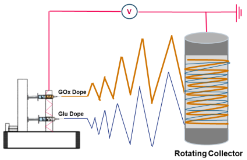
2.2. GOx Encapsulation: Simultaneous Electrospinning of PVA/GOx and PVA/Glucose

PVA (10%) solution was prepared by dissolving PVA powder in phosphate buffer (10 mM, pH 6.8) at 80 °C with vigorous stirring for 3 h. After the solution was cooled down to room temperature, 4 mL of this solution was mixed with 1 mL of 2% GOx enzyme dissolved in phosphate buffer (10 mM, pH 6.8) to give a 4:1 volume ratio of PVA/GOx. Similarly, three sets of 4 mL of PVA solutions were mixed with 1 mL of each three different Glu concentrations (1, 3, and 5 mg/mL) dissolved in phosphate buffer (10 mM, pH 6.8) to give each a 4:1 volume ratio of PVA/Glu. The PVA/GOx and PVA/Glu solutions are loaded into two separate 5 mL syringes fitted with 21G blunt end stainless-steel needles with 0.5 mm inner diameter. Simultaneous electrospinning of the solutions was carried out in a Nanofiber Electro-Spinning Unit (JYI GOANG ENTERPRISE Co., Ltd, New Taipei City, Taiwan) fitted with a high voltage supply (COSMI SC-PME50, New Taipei City, Taiwan) by charging the solutions at 15.5 kV–18.5 kV and the distance between the needle tips and the rotating drum collector (30 rpm) was set to be 110 mm. The two solutions (PVA/GOx and PVA/Glu) were dispensed simultaneously at a flow rate of 9 μL/min. Samples were first placed in an oven at 45 °C for around 30 min to 1 h for complete drying before experiments.

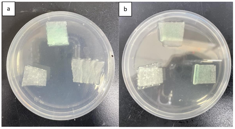
2.3. GA Vapor Cross-Linking

The cross-linking procedure was carried out with GA vapor in a sealed vessel. The co-electrospun GOx/Glu nanofibrous membrane was exposed to GA vapor prepared with 2 M GA solution containing 1 M HCl as a catalyst for cross-linking reaction in a 3:1 volume ratio GA:HCl for 12 h.

2.4. Characterization

The physical morphology of the co-electrospun GOx/Glu nanofibrous membrane and the cross-linked one was characterized by a field-emission scanning electron microscope (FE-SEM, JEOL JSM-6500 F, Taipei, Taiwan) after sputter coated with platinum for 80 s. The average nanofibers diameter distribution was measured by analyzing the SEM image using ImageJ software.

2.5. Hydrogen Peroxide Generation Detection and Sustainability Tests

GOx and Glu co-electrospun nanofibrous membrane was tested for its H2O2 generation capacity using two different colorimetric assays. Qualitatively, the generation of H2O2 was determined by the reaction with DA-64 and HRP to produce a green color. As for the H2O2 production sustainability, three different nanofibrous membranes (PVA only, PVA/GOX, and co-electrospun PVA (GOx/Glu)) without UV-sterilization pretreatment were placed on the surface of agar plate containing glucose for one week. TMB assay using 3,3′,5,5′-tetramethylbenzidine dye was employed to quantify the H2O2 concentration generated by the membrane. Briefly, reaction solution prepared by mixing sodium acetate buffer (0.1 M, pH 4.5), TMB solution (0.025 M in acetate buffer), and HRP (60 U/mL) with a volumetric ratio of 85:10:5. H2O2 solutions of different concentrations were added to the reaction solution for establishing a calibration curve. For the study of sustainable H2O2 production from the membrane, membrane samples (1 cm × 1 cm; ± 4 mg) were incubated in 20 mL DI water, and an aliquot of the sample solution (volume of reaction solution: sample solution = 10:1) was taken for H2O2 determination at different time. The stability of the membrane for H2O2 generation was investigated by storing the membranes in dry conditions at room temperature. At different times, several pieces of stored membranes were taken to incubate in DI water for 4 h and the H2O2 concentration in the water solution was then measured.

2.6. Antimicrobial Activity of the GOx Encapsulated PVA Nanofibrous Mat

Gram-negative (Escherichia coli) and Gram-positive (Staphylococcus aureus) were taken as model strains to examine the antimicrobial activity of the as-electrospun nanofibrous mat. E. coli was cultured in LB nutrient culture media and S. aureus was cultured in TSB culture media and both cultures were incubated with a shaker for 24 h at 37 °C. The antimicrobial activity of the nanofibrous mat was tested using the inhibition zone method where the bacterial concentration of OD600 = 0.5 inside PBS buffer was then plated on the agar plates. To determine the effect of glucose, four different agars (LB, LB with glucose supplement, TSB, TSB without glucose supplement) were used for the two bacteria. The nanofibrous membrane was then plated onto the agar plates and incubated for 24 h at 37 °C. Alternatively, an antimicrobial test via CFU/mL (viable cell counting) was made in order to ascertain the antimicrobial activity of the membranes quantitatively. In a 96-well plate, small cut-out pieces of the membranes (approx. 0.5 cm × 0.5 cm) were placed in six different wells for each bacteria and 0.1 mL of each bacterial solution was transferred into the wells. Six wells were used to measure three different contact times, which were 30 min, 1 h, and 2 h for each bacterial solution. After reaching the supposed contact times, the membranes were taken out of the solution, and serial dilution was conducted in PBS buffer in an attempt to reach countable amounts of bacterial colonies. One hundred microliters of this solution was then plated on each designated agar plate three times, which were then incubated for the same amount of time to the disk diffusion antimicrobial step. In this case, the antimicrobial activity values were expressed as CFU (dilution 10−4 and 10−6, three times each). Before the tests were conducted, all the as-electrospun membranes were sterilized with UV light for approximately 20 min.

3. Results and Discussion

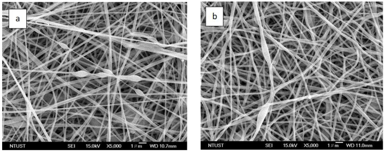
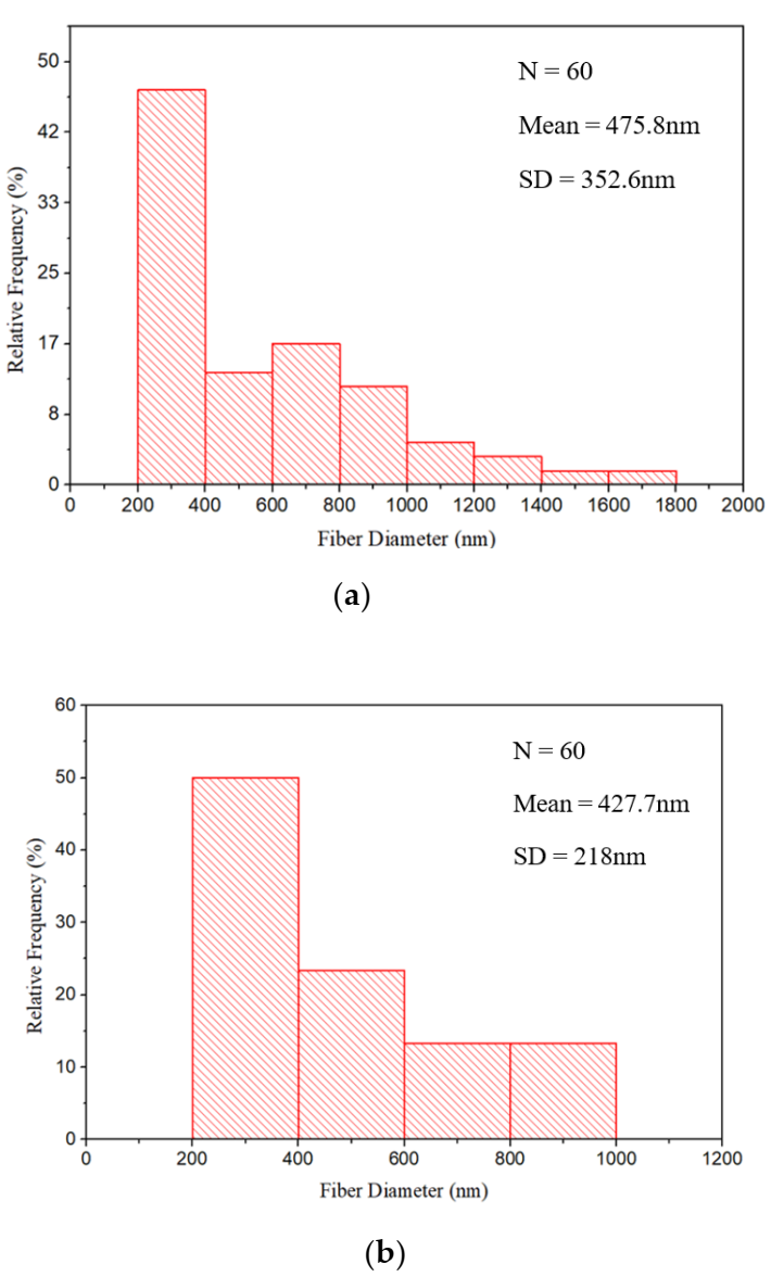
3.1. GOx Encapsulation

Due to their complex three-dimensional structure, size, and homogenous form-factor, most biologically active proteins (such as enzymes) are not easily electrospun into nanofibers. Most often, this issue is overcome by blending enzymes with an appropriate electrospinable polymer. PVA dope prepared in an aqueous solution has been demonstrated to be easily electrospun into nanofibers and used as an appropriate matrix to encapsulate enzymes, such as GOx due to its hydrophilicity, biocompatibility, and non-toxicity [39]. In addition, polymeric microencapsulation is highly beneficial for high protein loading and protection against denaturing agents [40]. As shown in Figure 2, the co-electrospinning of PVA/GOx and PVA/Glu dopes could produce a nanofibrous membrane consisting of fully entangled nanofibers. Since the feed streams of these two dopes were separated from each other, GOx and Glu nanofibers should be obtained as separated nanofibers, unable to react with each other to generate hydrogen peroxide in its dry state. The morphology of the as-prepared membrane was examined using FE-SEM, as shown in Figure 2, two noticeably different kinds of nanofibers could be observed. One is a straight thinner fiber, the other is a larger size fiber with shuttle-shaped beads. As shown in Figure 3a, fibers with a size of 200–400 nm consisted of 45% of the membrane. The other fibers have a much broader size distribution with a range of 400–1800 nm. Due to the complicated three-dimensional structure and strong inter- and intra-molecular forces of GOx, GOx/PVA dope will be electrospun into uneven fibers and often carries some shuttle-shaped beads structure [39]. The thinner size fibers resulted from Glu/PVA dope because PVA dope alone could always be electrospun into smooth fibers with a narrow size distribution. GA vapor crosslinking treatment was expected to prevent the PVA nanofibers from dissolving in water but it affected the morphology of the membrane. As demonstrated in our previous work [38], PVA crosslinked by GA has been confirmed by FTIR analysis. Figure 2b shows that PVA nanofibers curled up slightly and the shuttle-shaped beads on the PVA strings collapsed after GA vapor crosslinking (Supplementary Materials Figure S3). This possibly occurs due to the high GA concentration (2 M) used for the vapor-crosslinking. The acid-catalyzed GA crosslinking between the PVA chains compacts the three-dimensional network of PVA, which leads to a reduction in free volume between PVA chains for water absorption [41]. Shrinking results from crosslinking were shown by the reducing size of nanofibers appreciably, as shown in Figure 3b, where even though fibers with a size of 200–400 nm still consisted of ca 50% of the membrane, the size distribution was narrowed to 400–1000 nm. Evidently, PVA chains in electrospun nanofibers were indeed crosslinked leading to the shrinkage of nanofibers structure compared to the original ones.

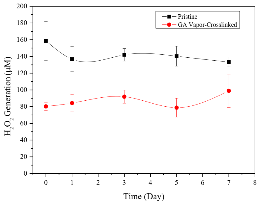
3.2. Sustained Release of Hydrogen Peroxide

The release of H2O2 from the pristine membrane, in which 3 mg/mL GOx and Glu were separately included in PVA dope for simultaneous electrospinning, was first qualitatively detected by colorimetric method using DA-64 as H2O2-sensitive dye. To handle the very light membrane, the membrane was placed on top of a paper towel which had been casted and dried with DA-64 and HRP mixture solution. Then, the membrane-loaded paper towel was placed on the surface of an agar plate. Moisture from agar would wet both paper towel and membrane so that Glu can be released to react with GOx to produce H2O2. H2O2 generated would then react with DA-64 with the help of HRP to produce Bindschedler’s green color [42] as shown in Figure 4. The color intensities developed on agar plates with and without additional supplement of 0.8% w/v glucose were compared. Evidently, the PVA-only membrane did not develop any colors on both plates. The GOx membrane, on the other hand, did not produce a green color (Figure 4a bottom right) on the glucose-free agar plate but did develop a green color on the glucose plate. In contrast, the pristine GOx/Glu membranes developed a green color on both agar plates which means H2O2 could be generated without supplementary Glu.
Quantitatively, generated H2O2 was also determined using TMB/HRP colorimetric assay due to its rapid visual detection of color development and needlessly sophisticated instrumentation [43]. The assay produced a blue-green colored solution with strong peak absorption at 650 nm (Supplementary Materials Figure S1). A calibration curve of H2O2 concentration was first established (Supplementary Materials Figure S2) before further experiments were conducted. In order to find the optimal ratio of GOx/Glu encapsulated in the membrane, the GOx amount was fixed at 3 mg/mL in PVA dope. The glucose concentration in the PVA dope was varied from 1 mg/mL to 5 mg/mL. The time courses of H2O2 released from the simultaneously electrospun pristine membranes into water solution are shown in Figure 5. It seems that the glucose loading concentration was proportional to the amount of H2O2 generated. After 8 h incubation, ca. 50 μM H2O2 was released from 1 mg/mL Glu membrane while 150 μM was obtained for 3 mg/mL Glu membranes. With the glucose concentration increased to 5 mg/mL, a faster initial H2O2 releasing rate was obtained. However, a limiting H2O2 level ca 150 μM was achieved after 4 h. In other words, 3 and 5 mg/mL Glu membranes possessed a similar H2O2 generation capacity. Probably, the dissolved oxygen concentration limited the GOx reaction when a higher glucose concentration was employed as in the case of 5 mg/mL Glu membrane [44]. Therefore, 3 mg/mL Glu membrane was chosen for further self-sustained H2O2 generation studies. In order to release H2O2 from the membrane and sustain this for a longer time, the pristine membrane was furtherly crosslinked with GA vapor. As expected, as shown in Figure 5, the H2O2 releasing rate was significantly reduced after crosslinking, approximately 100 µm H2O2 was obtained after 8 h incubation. After 24 h, the crosslinked membrane still could produce H2O2 to 160 µM. Evidently, GA vapor crosslinking did not damage GOx activity and the crosslinked PVA structure prevented the membrane from dissolving when in contact with water. The crosslinked PVA chains also slowed down the diffusion of glucose allowing H2O2 to be produced for a longer time.
The stability of H2O2 generation from GOx/Glu membrane stored at room temperature is shown in Figure 6. The membrane of equal size was incubated with an aqueous solution for 4 h before the H2O2 concentration was measured. The membrane showed a very stable capacity for H2O2 generation in which approximately the same level of H2O2 (~140 μM) was obtained after 7 days stored at room temperature. The GA-crosslinked membrane also showed similar stability but with a lower level of H2O2 (~90 μM) production. Apparently, the lower level of H2O2 obtained resulted from the GA crosslinking that slowed down the mass transfer rate of glucose, and therefore, reduced the H2O2 generation.

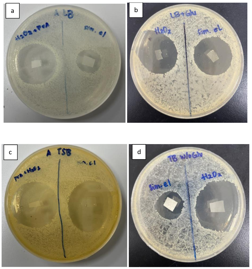
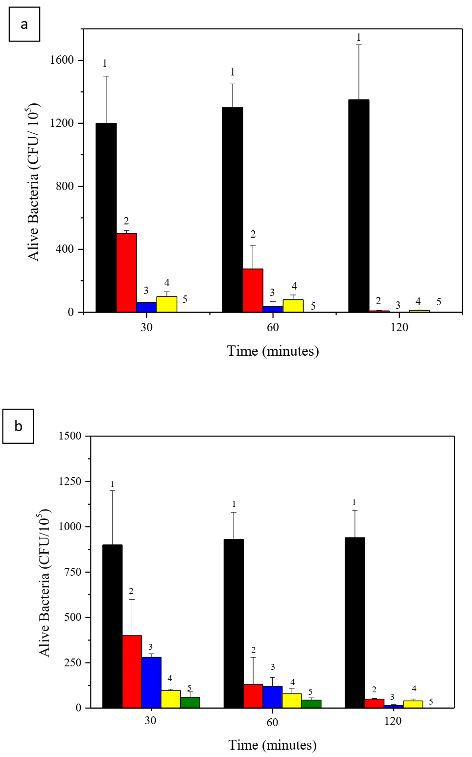
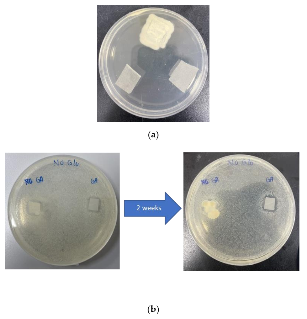
3.3. Antimicrobial Activity

The antimicrobial activity of the pristine GOx/Glu nanofibrous membrane was assessed and showed strong antimicrobial activity against Gram-negative (Escherichia coli; BL21) and Gram-positive (Staphylococcus aureus; ATCC65389) bacteria as shown by the disk diffusion inhibition assay. As shown in Figure 7, the antimicrobial activity of the GOx/Glu membrane resulted in large inhibition zones for both bacteria. As a reference for the antimicrobial activity of the membrane, 1% of H2O2 was cast into the PVA-only membrane side by side with the sample membranes. In the normal LB plate which contained no glucose, the GOx/Glu membrane displayed an inhibition zone against E. coli much smaller than the reference 1% H2O2 membrane (34 mm vs 20 mm; Figure 7a). With a supplement of 0.8% w/v glucose in the LB plate, the sample membrane demonstrated a similar size of inhibition zone as the reference membrane (32.5 mm vs 30 mm; Figure 7b). This shows that with the supplemented glucose the pristine GOx/Glu membrane has the same antimicrobial activity as 1% H2O2 against E. coli. For the antimicrobial activity assay against Gram-positive S. aureus, TSB agar is often used, however, 0.8% w/v or more glucose is usually included. In the normal TSB plate containing glucose, the GOx/Glu membrane displayed an inhibition zone with a size similar to that of the 1% H2O2 reference membrane (Figure 7c). In contrast, the size of the inhibition zone of the GOx/Glu membrane against S. aureus was much smaller than that of the reference H2O2 membrane on the TSB plate without glucose supplement (23 mm vs 37.5 mm; Figure 7d). Again, this demonstrates that the GOx/Glu membrane does possess strong antimicrobial activity against both Gram-positive and Gram-negative bacteria but the supplemental glucose from the surrounding environment could significantly enhance its antimicrobial activity with a capability equivalent to that of 1% H2O2.
The quantitative analysis of antimicrobial activity was also assessed through viable cell forming units (CFU/mL) reduction. To investigate the antimicrobial sustainability of the membranes, bacteria were cultured under shaking in contact with the membranes for 30 m, 1 h, and 2 h. Glucose of 200 mg/dL in PBS was also supplemented as a culture solution to emphasize the possibility of treating diabetic patients. The initial bacterial concentration was OD = 0.5, the sampled bacteria suspensions were serially diluted in PBS buffer until 10−4 and 10−6 dilution rate (three times each). As shown in Figure 8, the CFU of both bacteria decreased significantly after contacting the membranes for 30 min. As incubation time increased, the remaining CFU in solution furtherly decreased. It seems that S. aureus is more susceptible to H2O2 to such an extent that nearly no viable cells (CFU) could be detected after 30 min for the pristine membrane with supplemented glucose in solution (Figure 8a). In contrast, appreciable amounts of E. coli remained alive after 30 min. As expected, the pristine membrane without GA crosslinking is most effective for killing both bacteria in the presence of supplemented glucose. This is because without crosslinking, GOx would effectively release into the culture suspension with time and react with the supplemented glucose to generate H2O2 for killing bacteria. As for the GA-crosslinked membrane, even in the environment without supplemented glucose, its antimicrobial activity was still extraordinary in which nearly all viable S. aureus were killed after 2 h. However, for the Gram-negative E. coli cells, there were still small amounts of viable cells remaining. Supplemented glucose in the bacteria suspensions contributed a significantly enhanced antimicrobial activity of the crosslinked membranes against S. aureus (>80% reduction in CFU) as compared with E. coli (<20% reduction) for the incubation time of 30 min and 1 h. Gram-positive bacteria have only one cell membrane layer which makes them more vulnerable to antimicrobial agents as compared with Gram-negative bacteria which have an inner and outer cell membrane that afford them better protection against antimicrobial agents. The same antimicrobial trends of these two types of bacteria against GOx-based nanoparticles and direct H2O2 disinfectants have also been reported previously [45].
For the purpose of antimicrobial sustainability tests, pristine GOx/Glu membrane and PVA-only membrane without further sterilization were placed on top of an agar surface. To enhance and confirm the antimicrobial activity, hands were also not washed before handling these membranes. As shown in Figure 9, after 3 days of incubation, microorganisms started to grow on top of the PVA-only membrane while the other two pristine membranes showed no sign of microorganism growth even after 2 weeks of placement. This again showed that the continuous production of H2O2 from the GOx/Glu membrane can sterilize the membrane itself. Additionally, the sustainability effect of GA crosslinking for the GOx/Glu membrane was also analyzed. Initially, inhibition zones were observed for both pristine and GA-crosslinked membranes. Interestingly, contaminating microorganisms could grow (2 weeks after testing) on the pristine membrane (without GA crosslinking), though possessing a larger initial inhibition zone. In contrast, the GA-crosslinked membrane did not show any contamination and the inhibition zone could still be clearly observed. This shows that the release of in situ-generated H2O2 from the crosslinked membrane can last for a longer time to protect the membrane from contamination. On the other hand, the pristine membrane could generate H2O2 faster for bacterial killing but once H2O2 was exhausted it could not protect itself from bacteria contamination.
In detail, the antimicrobial activity of the as-prepared nanofibrous membrane was mainly due to cytotoxicity of in situ reaction products of GOx catalyzed glucose oxidation. In this nanofibrous membrane antimicrobial system, glucose can be offered either in situ or supplemented externally. Glucose supplemented from the surrounding can enhance H2O2 generation from the system. Therefore, the GOx/Glu membrane would be useful not only for a normal wound dressing but also a more potent antimicrobial wound dressing for diabetic patients who usually have a higher level of blood sugar. The H2O2 generated from the membrane will act as a reactive oxygen species (ROS), producing hydroxyl free radicals (•OH), attacking crucial cellular constituents, such as proteins, lipids, and nucleic acids, and thereby killing the bacterial cells through peroxidation and disruption of cell membranes.

4. Conclusions

A self-sustained hydrogen peroxide-generating membrane was successfully pre-pared by separately encapsulating GOx and glucose in PVA nanofibers via simultaneous electrospinning of PVA/GOx and PVA/Glu dope. GA vapor cross-linking made the membrane water-resistance and slowed down its H2O2 releasing rate. The H2O2 production level in an aqueous solution could be well maintained even after 7 days of incubation of the dry membrane at room temperature. Because of its extremely high specific surface area, PVA nanofibrous membrane could respond swiftly to an aqueous environment, and as a result, glucose was released from PVA nanofibers and reacted with encapsulated GOx to generate H2O2. The sustained release of H2O2 from the membrane was shown to possess strong antimicrobial activity against Gram(−) E. coli and Gram(+) S. aureus, with and without the presence of glucose in their environment. In addition, the sustained release of H2O2 also demonstrated that the as-prepared membrane was self-sterilizable and that most microorganisms contaminated on the membrane could be effectively killed continuously. This GOx/Glu membrane may find potential application as versatile antimicrobial materials in the field of biomedical, food, and health industries, especially in facing challenges relating to wound healing in diabetic patients.

Supplementary Materials

The following are available online at https://www.mdpi.com/article/10.3390/membranes11120997/s1, Figure S1: UV-vis absorption spectra of HRP-TMB solution containing different H2O2 concentrations and samples, Figure S2: Standard curve showing the relation between H2O2 concentration and absorbance at λ = 650 nm, Figure S3: FE-SEM micrographs of simultaneously co-electrospun GOx/Glu nanofibrous membrane; (a): before GA vapor crosslinking, (b): after GA Vapor crosslinking under lower magnification.

Author Contributions

Conceptualization, C.-K.L.; Investigation F.L. and C.-K.L.; Writing original draft preparation, F.L. and C.-K.L.; Review and editing, F.L. and C.-K.L.; Resources and supervision, C.-K.L. All authors have read and agreed to the published version of the manuscript.

Funding

This research received no external funding.

Acknowledgments

Author Fernaldy Leonarta is greatly indebted to Department of Chemical Engineering of National Taiwan University of Science and Technology for the graduate study scholarship grant.

Conflicts of Interest

The authors declare no conflict of interest.

References

  1. Hanušová, K.; Vápenka, L.; Dobiáš, J.; Mišková, L. Development of Antimicrobial Packaging Materials with Immobilized Glucose Oxidase and Lysozyme. Cent. Eur. J. Chem. 2013, 11, 1066–1078. [Google Scholar] [CrossRef]
  2. Thallinger, B.; Prasetyo, E.N.; Nyanhongo, G.S.; Guebitz, G.M. Antimicrobial Enzymes: An Emerging Strategy to Fight Microbes and Microbial Biofilms. Biotechnol. J. 2013, 8, 97–109. [Google Scholar] [CrossRef] [PubMed]
  3. Fuglsang, C.C.; Johansen, C.; Christgau, S.; Adler-Nissen, J. Antimicrobial Enzymes: Applications and Future Potential in the Food Industry. Trends Food Sci. Technol. 1995, 6, 390–396. [Google Scholar] [CrossRef]
  4. Zia, M.A.; Riaz, A.; Rasul, S.; Abbas, R.Z. Evaluation of Antimicrobial Activity of Glucose Oxidase from Aspergillus Niger EBL-A and Penicillium Notatum. Braz. Arch. Biol. Technol. 2013, 56, 956–961. [Google Scholar] [CrossRef] [Green Version]
  5. Bankar, S.B.; Bule, M.V.; Singhal, R.S.; Ananthanarayan, L. Glucose Oxidase—An Overview. Biotechnol. Adv. 2009, 27, 489–501. [Google Scholar] [CrossRef]
  6. Tiina, M. Antibacterial Effect of the Glucose Oxidase-Glucose System on Food-Poisoning Organisms. Int. J. Food Microbiol. 1989, 8, 165–174. [Google Scholar] [CrossRef]
  7. Ríos-Castillo, A.G.; González-Rivas, F.; Rodríguez-Jerez, J.J. Bactericidal Efficacy of Hydrogen Peroxide-Based Disinfectants Against Gram-Positive and Gram-Negative Bacteria on Stainless Steel Surfaces. J. Food Sci. 2017, 82, 2351–2356. [Google Scholar] [CrossRef]
  8. Ramachandran, S.; Fontanille, P.; Pandey, A.; Larroche, C. Gluconic Acid: Properties, Applications and Microbial Production. Food Technol. Biotechnol. 2006, 44, 185–195. [Google Scholar]
  9. Zhao, L.; Wang, L.; Zhang, Y.; Xiao, S.; Bi, F.; Zhao, J.; Gai, G.; Ding, J. Glucose Oxidase-Based Glucose-Sensitive Drug Delivery for Diabetes Treatment. Polymers 2017, 9, 255. [Google Scholar] [CrossRef]
  10. Anusha, J.R.; Raj, C.J.; Cho, B.-B.; Fleming, A.T.; Yu, K.-H.; Kim, B.C. Amperometric Glucose Biosensor Based on Glucose Oxidase Immobilized over Chitosan Nanoparticles from Gladius of Uroteuthis Duvauceli. Sens. Actuators B Chem. 2015, 215, 536–543. [Google Scholar] [CrossRef]
  11. Liu, Y.; Nan, X.; Shi, W.; Liu, X.; He, Z.; Sun, Y.; Ge, D. A Glucose Biosensor Based on the Immobilization of Glucose Oxidase and Au Nanocomposites with Polynorepinephrine. RSC Adv. 2019, 9, 16439–16446. [Google Scholar] [CrossRef] [Green Version]
  12. Liu, X.; Cai, Z.; Gao, N.; Ye, S.; Tao, T.; He, H.; Chang, G.; He, Y. Controllable Preparation of (200) Facets Preferential Oriented Silver Nanowires for Non-Invasive Detection of Glucose in Human Sweat. Smart Mater. Med. 2021, 2, 150–157. [Google Scholar] [CrossRef]
  13. Martins, R.F.; Plieva, F.M.; Santos, A.; Hatti-Kaul, R. Integrated Immobilized Cell Reactor-Adsorption System for β-Cyclodextrin Production: A Model Study Using PVA-Cryogel Entrapped Bacillus Agaradhaerens Cells. Biotechnol. Lett. 2003, 25, 1537–1543. [Google Scholar] [CrossRef]
  14. Soto, J.; Hughes, T.; Li, Y.S. Silicon-Based Glucose Oxidase Working Electrode for Glucose Sensing. ACS Omega 2019, 4, 18312–18316. [Google Scholar] [CrossRef] [Green Version]
  15. Hecht, H.J.; Schomburg, D.; Kalisz, H.; Schmid, R.D. The 3D Structure of Glucose Oxidase from Aspergillus Niger. Implications for the Use of GOD as a Biosensor Enzyme. Biosens. Bioelectron. 1993, 8, 197–203. [Google Scholar] [CrossRef]
  16. Wang, Z.-G.; Wan, L.-S.; Liu, Z.-M.; Huang, X.-J.; Xu, Z.-K. Enzyme Immobilization on Electrospun Polymer Nanofibers: An Overview. J. Mol. Catal. B Enzym. 2009, 56, 189–195. [Google Scholar] [CrossRef]
  17. Puttananjegowda, K.; Taksi, A.; Thomas, S. Perspective—Electrospun Nanofibrous Structures for Electrochemical Enzymatic Glucose Biosensing: A Perspective. J. Electrochem. Soc. 2020, 167, 037553. [Google Scholar] [CrossRef]
  18. Guilbault, G.G.; Lubrano, G.J. An Enzyme Electrode for the Amperometric Determination of Glucose. Anal. Chim. Acta 1973, 64, 439–455. [Google Scholar] [CrossRef]
  19. Senthamizhan, A.; Balusamy, B.; Uyar, T. Glucose Sensors Based on Electrospun Nanofibers: A Review. Anal. Bioanal. Chem. 2016, 408, 1285–1306. [Google Scholar] [CrossRef]
  20. Zhao, Y.-T.; Zhang, J.; Gao, Y.; Liu, X.-F.; Liu, J.-J.; Wang, X.-X.; Xiang, H.-F.; Long, Y.-Z. Self-Powered Portable Melt Electrospinning for in Situ Wound Dressing. J. Nanobiotechnol. 2020, 18, 111. [Google Scholar] [CrossRef]
  21. Massarelli, E.; Silva, D.; Pimenta, A.F.R.; Fernandes, A.I.; Mata, J.L.G.; Armês, H.; Salema-Oom, M.; Saramago, B.; Serro, A.P. Polyvinyl Alcohol/Chitosan Wound Dressings Loaded with Antiseptics. Int. J. Pharm. 2021, 593, 120110. [Google Scholar] [CrossRef]
  22. Arampatzis, A.S.; Kontogiannopoulos, K.N.; Theodoridis, K.; Aggelidou, E.; Rat, A.; Willems, A.; Tsivintzelis, I.; Papageorgiou, V.P.; Kritis, A.; Assimopoulou, A.N. Electrospun Wound Dressings Containing Bioactive Natural Products: Physico-Chemical Characterization and Biological Assessment. Biomater. Res. 2021, 25, 23. [Google Scholar] [CrossRef]
  23. Azimi, B.; Maleki, H.; Zavagna, L.; de la Ossa, J.G.; Linari, S.; Lazzeri, A.; Danti, S. Bio-Based Electrospun Fibers for Wound Healing. J. Funct. Biomater. 2020, 11, 67. [Google Scholar] [CrossRef]
  24. Samadian, H.; Zamiri, S.; Ehterami, A.; Farzamfar, S.; Vaez, A.; Khastar, H.; Alam, M.; Ai, A.; Derakhshankhah, H.; Allahyari, Z.; et al. Electrospun Cellulose Acetate/Gelatin Nanofibrous Wound Dressing Containing Berberine for Diabetic Foot Ulcer Healing: In Vitro and in Vivo Studies. Sci. Rep. 2020, 10, 8312. [Google Scholar] [CrossRef]
  25. Huang, C.-L.; Lee, K.-M.; Liu, Z.-X.; Lai, R.-Y.; Chen, C.-K.; Chen, W.-C.; Hsu, J.-F. Antimicrobial Activity of Electrospun Polyvinyl Alcohol Nanofibers Filled with Poly[2-(Tert-Butylaminoethyl) Methacrylate]-Grafted Graphene Oxide Nanosheets. Polymers 2020, 12, 1449. [Google Scholar] [CrossRef]
  26. Wei, Z.; Wang, L.; Zhang, S.; Chen, T.; Yang, J.; Long, S.; Wang, X. Electrospun Antibacterial Nanofibers for Wound Dressings and Tissue Medicinal Fields: A Review. J. Innov. Opt. Health Sci. 2020, 13, 2030012. [Google Scholar] [CrossRef]
  27. Mendes, L.G.; Ferreira, F.V.; Sielski, M.S.; Livi, S.; Rocco, S.A.; Sforça, M.L.; Burga-Sánchez, J.; Vicente, C.P.; Mei, L.H.I. Electrospun Nanofibrous Architectures of Thrombin-Loaded Poly(Ethylene Oxide) for Faster in Vivo Wound Clotting. ACS Appl. Bio Mater. 2021, 4, 5240–5250. [Google Scholar] [CrossRef]
  28. Gao, C.; Zhang, L.; Wang, J.; Jin, M.; Tang, Q.; Chen, Z.; Cheng, Y.; Yang, R.; Zhao, G. Electrospun Nanofibers Promote Wound Healing: Theories, Techniques, and Perspectives. J. Mater. Chem. B 2021, 9, 3106–3130. [Google Scholar] [CrossRef] [PubMed]
  29. Liu, X.; Xu, H.; Zhang, M.; Yu, D.-G. Electrospun Medicated Nanofibers for Wound Healing: Review. Membranes 2021, 11, 770. [Google Scholar] [CrossRef] [PubMed]
  30. Rivera-Hernández, G.; Antunes-Ricardo, M.; Martínez-Morales, P.; Sánchez, M.L. Polyvinyl Alcohol Based-Drug Delivery Systems for Cancer Treatment. Int. J. Pharm. 2021, 600, 120478. [Google Scholar] [CrossRef]
  31. Jensen, B.E.B.; Dávila, I.; Zelikin, A.N. Poly(Vinyl Alcohol) Physical Hydrogels: Matrix-Mediated Drug Delivery Using Spontaneously Eroding Substrate. J. Phys. Chem. B 2016, 120, 5916–5926. [Google Scholar] [CrossRef]
  32. Li, J.K.; Wang, N.; Wu, X.S. Poly(Vinyl Alcohol) Nanoparticles Prepared by Freezing–Thawing Process for Protein/Peptide Drug Delivery. J. Control. Release 1998, 56, 117–126. [Google Scholar] [CrossRef]
  33. Piacentini, E.; Bazzarelli, F.; Poerio, T.; Albisa, A.; Irusta, S.; Mendoza, G.; Sebastian, V.; Giorno, L. Encapsulation of Water-Soluble Drugs in Poly (Vinyl Alcohol) (PVA)- Microparticles via Membrane Emulsification: Influence of Process and Formulation Parameters on Structural and Functional Properties. Mater. Today Commun. 2020, 24, 100967. [Google Scholar] [CrossRef]
  34. Karim, A.A.; Tan, E.; Loh, X.J. Encapsulation of Vitamin C with Its Protection from Oxidation by Poly(Vinyl Alcohol). J. Mol. Eng. Mater. 2017, 5, 1750013. [Google Scholar] [CrossRef]
  35. Jia, Y.-T.; Gong, J.; Gu, X.-H.; Kim, H.-Y.; Dong, J.; Shen, X.-Y. Fabrication and Characterization of Poly (Vinyl Alcohol)/Chitosan Blend Nanofibers Produced by Electrospinning Method. Carbohydr. Polym. 2007, 67, 403–409. [Google Scholar] [CrossRef]
  36. Wu, L.; Yuan, X.; Sheng, J. Immobilization of Cellulase in Nanofibrous PVA Membranes by Electrospinning. J. Membr. Sci. 2005, 250, 167–173. [Google Scholar] [CrossRef]
  37. Hulupi, M.; Haryadi, H. Synthesis and Characterization of Electrospinning PVA Nanofiber-Crosslinked by Glutaraldehyde. Mater. Today: Proc. 2019, 13, 199–204. [Google Scholar] [CrossRef]
  38. Destaye, A.G.; Lin, C.-K.; Lee, C.-K. Glutaraldehyde Vapor Cross-Linked Nanofibrous PVA Mat with in Situ Formed Silver Nanoparticles. ACS Appl. Mater. Interfaces 2013, 5, 4745–4752. [Google Scholar] [CrossRef]
  39. Ren, G.; Xu, X.; Liu, Q.; Cheng, J.; Yuan, X.; Wu, L.; Wan, Y. Electrospun Poly(Vinyl Alcohol)/Glucose Oxidase Biocomposite Membranes for Biosensor Applications. React. Funct. Polym. 2006, 66, 1559–1564. [Google Scholar] [CrossRef]
  40. Wang, Y.; Hsieh, Y.L. Immobilization of Lipase Enzyme in Polyvinyl Alcohol (PVA) Nanofibrous Membranes. J. Membr. Sci. 2008, 309, 73–81. [Google Scholar] [CrossRef]
  41. Long, J.; Nand, A.V.; Bunt, C.; Seyfoddin, A. Controlled Release of Dexamethasone from Poly(Vinyl Alcohol) Hydrogel. Pharm. Dev. Technol. 2019, 24, 839–848. [Google Scholar] [CrossRef]
  42. Begander, B.; Huber, A.; Döring, M.; Sperl, J.; Sieber, V. Development of an Improved Peroxidase-Based High-Throughput Screening for the Optimization of D-Glycerate Dehydratase Activity. Int. J. Mol. Sci. 2020, 21, 335. [Google Scholar] [CrossRef] [PubMed] [Green Version]
  43. Chen, Y.; Zhong, Q.; Wang, Y.; Yuan, C.; Qin, X.; Xu, Y. Colorimetric Detection of Hydrogen Peroxide and Glucose by Exploiting the Peroxidase-like Activity of Papain. RSC Adv. 2019, 9, 16566–16570. [Google Scholar] [CrossRef] [Green Version]
  44. Nicol, M.J.; Duke, F.R. Substrate Inhibition with Glucose Oxidase. J. Biol. Chem. 1966, 241, 4292–4293. [Google Scholar] [CrossRef]
  45. Lee, I.; Cheon, H.J.; Adhikari, M.D.; Tran, T.D.; Yeon, K.-M.; Kim, M.I.; Kim, J. Glucose Oxidase-Copper Hybrid Nanoflowers Embedded with Magnetic Nanoparticles as an Effective Antibacterial Agent. Int. J. Biol. Macromol. 2020, 155, 1520–1531. [Google Scholar] [CrossRef] [PubMed]
Scheme 1. Schematic representation of GOx catalytic reaction with glucose for in-situ generation of hydrogen peroxide as antimicrobial agent.
Membranes 11 00997 sch001
Figure 1. Simultaneous electrospinning of GOx/PVA and Glu/PVA interwoven nanofibrous membrane.
Membranes 11 00997 g001
Figure 2. FE-SEM micrographs of simultaneously co-electrospun PVA/GOx-PVA/Glu nanofibrous membrane; (a) before GA vapor crosslinking; (b) after GA vapor crosslinking.
Membranes 11 00997 g002
Figure 3. Diameter size distribution of nanofibers consisted of the simultaneous electrospun nanofibrous membrane; (a) before GA vapor-crosslinking; (b) after GA vapor-crosslinking.
Membranes 11 00997 g003
Figure 4. Qualitative test of H2O2 generation from pristine membranes; (top) co-electrospun GOx/Glu/PVA; (bottom left) PVA only; (bottom right) PVA/Gox. DA-64 and HRP colorimetric assay employed for producing Bindschedler’s green on agar plate (a) without; (b) with 0.8% glucose supplement in the agar.
Membranes 11 00997 g004
Figure 5. Effect of glucose concentration loaded in Glu/PVA dope and GA crosslinking on the time course of H2O2 generation from the GOx/Glu membranes.
Membranes 11 00997 g005
Figure 6. Stability GOx encapsulated in PVA nanofibrous membrane (with and without vapor-crosslinking) with Glu 3 mg/mL in Glu/PVA dope as measured by its hydrogen peroxide generation capacity at 4 h reaction time after being kept at room temperature for different days.
Membranes 11 00997 g006
Figure 7. Inhibition zone against Escherichia coli (a) without; (b) with glucose supplement in agar plate, and Staphylococcus aureus (c) with glucose; (d) without glucose supplement in agar plate. Addition of 1% H2O2 solution onto PVA-only membranes were employed as antimicrobial reference.
Membranes 11 00997 g007
Figure 8. Effect of GA crosslinking and extra glucose addition in culture medium on colony-forming units (CFU) of bacteria cultured with membrane samples for different times; 1: control; 2: crosslinked/Glu (−); 3: crosslinked/Glu (+); 4: pristine/Glu (−); 5: pristine/Glu (+); (a) Staphylococcus aureus; (b) Escherichia coli.
Membranes 11 00997 g008
Figure 9. Self-sustained antimicrobial activity test of unsterilized nanofibrous membranes. (a) Pristine membrane (bottom left and right) compared with PVA-only membrane (top); (b) GA-crosslinking effect on sterilization of membrane after 2 weeks incubation on agar plate.
Membranes 11 00997 g009
Publisher’s Note: MDPI stays neutral with regard to jurisdictional claims in published maps and institutional affiliations.

