Review

Oxylipins and Reactive Carbonyls as Regulators of the Plant Redox and Reactive Oxygen Species Network under Stress

Biochemistry and Physiology of Plants, Faculty of Biology, Bielefeld University, 33615 Bielefeld, Germany
*
Author to whom correspondence should be addressed.
Antioxidants 2023, 12(4), 814; https://doi.org/10.3390/antiox12040814
Submission received: 1 March 2023 / Revised: 20 March 2023 / Accepted: 22 March 2023 / Published: 27 March 2023
(This article belongs to the Special Issue Antioxidant Research in Germany)

Abstract

:
Reactive oxygen species (ROS), and in particular H2O2, serve as essential second messengers at low concentrations. However, excessive ROS accumulation leads to severe and irreversible cell damage. Hence, control of ROS levels is needed, especially under non-optimal growth conditions caused by abiotic or biotic stresses, which at least initially stimulate ROS synthesis. A complex network of thiol-sensitive proteins is instrumental in realizing tight ROS control; this is called the redox regulatory network. It consists of sensors, input elements, transmitters, and targets. Recent evidence revealed that the interplay of the redox network and oxylipins–molecules derived from oxygenation of polyunsaturated fatty acids, especially under high ROS levels–plays a decisive role in coupling ROS generation and subsequent stress defense signaling pathways in plants. This review aims to provide a broad overview of the current knowledge on the interaction of distinct oxylipins generated enzymatically (12-OPDA, 4-HNE, phytoprostanes) or non-enzymatically (MDA, acrolein) and components of the redox network. Further, recent findings on the contribution of oxylipins to environmental acclimatization will be discussed using flooding, herbivory, and establishment of thermotolerance as prime examples of relevant biotic and abiotic stresses.

Graphical Abstract

1. Introduction

About 2.4 billion years ago, molecular oxygen was first introduced into the earth’s atmosphere by oxygenic photosynthesis [1]. Alongside respiratory and photosynthetic electron transport and relevant enzyme activities, reactive oxygen species (ROS), derivatives of O2, originated as permanently formed byproducts of metabolism. To survive and grow under fluctuating environmental conditions, especially in the context of ongoing climate change and global warming, plants have evolved a complex system of molecular mechanisms and pathways of sensing, transmitting, and responding to optimal acclimatization [2,3]. The key components are ROS, in particular superoxide anions (O2•−) and hydroxyl radicals (OH), which are free radicals containing an unpaired electron of varying reactivity, singlet oxygen (1O2), which is an excited non-radical derived from molecular oxygen by spin inversion, and hydrogen peroxide (H2O2), originating from O2•− dismutation or directly from two electron transfer reactions. These components accumulate under suboptimal growth conditions [2,4,5,6]. Generation of ROS occurs in plastids, peroxisomes, endoplasmic reticulum (ER), mitochondria, apoplast, and cytosol [2,3,4,5,6,7]. While they are generated constantly by housekeeping enzymes and as byproducts of metabolism, biotic and abiotic stresses enhance ROS synthesis distinctly [2,4,8]. Overall, 1–2% of the total oxygen consumed by plants has been proposed to account for ROS formation [9,10,11,12].
On the one hand, ROS serve as second messengers, modulating stress defense, hormone signaling, development, and growth processes. Resting H2O2 concentrations in plasmatic compartments are in the range of 10–30 nM [13,14]. On the other hand, ROS are highly reactive, hence posing a significant oxidative threat at high concentrations [2,4,5,6,7,15]. Therefore, plant cells monitor and modulate ROS levels vigorously to maintain their signaling function while preventing oxidative damage. The basic processes of this modulation of both ROS and all reactive molecular species (RMS) include regulation of (1) RMS synthesis, (2) RMS sensing and signal processing, and (3) RMS degradation [2,16,17]. These processes are implemented by the redox-regulatory network, which controls the total antioxidant capacity of the cell.

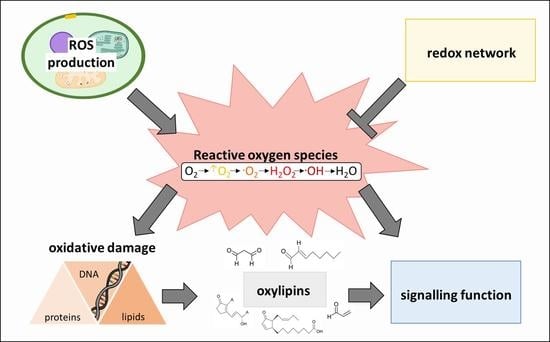
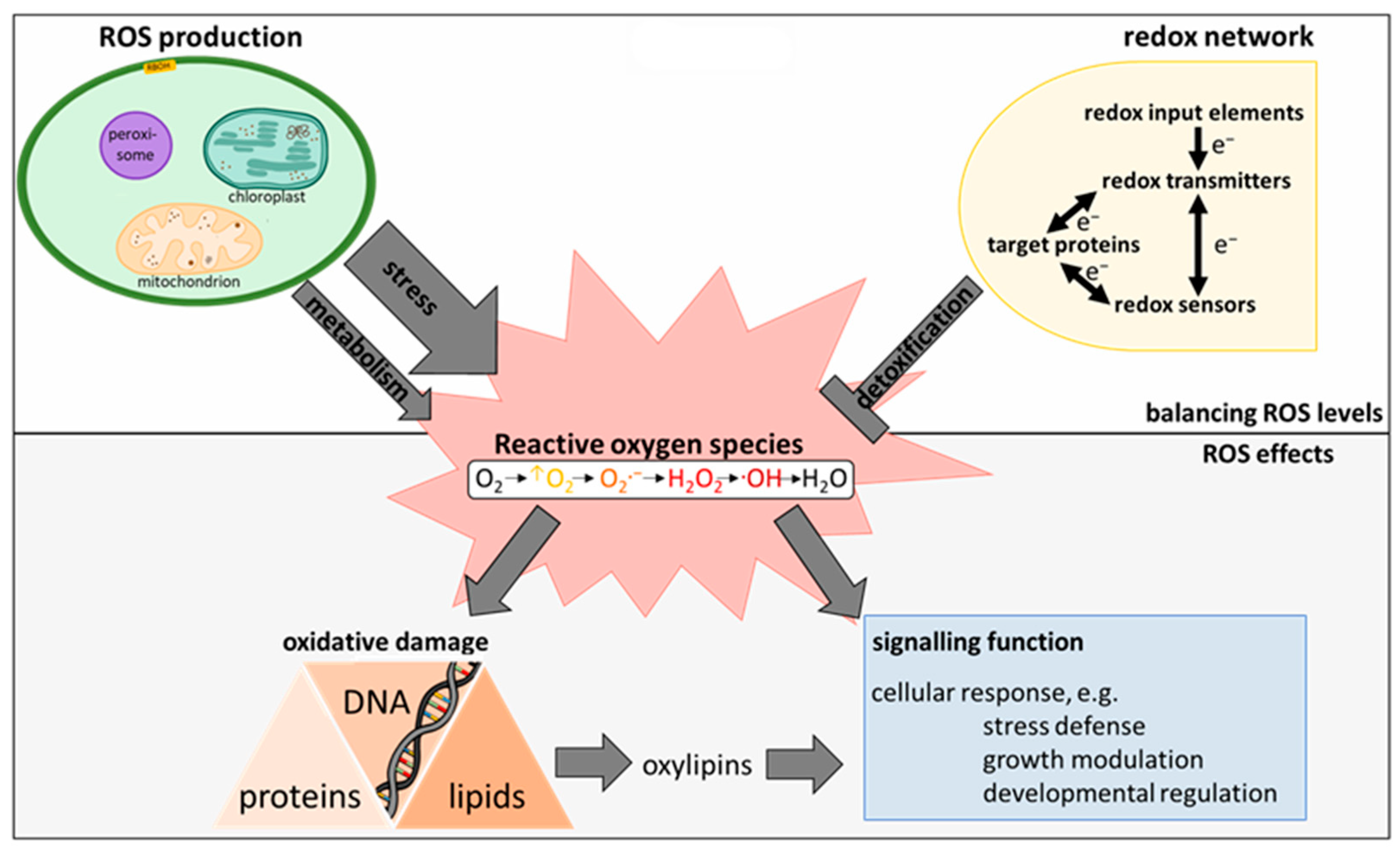
2. The Plant ROS and Redox Network

ROS homeostasis as influenced by ROS generation on the one hand and decomposition on the other shapes the way ROS affect plant cells and will therefore constitute the first chapter of this review. Subsequently, oxylipins, products of ROS-induced disruption of lipid membranes, will be introduced and their influence on the redox network characterized (Figure 1).

2.1. Synthesis of ROS in Higher Plants

The major organelles contributing to stress-induced ROS formation are chloroplasts, followed by mitochondria due to their electron transport chains [2,6,11]. For instance, the electron transport chain of cellular respiration is leaky, with an estimated percentage of 0.1% to 2% of electrons passing the chain being released, thereby causing generation of O2•− [5]. Plastid generation of O2 is achieved by the univalent reduction of molecular O2, primarily in the photosystem I [2,6]. O2•− does not only serve as a precursor for other ROS; it also impedes ROS scavenging by antioxidant enzymes (e.g., peroxidases) [6,8,11,18]. Further, regarding plastid ROS synthesis, 1O2, another common and highly reactive ROS, is produced in both photosystems by transfer of energy from excited chromophores to O2 [6]. In the apoplast, O2•- and H2O2 are formed by respiratory burst oxidase homologues (RBOHs), polyamine oxidases, pH-dependent peroxidases, and copper amine oxidases. O2•−, which is unstable and non-permeable to membranes, further undergoes protonation and dismutation and hence contributes to the extracellular H2O2 pool [5]. H2O2 is imported into the cell by aquaporins [2]. Additionally, H2O2 is directly produced by SOD-mediated or spontaneous dismutation of O2•− and by enzyme-catalyzed two electron transfer, e.g., by xanthine or glycolate oxidases, the latter as part of peroxisomal photorespiration [2,4,6]. As photorespiration is a major source of H2O2, contributing about 70% of generated H2O2 (as studied in wheat with a supposed flux of 10% of photosynthetic electrons to photorespiration), peroxisomes contribute distinctly to cellular oxidative metabolism [19,20,21]. Besides H2O2, peroxisomal metabolism supplies essential signaling molecules, such as reactive nitrogen species including nitric oxide (NO) and jasmonic acid (JA, see Section 3.1.2) [20]. More detailed insights into peroxisomal metabolism can be found in other reviews [20,21,22,23].
ROS can act locally, but they can also be transported to different organelles and serve as signals with maximal migration distances ranging from 1 nm (OH) to more than 1 µm (H2O2) [8,16]. Their levels in different cell compartments is adjusted by local synthesis in combination with import, export, and degradation differ between different stress conditions, creating unique ROS signatures [4].

2.2. ROS Homeostasis as Key Mechanism to Avoid Oxidative Stress

As oxidizing agents, ROS react with proteins, especially reduced and deprotonated thiolate residues. This alters catalytic activity of thiol-switch proteins which are essential in a plethora of plant processes, including metabolism and regulation of the antioxidant pool. In the case of H2O2, one of the most reactive ROS (only OH, the hydroxyl radical, is more reactive), reaction with thiolates leads to generation of sulfenic, sulfinic, and sulfonic acid derivatives; only the first two of these can be reversed (non-)enzymatically [2,8]. Furthermore, ROS can fragmentize peptide chains and increase protein aggregation and degradation [5]. As oxidation of proteins by ROS is essential for ROS sensing and signaling, a highly complex network of oxidation-sensitive proteins, the thiol-redox network, is dedicated to modulating ROS function and level. This network will further be characterized in Section 2.2.1.
Additional targets of ROS-induced oxidative damage are DNA and RNA; these are prone to modification of nucleotide bases and breakage of single- and double-strands, e.g., by rupture of nucleosomes [8,11].
Finally, they are linked to lipid peroxidation, thereby contributing to synthesis of oxylipins. Oxylipins are regulatory compounds derived from the oxidation of polyunsaturated fatty acids (PUFA) [8]. As ROS synthesis often occurs close to membranes, lipids are regarded as the primary target of ROS [9,24]. More detailed information on lipid peroxidation, subsequent formation of oxylipins, and their highly diverse functions in basic plant processes will be provided in Section 3 and 4 of this review.

2.2.1. Structure of the Redox-Regulatory Network

In general, the redox network can be divided hierarchically into several regulatory levels, starting with electron flow from metabolic and photosynthetic activity to redox input elements such as ferredoxin, NADPH, and γ-glutamyl-cysteinyl-glycine (glutathione, GSH). These input elements transfer electrons to redox transmitters, involving dedicated enzymes such as ferredoxin-thioredoxin reductases [25]. Redox transmitters reduce ROS sensors or target proteins as a next step. The group of target proteins includes a high variety of enzymes able to influence gene expression, metabolism, ROS detoxification, and protein turnover among other cellular processes [26]. Thiol switches (proteins characterized by redox-dependency of their catalytic or binding activity as based on cysteine residues) and thiol/disulfide exchange cascades are the main mechanistic elements of target proteins and the overall redox-regulatory network [2].
An example of a target protein is cyclophilin 20-3 (Cyp20-3), which contributes to thiol synthesis. Cyp20-3 is the only cyclophilin localized in the chloroplast stroma and acts as a regulatory hub between stress signaling of high light stress and wounding [27]. Under light conditions, Cyp20-3 is reduced by thioredoxins (TRX). In its reduced state, Cyp20-3 interacts with serine acetyl transferase 1 (SAT1), enabling formation of the cysteine synthase complex and subsequent production of the cysteine precursor O-acetyl serine [28,29,30]. Consequently, the cellular reduction state increases, changing the redox potential of the cell and allowing for alteration of gene expression and increased detoxification of ROS by peroxidases such as the ROS sensor 2-cysteine peroxiredoxin (2-CysPRX). Besides regulation of thiol synthesis, electrons from active Cyp20-3 can also be transferred to 2-CysPRX to enhance H2O2 detoxification [29].

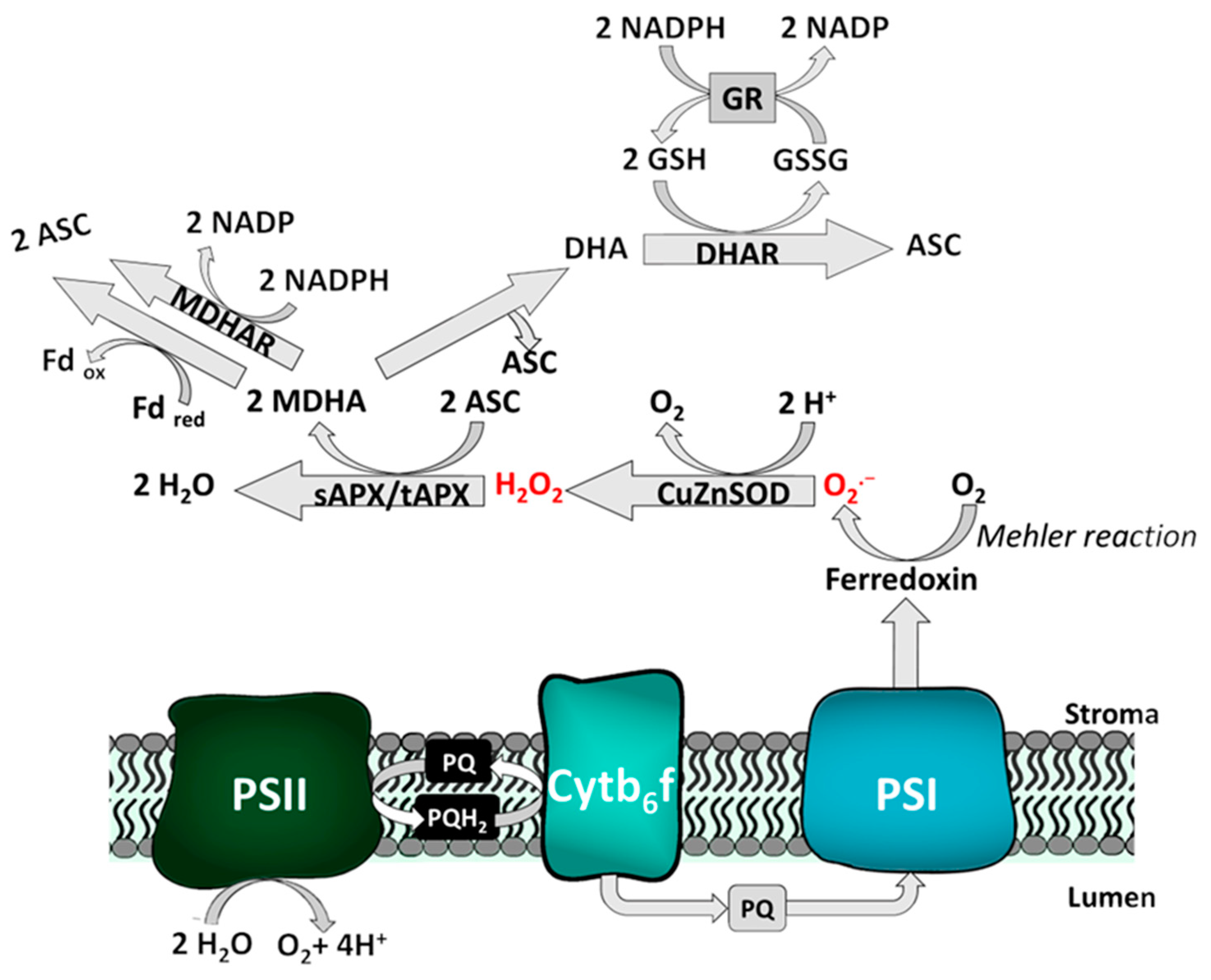
2.2.2. Exemplary Function of the Redox Network: The Water-Water-Cycle

An example of the interplay of ROS and the redox network is the plastid water-water-cycle, depicted in Figure 2. As previously mentioned, ROS generation in chloroplasts is especially high under non-optimal conditions due to univalent reduction of O2 by the redox input element ferredoxin in the course of the Mehler reaction [31,32]. The superoxide radical is rapidly converted into H2O2 by a (membrane-bound) Cu/Zn SOD, which acts as redox sensor [33,34]. Subsequently, H2O2 is detoxified by ascorbate peroxidases (APX) under consumption of ascorbate (ASC) [33,35]. The peroxidases, in this case, act as ROS scavenging enzymes; however, they additionally serve as redox sensors in plant cells [2]. Coupling of the water-water-cycle to the ascorbate-glutathione pathway (Asada-Halliwell pathway) ensures regeneration of oxidized dehydroascorbate [36]. Reduction of monodehydroascorbate (MDHA) occurs directly by ferredoxin oxidation or NADPH oxidation catalyzed by monodehydroascorbate reductase (MDHAR). Alternatively, MDHA can be further oxidized to didehydroascorbate (DHA), yielding one molecule of ascorbate. MDHA is then reduced to ASC by GSH-dependent dehydroascorbate reductase (DHAR); GSH, in turn, is regenerated by glutathione reductase (GR), using NADPH+ H+ [34].

2.3. Decomposition of ROS
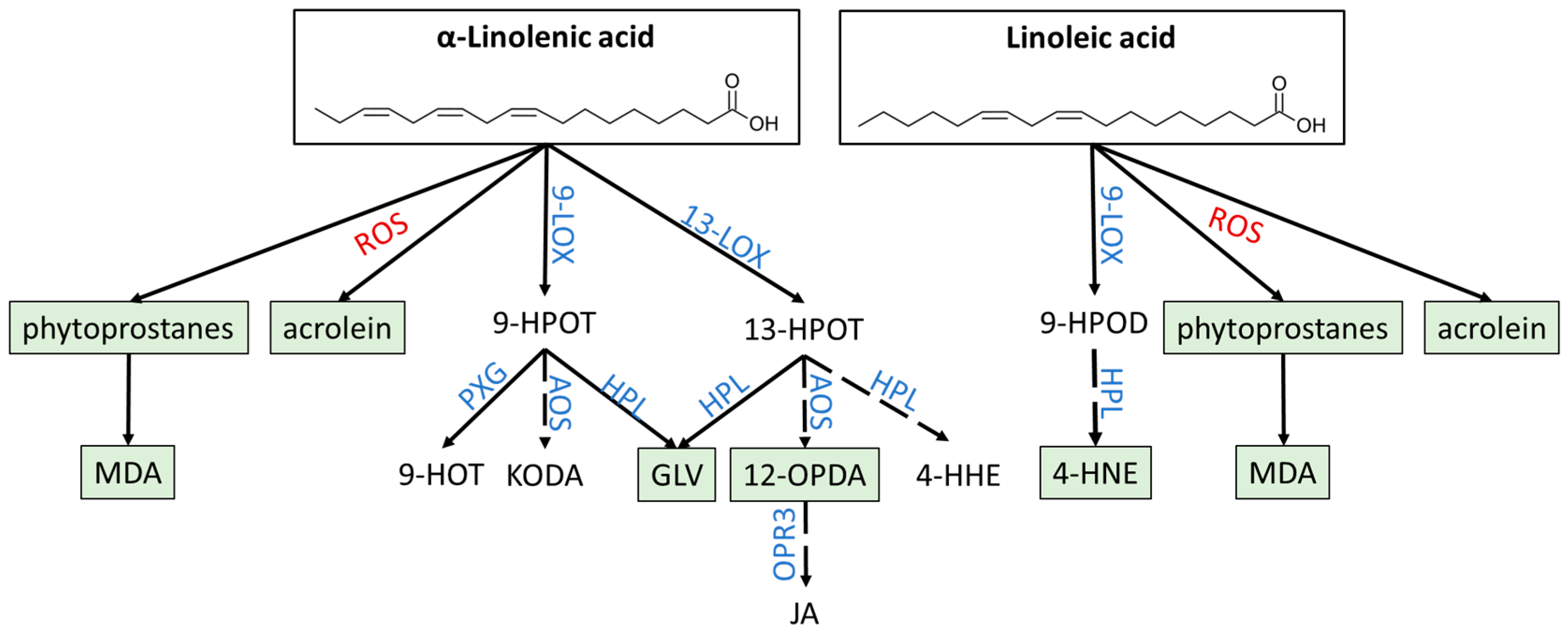
Due to their high reactivity, ROS typically have short half-life times ranging from 10−3 to 10−9 s [5,6,9,15,16,24,37]. Although dismutation of ROS also occurs spontaneously, dedicated enzymes as catalysts (e.g., SODs) increase the reaction speed by a factor of 104 [6,8]. Hence, detoxification of ROS by enzymatic antioxidants is achieved by a variety of stress-induced enzymes including SODs, catalases (CATs), and various peroxidases, including APXs, glutathione peroxidases (GPXs), glutathione-like peroxidases (GPXLs), and peroxiredoxins (PRX). Fine-tuning of ROS detoxification and, concomitantly, the ROS signature are realized by differential localization and the abundance of enzymes depending on developmental stage and tissue [6,8]. CATs, for instance, are thought to detoxify H2O2 in peroxisomes, but not in chloroplasts [6,8].
In addition, peroxidases serve as both a detoxification mechanism and as redox sensors; as such, they contribute distinctly to ROS signaling [2,4]. Key components of non-enzymatic ROS detoxification are GSH, ASC, carotenoids, tocopherols, and flavonoids [6,36]. As the primary ROS producer, chloroplasts are prone to oxidative damage. To counteract this threat, both enzymatic and non-enzymatic antioxidants accumulate in chloroplasts at high levels. Between 30% and 40% of the total cellular ASC content is stored in chloroplasts [6,11,38].

3. Oxylipins

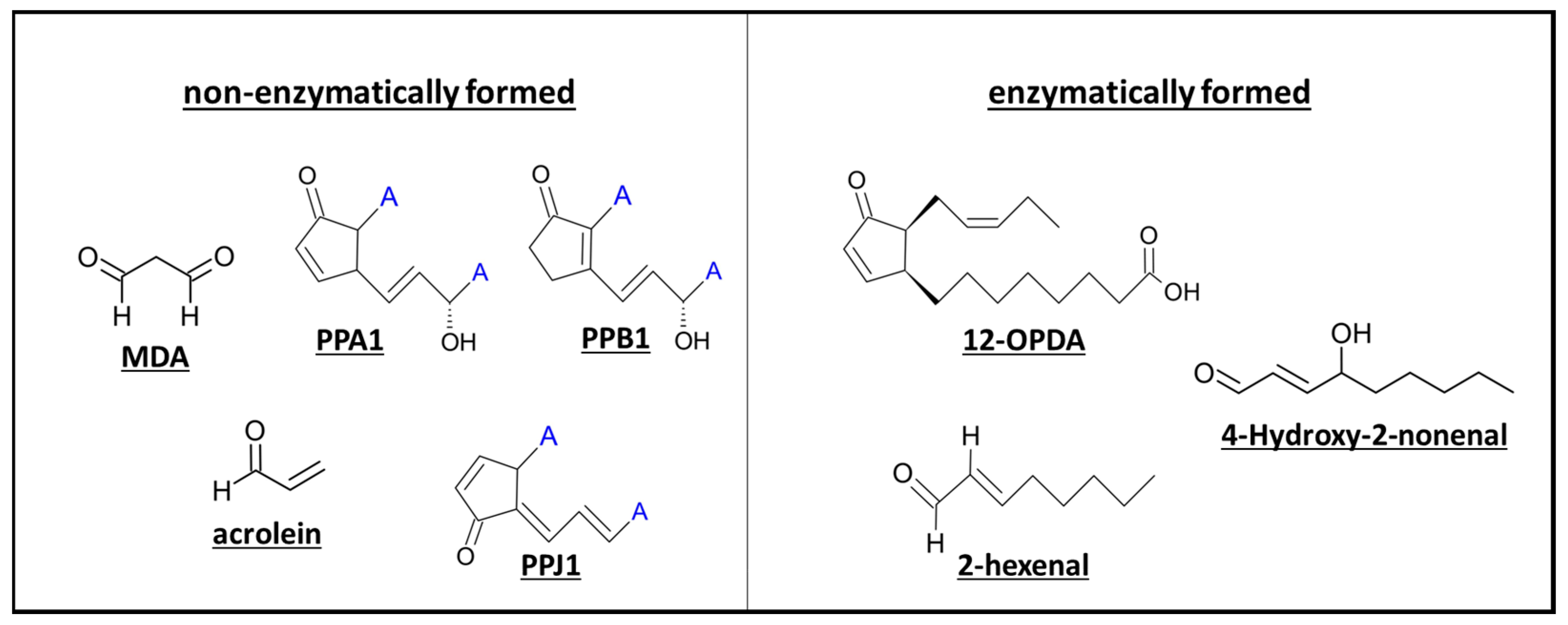
3.1. Non-Enzymatic and Enzymatic Lipid Peroxidation Yields Highly Diverse Oxylipins

Oxylipins are bioactive lipid derivatives generated by oxidation of PUFAs that regulate plant growth, development, and stress defense [39,40,41,42]. While they are produced in all domains of life, species from each kingdom differ in the usage of PUFAs as a starting substrate: in plants, enzymatic oxylipin synthesis starts from α-linolenic acid (α-LeA) and linoleic acid, while animals commonly use arachidonic acid and eicosapentaenoic acid [40,43]. While the set of substrates for oxylipin synthesis is limited, a variety of products are generated by different pathways following PUFA oxidation (Figure 3).
In plants, oxygenation of α-LeA yields allene oxides and α-hydroxy PUFAs by the action of lipoxygenases (LOXs) and α-dioxygenases, respectively [40,41,44,45]. LOXs can be divided into two groups: 9-LOX and 13-LOX. Both groups catalyze oxygenation of PUFAs; however, their regiospecificity varies with 9-LOX introducing oxygen at the C9 carbon atom and 13-LOX at position C13 [40,44,46]. Hence, either (9S,10E,12E,15E)-9-hydroperoxyoctadeca-10,12,15-trienoic acid (9-HPOT) or (9Z,11E,13S,15Z)-13-hydroperoxyoctadeca-9,11,15-trienoic acid (13-HPOT) are generated and can be further metabolized by multiple enzymes to yield diverse oxylipins. A total of seven different pathways of 9-/13-HPOT derivatization has been described; a simplified overview of the synthetic pathways important for this review is provided in Figure 3 [41]. This review will focus (primarily) on oxylipins of three pathways: 12-OPDA as an example for the group of jasmonates, derived by the allene oxide synthase (AOS) pathway; 4-HNE and green leaf volatiles (GLVs) generated by the hydroperoxide lyase (HPL) pathway; and phytoprostanes, MDA, and acrolein as example of non-enzymatically derived oxylipins. 12-OPDA serves as both a signaling and regulator molecule on its own and as a precursor of JA. JA and its derivatives will not be included in this review, since extensive information on JA function can be found in recent reviews [47,48,49].
In addition to enzymatically formed oxylipins, a substantial amount of oxylipins is generated non-enzymatically by lipid peroxidation through free radicals [41,50]. Membrane lipids are the most prominent targets of free radicals in plant cells [51]; this is why lipid peroxidation serves as an early sign of oxidative stress. Important oxylipins of this pathway are phytoprostanes and their derivative (MDA), as well as acrolein [41,52,53]. Interestingly, the base concentration of diverse non-enzymatically formed oxylipins has been reported to be much higher than that of their enzyme-derived counterparts [54]. For instance, MDA levels surpass those of JA up to 50-fold [52].
Independent of their synthesis, oxylipins can be separated into two groups: reactive electrophile oxylipins (RES-oxylipins), also referred to as reactive carbonyl species (RCS), and non-reactive oxylipins [24,52,55]. Reactive electrophiles share a structural similarity, specifically the α, β-unsaturated carbonyl moiety, often as part of a cyclopentenone ring (Figure 4). This structure enables them to react with free thiol groups by Michael addition, regulating protein activity and the available pool of antioxidant molecules such as glutathione [27,56,57,58,59]. Hence, modification of thiol groups is a major mechanism of action shared by phytoprostanes, GLVs, non-GLV aldehydes, and JAs.
In general, RES can significantly damage plant cells and act as toxic and mutagenic agents [60]. For example, MDA damages DNA through reaction with guanine [53,60,61]. However, RES play important roles in signal transduction and defense mechanisms against biotic and abiotic stress; as such, they are regarded as a “REScue” mechanism of cells [55,60,62].

3.1.1. Phytoprostanes Are Evolutionary Ancient Oxylipins

Phytoprostanes derive non-enzymatically from α-LeA and are precursors of MDA [51,52,63]. Through free radical activity, α-LeA is oxidized and a linolenate radical is generated. After additional autoxidation and cyclization, this radical forms phytoprostanes of two regioisomeric classes: the 9- and 16-series of phytoprostanes [51]. With a total of 32 isomers, the phytoprostane group contains a diverse set of oxylipins [63].
As they appear to have evolved distinctly earlier than other oxylipins and are abundant in most organisms, they are considered as an evolutionary ancient mechanism against oxidative stress [64]. Interestingly, cyclopentenone-containing phytoprostanes induce the expression of stress defense-related genes; these include glutathione-S-transferases (GSTs) which catalyze binding of RES to glutathione, one of the most abundant antioxidant molecules of plants [64,65]. Moreover, phytoprostanes stimulate production of phytoalexins, antimicrobial secondary metabolites, and thus enhance biotic stress tolerance [54,64]. This feature, as well as the regulation of gene expression and subsequent upregulation of GSTs, is not unique to phytoprostanes, but rather a common trait of oxylipins shared also by 12-OPDA, MDA, acrolein, and E-2-hexenal, the latter of which belongs to the group of GLVs [57,64,66,67,68,69,70,71].

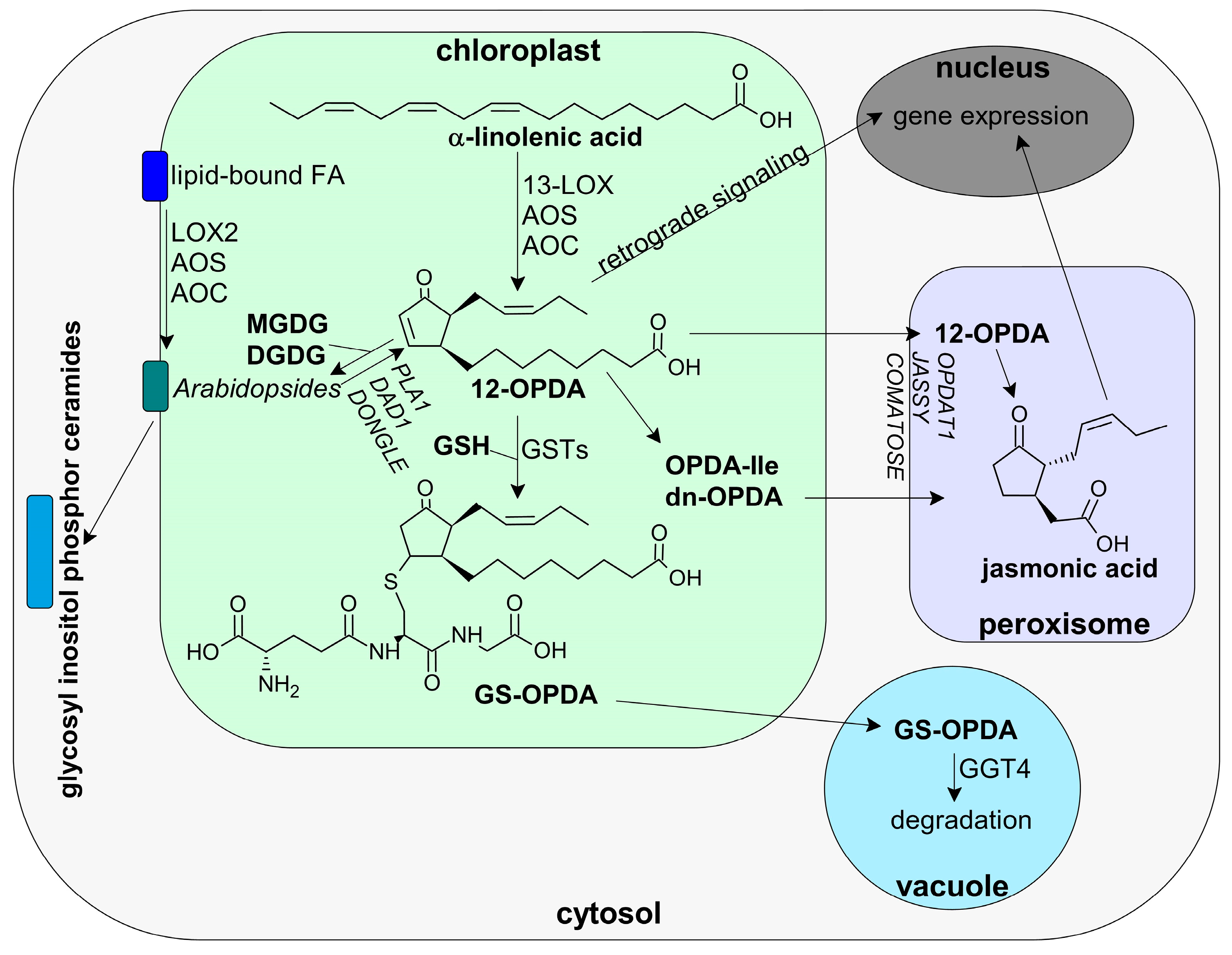
3.1.2. 12-OPDA and OPDAylation as Potent PTM

Cis-12-OPDA, which belongs to the group of jasmonates, is derived by the action of 13-LOX and the AOS pathway, which yields (9Z)-(13S-)-12,13-epoxyoctadeca-9,11,15-trienoate (12,13-EOT). As 12,13-EOT is highly unstable, it is rapidly converted to cis-(+)-12-OPDA by allene oxide cyclase (AOC), or, if there is no AOC available, to α- and γ-ketols and racemic 9R-13R-12-OPDA [27,56,72,73]. After enzymatic synthesis of 12-OPDA, it can either function as a potent signaling molecule and phytohormone or be transported to the peroxisomes, where it is used to generate JA [27,47,74]. JA, in turn, is further modified, e.g., by conjugation to amino acids such as isoleucine (yielding JA-Ile), as catalyzed by JASMONIC ACID RESISTANT 1 (JAR1) [47,75]. JA-Ile and cis-12-OPDA are regarded as the biologically active forms of JA and 12-OPDA, respectively [76,77]. Recently, conversion of 12-OPDA to dinor-OPDA (dn-OPDA), a homologue of OPDA with an acyl residue shortened by two carbons, usually synthesized from hexadecatrienoic acid, was detected in Arabidopsis thaliana [78,79]. An overview of OPDA synthesis and metabolic conversion is provided in Figure 5.
Under stress conditions such as wounding, high light or pathogen attack, 12-OPDA synthesis is highly stimulated [27]. Using transgenic A. thaliana lines which express the Pseudomonas syringae avirulence peptide AvrRpm1 depending on dexamethasone, Andersson et al. observed an increase of 12-OPDA from basal concentration of 2 µM to 36 µM after 4 h of AvrRpm1 expression. Interestingly, accumulation of lipid-bound 12-OPDA, so-called Arabidopsides was even more pronounced with a 200-fold increase [42].
Arabidopsides are cyclo-oxylipin-galactolipids in which 12-OPDA is bound to complex membrane lipids such as mono- and digalactosyldiacylglycerol (MGDG/DGDG) [40,41,80,81]. They are unique to certain species, primarily of the Brassicaceae family (e.g., A. thaliana, Camelina microcarpa, Capsella rubella), but also Poaceae (Hordeum vulgare), Convolvulaceae (Ipomoea tricolor), and Lamiaceae (Melissa officinalis) [39,40,76,80,81,82,83,84]. Concerning synthesis of Arabidopsides, incorporation of free 12-OPDA to membrane lipids as well as direct membrane-bound generation from α-linolenic acid are discussed, with the existence of membrane-bound 13-HPOT in Arabidopsis arguing for the latter [42,84,85,86]. While increases in Arabidopside content due to unfavorable environmental conditions could be observed in several studies, their function in plants still remains mostly unknown [42,80,82,85,87]. On the one hand, they have been proposed to serve as storage pools for the fast release of 12-OPDA under stress, either as a means of rapidly increasing OPDA signaling or increasing synthesis to JA [42,82,86,87]. Several enzymes have been linked to this release of 12-OPDA, including acylhydrolase, phospholipase 1 (PLAI), DEFECTIVE IN ANTHER DEHISCENCE1 (DAD1), and DONGLE (DGL) [27,88,89]. On the other hand, Arabidopside E and G possess antimicrobial functions; Arabidopside A stimulates senescence in barley leaves and Arabidopside A and B inhibit root growth, indicating the possibility that Arabidopsides themselves are functional molecules of stress signaling and defense [42,86,90]. Furthermore, Arabidopsides might interact with glycosyl inositol phosphor ceramides, i.e., major sphingolipids in plants linked to stress defense [91,92].
Similar to phytoprostanes, jasmonates play an essential role in plant defense against different abiotic and biotic stresses [27,93,94]. Additionally, they influence plant growth and reproduction. For instance, jasmonates regulate pollen maturation, elongation of stamen filament, and pollen release as well as leaf movement and fruit ripening [76,93,94,95]. Further overviews on the role of 12-OPDA in plants can be found in recent reviews [27,96,97].

3.1.3. Oxylipin Aldehydes

Similar to ROS, high concentrations of oxylipin aldehydes can lead to extensive cell damage, while lower concentrations contribute to cell signaling processes in the context of abiotic stress [98,99,100]. In general, lipid-peroxide derived aldehydes inhibit seed germination, CO2 photoreduction, and plant growth and senescence [98,101,102].
Unlike the oxylipins mentioned so far, 4-hydroxy-2-nonenal (4-HNE) is a lipid aldehyde that derives from linoleic acid by action of 9-LOX, hydroperoxide lyase (HPL), an alkenal oxygenase, and hydroperoxide peroxygenase [103]. 4-HNE has been regarded as the most toxic and most abundant product of lipid peroxidation and primarily accumulates in biomembranes [103]. Besides thiol binding by Michael addition, binding of 4-HNE to Arg residues leading to 2-pentylpyrrole adducts has been observed [104]. As the latter has only been documented in animals, a similar effect of 4-HNE remains to be proven in planta.
MDA is the smallest molecule discussed in this review and only consists of three carbon molecules and two aldehyde groups [105]. Nevertheless, it is highly reactive and commonly regarded as biomarker of oxidative stress [105]. For instance, accumulation of MDA under heat stress negatively correlates with functionality of the photosynthetic electron transport chain [99,105]. However, as with ROS and oxylipin aldehydes in general, MDA fulfills a dual role in plants, acting as signaling molecule in stress defense and acclimation processes as long as its concentration is correctly balanced [105].
The C6 aldehyde 2-hexenal belongs to the subgroup of GLVs and the bigger group of biogenic volatile organic compounds (BVOCs) [41,106]. Emission of VOCs accounts for up to 10% of total fixated carbon in plants with 109 tons VOCs per year [107,108]. Production and release of VOCs is tightly regulated by biotic factors (pollination status, herbivore infestation) and abiotic factors (light intensity, atmospheric CO2, temperature, humidity, nutrition) in a spatial, temporal, development-specific, and species-dependent manner [107,108]. To be released, VOCs must cross membranes, the cell wall, and (depending on the tissue) also the cuticle, a major barrier in most plant cells [107,108].
Due to their high lipophilicity, as evident from their high octanol-water partition coefficient, VOCs are primarily found in hydrophobic environments where they can significantly damage cellular structures [107,108]. To ameliorate this damaging effect, the cuticle serves as a volatile sink and storage pool, primarily for VOCs with low volatility [108]. As a consequence, VOC emission depends on the composition and thickness of the cuticle [107].

3.2. Oxylipin Signature

The oxylipin composition of cells and tissues, the oxylipin signature, varies depending on plant species, organ and tissue, developmental stage, and environmental conditions [109,110,111]. By fine-tuning oxylipin concentrations, different signaling pathways and plant processes can be targeted. An example of a species-dependent oxylipin signature is the induction of phytoalexin synthesis. In rice and tobacco, both JA and 12-OPDA stimulate phytoalexin production in leaves (as measured as sakuranetin and scopoletin contents); however, in soybean, only OPDA can activate phytoalexin synthesis [112,113,114]. Species-dependent oxylipin signatures can be traced back to both differing oxylipin functions and to differences in the oxylipin concentrations needed for their functionality. For instance, high concentrations of jasmonates could be measured in sorbitol-treated Hordeum vulgare with 5 nmol g−1 FW of 12-OPDA (1462 ng g−1 FW) and 2.2 nmol g−1 FW of JA (462 ng g−1 FW) after 24 h [115]. In contrast, osmotic stress caused by sorbitol treatment of A. thaliana for the same time span results in a lower accumulation of 12-OPDA and JA with 462 and 256 ng g−1 FW, respectively [116]. As sorbitol was employed at twice the concentration in the first study when compared to the second study (1 M and 0.5 M, respectively), one might argue that OPDA accumulates significantly due to enhanced osmotic stress caused by high sorbitol concentrations. Nevertheless, dependency of oxylipin composition on plant species should be considered in this case as well.
Interestingly, the type of attacking insects also influences the oxylipin signature under biotic stress. For instance, piercing-sucking and chewing insects both stimulate emission of LOX-derived volatiles in distinctly different degrees. After aphid attack, LOX-derived volatiles accounted for 8.9% of total volatiles; chewing insects, meanwhile, caused an increase of this percentage to 53%. Additionally, the composition of volatiles varies, with C6-volatiles being more strongly emitted after aphid infection and C9-aldehydes (as measured on nonanal) after infestation with chewing insects [117].
Consequently, regulation of oxylipin signatures is highly complex and more detailed studies, concentrating not only on single oxylipins but on the broad profile simultaneously, are needed to understand how oxylipins affect plant processes [50].
Modulation of oxylipin composition might be achieved by differences in the ROS level (concerning non-enzymatically derived oxylipins) and regulation of the activity of different enzymes such as LOXs. For example, if 13-LOX is up- and 9-LOX downregulated, the ratio of 13-HPOT to 9-HPOT increases. The changed ratio results in enhanced synthesis of jasmonates relative to GLVs.

4. Influence of Oxylipins on the Redox-Regulatory Network

4.1. Modulation of Thiol-Sensitive Proteins

Oxylipin RES impact the redox regulatory network at all levels, from redox input elements to redox sensors and target proteins, by influencing gene expression, protein synthesis, and catalytic activity. Using an anti-HNE antibody, Mano et al. described a subset of 34 proteins modified by, as they assumed, by both 4-HNE RCS in general. This subset contains essential proteins of ROS generation and the redox network such as peroxidase 34, a cell wall peroxidase linked to oxidative burst, or Cyp20-3 and cysteine synthase, both dedicated to thiol synthesis [118].
Synthesis of heat shock proteins, stress-induced chaperones, is upregulated not only by 12-OPDA and phytoprostanes but also by MDA [66,119], while, after synthesis, they are targets of 4-HNE [103]. Other universal targets of RCS include GSTs, as mentioned earlier, and, interestingly, enzymes usually linked to carbon metabolism, more specifically glycolysis. NAD-dependent glyceraldehyde-3-phosphate dehydrogenase (GAPDH) is not only dedicated to glycolysis but also exerts moonlighting functions including autophagy, apoptosis, and translation by RNA binding [120,121,122]. As its function strongly depends on its cysteinyl residue redox state, GAPDH belongs to the group of target proteins [123,124]. Oxylipins play a dual role in altering GAPDH levels and activity. Covalent binding of 4-HNE to GAPDH and the subsequent inhibition of catalytic activity has already been shown by Uchida and Stadman in 1993 [125]. Binding of acrolein also decreases GAPDH activity [102,126]. Moreover, binding of 4-HNE as well as 4-HHE appears to trigger GAPDH degradation [127].
On the other hand, a stimulatory effect of 12-OPDA on GAPDH expression has been proposed based on studies with Physcomitrella patens mutant lines lacking AOS activity and 12-OPDA-treated Pohlia nutans [128,129]. Assessment of differentially expressed genes due to 12-OPDA treatment in various organisms (P. nutans, variegated Epipremnum aureum, and the algae Klebsormidium nitens) shows upregulation of gene orthologues of A. thaliana GAPDH isoforms (GAPCp1, GAPC2), whereas GAPA2 and GAPB orthologues are downregulated [79,129,130]. The only direct study of 12-OPDA-responsive genes (ORGs) in A. thaliana, conducted by Taki et al., does not show significant regulation of GAPDH expression by 12-OPDA. However, although not fulfilling the criteria for ORGs (which were quite stringent, with a minimum of 3-fold relative expression increase in this study), gene expression of GAPC1 shows a trend towards upregulation with an approximately twofold increase [66]. Altogether, one might tentatively expect a stimulatory effect of 12-OPDA on GAPDH.
In vitro, OPDAylation of GAPDH (as tested using GAPC2) only showed minor inhibition on its NADH oxidation activity under physiological 12-OPDA concentrations [56]. Modulation of GAPDH signaling by oxylipins might influence carbon metabolism and the energetic state of the cell. Stimulation of GAPDH levels by 12-OPDA could serve as a means of counteracting energy consumption due to stress defense mechanisms. On the other hand, GAPDH also exerts RNA binding functions, hence modulating protein synthesis [120]. RNA binding motifs of GAPDH are primarily found in high light-induced transcripts [131]. Therefore, modulation of GAPDH levels and oxidation state might help in steering protein synthesis in the direction of stress signaling and defense, similar to the induction of ORGs by interaction with Cyp20-3. Similar to GAPDH, aldehydes such as acrolein target and inactivate fructose-bisphosphate aldolase (FBPase), another glycolytic enzyme and redox target protein for which RNA binding activity has been described [98,102,120]. However, in plants, only nonspecific binding of plastid FBA1 to RNA coding for a subunit of the cytochrome b6/f complex has been shown [132].
Modulation of Cyp20-3 activity by oxylipins, more specifically non-covalent binding of 12-OPDA, has been shown to enhance Cyp20-3 activity. Consequently, thiol synthesis is stimulated, strengthening the redox capacity of the cell. This change in redox state in turn alters the expression of 12-OPDA-responsive genes [27,28,96,133]. Contrary to Cyp20-3 stimulation, OPDAylation diminishes the H2O2 scavenging function of 2-CysPRX [133]. OPDAylation of TRXs drastically impedes their activity as redox transmitters, thereby decreasing the rate of TRX-dependent Cyp20-3 reduction and glutathione peroxidase (GPXL) regeneration. The overall functionality of this inhibition might be explained by different modes. First, inhibition of TRX activity and subsequently increased ROS productions might enhance ROS-induced stress signaling [56,134,135]. Second, inhibition of chloroplast TRXs also diminishes TRX-dependent FBPase reduction. As a consequence, CO2 assimilation and starch synthesis are inhibited, reducing the energy consumption by the Calvin–Benson cycle [133].
SODs are additional targets of oxylipins. While 4-HNE has been shown to bind to Mn SODs, 12-OPDA downregulates gene expression of Cu/Zn SODs; these are part of the water-water cycle [103,136]. Since acrolein releases Zn2+ from different proteins, an inhibitory effect of this RCS on Cu/Zn SODs followed by upregulation of ROS levels has been proposed [100,137].
Exogenously supplied acrolein, 4-HNE, and HHE increase catalase and APX catalytic activity in A. thaliana [138]. Moreover, 4-HNE interacts with MDHAR. An oxidizing effect might be assumed, but remains to be proven, whereas 12-OPDA enhances DHAR synthesis [103,139]. An exemplary overview of oxylipin-dependent regulation of redox network proteins can be found in Table 1.

4.2. Interaction with Non-Protein Thiols

Non-protein targets of oxylipins include ASC and GSH. Both compounds constitute abundant cellular redox buffers that are essential, e.g., in the Asada–Halliwell pathway [27,36]. Glutathionylation of RES, including cyclopentenone phytoprostanes, inhibits their activity and thus protects cells from damage such as the inactivation of thiol switch proteins [65,68,140]. Regarding the main oxylipins included in this review, only 4-HNE, acrolein, and 12-OPDA have been shown to undergo GSH binding so far (either spontaneously or catalyzed by GST6, respectively) [70,119,141]. However, the fate of these adducts remains mostly unknown; it is expected that adducts are transported to the vacuole for degradation, as is also the case for GSH-OPDA adducts [67].
Possible new functions of the GS-adducts in vivo await elucidation. In animals, studies indicate a unique ability of GS-HNE in stress defense modulation when compared to free 4-HNE, with a function in regulating the transcription factor NF-κB [142]. Concerning cyclopentenone prostaglandins PGA2 and PGJ2, GSH binding serves as a “shuttle”, enabling transport of otherwise highly hydrophobic molecules into different cell organelles. After reaching their target destination, PGs might be released from GSH (either spontaneously or enzymatically catalyzed) by undergoing retro-Michael Addition and binding to higher affinity protein thiols [143]. This mechanism, based on increasing hydrophilicity after GSH binding, is likely transferable to plant oxylipins and broadens the spectrum of cell organelles containing proteins sensitive to OPDAylation and OPDA-dependent regulation.
To this day, the only effect of GS-OPDA adduct formation concerns the 12-OPDA localization by import into the vacuole, where it undergoes degradation [67]. Hence, this might be considered as a purely protective function to remove this compound from the cytoplasm. However, this process should be studied more extensively in order to clarify whether GSH binding has additional functions in cellular regulation.
GSH concentrations surpass those of typical oxylipins several-fold. GSH concentrations in plasmatic compartments are in the mM range, whereas oxylipins, including 12-OPDA, reach µM values. Given these molar ratios, the question of how free oxylipin functions are maintained despite their reactivity with GSH must be discussed. First, the reaction kinetics between oxylipins and proteins on the one hand and oxylipins and GSH on the other hand differ greatly in dependence on the pKA values of the Cys residues. Acrolein binds rapidly to protein thiols; however, when compared to HHE and 4-HNE, GSH-acrolein adducts form 70- to 110-times faster, respectively [53,100,126]. For example, studies on the interaction of 4-HNE and GSH revealed a half-life time of 2 min for 4-HNE in the presence of 5 mM GSH [144]. Second, adduct formation of GSH and oxylipins is a reversible process that includes characteristic adduct stabilities for each oxylipin. For instance, whereas acrolein-GSH adducts are very stable with a half-life time of 4.6 days, hexenal-GSH adducts decayed with a half-life time of 4 to 6.3 h [145].
Further, oxylipins influence the cellular GSH pool and synthesis and activity of GSTs. Trans-2-hexenal inhibits pumpkin GST, though only at high concentrations (mM range) [70]. Aldehyde dehydrogenases (ALDHs) function as important enzymes by maintaining the glutathione pool in a reduced state and detoxifying RCS. ALDHs are targets of 4-HNE [101,103,146]. Depletion of chloroplast GSH content is a common feature of lipid-peroxide derived aldehydes [98].
To conclude, oxylipins commonly decrease cellular GSH levels. High concentrations of acrolein even deplete the cell of GSH and ascorbate. This might be due to inhibition of GSH synthesis as well as stimulation of GSTs and binding of GSH to the oxylipins themselves. Contrary to this, 12-OPDA influences GSH levels positively by enhancing Cyp20-3-mediated thiol synthesis. Whether oxylipins are completely inactivated after binding to GSH and whether adducts gain additional functions or only serve as shuttles remains to be elucidated.

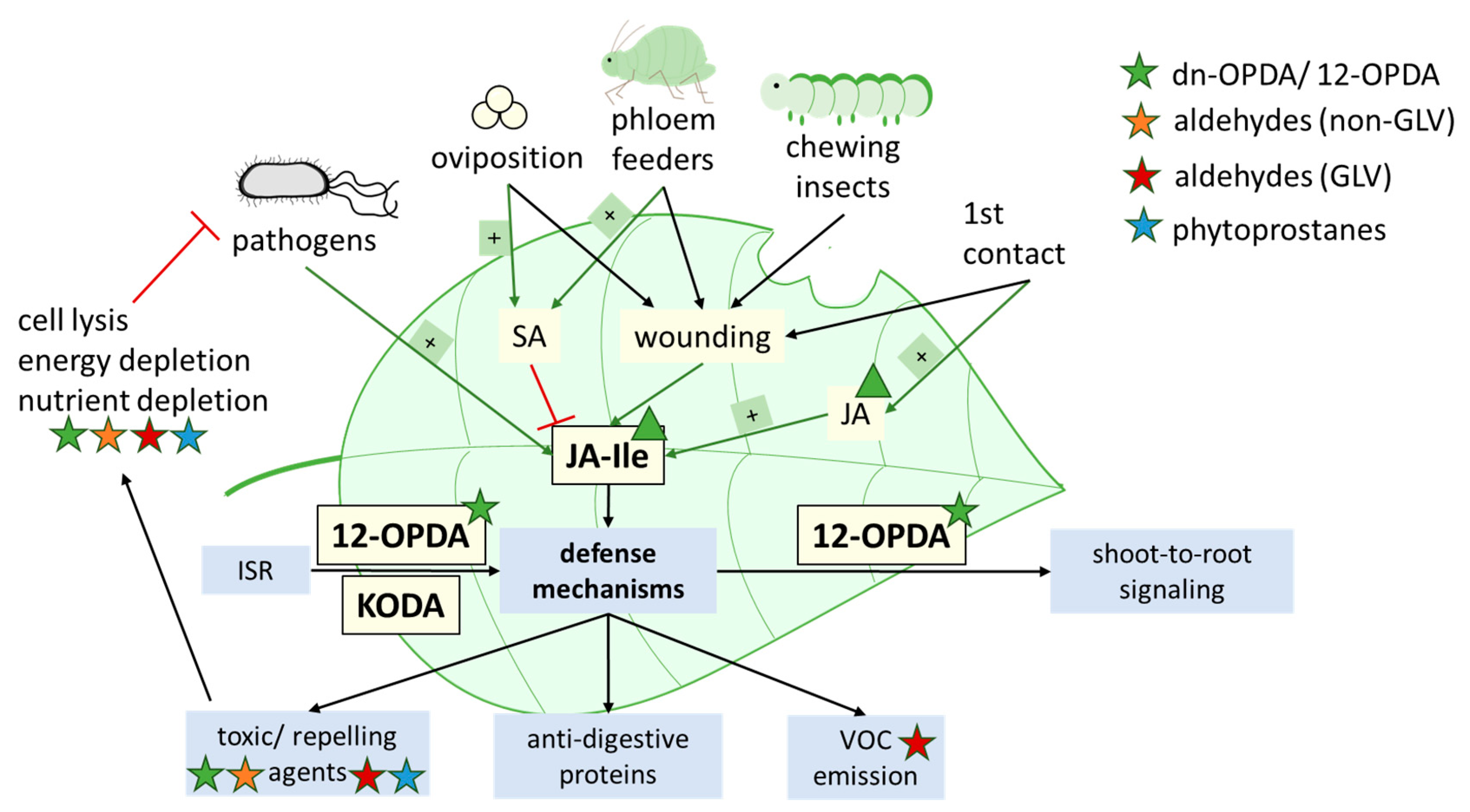
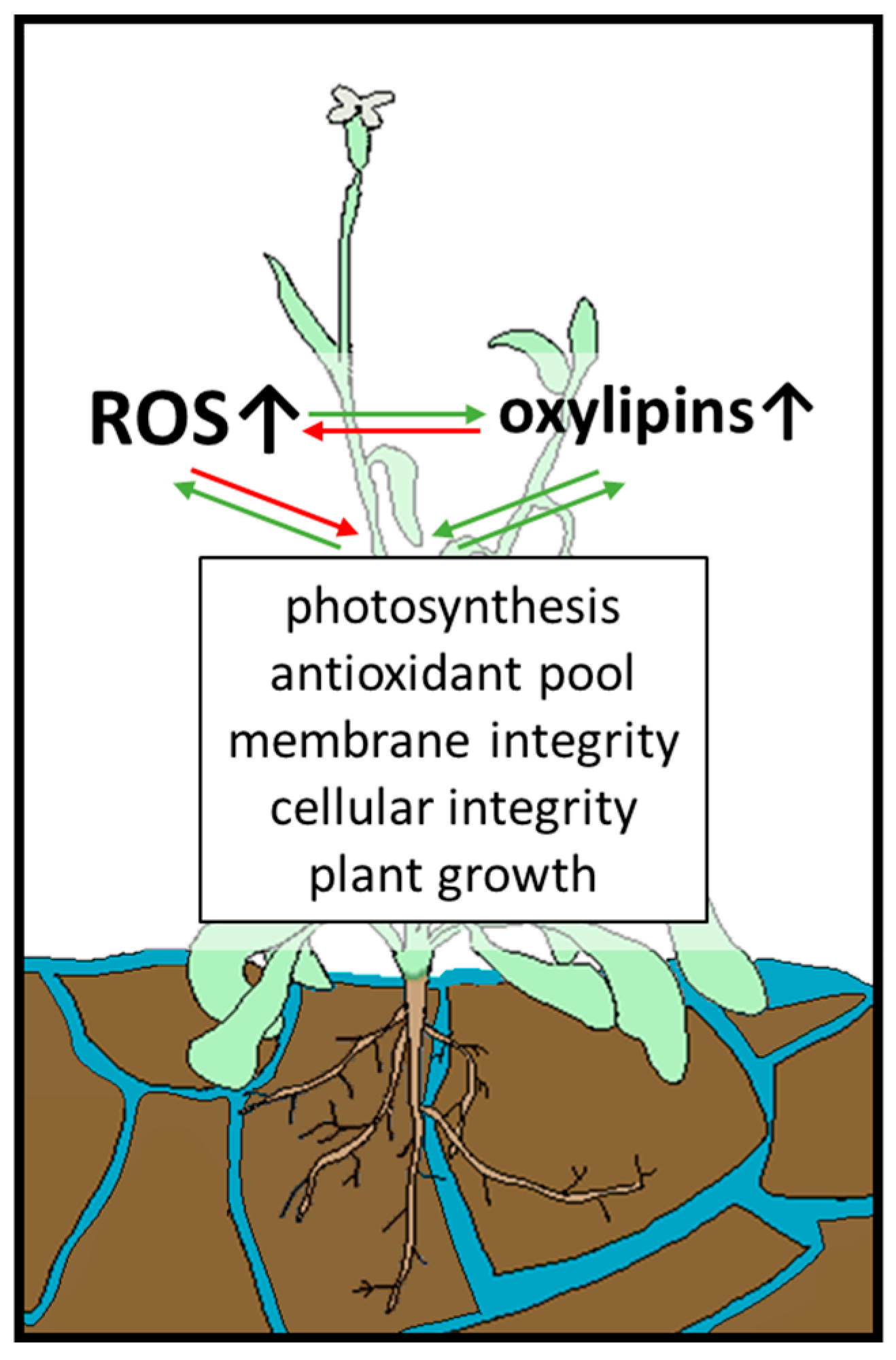
4.3. Contribution of Oxylipins to Environmental Acclimatization

Due to climate change and global warming, plants consistently face changing environmental conditions and extended periods of severe biotic and abiotic stress. On the one hand, species dominance shifts by changes in land use, and more extensive outbreaks of insects are expected, especially in forests [106,147,148]. Simultaneously, temperature variation becomes more extreme with the mean global temperature rising; however, more pronounced cold periods may occur, while the frequency of flooding and drought events rises [106,147,148,149]. Not only is soil nutrient availability changing, but the pollution of soil (as well as air and water) is constantly increasing as a result of anthropogenic activities [106,149].
All these stresses threaten yield formation as plants invest more resources in defence as a trade-off to survive. For instance, under UV stress, plants might encounter morphological changes and altered genome stability due to DNA damage [108]. Oxylipins and redox state are major players in the plant-environment interaction, e.g., the adaptive mechanisms include upregulation of cuticle synthesis. This upregulation, in turn, changes VOC emission, their distribution in planta, and their biosynthesis [108]. Similar effects on cuticle synthesis can be observed under drought stress, flooding stress, high salinity, and high and low temperature [150,151]. Abiotic stress, such as intense heat or drought periods, and severe climatic events, such as flooding, require a constant evolutionary adaption and flexible acclimatization of plants to their environment.

4.3.1. Thermotolerance

Changing temperature and air pollution disturbs signalling through GLVs. Air pollution results in a decrease of maximum downwind distance of reactive volatiles, while cold temperatures cause GLVs to condense and reside on plant surfaces instead of emitting into the atmosphere [106,147]. Hence, pollination of plants deteriorates as the searching and foraging efficiency of pollinators decreases. At the same time, the diminished attraction of predators prevents anti-herbivore defence and might lead to more extensive wounding of plants with increased synthesis of oxylipins. This adds to a generally increased severity of biotic stress, which is even further pronounced due to the rising severity of insect outbreaks [147].
On the other hand, emission of BVOCs, including GLVs, has already increased by 10% over the last three decades due to climate change; this is estimated to increase by a further 30–45% as global temperature rises by another 2–3 °C [106]. This increase might be traced back to indirect and direct effects on BVOC emission, which is characterized not only by their generation, but also by their physicochemical properties (solubility, volatility, diffusivity). Emission rates are affected indirectly by prolongation of plant growth phases and directly by affecting biochemical generation of BVOCs, both due to increasing temperature [106]. Considering high temperatures as the only factor, emission of BVOCs is proposed to be more potent, as their vapour pressure and their volatility enhance their diffusivity at the same time [106]. However, as mentioned before, cold temperatures and air pollution might counteract these effects.
RES oxylipins are proposed to act as an ancient system of stress defense regarding thermotolerance, which evolved distinctly before JA signaling [79]. To counteract heat stress, 12-OPDA, phytoprostanes, and MDA induce synthesis of heat shock proteins, major chaperones involved in heat stress acclimation [66,96,119].
As with heat stress, cold stress severely limits plant performance and may ultimately lead to plant death in non-adapted species. Tolerance to both stresses, moreover, involves JA and SA signalling [152]. In rice, under cold stress, several genes of jasmonate biosynthesis are upregulated, including OsLOX2, OsAOS1, and OsOPR1, resulting in accumulation of JA (and possibly also 12-OPDA) [152]. JA signalling increases plant endurance and freezing tolerance [152,153]. However, as shown for A. thaliana, this upregulation of jasmonates appears to be limited to the first exposure to cold stress and dramatically decreases in primed plants [154]. This might be explained as a protection mechanism to reduce costs for gene expression of cold-tolerance genes under fluctuating weather conditions, for example in spring [154].

4.3.2. Pathogen Infection and Induced Systemic Resistance (ISR)

Besides abiotic stress, biotic stress, as caused by pathogen infection, insect attack, or intraspecific competition, significantly impacts pre- and postharvest crop yield [155,156]. In regards to most prominent food crops, including wheat, rice, maize, and potato, biotic stress has been reported to account for roughly 28%, 37%, 31%, and 40% of yield losses from 2001 to 2003, respectively [157,158]. In general, biotic stress is thought to cause over 40% of yield loss in global food production [157,159,160].
As recently reviewed, JA and its derivate JA-Ile are major regulators of herbivory- and pathogen-induced defence mechanisms in plants [161,162]. However, other oxylipins, besides JA and JA-Ile, further contribute to herbivory resistance. In organisms devoid of JA signalling, such as Marchantia polymorpha, dinor-OPDA, instead of JA, mediates herbivory defence signalling together with salicylic acid (SA) [163]. Moreover, 12-OPDA, possibly in concert with dn-OPDA, maintains plant resistance against insects and fungi in absence of JA in A. thaliana [164].
Feeding preferences of herbivores depend on cis-3-hexenal, a GLV that stimulates feeding activity [165]. In A. thaliana, signalling from wounded shoot tissue to distal parts of the plant depends on the transport of 12-OPDA through the phloem from shoot-to-root, where it then mediates activation of JA and JA-Ile signalling [166]. GLVs are important signalling molecules involved in intra- and interspecies communication, even across kingdoms. For instance, after herbivorous damage, there exist a means of attracting predators of herbivorous insects and enhancing plant defence against pathogens and nematodes [68]. Further, they serve as warning molecules for neighbouring plants. GLVs that are emitted from plants subjected to biotic stress can be perceived by surrounding plants. This induces stress defence mechanisms or primes the plant, resulting in a pre-defence state in which stress defence induction proceeds more quickly and strongly [68,167]. The mechanism of GLV perception in plants, however, is still largely unknown [68]. Lastly, GLVs are essential molecules for attracting pollinators and, thereby, maintain plant reproduction [68,106,168,169].
Oxylipins (phytoprostanes, OPDA) distinctly enhance phytoalexin synthesis [54]. Furthermore, due to their chemical structure (dependent on carbon chain length, double bonds, and hydroperoxyl groups), oxylipins display antimicrobial effects. They cause cell lysis and subsequent electrolyte leakage in gram-positive and gram-negative bacterial cells and inhibit enzymes, including those dedicated to nutrient uptake, ATP synthesis, and the respiratory ETC [170,171,172,173,174,175,176,177].
Symbiosis of plant roots and rhizobacteria not only benefit for plant growth but can also trigger induced systemic resistance (ISR). ISR is a type of defence mechanism against pathogens and herbivores that relies on signalling molecules transported from roots to aboveground plant tissues (root-to-shoot signal molecules) [178,179]. In maize roots colonized by Trichoderma virens, ISR is activated by fungal secretion of oxylipin synthesis inhibitors.
While Sm1, a hydrophobin-like elicitor, is secreted by T. virens, the production of 9-HPOT by LOX3 is inhibited. Although seven 9-LOX are produced in maize, LOX3 appears to be the only LOX responsible for regulation of ISR, as LOX3 knockout-mutants show constitutive ISR [180,181,182]. However, plant lines deficient in LOX5 and LOX10 also display enhanced resistance to parasite infection, which raises the question of whether they might be inhibited in the absence of LOX3 activity [180,183,184]. Recently, 12-OPDA and α-ketol-octadecadienoic acid (KODA) have been identified as root-to-shoot signals in Zea mays [185]. Both depend on the AOS pathway of oxylipin synthesis; however, 12-OPDA is synthesized from 13-HPOT and KODA from 9-HPOT. As a result, activity of both 9- and 13-LOX is required. This might appear contradictory with the involvement of LOX3 in ISR. However, maize plants deficient in LOX3 activity characteristically overexpress genes of jasmonate biosynthesis such as LOX10 [186]. Production of KODA in maize has not been traced back to a specific 9-LOX so far, excluding LOX3, so KODA can still by synthesized by the other six 9-LOX.
All in all, while JA is the primary oxylipin involved in herbivory, the oxylipins that we focus on in this review fulfil additional and essential functions, such as root-to-shoot signalling and ISR. Furthermore, they contribute to pathogen defence and are able to substitute JA signalling in the absence of JA (depicted in Figure 6), as indicated by studies in different species.
Another important plant stress defense mechanisms against pathogen infection is hypersensitive response (HR): local necrosis, which prohibits further spread of infection to healthy tissue [42,187,188]. HR is driven by recognition of small elicitor molecules containing specific patterns, so-called pathogen-associated molecular patterns (PAMPs), by dedicated receptors (pattern recognition receptors, PRRs) as well as detection of avirulence (avr) proteins by nucleotide-binding and leucine-rich repeat receptors (NLRs) and resistance (R)-proteins [187,189,190,191,192]. After recognition of PAMPs or avr proteins, a signalling cascade involving NADPH oxidases (RBOH) is initiated that causes an oxidative burst and subsequently programmed cell death (PCD) [42,193,194].
As regulation of HR constitutes a complex network of transcriptional, post-transcriptional, and post-translational modifications, this review will only provide limited insight as related to oxylipin-linked HR. While concrete mechanisms and redox-regulatory pathways behind HR-associated PCD still await further elucidation, the overall state of current knowledge on HR has been focus of recent reviews [187,192,195,196]. One protein involved in post-translational modification of HR is HSP90, which, together with additional chaperones, stabilizes NLRs [187,197]. Moreover, disturbance of APX and MDHAR levels and activities has been linked to generation of the oxidative burst of PCD [198,199]. Consequently, as RBOHS, MDHAR, APX, and HSP90 have been shown to be affected by oxylipins, as described in Section 4.1 and Section 4.2, they might contribute possible links to oxylipin-modulated HR. As TRXs negatively regulate HR-driven PCD, inhibition of TRX activity by 12-OPDA might contribute to the rapid induction of PCD under stress conditions, as 12-OPDA synthesis is stimulated in the course of HR [27,200].
Another possible interaction node of HR signaling are ceramides, which are hypothesized to interact with Arabidopsides as well as the phytoalexin pool [201]. Moreover, Arabidopsides, specifically Arabidopside E, are thought to serve as protection against secondary infections in already dead tissue after PCD [42]. Although studies on the effect of oxylipins on HR are scarce, oxylipins have been proposed as determinants of HR [201]. For instance, stimulation of ROS-triggered programmed cell death (PCD) by oxylipins (acrolein, 4-HNE) after pathogen infection has been linked to depletion of the glutathione pool and lethal depletion of ASC, explained by stimulation of caspase-like proteases [202]. Negative regulation of HR by oxylipins has also been observed in A. thaliana cells treated with phytoprostanes, possibly due to activation and increased synthesis of GSTs, MAPK, and antimicrobial compounds [201,203]. Overall, both the mechanisms of HR-induced PCD and the possible involvement of oxylipins should be studied more extensively.

4.3.3. Flooding

Another effect of climate change is more frequent (temporary) flooding, causing not only submergence (hypoxia) stress, but reoxygenation stress as an additional challenge. During submergence, oxygen availability decreases drastically, the composition of soil and accessibility of nutrients and microbial environment changes, and toxic compounds are formed [204]. Consequently, the photosynthesis rate of plants decreases as well as their energy and carbon levels, resulting in growth retardation. Temporary flooding is followed by reoxygenation, which leads to distinct oxidative stress in plants [204,205,206].
Oxylipins, especially 12-OPDA and C6 aldehydes of the group of GLVs, ameliorate flooding stress in plants and increase plant survival as depicted in Figure 7 [204,207]. By using different A. thaliana wild type and transgenic lines, Savchenko et al. analysed the function of the HPL and the AOS pathway, both dependent on 13-HPOT [208]. They showed a protective effect under waterlogging stress concerning biomass accumulation, membrane integrity, photosynthesis rate, and overall submergence survival rate. This effect relies not only on the combined action of both pathways, but also on the unique action of products of the HPL or AOS branch. For instance, the AOS branch appeared to be more critical for defence against membrane damage during reoxygenation as measured by electrolyte leakage and lipid peroxidation. However, in both cases, the combination of HPL and AOS activity resulted in the least damaged membrane.
Furthermore, the decline in photosystem II activity was less pronounced in the presence of oxylipins. This is in accordance with previous studies that show a protective effect of HPL on the photosynthetic apparatus under high light stress [209]. Overall, the survival rate of plants under submergence stress increases from ~50% when only one of the two pathways is active to ~77% with both pathways functional. In a double knockout mutant displaying neither AOS nor HPL activity, survival rate drops to ~30%, supporting the importance of oxylipins under flooding stress [204].
In plants displaying HPL and AOS activity, 12-OPDA accumulated after submergence stress. Since earlier studies reported transgenic A. thaliana lines deficient in JA biosynthesis and/or signalling to show increased sensitivity towards reoxygenation, and no increase of JA could be detected in this study, an essential role of 12-OPDA in flooding tolerance can be concluded. 12-OPDA could either be the only AOS-derived key regulator, or act together with basal concentrations of JA [204]. Additionally, 12-OPDA accumulates under combined flooding and heavy metal stress and has been proposed to enhance stress defense by upregulation of thiol production and tuning gene expression under these stress conditions [207].
In total, an essential role in plant survival under flooding stress can be attributed to oxylipins with 12-OPDA acting as a primary signalling molecule.

5. Conclusions

Under non-optimal growth conditions and severe stress such as flooding and heat, which are expected to further increase due to global warming over the next decades, plants are faced with an accumulation of ROS. As these can cause extensive damage, a complex array of proteins, the redox-regulatory network, is dedicated to maintaining ROS homeostasis. Products of ROS-induced damage, oxylipins, and the function of the redox network are strongly linked. This interplay awaits further elucidation, as studies on oxylipins are generally rather scarce, especially concerning studies on oxylipin signature instead of single oxylipins. Nevertheless, the strong effect of oxylipins on redox homeostasis in plant cells has been widely recognized; this interference affects plant performance in a larger context and in environmental acclimatization. The interaction between glutathione and oxylipins, especially in the context of adduct formation, function, and cleavage, should be further characterized, particularly concerning 12-OPDA, a precursor of jasmonic acid, that, in recent years, has emerged as a potent phytohormone on its own.

Author Contributions

Ideation, A.V., K.-J.D., M.K.; literature search, M.K., writing—original draft preparation, M.K.; writing—review and editing, A.V., K.-J.D., M.K. All authors have read and agreed to the published version of the manuscript.

Funding

This research was funded by the German Science Foundation (Deutsche Forschungsgemeinschaft), grant number DI 346/22-1 and /46-23-1.

Institutional Review Board Statement

Not applicable.

Informed Consent Statement

Not applicable.

Data Availability Statement

Data sharing not applicable.

Conflicts of Interest

The authors declare no conflict of interest.

References

  1. Holland, H.D. The oxygenation of the atmosphere and oceans. Philos. Trans. R. Soc. Lond. B Biol. Sci. 2006, 361, 903–915. [Google Scholar] [CrossRef] [PubMed] [Green Version]
  2. Vogelsang, L.; Dietz, K.-J. Plant thiol peroxidases as redox sensors and signal transducers in abiotic stress acclimation. Free Radic. Biol. Med. 2022, 193, 764–778. [Google Scholar] [CrossRef] [PubMed]
  3. Turkan, I. Emerging roles for ROS and RNS—Versatile molecules in plants. J. Exp. Bot. 2017, 68, 4413–4416. [Google Scholar] [CrossRef] [PubMed] [Green Version]
  4. Mittler, R.; Zandalinas, S.I.; Fichman, Y.; van Breusegem, F. Reactive oxygen species signalling in plant stress responses. Nat. Rev. Mol. Cell Biol. 2022, 23, 663–679. [Google Scholar] [CrossRef]
  5. Juan, C.A.; La Pérez de Lastra, J.M.; Plou, F.J.; Pérez-Lebeña, E. The Chemistry of Reactive Oxygen Species (ROS) Revisited: Outlining Their Role in Biological Macromolecules (DNA, Lipids and Proteins) and Induced Pathologies. Int. J. Mol. Sci. 2021, 22, 4642. [Google Scholar] [CrossRef]
  6. Khan, M.; Ali, S.; Al Azzawi, T.N.I.; Saqib, S.; Ullah, F.; Ayaz, A.; Zaman, W. The Key Roles of ROS and RNS as a Signaling Molecule in Plant–Microbe Interactions. Antioxidants 2023, 12, 268. [Google Scholar] [CrossRef]
  7. Tripathy, B.C.; Oelmüller, R. Reactive oxygen species generation and signaling in plants. Plant Signal. Behav. 2012, 7, 1621–1633. [Google Scholar] [CrossRef] [Green Version]
  8. Dumanović, J.; Nepovimova, E.; Natić, M.; Kuča, K.; Jaćević, V. The Significance of Reactive Oxygen Species and Antioxidant Defense System in Plants: A Concise Overview. Front. Plant Sci. 2020, 11, 552969. [Google Scholar] [CrossRef]
  9. Asada, K. Production and scavenging of reactive oxygen species in chloroplasts and their functions. Plant Physiol. 2006, 141, 391–396. [Google Scholar] [CrossRef] [Green Version]
  10. Farooq, M.A.; Niazi, A.K.; Akhtar, J.; Saifullah; Farooq, M.; Souri, Z.; Karimi, N.; Rengel, Z. Acquiring control: The evolution of ROS-Induced oxidative stress and redox signaling pathways in plant stress responses. Plant Physiol. Biochem. 2019, 141, 353–369. [Google Scholar] [CrossRef]
  11. Gill, S.S.; Tuteja, N. Reactive oxygen species and antioxidant machinery in abiotic stress tolerance in crop plants. Plant Physiol. Biochem. 2010, 48, 909–930. [Google Scholar] [CrossRef]
  12. Bhattacharjee, S. Reactive oxygen species and oxidative burst: Roles in stress, senescence and signal transduction in plants. Curr. Sci. 2005, 89, 1113–1121. [Google Scholar]
  13. Gerken, M.; Kakorin, S.; Chibani, K.; Dietz, K.-J. Computational simulation of the reactive oxygen species and redox network in the regulation of chloroplast metabolism. PLoS Comput. Biol. 2020, 16, e1007102. [Google Scholar] [CrossRef] [Green Version]
  14. Sies, H. Oxidative eustress: On constant alert for redox homeostasis. Redox Biol. 2021, 41, 101867. [Google Scholar] [CrossRef]
  15. Khorobrykh, S.; Havurinne, V.; Mattila, H.; Tyystjärvi, E. Oxygen and ROS in Photosynthesis. Plants 2020, 9, 91. [Google Scholar] [CrossRef] [Green Version]
  16. Mittler, R. ROS Are Good. Trends Plant Sci. 2017, 22, 11–19. [Google Scholar] [CrossRef] [Green Version]
  17. Mittler, R.; Vanderauwera, S.; Gollery, M.; van Breusegem, F. Reactive oxygen gene network of plants. Trends Plant Sci. 2004, 9, 490–498. [Google Scholar] [CrossRef]
  18. Fridovich, I. Biological effects of the superoxide radical. Arch. Biochem. Biophys. 1986, 247, 1–11. [Google Scholar] [CrossRef]
  19. Noctor, G.; Veljovic-Jovanovic, S.; Driscoll, S.; Novitskaya, L.; Foyer, C.H. Drought and oxidative load in the leaves of C3 plants: A predominant role for photorespiration? Ann. Bot. 2002, 89, 841–850. [Google Scholar] [CrossRef]
  20. Corpas, F.J.; Barroso, J.B. Peroxisomal plant metabolism—An update on nitric oxide, Ca2+ and the NADPH recycling network. J. Cell Sci. 2018, 131. [Google Scholar] [CrossRef] [Green Version]
  21. Corpas, F.J.; González-Gordo, S.; Palma, J.M. Plant Peroxisomes: A Factory of Reactive Species. Front. Plant Sci. 2020, 11, 853. [Google Scholar] [CrossRef] [PubMed]
  22. Corpas, F.J.; Barroso, J.B.; Palma, J.M.; Rodriguez-Ruiz, M. Plant peroxisomes: A nitro-oxidative cocktail. Redox Biol. 2017, 11, 535–542. [Google Scholar] [CrossRef] [PubMed]
  23. Corpas, F.J.; Del Río, L.A.; Palma, J.M. Plant peroxisomes at the crossroad of NO and H2O2 metabolism. J. Integr. Plant Biol. 2019, 61, 803–816. [Google Scholar] [CrossRef] [PubMed] [Green Version]
  24. Mano, J.; Biswas, M.S.; Sugimoto, K. Reactive Carbonyl Species: A Missing Link in ROS Signaling. Plants 2019, 8, 391. [Google Scholar] [CrossRef] [Green Version]
  25. Yoshida, K.; Yokochi, Y.; Tanaka, K.; Hisabori, T. The ferredoxin/thioredoxin pathway constitutes an indispensable redox-signaling cascade for light-dependent reduction of chloroplast stromal proteins. J. Biol. Chem. 2022, 298, 102650. [Google Scholar] [CrossRef]
  26. Liebthal, M.; Maynard, D.; Dietz, K.-J. Peroxiredoxins and Redox Signaling in Plants. Antioxid. Redox Signal. 2018, 28, 609–624. [Google Scholar] [CrossRef] [Green Version]
  27. Maynard, D.; Gröger, H.; Dierks, T.; Dietz, K.-J. The function of the oxylipin 12-oxophytodienoic acid in cell signaling, stress acclimation, and development. J. Exp. Bot. 2018, 69, 5341–5354. [Google Scholar] [CrossRef]
  28. Park, S.-W.; Li, W.; Viehhauser, A.; He, B.; Kim, S.; Nilsson, A.K.; Andersson, M.X.; Kittle, J.D.; Ambavaram, M.M.R.; Luan, S.; et al. Cyclophilin 20-3 relays a 12-oxo-phytodienoic acid signal during stress responsive regulation of cellular redox homeostasis. Proc. Natl. Acad. Sci. USA 2013, 110, 9559–9564. [Google Scholar] [CrossRef] [Green Version]
  29. Laxa, M.; König, J.; Dietz, K.-J.; Kandlbinder, A. Role of the cysteine residues in Arabidopsis thaliana cyclophilin CYP20-3 in peptidyl-prolyl cis-trans isomerase and redox-related functions. Biochem. J. 2007, 401, 287–297. [Google Scholar] [CrossRef] [Green Version]
  30. Feldman-Salit, A.; Wirtz, M.; Hell, R.; Wade, R.C. A mechanistic model of the cysteine synthase complex. J. Mol. Biol. 2009, 386, 37–59. [Google Scholar] [CrossRef]
  31. Asada, K.; Kiso, K.; Yoshikawa, K. Univalent Reduction of Molecular Oxygen by Spinach Chloroplasts on Illumination. J. Biol. Chem. 1974, 249, 2175–2181. [Google Scholar] [CrossRef]
  32. Mehler, A.H. Studies on reactions of illuminated chloroplasts. I. Mechanism of the reduction of oxygen and other Hill reagents. Arch. Biochem. Biophys. 1951, 33, 65–77. [Google Scholar] [CrossRef]
  33. Mano, J.; Endo, T.; Miyake, C. How do photosynthetic organisms manage light stress? A tribute to the late Professor Kozi Asada. Plant Cell Physiol. 2016, 57, 1351–1353. [Google Scholar] [CrossRef] [Green Version]
  34. Ishikawa, T.; Maruta, T.; Ogawa, T.; Yoshimura, K.; Shigeoka, S. Redox Balance in Chloroplasts as a Modulator of Environmental Stress Responses: The Role of Ascorbate Peroxidase and Nudix Hydrolase in Arabidopsis. In Redox State as a Central Regulator of Plant-Cell Stress Responses; Springer: Cham, Switzerland, 2016; pp. 51–70. [Google Scholar]
  35. Nakano, Y.; Asada, K. Hydrogen Peroxide is Scavenged by Ascorbate-specific Peroxidase in Spinach Chloroplasts. Plant Cell Physiol. 1981, 22, 867–880. [Google Scholar] [CrossRef]
  36. Hasanuzzaman, M.; Bhuyan, M.H.M.B.; Anee, T.I.; Parvin, K.; Nahar, K.; Mahmud, J.A.; Fujita, M. Regulation of Ascorbate-Glutathione Pathway in Mitigating Oxidative Damage in Plants under Abiotic Stress. Antioxidants 2019, 8, 384. [Google Scholar] [CrossRef] [Green Version]
  37. Krasnovsky, A.A. Singlet molecular oxygen in photobiochemical systems: IR phosphorescence studies. Membr. Cell Biol. 1998, 12, 665–690. [Google Scholar]
  38. Foyer, C.H.; Noctor, G. Redox homeostasis and antioxidant signaling: A metabolic interface between stress perception and physiological responses. Plant Cell 2005, 17, 1866–1875. [Google Scholar] [CrossRef] [Green Version]
  39. Mosblech, A.; Feussner, I.; Heilmann, I. Oxylipins: Structurally diverse metabolites from fatty acid oxidation. Plant Physiol. Biochem. 2009, 47, 511–517. [Google Scholar] [CrossRef]
  40. Böttcher, C.; Pollmann, S. Plant oxylipins: Plant responses to 12-oxo-phytodienoic acid are governed by its specific structural and functional properties. FEBS J. 2009, 276, 4693–4704. [Google Scholar] [CrossRef] [Green Version]
  41. Yan, Y.; Borrego, E.; Kolomiets, M.V. Jasmonate Biosynthesis, Perception and Function in Plant Development and Stress Responses. In Lipid Metabolism; InTech: Rijeka, Croatia, 2013; pp. 393–442. [Google Scholar]
  42. Andersson, M.X.; Hamberg, M.; Kourtchenko, O.; Brunnström, A.; McPhail, K.L.; Gerwick, W.H.; Göbel, C.; Feussner, I.; Ellerström, M. Oxylipin profiling of the hypersensitive response in Arabidopsis thaliana. Formation of a novel oxo-phytodienoic acid-containing galactolipid, arabidopside E. J. Biol. Chem. 2006, 281, 31528–31537. [Google Scholar] [CrossRef] [Green Version]
  43. Smith, T.; McCracken, J.; Shin, Y.K.; DeWitt, D. Arachidonic acid and nonsteroidal anti-inflammatory drugs induce conformational changes in the human prostaglandin endoperoxide H2 synthase-2 (cyclooxygenase-2). J. Biol. Chem. 2000, 275, 40407–40415. [Google Scholar] [CrossRef] [PubMed] [Green Version]
  44. Feussner, I.; Wasternack, C. The lipoxygenase pathway. Annu. Rev. Plant Biol. 2002, 53, 275–297. [Google Scholar] [CrossRef] [PubMed]
  45. Hamberg, M.; Sanz, A.; Castresana, C. alpha-oxidation of fatty acids in higher plants. Identification of a pathogen-inducible oxygenase (piox) as an alpha-dioxygenase and biosynthesis of 2-hydroperoxylinolenic acid. J. Biol. Chem. 1999, 274, 24503–24513. [Google Scholar] [CrossRef] [PubMed] [Green Version]
  46. Liavonchanka, A.; Feussner, I. Lipoxygenases: Occurrence, functions and catalysis. J. Plant Physiol. 2006, 163, 348–357. [Google Scholar] [CrossRef] [PubMed]
  47. Ruan, J.; Zhou, Y.; Zhou, M.; Yan, J.; Khurshid, M.; Weng, W.; Cheng, J.; Zhang, K. Jasmonic Acid Signaling Pathway in Plants. Int. J. Mol. Sci. 2019, 20, 2479. [Google Scholar] [CrossRef] [Green Version]
  48. Ghorbel, M.; Brini, F.; Sharma, A.; Landi, M. Role of jasmonic acid in plants: The molecular point of view. Plant Cell Rep. 2021, 40, 1471–1494. [Google Scholar] [CrossRef]
  49. Wang, J.; Song, L.; Gong, X.; Xu, J.; Li, M. Functions of Jasmonic Acid in Plant Regulation and Response to Abiotic Stress. Int. J. Mol. Sci. 2020, 21, 1446. [Google Scholar] [CrossRef] [Green Version]
  50. Göbel, C.; Feussner, I. Methods for the analysis of oxylipins in plants. Phytochemistry 2009, 70, 1485–1503. [Google Scholar] [CrossRef]
  51. Collado-González, J.; Durand, T.; Ferreres, F.; Medina, S.; Torrecillas, A.; Gil-Izquierdo, Á. Phytoprostanes. Lipid Technol. 2015, 27, 127–130. [Google Scholar] [CrossRef]
  52. Mueller, M.J. Archetype signals in plants: The phytoprostanes. Curr. Opin. Plant Biol. 2004, 7, 441–448. [Google Scholar] [CrossRef]
  53. Esterbauer, H.; Schaur, R.J.; Zollner, H. Chemistry and biochemistry of 4-hydroxynonenal, malonaldehyde and related aldehydes. Free. Radic. Biol. Med. 1991, 11, 81–128. [Google Scholar] [CrossRef]
  54. Thoma, I.; Loeffler, C.; Sinha, A.K.; Gupta, M.; Krischke, M.; Steffan, B.; Roitsch, T.; Mueller, M.J. Cyclopentenone isoprostanes induced by reactive oxygen species trigger defense gene activation and phytoalexin accumulation in plants. Plant J. 2003, 34, 363–375. [Google Scholar] [CrossRef]
  55. Findling, S.; Stotz, H.U.; Zoeller, M.; Krischke, M.; Zander, M.; Gatz, C.; Berger, S.; Mueller, M.J. TGA2 signaling in response to reactive electrophile species is not dependent on cysteine modification of TGA2. PLoS ONE 2018, 13, e0195398. [Google Scholar] [CrossRef] [Green Version]
  56. Knieper, M.; Vogelsang, L.; Guntelmann, T.; Sproß, J.; Gröger, H.; Viehhauser, A.; Dietz, K.-J. OPDAylation of Thiols of the Redox Regulatory Network In Vitro. Antioxidants 2022, 11, 855. [Google Scholar] [CrossRef]
  57. Davoine, C.; Falletti, O.; Douki, T.; Iacazio, G.; Ennar, N.; Montillet, J.-L.; Triantaphylidès, C. Adducts of Oxylipin Electrophiles to Glutathione Reflect a 13 Specificity of the Downstream Lipoxygenase Pathway in the Tobacco Hypersensitive Response. Plant Physiol. 2006, 140, 1484–1493. [Google Scholar] [CrossRef] [Green Version]
  58. Farmer, E.E.; Davoine, C. Reactive electrophile species. Curr. Opin. Plant Biol. 2007, 10, 380–386. [Google Scholar] [CrossRef]
  59. Stotz, H.U.; Mueller, S.; Zoeller, M.; Mueller, M.J.; Berger, S. TGA transcription factors and jasmonate-independent COI1 signalling regulate specific plant responses to reactive oxylipins. J. Exp. Bot. 2013, 64, 963–975. [Google Scholar] [CrossRef]
  60. Mueller, M.J.; Berger, S. Reactive electrophilic oxylipins: Pattern recognition and signalling. Phytochemistry 2009, 70, 1511–1521. [Google Scholar] [CrossRef]
  61. Del Rio, D.; Stewart, A.J.; Pellegrini, N. A review of recent studies on malondialdehyde as toxic molecule and biological marker of oxidative stress. Nutr. Metab. Cardiovasc. Dis. NMCD 2005, 15, 316–328. [Google Scholar] [CrossRef]
  62. Farmer, E.E.; Mueller, M.J. ROS-mediated lipid peroxidation and RES-activated signaling. Annu. Rev. Plant Biol. 2013, 64, 429–450. [Google Scholar] [CrossRef]
  63. Ahmed, O.S.; Galano, J.-M.; Pavlickova, T.; Revol-Cavalier, J.; Vigor, C.; Lee, J.C.-Y.; Oger, C.; Durand, T. Moving forward with isoprostanes, neuroprostanes and phytoprostanes: Where are we now? Essays Biochem. 2020, 64, 463–484. [Google Scholar] [CrossRef] [PubMed]
  64. Thoma, I.; Krischke, M.; Loeffler, C.; Mueller, M.J. The isoprostanoid pathway in plants. Chem. Phys. Lipids 2004, 128, 135–148. [Google Scholar] [CrossRef] [PubMed]
  65. Baillie, T.A.; Slatter, J.G. Glutathione: A vehicle for the transport of chemically reactive metabolites in vivo. Acc. Chem. Res. 1991, 24, 264–270. [Google Scholar] [CrossRef]
  66. Taki, N.; Sasaki-Sekimoto, Y.; Obayashi, T.; Kikuta, A.; Kobayashi, K.; Ainai, T.; Yagi, K.; Sakurai, N.; Suzuki, H.; Masuda, T.; et al. 12-oxo-phytodienoic acid triggers expression of a distinct set of genes and plays a role in wound-induced gene expression in Arabidopsis. Plant Physiol. 2005, 139, 1268–1283. [Google Scholar] [CrossRef] [Green Version]
  67. Ohkama-Ohtsu, N.; Sasaki-Sekimoto, Y.; Oikawa, A.; Jikumaru, Y.; Shinoda, S.; Inoue, E.; Kamide, Y.; Yokoyama, T.; Hirai, M.Y.; Shirasu, K.; et al. 12-oxo-phytodienoic acid-glutathione conjugate is transported into the vacuole in Arabidopsis. Plant Cell Physiol. 2011, 52, 205–209. [Google Scholar] [CrossRef]
  68. Scala, A.; Allmann, S.; Mirabella, R.; Haring, M.A.; Schuurink, R.C. Green leaf volatiles: A plant’s multifunctional weapon against herbivores and pathogens. Int. J. Mol. Sci. 2013, 14, 17781–17811. [Google Scholar] [CrossRef] [Green Version]
  69. Mirabella, R.; Rauwerda, H.; Struys, E.A.; Jakobs, C.; Triantaphylidès, C.; Haring, M.A.; Schuurink, R.C. The Arabidopsis her1 mutant implicates GABA in E-2-hexenal responsiveness. Plant J. 2008, 53, 197–213. [Google Scholar] [CrossRef]
  70. Fujita, M.; Hossain, M.Z. Modulation of pumpkin glutathione S-transferases by aldehydes and related compounds. Plant Cell Physiol. 2003, 44, 481–490. [Google Scholar] [CrossRef] [Green Version]
  71. Vollenweider, S.; Weber, H.; Stolz, S.; Chételat, A.; Farmer, E.E. Fatty acid ketodienes and fatty acid ketotrienes: Michael addition acceptors that accumulate in wounded and diseased Arabidopsis leaves. Plant J. 2000, 24, 467–476. [Google Scholar] [CrossRef]
  72. Wasternack, C.; Feussner, I. The Oxylipin Pathways: Biochemistry and Function. Annu. Rev. Plant Biol. 2018, 69, 363–386. [Google Scholar] [CrossRef]
  73. Yoeun, S.; Cho, K.; Han, O. Structural Evidence for the Substrate Channeling of Rice Allene Oxide Cyclase in Biologically Analogous Nazarov Reaction. Front. Chem. 2018, 6, 500. [Google Scholar] [CrossRef]
  74. Vick, B.A.; Zimmerman, D.C. Characterization of 12-oxo-phytodienoic Acid reductase in corn: The jasmonic Acid pathway. Plant Physiol. 1986, 80, 202–205. [Google Scholar] [CrossRef] [Green Version]
  75. Staswick, P.E.; Tiryaki, I. The oxylipin signal jasmonic acid is activated by an enzyme that conjugates it to isoleucine in Arabidopsis. Plant Cell 2004, 16, 2117–2127. [Google Scholar] [CrossRef] [Green Version]
  76. Acosta, I.F.; Farmer, E.E. Jasmonates. Arab. Book 2010, 8, e0129. [Google Scholar] [CrossRef]
  77. Tang, G.; Ma, J.; Hause, B.; Nick, P.; Riemann, M. Jasmonate is required for the response to osmotic stress in rice. Environ. Exp. Bot. 2020, 175, 104047. [Google Scholar] [CrossRef]
  78. Chini, A.; Monte, I.; Zamarreño, A.M.; Hamberg, M.; Lassueur, S.; Reymond, P.; Weiss, S.; Stintzi, A.; Schaller, A.; Porzel, A.; et al. An OPR3-independent pathway uses 4,5-didehydrojasmonate for jasmonate synthesis. Nat. Chem. Biol. 2018, 14, 171–178. [Google Scholar] [CrossRef] [Green Version]
  79. Monte, I.; Kneeshaw, S.; Franco-Zorrilla, J.M.; Chini, A.; Zamarreño, A.M.; García-Mina, J.M.; Solano, R. An Ancient COI1-Independent Function for Reactive Electrophilic Oxylipins in Thermotolerance. Curr. Biol. 2020, 30, 962–971.e3. [Google Scholar] [CrossRef]
  80. Buseman, C.M.; Tamura, P.; Sparks, A.A.; Baughman, E.J.; Maatta, S.; Zhao, J.; Roth, M.R.; Esch, S.W.; Shah, J.; Williams, T.D.; et al. Wounding stimulates the accumulation of glycerolipids containing oxophytodienoic acid and dinor-oxophytodienoic acid in Arabidopsis leaves. Plant Physiol. 2006, 142, 28–39. [Google Scholar] [CrossRef] [Green Version]
  81. Dave, A.; Graham, I.A. Oxylipin Signaling: A Distinct Role for the Jasmonic Acid Precursor cis-(+)-12-Oxo-Phytodienoic Acid (cis-OPDA). Front. Plant Sci. 2012, 3, 42. [Google Scholar] [CrossRef] [Green Version]
  82. Böttcher, C.; Weiler, E.W. cyclo-Oxylipin-galactolipids in plants: Occurrence and dynamics. Planta 2007, 226, 629–637. [Google Scholar] [CrossRef]
  83. Hartley, S.E.; Eschen, R.; Horwood, J.M.; Gange, A.C.; Hill, E.M. Infection by a foliar endophyte elicits novel arabidopside-based plant defence reactions in its host, Cirsium arvense. New Phytol. 2015, 205, 816–827. [Google Scholar] [CrossRef] [PubMed] [Green Version]
  84. Nilsson, A.K.; Johansson, O.N.; Fahlberg, P.; Kommuri, M.; Töpel, M.; Bodin, L.J.; Sikora, P.; Modarres, M.; Ekengren, S.; Nguyen, C.T.; et al. Acylated monogalactosyl diacylglycerol: Prevalence in the plant kingdom and identification of an enzyme catalyzing galactolipid head group acylation in Arabidopsis thaliana. Plant J. 2015, 84, 1152–1166. [Google Scholar] [CrossRef] [PubMed] [Green Version]
  85. Nilsson, A.K.; Fahlberg, P.; Ellerström, M.; Andersson, M.X. Oxo-phytodienoic acid (OPDA) is formed on fatty acids esterified to galactolipids after tissue disruption in Arabidopsis thaliana. FEBS Lett. 2012, 586, 2483–2487. [Google Scholar] [CrossRef] [PubMed] [Green Version]
  86. Kourtchenko, O.; Andersson, M.X.; Hamberg, M.; Brunnström, A.; Göbel, C.; McPhail, K.L.; Gerwick, W.H.; Feussner, I.; Ellerström, M. Oxo-phytodienoic acid-containing galactolipids in Arabidopsis: Jasmonate signaling dependence. Plant Physiol. 2007, 145, 1658–1669. [Google Scholar] [CrossRef] [Green Version]
  87. Stelmach, B.A.; Müller, A.; Hennig, P.; Gebhardt, S.; Schubert-Zsilavecz, M.; Weiler, E.W. A novel class of oxylipins, sn1-O-(12-oxophytodienoyl)-sn2-O-(hexadecatrienoyl)-monogalactosyl Diglyceride, from Arabidopsis thaliana. J. Biol. Chem. 2001, 276, 12832–12838. [Google Scholar] [CrossRef] [Green Version]
  88. Ellinger, D.; Stingl, N.; Kubigsteltig, I.I.; Bals, T.; Juenger, M.; Pollmann, S.; Berger, S.; Schuenemann, D.; Mueller, M.J. DONGLE and DEFECTIVE IN ANTHER DEHISCENCE1 lipases are not essential for wound- and pathogen-induced jasmonate biosynthesis: Redundant lipases contribute to jasmonate formation. Plant Physiol. 2010, 153, 114–127. [Google Scholar] [CrossRef] [Green Version]
  89. Wang, K.; Guo, Q.; Froehlich, J.E.; Hersh, H.L.; Zienkiewicz, A.; Howe, G.A.; Benning, C. Two Abscisic Acid-Responsive Plastid Lipase Genes Involved in Jasmonic Acid Biosynthesis in Arabidopsis thaliana. Plant Cell 2018, 30, 1006–1022. [Google Scholar] [CrossRef] [Green Version]
  90. Hisamatsu, Y.; Goto, N.; Hasegawa, K.; Shigemori, H. Senescence-promoting effect of arabidopside A. Z. Naturforsch. C J. Biosci. 2006, 61, 363–366. [Google Scholar] [CrossRef] [Green Version]
  91. Gronnier, J.; Germain, V.; Gouguet, P.; Cacas, J.-L.; Mongrand, S. GIPC: Glycosyl Inositol Phospho Ceramides, the major sphingolipids on earth. Plant Signal. Behav. 2016, 11, e1152438. [Google Scholar] [CrossRef] [Green Version]
  92. Li, J.; Yin, J.; Wu, J.-X.; Wang, L.-Y.; Liu, Y.; Huang, L.-Q.; Wang, R.-H.; Yao, N. Ceramides regulate defense response by binding to RbohD in Arabidopsis. Plant J. 2022, 109, 1427–1440. [Google Scholar] [CrossRef]
  93. Dar, T.A.; Uddin, M.; Khan, M.M.A.; Hakeem, K.R.; Jaleel, H. Jasmonates counter plant stress: A Review. Environ. Exp. Bot. 2015, 115, 49–57. [Google Scholar] [CrossRef]
  94. Wasternack, C. Action of jasmonates in plant stress responses and development—Applied aspects. Biotechnol. Adv. 2014, 32, 31–39. [Google Scholar] [CrossRef]
  95. Wasternack, C.; Hause, B. Jasmonates and octadecanoids: Signals in plant stress responses and development. Prog. Nucleic Acid Res. Mol. Biol. 2002, 72, 165–221. [Google Scholar] [CrossRef]
  96. Liu, W.; Park, S.-W. 12-oxo-Phytodienoic Acid: A Fuse and/or Switch of Plant Growth and Defense Responses? Front. Plant Sci. 2021, 12, 724079. [Google Scholar] [CrossRef]
  97. Jimenez Aleman, G.H.; Thirumalaikumar, V.P.; Jander, G.; Fernie, A.R.; Skirycz, A. OPDA, more than just a jasmonate precursor. Phytochemistry 2022, 204, 113432. [Google Scholar] [CrossRef]
  98. Liang, X.; Qian, R.; Wang, D.; Liu, L.; Sun, C.; Lin, X. Lipid-Derived Aldehydes: New Key Mediators of Plant Growth and Stress Responses. Biology 2022, 11, 1590. [Google Scholar] [CrossRef]
  99. Yamauchi, Y.; Furutera, A.; Seki, K.; Toyoda, Y.; Tanaka, K.; Sugimoto, Y. Malondialdehyde generated from peroxidized linolenic acid causes protein modification in heat-stressed plants. Plant Physiol. Biochem. 2008, 46, 786–793. [Google Scholar] [CrossRef]
  100. Yalcinkaya, T.; Uzilday, B.; Ozgur, R.; Turkan, I.; Mano, J. Lipid peroxidation-derived reactive carbonyl species (RCS): Their interaction with ROS and cellular redox during environmental stresses. Environ. Exp. Bot. 2019, 165, 139–149. [Google Scholar] [CrossRef]
  101. Mano, J. Reactive carbonyl species: Their production from lipid peroxides, action in environmental stress, and the detoxification mechanism. Plant Physiol. Biochem. 2012, 59, 90–97. [Google Scholar] [CrossRef]
  102. Mano, J.; Miyatake, F.; Hiraoka, E.; Tamoi, M. Evaluation of the toxicity of stress-related aldehydes to photosynthesis in chloroplasts. Planta 2009, 230, 639–648. [Google Scholar] [CrossRef]
  103. Winger, A.M.; Taylor, N.L.; Heazlewood, J.L.; Day, D.A.; Millar, A.H. The Cytotoxic lipid peroxidation product 4-hydroxy-2-nonenal covalently modifies a selective range of proteins linked to respiratory function in plant mitochondria. J. Biol. Chem. 2007, 282, 37436–37447. [Google Scholar] [CrossRef] [Green Version]
  104. Isom, A.L.; Barnes, S.; Wilson, L.; Kirk, M.; Coward, L.; Darley-Usmar, V. Modification of Cytochrome c by 4-hydroxy- 2-nonenal: Evidence for histidine, lysine, and arginine-aldehyde adducts. J. Am. Soc. Mass Spectrom. 2004, 15, 1136–1147. [Google Scholar] [CrossRef] [PubMed] [Green Version]
  105. Morales, M.; Munné-Bosch, S. Malondialdehyde: Facts and Artifacts. Plant Physiol. 2019, 180, 1246–1250. [Google Scholar] [CrossRef] [PubMed] [Green Version]
  106. Peñuelas, J.; Staudt, M. BVOCs and global change. Trends Plant Sci. 2010, 15, 133–144. [Google Scholar] [CrossRef] [PubMed]
  107. Widhalm, J.R.; Jaini, R.; Morgan, J.A.; Dudareva, N. Rethinking how volatiles are released from plant cells. Trends Plant Sci. 2015, 20, 545–550. [Google Scholar] [CrossRef] [Green Version]
  108. Liao, P.; Ray, S.; Boachon, B.; Lynch, J.H.; Deshpande, A.; McAdam, S.; Morgan, J.A.; Dudareva, N. Cuticle thickness affects dynamics of volatile emission from petunia flowers. Nat. Chem. Biol. 2021, 17, 138–145. [Google Scholar] [CrossRef]
  109. Weber, H.; Vick, B.A.; Farmer, E.E. Dinor-oxo-phytodienoic acid: A new hexadecanoid signal in the jasmonate family. Proc. Natl. Acad. Sci. USA 1997, 94, 10473–10478. [Google Scholar] [CrossRef] [Green Version]
  110. Blée, E. Impact of phyto-oxylipins in plant defense. Trends Plant Sci. 2002, 7, 315–322. [Google Scholar] [CrossRef]
  111. Ghanem, M.E.; Ghars, M.A.; Frettinger, P.; Pérez-Alfocea, F.; Lutts, S.; Wathelet, J.-P.; Du Jardin, P.; Fauconnier, M.-L. Organ-dependent oxylipin signature in leaves and roots of salinized tomato plants (Solanum lycopersicum). J. Plant Physiol. 2012, 169, 1090–1101. [Google Scholar] [CrossRef]
  112. Fliegmann, J.; Schüler, G.; Boland, W.; Ebel, J.; Mithöfer, A. The role of octadecanoids and functional mimics in soybean defense responses. Biol. Chem. 2003, 384, 437–446. [Google Scholar] [CrossRef] [Green Version]
  113. Iqbal, M.; Evans, P.; Lledó, A.; Verdaguer, X.; Pericàs, M.A.; Riera, A.; Loeffler, C.; Sinha, A.K.; Mueller, M.J. Total synthesis and biological activity of 13,14-dehydro-12-oxo-phytodienoic acids (deoxy-J1-phytoprostanes). Chembiochem A Eur. J. Chem. Biol. 2005, 6, 276–280. [Google Scholar] [CrossRef]
  114. Tamogami, S.; Kodama, O. Coronatine elicits phytoalexin production in rice leaves (Oryza sativa L.) in the same manner as jasmonic acid. Phytochemistry 2000, 54, 689–694. [Google Scholar] [CrossRef]
  115. Kramell, R.; Miersch, O.; Atzorn, R.; Parthier, B.; Wasternack, C. Octadecanoid-derived alteration of gene expression and the “oxylipin signature” in stressed barley leaves. Implications for different signaling pathways. Plant Physiol. 2000, 123, 177–188. [Google Scholar] [CrossRef] [Green Version]
  116. Seltmann, M.A.; Stingl, N.E.; Lautenschlaeger, J.K.; Krischke, M.; Mueller, M.J.; Berger, S. Differential Impact of Lipoxygenase 2 and Jasmonates on Natural and Stress-Induced Senescence in Arabidopsis. Plant Physiol. 2010, 152, 1940–1950. [Google Scholar] [CrossRef] [Green Version]
  117. Gosset, V.; Harmel, N.; Göbel, C.; Francis, F.; Haubruge, E.; Wathelet, J.-P.; Du Jardin, P.; Feussner, I.; Fauconnier, M.-L. Attacks by a piercing-sucking insect (Myzus persicae Sultzer) or a chewing insect (Leptinotarsa decemlineata Say) on potato plants (Solanum tuberosum L.) induce differential changes in volatile compound release and oxylipin synthesis. J. Exp. Bot. 2009, 60, 1231–1240. [Google Scholar] [CrossRef] [Green Version]
  118. Mano, J.; Nagata, M.; Okamura, S.; Shiraya, T.; Mitsui, T. Identification of oxidatively modified proteins in salt-stressed Arabidopsis: A carbonyl-targeted proteomics approach. Plant Cell Physiol. 2014, 55, 1233–1244. [Google Scholar] [CrossRef] [Green Version]
  119. Mueller, S.; Hilbert, B.; Dueckershoff, K.; Roitsch, T.; Krischke, M.; Mueller, M.J.; Berger, S. General detoxification and stress responses are mediated by oxidized lipids through TGA transcription factors in Arabidopsis. Plant Cell 2008, 20, 768–785. [Google Scholar] [CrossRef] [Green Version]
  120. Wegener, M.; Dietz, K.-J. The mutual interaction of glycolytic enzymes and RNA in post-transcriptional regulation. RNA 2022, 28, 1446–1468. [Google Scholar] [CrossRef]
  121. Godbole, A.; Dubey, A.K.; Reddy, P.S.; Udayakumar, M.; Mathew, M.K. Mitochondrial VDAC and hexokinase together modulate plant programmed cell death. Protoplasma 2013, 250, 875–884. [Google Scholar] [CrossRef]
  122. Henry, E.; Fung, N.; Liu, J.; Drakakaki, G.; Coaker, G. Beyond glycolysis: GAPDHs are multi-functional enzymes involved in regulation of ROS, autophagy, and plant immune responses. PLoS Genet. 2015, 11, e1005199. [Google Scholar] [CrossRef] [Green Version]
  123. Hildebrandt, T.; Knuesting, J.; Berndt, C.; Morgan, B.; Scheibe, R. Cytosolic thiol switches regulating basic cellular functions: GAPDH as an information hub? Biol. Chem. 2015, 396, 523–537. [Google Scholar] [CrossRef] [PubMed]
  124. Schneider, M.; Knuesting, J.; Birkholz, O.; Heinisch, J.J.; Scheibe, R. Cytosolic GAPDH as a redox-dependent regulator of energy metabolism. BMC Plant Biol. 2018, 18, 184. [Google Scholar] [CrossRef] [PubMed] [Green Version]
  125. Uchida, K.; Stadtman, E.R. Covalent attachment of 4-hydroxynonenal to glyceraldehyde-3-phosphate dehydrogenase. A possible involvement of intra- and intermolecular cross-linking reaction. J. Biol. Chem. 1993, 268, 6388–6393. [Google Scholar] [CrossRef] [PubMed]
  126. Sauerland, M.; Mertes, R.; Morozzi, C.; Eggler, A.L.; Gamon, L.F.; Davies, M.J. Kinetic assessment of Michael addition reactions of alpha, beta-unsaturated carbonyl compounds to amino acid and protein thiols. Free Radic. Biol. Med. 2021, 169, 1–11. [Google Scholar] [CrossRef] [PubMed]
  127. Tsuchiya, Y.; Yamaguchi, M.; Chikuma, T.; Hojo, H. Degradation of glyceraldehyde-3-phosphate dehydrogenase triggered by 4-hydroxy-2-nonenal and 4-hydroxy-2-hexenal. Arch. Biochem. Biophys. 2005, 438, 217–222. [Google Scholar] [CrossRef]
  128. Luo, W.; Komatsu, S.; Abe, T.; Matsuura, H.; Takahashi, K. Comparative Proteomic Analysis of Wild-Type Physcomitrella Patens and an OPDA-Deficient Physcomitrella Patens Mutant with Disrupted PpAOS1 and PpAOS2 Genes after Wounding. Int. J. Mol. Sci. 2020, 21, 1417. [Google Scholar] [CrossRef] [Green Version]
  129. Liu, S.; Li, T.; Zhang, P.; Zhao, L.; Yi, D.; Zhang, Z.; Cong, B. Insights into the Jasmonate Signaling in Basal Land Plant Revealed by the Multi-Omics Analysis of an Antarctic Moss Pohlia nutans Treated with OPDA. Int. J. Mol. Sci. 2022, 23, 13507. [Google Scholar] [CrossRef]
  130. Sun, Y.-H.; Hung, C.-Y.; Qiu, J.; Chen, J.; Kittur, F.S.; Oldham, C.E.; Henny, R.J.; Burkey, K.O.; Fan, L.; Xie, J. Accumulation of high OPDA level correlates with reduced ROS and elevated GSH benefiting white cell survival in variegated leaves. Sci. Rep. 2017, 7, 44158. [Google Scholar] [CrossRef] [Green Version]
  131. Moore, M.; Smith, A.B.; Wegener, M.; Wesemann, C.; Schmidtpott, S.; Farooq, M.A.; Ganguly, D.R.; Seidel, T.; Pogson, B.J.; Dietz, K.-J. Retrograde Control of Cytosolic Translation Targets Synthesis of Plastid Proteins and Nuclear Responses for High-Light Acclimation. bioRxiv 2021. [Google Scholar] [CrossRef]
  132. Stenger, D.; Gruissem, W.; Baginsky, S. Mass spectrometric identification of RNA binding proteins from dried EMSA gels. J. Proteome Res. 2004, 3, 662–664. [Google Scholar] [CrossRef]
  133. Maynard, D.; Viehhauser, A.; Knieper, M.; Dreyer, A.; Manea, G.; Telman, W.; Butter, F.; Chibani, K.; Scheibe, R.; Dietz, K.-J. The In Vitro Interaction of 12-Oxophytodienoic Acid and Related Conjugated Carbonyl Compounds with Thiol Antioxidants. Biomolecules 2021, 11, 457. [Google Scholar] [CrossRef]
  134. Meyer, A.J.; Dreyer, A.; Ugalde, J.M.; Feitosa-Araujo, E.; Dietz, K.-J.; Schwarzländer, M. Shifting paradigms and novel players in Cys-based redox regulation and ROS signaling in plants—And where to go next. Biol. Chem. 2021, 402, 399–423. [Google Scholar] [CrossRef]
  135. Dietz, K.-J. Synergism and antagonism in plant acclimation to abiotic stress combinations. Turk. J. Bot. 2021, 45, 587–600. [Google Scholar] [CrossRef]
  136. Sham, A.; Moustafa, K.; Al-Ameri, S.; Al-Azzawi, A.; Iratni, R.; AbuQamar, S. Identification of Arabidopsis candidate genes in response to biotic and abiotic stresses using comparative microarrays. PLoS ONE 2015, 10, e0125666. [Google Scholar] [CrossRef] [Green Version]
  137. Hao, Q.; Maret, W. Aldehydes release zinc from proteins. A pathway from oxidative stress/lipid peroxidation to cellular functions of zinc. FEBS J. 2006, 273, 4300–4310. [Google Scholar] [CrossRef]
  138. Yalcinkaya, T.; Uzilday, B.; Ozgur, R.; Turkan, I. The roles of reactive carbonyl species in induction of antioxidant defence and ROS signalling in extreme halophytic model Eutrema parvulum and glycophytic model Arabidopsis thaliana. Environ. Exp. Bot. 2019, 160, 81–91. [Google Scholar] [CrossRef]
  139. Dueckershoff, K.; Mueller, S.; Mueller, M.J.; Reinders, J. Impact of cyclopentenone-oxylipins on the proteome of Arabidopsis thaliana. Biochim. Biophys. Acta 2008, 1784, 1975–1985. [Google Scholar] [CrossRef]
  140. Nianiou-Obeidat, I.; Madesis, P.; Kissoudis, C.; Voulgari, G.; Chronopoulou, E.; Tsaftaris, A.; Labrou, N.E. Plant glutathione transferase-mediated stress tolerance: Functions and biotechnological applications. Plant Cell Rep. 2017, 36, 791–805. [Google Scholar] [CrossRef]
  141. Davoine, C.; Douki, T.; Iacazio, G.; Montillet, J.-L.; Triantaphylidès, C. Conjugation of keto fatty acids to glutathione in plant tissues. Characterization and quantification by HPLC-tandem mass spectrometry. Anal. Chem. 2005, 77, 7366–7372. [Google Scholar] [CrossRef]
  142. Yadav, U.C.S.; Ramana, K.V. Regulation of NF-κB-induced inflammatory signaling by lipid peroxidation-derived aldehydes. Oxidative Med. Cell. Longev. 2013, 2013, 690545. [Google Scholar] [CrossRef] [Green Version]
  143. Johansson, M.H. Reversible Michael additions: Covalent inhibitors and prodrugs. Mini Rev. Med. Chem. 2012, 12, 1330–1344. [Google Scholar] [CrossRef] [PubMed]
  144. Petersen, D.R.; Doorn, J.A. Reactions of 4-hydroxynonenal with proteins and cellular targets. Free. Radic. Biol. Med. 2004, 37, 937–945. [Google Scholar] [CrossRef]
  145. Esterbauer, H.; Zollner, H.; Scholz, N. Reaction of glutathione with conjugated carbonyls. Z. Naturforsch. C Biosci. 1975, 30, 466–473. [Google Scholar] [CrossRef] [PubMed]
  146. Missihoun, T.D.; Kotchoni, S.O.; Bartels, D. Aldehyde Dehydrogenases Function in the Homeostasis of Pyridine Nucleotides in Arabidopsis thaliana. Sci. Rep. 2018, 8, 2936. [Google Scholar] [CrossRef] [PubMed] [Green Version]
  147. Holopainen, J.K.; Gershenzon, J. Multiple stress factors and the emission of plant VOCs. Trends Plant Sci. 2010, 15, 176–184. [Google Scholar] [CrossRef]
  148. Riemann, M.; Dhakarey, R.; Hazman, M.; Miro, B.; Kohli, A.; Nick, P. Exploring Jasmonates in the Hormonal Network of Drought and Salinity Responses. Front. Plant Sci. 2015, 6, 1077. [Google Scholar] [CrossRef] [Green Version]
  149. Singh, S.; Rai, K.; Ansari, N.; Agrawal, S.B. Climate Change and Secondary Metabolism in Plants: Resilience to Disruption; Woodhead Publishing: Cambridge, UK, 2019. [Google Scholar]
  150. Lee, E.-J.; Kim, K.Y.; Zhang, J.; Yamaoka, Y.; Gao, P.; Kim, H.; Hwang, J.-U.; Suh, M.C.; Kang, B.; Lee, Y. Arabidopsis seedling establishment under waterlogging requires ABCG5-mediated formation of a dense cuticle layer. New Phytol. 2021, 229, 156–172. [Google Scholar] [CrossRef]
  151. Liu, L.; Wang, X.; Chang, C. Toward a smart skin: Harnessing cuticle biosynthesis for crop adaptation to drought, salinity, temperature, and ultraviolet stress. Front. Plant Sci. 2022, 13, 961829. [Google Scholar] [CrossRef]
  152. Sharma, M.; Laxmi, A. Jasmonates: Emerging Players in Controlling Temperature Stress Tolerance. Front. Plant Sci. 2015, 6, 1129. [Google Scholar] [CrossRef] [Green Version]
  153. Hu, Y.; Jiang, L.; Wang, F.; Yu, D. Jasmonate regulates the inducer of cbf expression-C-repeat binding factor/DRE binding factor1 cascade and freezing tolerance in Arabidopsis. Plant Cell 2013, 25, 2907–2924. [Google Scholar] [CrossRef] [Green Version]
  154. Bittner, A.; Hause, B.; Baier, M. Cold-priming causes dampening of oxylipin biosynthesis and signalling during the early cold- and light-triggering response of Arabidopsis thaliana. J. Exp. Bot. 2021, 72, 7163–7179. [Google Scholar] [CrossRef]
  155. Hill, J.; Becker, H.C.; Tigerstedt, P.M.A. Quantitative and Ecological Aspects of Plant Breeding; Chapman & Hall: London, UK, 1998; ISBN 9780412753909. [Google Scholar]
  156. Singla, J.; Krattinger, S.G. Biotic Stress Resistance Genes in Wheat. Encyclopedia of Food Grains; Elsevier: Amsterdam, The Netherlands, 2016; pp. 388–392. ISBN 9780123947864. [Google Scholar]
  157. Oerke, E.-C. Crop losses to pests. J. Agric. Sci. 2006, 144, 31–43. [Google Scholar] [CrossRef]
  158. Wang, M.; Zheng, Q.; Shen, Q.; Guo, S. The critical role of potassium in plant stress response. Int. J. Mol. Sci. 2013, 14, 7370–7390. [Google Scholar] [CrossRef] [Green Version]
  159. Malathi, V.M.; Lakshmi, M.A.; Charles, S. The Applications of Genomics and Transcriptomics Approaches for Biotic Stress Tolerance in Crops. In Principles and Practices of OMICS and Genome Editing for Crop Improvement; Prakash, C.S., Fiaz, S., Fahad, S., Eds.; Springer International Publishing: Cham, Switzerland, 2022; pp. 93–122. ISBN 978-3-030-96924-0. [Google Scholar]
  160. Savary, S.; Willocquet, L.; Pethybridge, S.J.; Esker, P.; McRoberts, N.; Nelson, A. The global burden of pathogens and pests on major food crops. Nat. Ecol. Evol. 2019, 3, 430–439. [Google Scholar] [CrossRef]
  161. Wang, Y.; Mostafa, S.; Zeng, W.; Jin, B. Function and Mechanism of Jasmonic Acid in Plant Responses to Abiotic and Biotic Stresses. Int. J. Mol. Sci. 2021, 22, 8568. [Google Scholar] [CrossRef]
  162. Mostafa, S.; Wang, Y.; Zeng, W.; Jin, B. Plant Responses to Herbivory, Wounding, and Infection. Int. J. Mol. Sci. 2022, 23, 7031. [Google Scholar] [CrossRef]
  163. Gimenez-Ibanez, S.; Zamarreño, A.M.; García-Mina, J.M.; Solano, R. An Evolutionarily Ancient Immune System Governs the Interactions between Pseudomonas syringae and an Early-Diverging Land Plant Lineage. Curr. Biol. 2019, 29, 2270–2281.e4. [Google Scholar] [CrossRef]
  164. Stintzi, A.; Weber, H.; Reymond, P.; Browse, J.; Farmer, E.E. Plant defense in the absence of jasmonic acid: The role of cyclopentenones. Proc. Natl. Acad. Sci. USA 2001, 98, 12837–12842. [Google Scholar] [CrossRef] [Green Version]
  165. Bosch, M.; Wright, L.P.; Gershenzon, J.; Wasternack, C.; Hause, B.; Schaller, A.; Stintzi, A. Jasmonic acid and its precursor 12-oxophytodienoic acid control different aspects of constitutive and induced herbivore defenses in tomato. Plant Physiol. 2014, 166, 396–410. [Google Scholar] [CrossRef] [Green Version]
  166. Schulze, A.; Zimmer, M.; Mielke, S.; Stellmach, H.; Melnyk, C.W.; Hause, B.; Gasperini, D. Wound-Induced Shoot-to-Root Relocation of JA-Ile Precursors Coordinates Arabidopsis Growth. Mol. Plant 2019, 12, 1383–1394. [Google Scholar] [CrossRef] [Green Version]
  167. Frost, C.J.; Mescher, M.C.; Carlson, J.E.; de Moraes, C.M. Plant Defense Priming against Herbivores: Getting Ready for a Different Battle. Plant Physiol. 2008, 146, 818–824. [Google Scholar] [CrossRef] [Green Version]
  168. Ton, J.; D’Alessandro, M.; Jourdie, V.; Jakab, G.; Karlen, D.; Held, M.; Mauch-Mani, B.; Turlings, T.C.J. Priming by airborne signals boosts direct and indirect resistance in maize. Plant J. 2007, 49, 16–26. [Google Scholar] [CrossRef] [PubMed] [Green Version]
  169. Heil, M.; Silva Bueno, J.C. Within-plant signaling by volatiles leads to induction and priming of an indirect plant defense in nature. Proc. Natl. Acad. Sci. USA 2007, 104, 5467–5472. [Google Scholar] [CrossRef] [PubMed] [Green Version]
  170. Pretorius, C.J.; Zeiss, D.R.; Dubery, I.A. The presence of oxygenated lipids in plant defense in response to biotic stress: A metabolomics appraisal. Plant Signal. Behav. 2021, 16, 1989215. [Google Scholar] [CrossRef] [PubMed]
  171. Desbois, A.P.; Smith, V.J. Antibacterial free fatty acids: Activities, mechanisms of action and biotechnological potential. Appl. Microbiol. Biotechnol. 2010, 85, 1629–1642. [Google Scholar] [CrossRef] [Green Version]
  172. Stulnig, T.M.; Huber, J.; Leitinger, N.; Imre, E.M.; Angelisova, P.; Nowotny, P.; Waldhausl, W. Polyunsaturated eicosapentaenoic acid displaces proteins from membrane rafts by altering raft lipid composition. J. Biol. Chem. 2001, 276, 37335–37340. [Google Scholar] [CrossRef] [Green Version]
  173. Kachroo, A.; Kachroo, P. Fatty Acid-derived signals in plant defense. Annu. Rev. Phytopathol. 2009, 47, 153–176. [Google Scholar] [CrossRef]
  174. Maia, M.R.G.; Chaudhary, L.C.; Bestwick, C.S.; Richardson, A.J.; McKain, N.; Larson, T.R.; Graham, I.A.; Wallace, R.J. Toxicity of unsaturated fatty acids to the biohydrogenating ruminal bacterium, Butyrivibrio fibrisolvens. BMC Microbiol. 2010, 10, 52. [Google Scholar] [CrossRef] [Green Version]
  175. Schieferle, S.; Tappe, B.; Korte, P.; Mueller, M.J.; Berger, S. Pathogens and Elicitors Induce Local and Systemic Changes in Triacylglycerol Metabolism in Roots and in Leaves of Arabidopsis thaliana. Biology 2021, 10, 920. [Google Scholar] [CrossRef]
  176. Lim, G.-H.; Singhal, R.; Kachroo, A.; Kachroo, P. Fatty Acid- and Lipid-Mediated Signaling in Plant Defense. Annu. Rev. Phytopathol. 2017, 55, 505–536. [Google Scholar] [CrossRef]
  177. Zheng, C.J.; Yoo, J.-S.; Lee, T.-G.; Cho, H.-Y.; Kim, Y.-H.; Kim, W.-G. Fatty acid synthesis is a target for antibacterial activity of unsaturated fatty acids. FEBS Lett. 2005, 579, 5157–5162. [Google Scholar] [CrossRef] [Green Version]
  178. Pieterse, C.M.J.; Zamioudis, C.; Berendsen, R.L.; Weller, D.M.; van Wees, S.C.M.; Bakker, P.A.H.M. Induced systemic resistance by beneficial microbes. Annu. Rev. Phytopathol. 2014, 52, 347–375. [Google Scholar] [CrossRef] [Green Version]
  179. Carella, P. Xylem-Mobile Oxylipins Are Critical Regulators of Induced Systemic Resistance in Maize. Plant Cell 2020, 32, 13–14. [Google Scholar] [CrossRef] [Green Version]
  180. Constantino, N.N.; Mastouri, F.; Damarwinasis, R.; Borrego, E.J.; Moran-Diez, M.E.; Kenerley, C.M.; Gao, X.; Kolomiets, M.V. Root-expressed maize lipoxygenase 3 negatively regulates induced systemic resistance to Colletotrichum graminicola in shoots. Front. Plant Sci. 2013, 4, 510. [Google Scholar] [CrossRef] [Green Version]
  181. Woldemariam, M.G.; Ahern, K.; Jander, G.; Tzin, V. A role for 9-lipoxygenases in maize defense against insect herbivory. Plant Signal. Behav. 2018, 13, e1422462. [Google Scholar] [CrossRef] [Green Version]
  182. Djonovic, S.; Vargas, W.A.; Kolomiets, M.V.; Horndeski, M.; Wiest, A.; Kenerley, C.M. A proteinaceous elicitor Sm1 from the beneficial fungus Trichoderma virens is required for induced systemic resistance in maize. Plant Physiol. 2007, 145, 875–889. [Google Scholar] [CrossRef] [Green Version]
  183. Park, Y.S. Diverse functions of the two segmentally duplicated 9-lipoxygenases ZmLOX4 and ZmLOX5 of maize: Diverse functions of the two segmentally duplicated 9-lipoxygenases ZmLOX4 and ZmLOX5 of maize. Doctoral Dissertation, Texas A&M University, College Station, TX, USA, 2012. [Google Scholar]
  184. Christensen, S.A. The Function of the Lipoxygenase ZmLOX10 in Maize Interactions with Insects and Pathogens: The Function of the Lipoxygenase ZmLOX10 in Maize Interactions with Insects and Pathogens. Doctoral Dissertation, Texas A&M University, College Station, TX, USA, 2011. [Google Scholar]
  185. Wang, K.-D.; Borrego, E.J.; Kenerley, C.M.; Kolomiets, M.V. Oxylipins Other Than Jasmonic Acid Are Xylem-Resident Signals Regulating Systemic Resistance Induced by Trichoderma virens in Maize. Plant Cell 2020, 32, 166–185. [Google Scholar] [CrossRef]
  186. Gao, X.; Starr, J.; Göbel, C.; Engelberth, J.; Feussner, I.; Tumlinson, J.; Kolomiets, M. Maize 9-lipoxygenase ZmLOX3 controls development, root-specific expression of defense genes, and resistance to root-knot nematodes. Mol. Plant-Microbe Interact. MPMI 2008, 21, 98–109. [Google Scholar] [CrossRef] [Green Version]
  187. Balint-Kurti, P. The plant hypersensitive response: Concepts, control and consequences. Mol. Plant Pathol. 2019, 20, 1163–1178. [Google Scholar] [CrossRef] [Green Version]
  188. Goodmann, R.N.; Novacky, A.J. The Hypersensitive Reaction in Plants to Pathogens: A Resistance Phenomenon; American Phytopathological Society (APS): St. Paul, MN, USA, 1994. [Google Scholar]
  189. Danon, A.; Delorme, V.; Mailhac, N.; Gallois, P. Plant programmed cell death: A common way to die. Plant Physiol. Biochem. 2000, 38, 647–655. [Google Scholar] [CrossRef]
  190. Naveed, Z.A.; Wei, X.; Chen, J.; Mubeen, H.; Ali, G.S. The PTI to ETI Continuum in Phytophthora-Plant Interactions. Front. Plant Sci. 2020, 11, 593905. [Google Scholar] [CrossRef] [PubMed]
  191. Nimchuk, Z.; Eulgem, T.; Holt, B.F.; Dangl, J.L. Recognition and response in the plant immune system. Annu. Rev. Genet. 2003, 37, 579–609. [Google Scholar] [CrossRef] [PubMed] [Green Version]
  192. Wang, J.; Chai, J. Structural Insights into the Plant Immune Receptors PRRs and NLRs. Plant Physiol. 2020, 182, 1566–1581. [Google Scholar] [CrossRef] [PubMed] [Green Version]
  193. Bigeard, J.; Colcombet, J.; Hirt, H. Signaling mechanisms in pattern-triggered immunity (PTI). Mol. Plant 2015, 8, 521–539. [Google Scholar] [CrossRef] [Green Version]
  194. Wu, L.; Chen, H.; Curtis, C.; Fu, Z.Q. Go in for the kill: How plants deploy effector-triggered immunity to combat pathogens. Corrected. Virulence 2014, 5, 710–721. [Google Scholar] [CrossRef]
  195. Ding, L.-N.; Li, Y.-T.; Wu, Y.-Z.; Li, T.; Geng, R.; Cao, J.; Zhang, W.; Tan, X.-L. Plant Disease Resistance-Related Signaling Pathways: Recent Progress and Future Prospects. Int. J. Mol. Sci. 2022, 23, 16200. [Google Scholar] [CrossRef]
  196. Frederickson Matika, D.E.; Loake, G.J. Redox Regulation in Plant Immune Function. Antioxid. Redox Signal. 2014, 21, 1373–1388. [Google Scholar] [CrossRef]
  197. Zhang, M.; Kadota, Y.; Prodromou, C.; Shirasu, K.; Pearl, L.H. Structural basis for assembly of Hsp90-Sgt1-CHORD protein complexes: Implications for chaperoning of NLR innate immunity receptors. Mol. Cell 2010, 39, 269–281. [Google Scholar] [CrossRef]
  198. de Pinto, M.C.; Locato, V.; de Gara, L. Redox regulation in plant programmed cell death. Plant Cell Environ. 2012, 35, 234–244. [Google Scholar] [CrossRef] [Green Version]
  199. Locato, V.; de Pinto, M.C.; de Gara, L. Different involvement of the mitochondrial, plastidial and cytosolic ascorbate-glutathione redox enzymes in heat shock responses. Physiol. Plant. 2009, 135, 296–306. [Google Scholar] [CrossRef]
  200. Mata-Pérez, C.; Spoel, S.H. Thioredoxin-mediated redox signalling in plant immunity. Plant Sci. 2019, 279, 27–33. [Google Scholar] [CrossRef]
  201. Biswas, M.S.; Mano, J. Reactive Carbonyl Species Activate Caspase-3-Like Protease to Initiate Programmed Cell Death in Plants. Plant Cell Physiol. 2016, 57, 1432–1442. [Google Scholar] [CrossRef] [Green Version]
  202. Mehta, S.; Chakraborty, A.; Roy, A.; Singh, I.K.; Singh, A. Fight Hard or Die Trying: Current Status of Lipid Signaling during Plant-Pathogen Interaction. Plants 2021, 10, 1098. [Google Scholar] [CrossRef]
  203. Walley, J.W.; Kliebenstein, D.J.; Bostock, R.M.; Dehesh, K. Fatty acids and early detection of pathogens. Curr. Opin. Plant Biol. 2013, 16, 520–526. [Google Scholar] [CrossRef] [Green Version]
  204. Savchenko, T.; Rolletschek, H.; Heinzel, N.; Tikhonov, K.; Dehesh, K. Waterlogging tolerance rendered by oxylipin-mediated metabolic reprogramming in Arabidopsis. J. Exp. Bot. 2019, 70, 2919–2932. [Google Scholar] [CrossRef]
  205. Yuan, L.-B.; Dai, Y.-S.; Xie, L.-J.; Yu, L.-J.; Zhou, Y.; Lai, Y.-X.; Yang, Y.-C.; Xu, L.; Chen, Q.-F.; Xiao, S. Jasmonate Regulates Plant Responses to Postsubmergence Reoxygenation through Transcriptional Activation of Antioxidant Synthesis. Plant Physiol. 2017, 173, 1864–1880. [Google Scholar] [CrossRef] [Green Version]
  206. Wang, X.; Komatsu, S. The Role of Phytohormones in Plant Response to Flooding. Int. J. Mol. Sci. 2022, 23, 6383. [Google Scholar] [CrossRef]
  207. Kumar, V.; Vogelsang, L.; Seidel, T.; Schmidt, R.; Weber, M.; Reichelt, M.; Meyer, A.; Clemens, S.; Sharma, S.S.; Dietz, K.-J. Interference between arsenic-induced toxicity and hypoxia. Plant Cell Environ. 2019, 42, 574–590. [Google Scholar] [CrossRef]
  208. Savchenko, T.; Kolla, V.A.; Wang, C.-Q.; Nasafi, Z.; Hicks, D.R.; Phadungchob, B.; Chehab, W.E.; Brandizzi, F.; Froehlich, J.; Dehesh, K. Functional convergence of oxylipin and abscisic acid pathways controls stomatal closure in response to drought. Plant Physiol. 2014, 164, 1151–1160. [Google Scholar] [CrossRef] [Green Version]
  209. Savchenko, T.; Yanykin, D.; Khorobrykh, A.; Terentyev, V.; Klimov, V.; Dehesh, K. The hydroperoxide lyase branch of the oxylipin pathway protects against photoinhibition of photosynthesis. Planta 2017, 245, 1179–1192. [Google Scholar] [CrossRef]
Figure 1. Regulation of ROS levels and their effect on plant cells. ROS are generated in cytosol, apoplast, plastids, mitochondria, and peroxisomes. ROS generation is strongly stimulated under stress conditions. Contrarily, decomposition of ROS is achieved by enzymatic and non-enzymatic antioxidants under control of the redox-regulatory network. As ROS inflict oxidative damage on proteins, DNA, and lipids (yielding e.g., oxylipins) while also fulfilling signaling functions, tight control of synthesis and degradation of ROS is essential for plant fitness and survival.
Antioxidants 12 00814 g001
Figure 2. Generation and scavenging of ROS in the water-water-cycle. Due to over-reduction of the photosynthetic electron transport chain, super oxide radical is produced during the Mehler reaction. A set of enzymes and antioxidants is dedicated to detoxification of O2•−, followed by regeneration of oxidized ascorbate and GSH. See text for further details.
Antioxidants 12 00814 g002
Figure 3. Synthesis pathways of oxylipins. Starting from oxygenation of α-linolenic acid or linoleic acid by LOX or reactive oxygen species, a variety of enzymes generates diverse oxylipins. Most important oxylipins covered in this review are marked in green. Blue: catalyzing enzymes; red: non-enzymatic conversion by ROS; dashed arrows: conversions that occur in multiple steps.
Antioxidants 12 00814 g003
Figure 4. Structure of different α, β-unsaturated carbonyl oxylipins. Oxylipins of different synthesis pathways (phytoprostanes (PP), MDA, acrolein (non-enzymatic synthesis), 12-OPDA (AOS pathway), and 2-hexenal, 4-hydroxy-nonenal (HPL pathway)) contain a reactive α,β-unsaturated carbonyl compound and can thus participate in Michael addition.
Antioxidants 12 00814 g004
Figure 5. Synthesis and metabolic conversion of 12-OPDA in plant cells. After synthesis from α-LeA by 13-LOX, AOS, and AOC, 12-OPDA can be transported to peroxisomes through the cytosol and converted to JA, which regulates gene expression in a COI-1 dependent matter. Further, 12-OPDA can be stored by binding to MGDG and DGDG, forming Arabidopsides. These storage pools can also be formed directly from lipid-bound fatty acids (FA) by LOX2, AOS, and AOC and might interact with the major sphingolipids GIPC in plant membranes. Further, 12-OPDA forms adducts with GSH which are degraded in vacuoles by γ-glutamyl transpeptidase 4 (GGT4). Without further conversion, 12-OPDA serves as regulator of retrograde signaling.
Antioxidants 12 00814 g005
Figure 6. Oxylipins as main mediators of biotic stress (alongside SA). Besides JA, 12-OPDA, (non-GLV)-aldehydes, and phytoprostanes influence plant defense against pathogens and herbivory. Processes influenced by these oxylipins are indicated by colored stars. Functions fulfilled by (dn-) OPDA in the absence of JA are marked by triangles.
Antioxidants 12 00814 g006
Figure 7. Waterlogging stress increases ROS content in plant cells, which impairs essential functions such as photosynthesis, and is counteracted by oxylipins. ROS accumulation can lead to extensive damage in plant cells, damaging, for example, membrane integrity, which leads to enhanced oxylipin synthesis. Oxylipins stimulate thiol synthesis and the subsequent increase of the antioxidant pool or regulate protein activity of detoxifying enzymes to ameliorate oxidative stress. Legend: green arrows: positive impact; red arrows: negative impact.
Antioxidants 12 00814 g007
Table 1. Interaction of oxylipins with proteins of ROS synthesis/scavenging and the redox network. Oxylipins modulate protein amounts of the redox network as well as their catalytic activity. Affected proteins include essential proteins of different functional categories of the redox network, such as TRXs as redox transmitter, as well as ROS scavenging enzymes such as peroxidases. Stimulatory effects of oxylipins on protein synthesis/activity is denoted as “↑”, inhibitory effects as “↓” and not yet characterized effects of detected oxylipin binding as “?".
ROS and RedoxProteinTAIRRegulation by OxylipinsReference
Network SynthesisActivity
Redox
transmitter
TRXAt5g42980
At1g45145
At3g02730
At3g15360
↓OPDA[56,133]
GRXAt1g28480
At5g40370
At4g28730
↑OPDA↓OPDA[56,66]
Redox
sensor
PRXIIBAt1g65980 ↓OPDA[56]
2-CysPRXAt3g11630 ↓OPDA[133]
APXAt1g07890 ↑4-HNE,
acrolein
[118,138]
GPXAt4g11600↑OPDA [66]
GAPDHAt1g12900↓OPDA
↑OPDA
↓OPDA
? 4-HNE
[56,103,129]
At1g79530
At3g04120
At1g13440
Redox target proteinCyp20-3At3g62030 ↑OPDA
? 4-HNE
[28,118,133]
Cysteine synthaseAt3g59760↑OPDA? 4-HNE[66,118]
GSTsAt2g29450
At1g02930
At2g30860
↑OPDA, acrolein, MDA? 4-HNE[66,71,118,119,129]
HSPAt3g12580
At3g46230
At5g12020
At4g10250
At5g12030
At1g525690
↑OPDA, PPA1? 4-HNE[66,103,118,119,136]
FBPaseAt1g43670 ↓acrolein[102]
DHAR2At1g19570↑OPDA [66,129]
MDHARAt1g63940 ? 4-HNE[103]
ROS synthesisMn SODAt3g10920 ? 4-HNE[103,118]
Cu/Zn SODAt2g28190
At1g08830
At2g28190
At5g23310
↓OPDA↑ 4-HNE, acrolein
↓4-HNE, acrolein
[66,136,138]
RBOHs
At5g47910
At1g64060
↑4-HNE, acrolein
↓4-HNE, acrolein
↑ OPDA, 4-HNE, acrolein[96,138]
ROS scavengingCatalaseAt4g35090
At1g20630
↑OPDA↑4-HNE, acrolein[118,129,138]
Peroxidase 34At3g49120 ? 4-HNE[118]
Disclaimer/Publisher’s Note: The statements, opinions and data contained in all publications are solely those of the individual author(s) and contributor(s) and not of MDPI and/or the editor(s). MDPI and/or the editor(s) disclaim responsibility for any injury to people or property resulting from any ideas, methods, instructions or products referred to in the content.

