Article

Eugenol Attenuates Transmissible Gastroenteritis Virus-Induced Oxidative Stress and Apoptosis Via ROS-NRF2-ARE Signaling

1
Institute of Animal Nutrition, Sichuan Agricultural University, Chengdu 611130, China
2
Key Laboratory of Animal Disease-Resistant Nutrition, Chengdu 611130, China
*
Author to whom correspondence should be addressed.
Antioxidants 2022, 11(9), 1838; https://doi.org/10.3390/antiox11091838
Submission received: 15 July 2022 / Revised: 9 September 2022 / Accepted: 13 September 2022 / Published: 18 September 2022
(This article belongs to the Section Health Outcomes of Antioxidants and Oxidative Stress)

Abstract

:
Transmissible gastroenteritis virus (TGEV), a coronavirus that causes severe diarrhea due to oxidative stress in the piglet intestine, is a major cause of economic loss in the livestock industry. However, limited interventions have been shown to be effective in the treatment of TGEV. Here, we demonstrate the therapeutic activity of eugenol in TGEV-induced intestinal oxidative stress and apoptosis. Our data show that eugenol supplementation protects intestine and IPEC-J2 cells from TGEV-induced damage. Mechanistically, eugenol reduces TGEV-induced oxidative stress in intestinal epithelial cells by reducing reactive oxygen species levels. Interestingly, eugenol also inhibits TGEV-induced intestinal cell apoptosis in vitro and in vivo. In conclusion, our data suggest that eugenol prevents TGEV-induced intestinal oxidative stress by reducing ROS-mediated damage to antioxidant signaling pathways. Therefore, eugenol may be a promising therapeutic strategy for TGEV infection.

Graphical Abstract

1. Introduction

The variability of coronaviruses and their cross-species spreading signature are thorny issues [1]. TGEV, a coronavirus with a length of 28.5 kb, has become a health threat to the global swine industry, and is the main pathogen causing swine gastroenteritis [2]. Vomiting, severe diarrhea and dehydration are the main clinical symptoms of TGEV infection [3]. It is reported that the mortality rate of TGEV-infected piglets at two weeks of age is up to 100% [4]. Even after the widespread use of vaccines, TGEV infection continues to cause substantial economic losses to the global pig industry [5]. Despite the health impact of the virus on pigs, unfortunately, drugs with evidence-based efficacy for TGEV and its associated complications are limited.
In the current literature, the harmful effects of TGEV are mainly due to oxidative stress caused by virus [6,7], but the exact molecular mechanism remains to be further studied. Oxidative stress is an imbalance between the production of oxidative substances and antioxidant defense in the body, and the main culprit of oxidative stress is ROS [8,9,10]. A large amount of evidence supports that one of the main causes of biological damage caused by TGEV is the generation of ROS [11]. In addition, ROS is an important inducer of apoptosis. When ROS accumulates in large quantities, intrinsic apoptosis will be initiated [12,13]. Oxidative stress and consequent apoptosis are the main features of virus-induced intestinal injury [14]. A previous study showed that TGEV infection could induce increased ROS levels in IPEC-J2 cells, leading to the occurrence of intestinal cell apoptosis [15].
Natural products provide effective resources for the discovery of potentially therapeutic drugs [16]. It has been shown that plant extracts from green barley (Hordeum vulgare) [17] and creosote bush (Larrea tridentata) [18] significantly increase the viability of human cells undergoing aggressive oxidative stress. Many natural polyphenols inhibit ROS production and interfere with the interaction with the Keap1-Nrf2 signaling pathway to maintain the physical balance of the body [19,20]. Eugenol is a natural plant essential oil with strong antioxidant properties [21]. Eugenol, also called clove oil, is the main constituent of extracted oil from clove (Syzygium aromaticum (L.)). Previous literature has shown that eugenol can achieve antioxidant effects by inhibiting intracellular levels of ROS, H2O2 and NO [22]. A similar result has also been shown in the work of Chen et al., indicating that eugenol attenuates colitis by activating the intestinal Keap1-Nrf2 signaling pathway to reduce oxidative stress [23]. The Keap1-Nrf2-ARE signaling pathway is the most important endogenous antioxidant pathway. Nrf2 is activated under oxidative conditions and induces numerous antioxidative genes and proteins to alleviate damage to cells, tissues, and organs [24,25]. Therefore, the Keap1-Nrf2-ARE signaling pathway is expected to significantly impact the prevention and treatment of the virus-induced intestinal oxidative injury. However, little is known about the physiological function of eugenol in viral infection. More importantly, no studies have demonstrated the critical regulatory role of eugenol in regulating oxidative stress during TGEV infection.
Therefore, this study focused on the effects of eugenol on the antioxidant capacity of intestinal epithelial cells of weaned piglets infected with TGEV, and reveals the underlying mechanism. Our results suggested that eugenol protects intestinal epithelial function by alleviating TGEV-induced oxidative stress and apoptosis, which may be related to the inhibition of TGEV-mediated ROS production.

2. Materials and Methods

2.1. Reagents

We obtained eugenol (≥98%, W246719) from Sigma-Aldrich. N-Acetyl-L-cysteine (NAC, HY-B0215) was purchased from MedChemExpress.

2.2. Virus, Cell Culture, and Treatment

TGEV TS strain (GenBank accession number: HQ462571.1) was preserved in our laboratory [26]. IPEC-J2 cell was obtained from ATCC. IPEC-J2 cells were cultured in DMEM/F12 with 10% FBS and 1% penicillin–streptomycin. Cells were placed in the 37 °C incubator with 5% CO2. The cells were pretreated with eugenol (200 μM) or NAC (5 mM) for 1 h and then infected with TGEV (MOI = 1).

2.3. Experimental Design and Diet

Twenty-one-day-old Duroc×Landrace×Yorkshire weaned piglets were acquired commercially from a pig farm (Mianyang, Sichuan, China). Piglets were divided into four groups: (1) control piglets; (2) eugenol piglets (400 mg/kg eugenol); (3) TGEV-infected piglets (model); (4) eugenol + TGEV-infected piglets (model + 400 mg/kg eugenol). The piglets were fed basal diets as shown in Table 1, and eugenol was additionally added to the diet. On day 15, all piglets were gavaged and the gavage was carried out by specialists. Briefly, 2.8 × 10⁹ PFU TGEV (TCID50 = 10−6.67/100 μL) was administered by gavage to piglets in the two infected groups, and the same dose of homologous medium was administered to piglets in the two uninfected groups. In order to avoid the death of piglets, all piglets were executed on day 18 (date of peak piglet diarrhea) to collect samples.

2.4. Sample Collection

On the 18th day of the experiment, blood was collected from the anterior vena cava of the experimental pigs. Whole blood was placed in the blood collection tubes and left to rest at room temperature for 30 min. The samples were centrifuged at 3000 r/min for 10 min and placed in 200 μL eppendorf tubes. The isolated serum was then stored at −20 °C. After the blood collection, the experimental pigs were euthanized. The middle section of piglet jejunum was cut, rinsed with normal saline, and dried with filter paper. The mucosal samples were scraped with glass slides and stored in sterile cryovials and stored at −80 °C.

2.5. Serum Antioxidant Indexes

Serum total antioxidant capacity (T-AOC) and malondialdehyde (MDA) concentrations were tested using the commercial kits (Nanjing Jiancheng Bioengineering Institute, Nanjing, China) following product instructions.

2.6. Western blotting and RT-PCR

After lysis in RIPA buffer at 4 °C, the protein concentration of the intestinal tissues and cells samples was determined using the BCA protein assay kit (Thermo Fisher, MA, USA, 23225). Western blotting was performed using primary antibodies against Keap1 (1:1000, proteintech, 10503-2-AP), Nrf2 (1:1000, proteintech, 16396-1-AP), HO-1 (1:1000, proteintech, 10701-1-AP), NQO1 (1:1000, proteintech, 67240-1-Ig), BAX (1:500, Santa Cruz, CA, USA, sc-7480), and β-actin (1:2000, CST, 3700). Image Lab 5.1 was used to perform the immunoblotting study quantification.
The extraction of total RNA from cells or tissue homogenates using Trizol reagent (Takara Bio, Dalian, China, 9109) was performed according to the manufacturer’s manual. Prime Script TM RT kit (Takara Bio, RR047A) was used to synthesize cDNA. The primer sequences are shown in Table 2. The 2−ΔΔCT method was used to calculate the relative expression of genes.

2.7. Intestinal Epithelial Cell Apoptosis and ROS Level Detection

Jejunum mucosal samples of piglets were cut into pieces in PBS at 4 °C with surgical scissors and centrifuged at 300 g for 5 min. The cell pellets were digested with trypsin for 5 min and fully shaken on a shaker. After centrifugation, the cell pellets were re-suspended with PBS to prepare cell suspension. The suspension was filtered by 70 μm cell sieve, and the cell concentration was adjusted to 1 × 10⁶ cells/mL with PBS.
Next, 100 μL cell suspension was mixed with 5 μL Annexin-V and 5 μL PI (BD, 556547), and then stained for 15 min at room temperature, followed by centrifugation at 300 g for 5 min at 4 °C. Cell precipitate was suspended with 500 μL PBS. Assays were performed by flow cytometry (FACSVerse, BD Biosciences, East Rutherfor, NJ, USA) and analyzed by FlowJo 10.0.7 software. Total apoptotic cells were defined as the sum of the early (Q3) and late (Q2) apoptotic subpopulations [27].
DCFH-DA (Solarbio, Beijing, China, 4091-99-0) was used to detect intracellular ROS levels. The cells were incubated with DCFH-DA working solution (5 μM) and 500 μL of the above regulated concentration cell suspension in the dark for 30 min. Then, the cell pellets were resuspended with 500 μL PBS at 300 g centrifugation at 4 °C for 5 min. FlowJo software was used to analyze the fluorescence intensity.

2.8. Cell Viability

The cells were seeded into 96-well plates for culture and various treatments. In the cell viability assay, cells were co-incubated with 10 μL of CCK8 reagent (Beyotime, Shanghai, China, C0038) for 2 h at 37 °C and absorbance was read at 450 nm with a microplate reader.

2.9. Statistical Analysis

Each of the experiments described here was performed in at least three independent biological replicates. GraphPad Prism 8.0 software was used for data analysis. Data are presented as mean ± standard error of mean (SEM). Statistical significance was determined by t-tests (two-tailed) for two groups or one-way ANOVA with Dunnett’s multiple comparisons test for more groups. Differences with p < 0.05 were considered significant.

3. Results

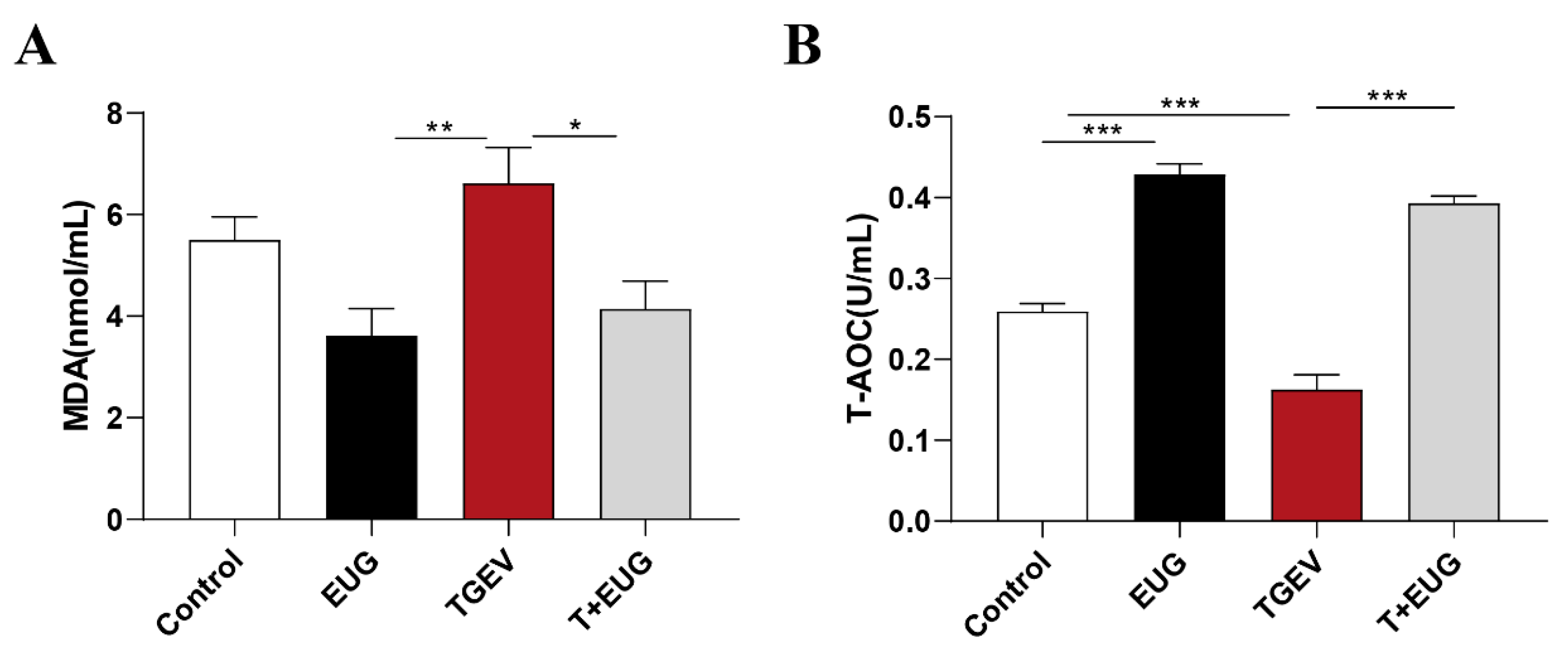
3.1. Effects of Eugenol on Serum Antioxidant Indicators in TGEV-Infected Weaned Piglets

As shown in Figure 1A,B, eugenol supplementation significantly increased serum T-AOC level, and TGEV infection significantly reduced serum T-AOC level. In addition, under the condition of TGEV infection, eugenol supplementation significantly reduced serum MDA and increased serum T-AOC content.

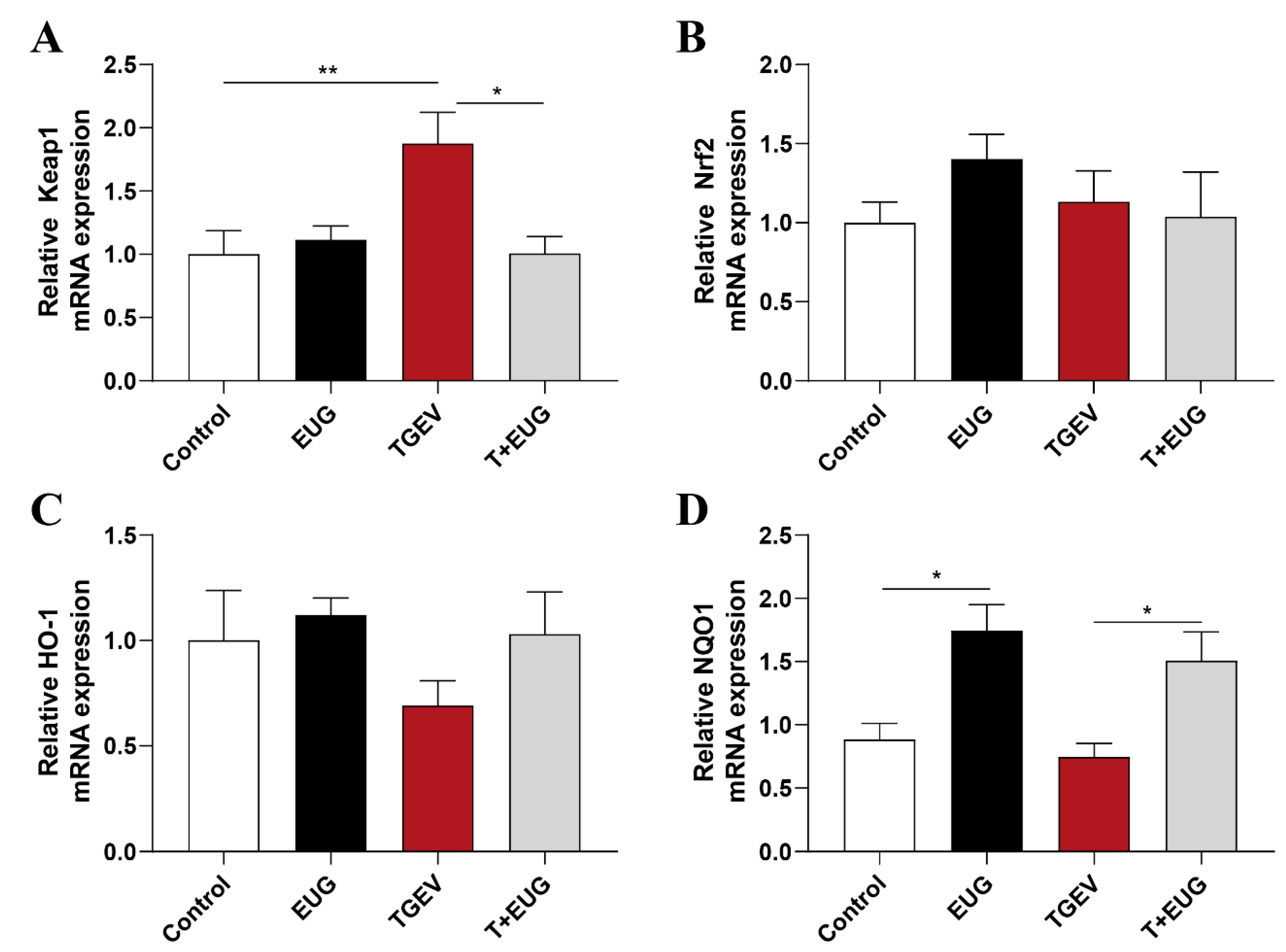
3.2. Effects of Eugenol on Jejunum Antioxidation-Related Genes in TGEV-Infected Weaned Piglets

Keap1-Nrf2-ARE signaling plays an important role in protecting cells from endogenous and exogenous stresses [28]. As shown in Figure 2A–D, TGEV infection increased the mRNA expressions of Keap1 in jejunum of weaned piglets, and eugenol supplementation increased the mRNA expressions of NQO1 in jejunum of weaned piglets. Of note, eugenol supplementation increases the mRNA expressions of NQO1 in jejunum of TGEV-infected piglets and alleviates the increase in Keap1 mRNA relative content.

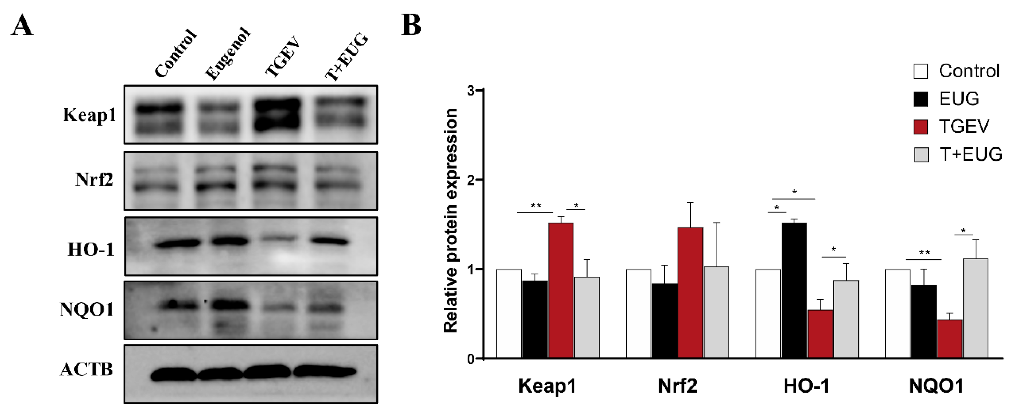
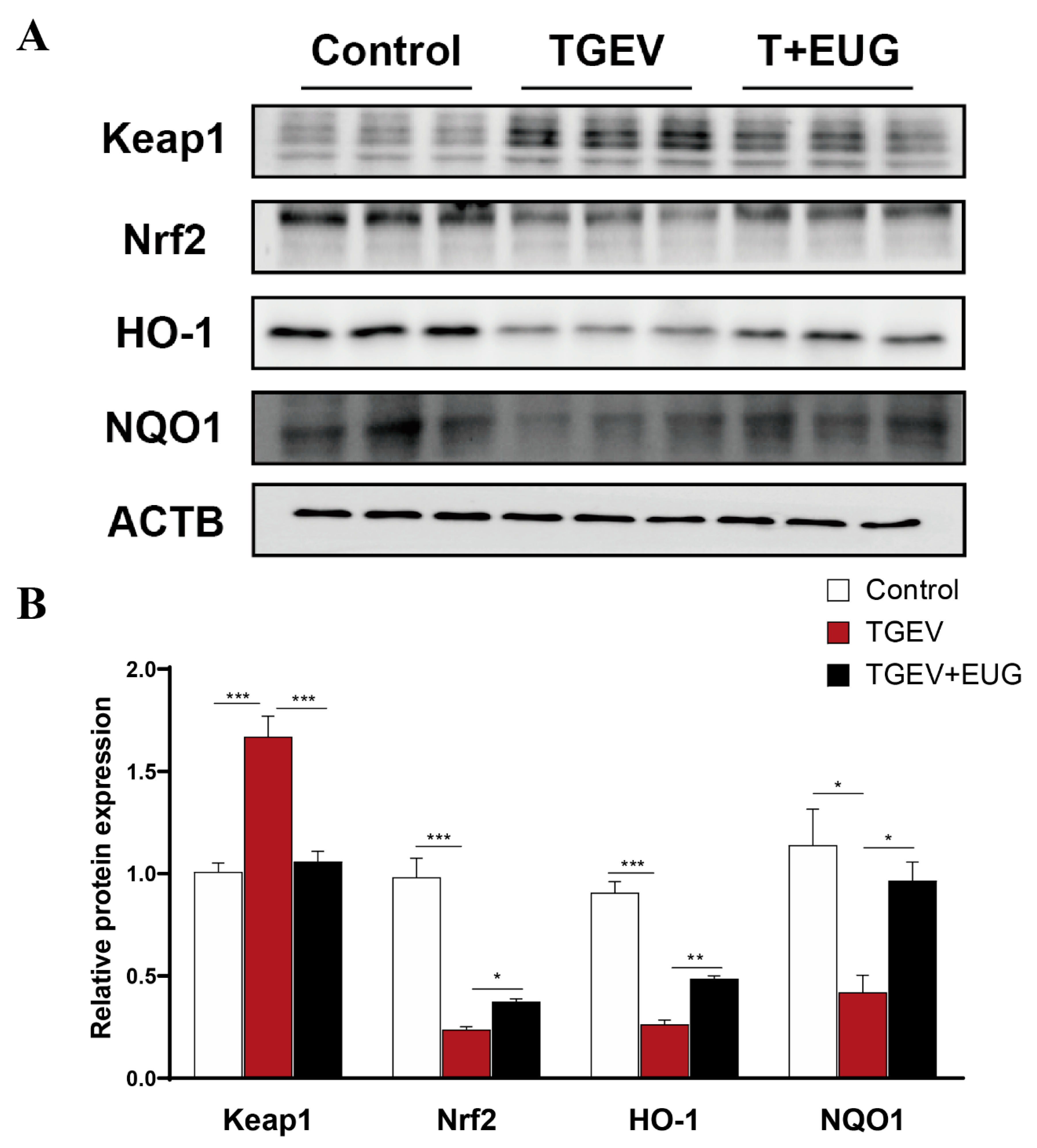
3.3. Effects of Eugenol on Jejunum Antioxidation-Related Proteins in TGEV-Infected Weaned Piglets

As shown in Figure 3A,B, TGEV infection significantly decreased the expression levels of HO-1 and NQO1 protein and increased the expression level of Keap1 protein in jejunum. In addition, eugenol supplementation significantly increased the TGEV-induced decrease in HO-1 and NQO1 protein levels and decreased the TGEV-induced increase in Keap1 protein level in jejunum.

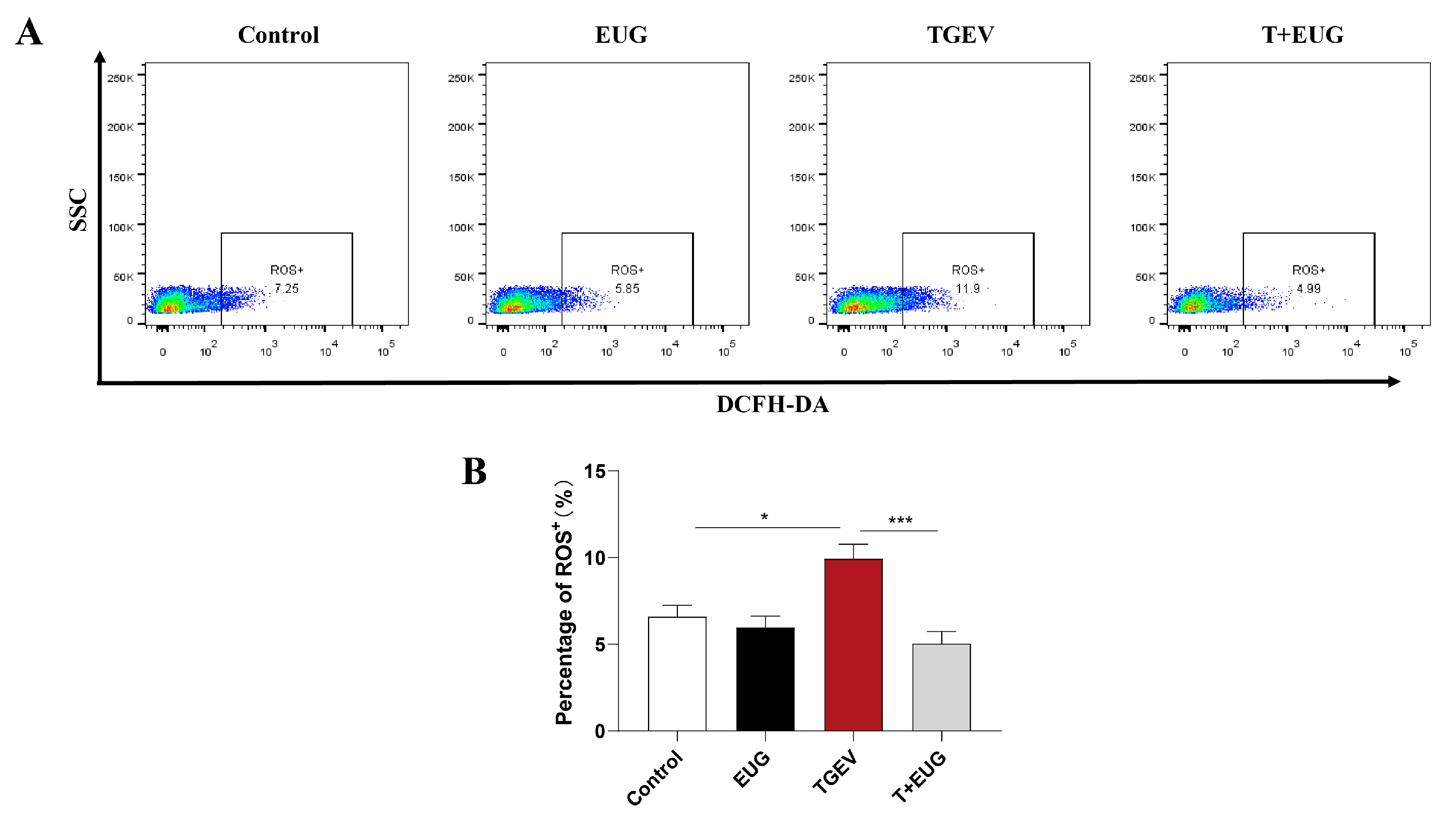
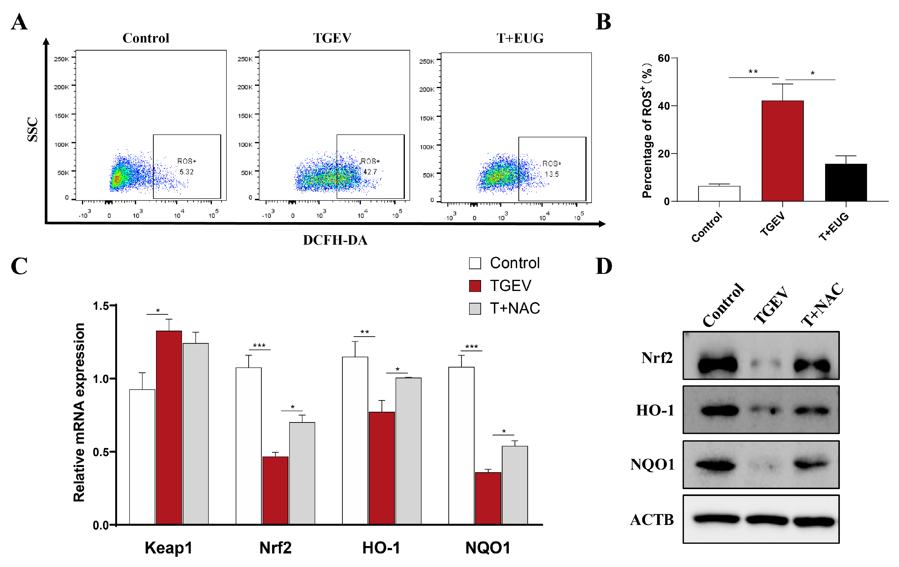
3.4. Eugenol Decreases TGEV-Induced ROS Increase in Jejunal Epithelial Cells of Weaned Piglets

Excessive production of ROS caused by abnormal redox regulation may be one of the causes of cell and tissue damage [29]. As shown in Figure 4A,B, notably, TGEV infection increased ROS level in the jejunum epithelial cells of weaned piglets. In addition, eugenol reduced ROS level in the jejunum epithelial cells of piglets induced by TGEV.

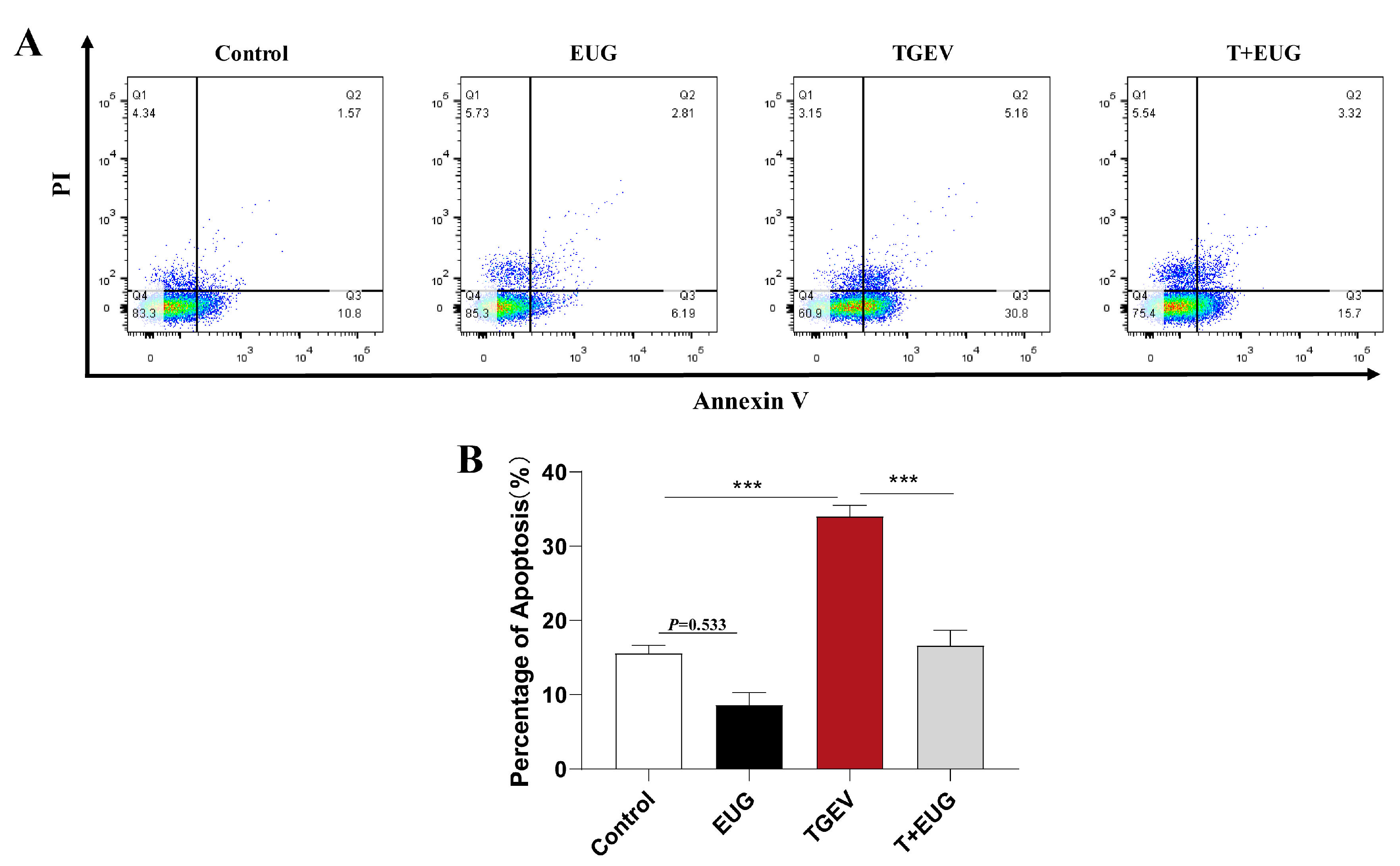
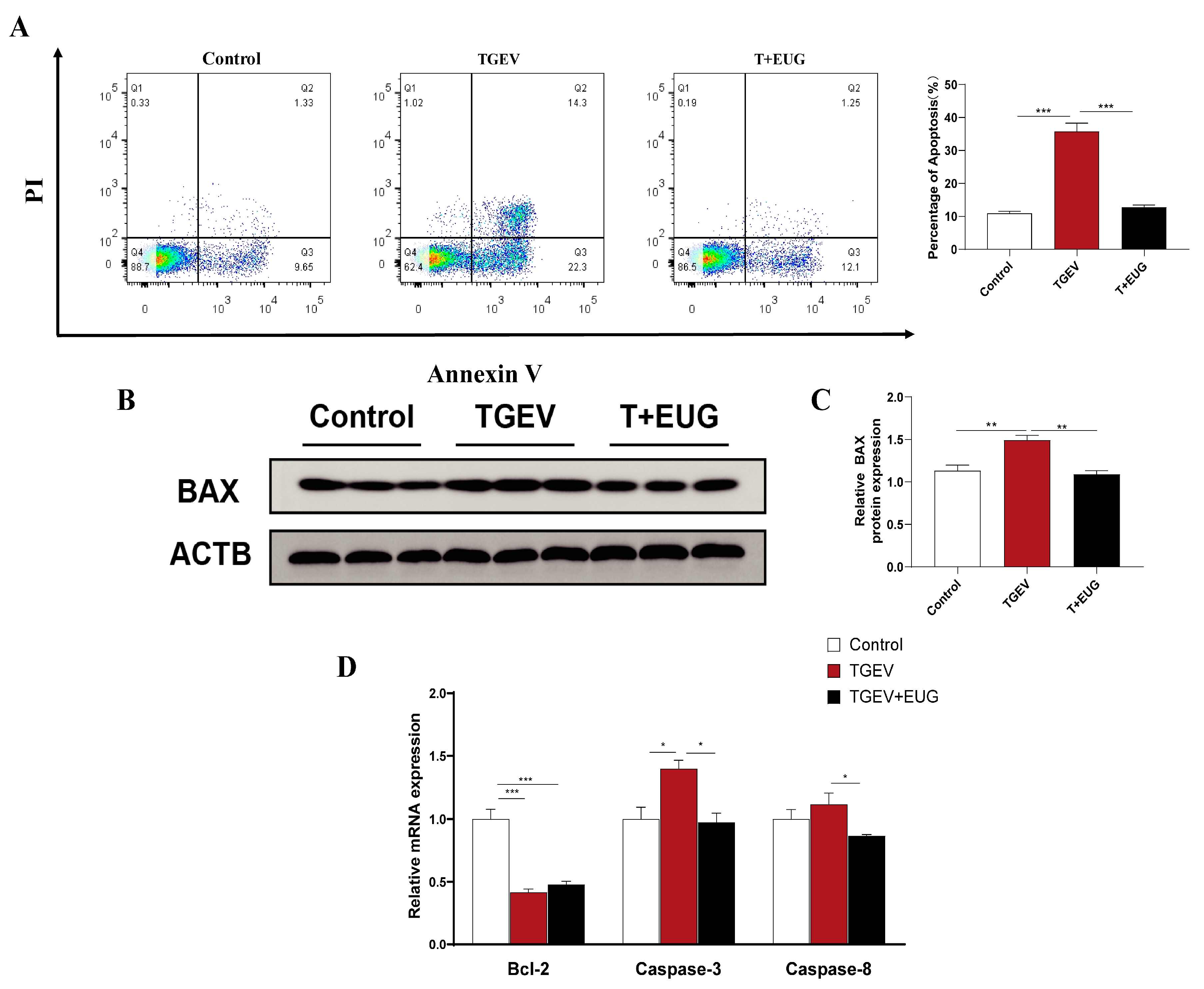
3.5. Eugenol Alleviates TGEV-Induced Jejunal Epithelial Cell Death in Piglets

Apoptosis is a programmed series of events dependent on energy [30]. As shown in Figure 5A,B, notably, TGEV infection increased the proportion of apoptosis in jejunum epithelial cells of weaned piglets, and eugenol supplementation tended to decrease the proportion of apoptosis. Interestingly, eugenol supplementation significantly alleviated the increase in jejunum epithelial cell apoptosis induced by TGEV infection.

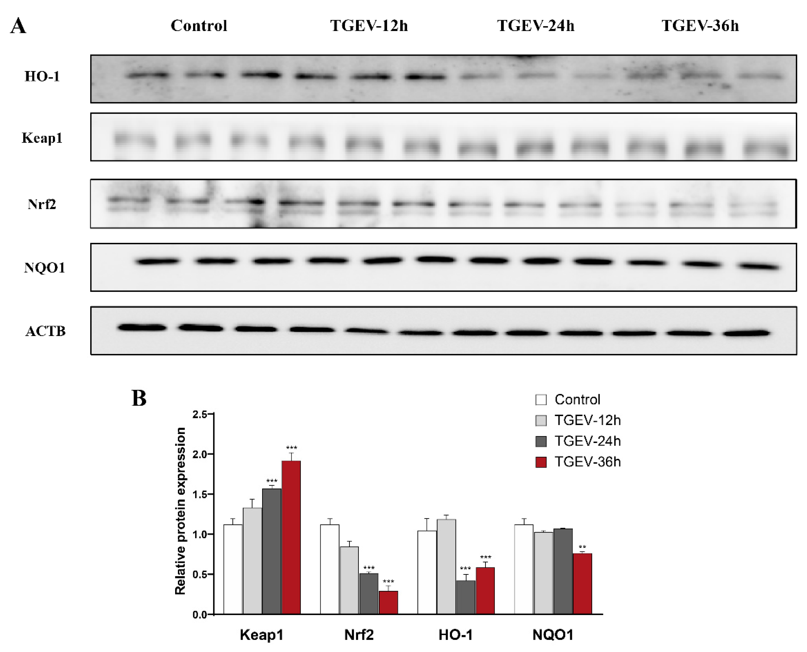
3.6. TGEV Damages the Antioxidant Capacity of IPEC-J2 Cells

To reveal the underlying mechanism by which eugenol attenuates TGEV infection in piglets, we further used IPEC-J2 cells infected with TGEV to construct an in vitro model. As shown in Figure 6A,B, with immunoblotting analysis, TGEV infection significantly increased Keap1 expression and decreased Nrf2, Ho-1 and NQO1 expression in IPEC-J2 cells for 36 h.

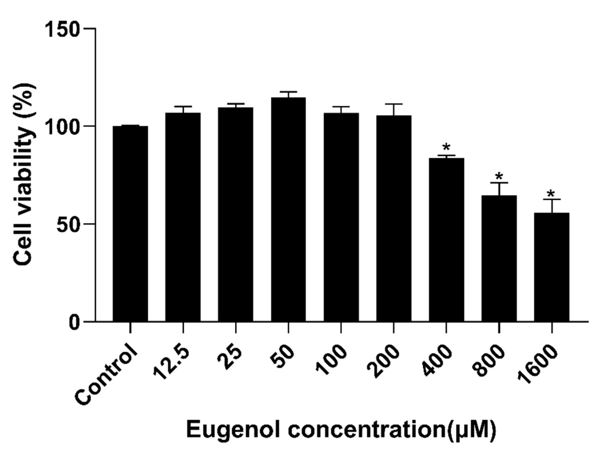
3.7. Effect of Eugenol on IPEC-J2 Cells Viability

As shown in Figure 7, after IPEC-J2 cells were treated with different levels of eugenol for 36 h, high concentrations (400, 800 and 1600 μM) of eugenol significantly reduced IPEC-J2 cells viability. Therefore, eugenol with a concentration of 200 μM was selected for follow-up study.

3.8. Eugenol Alleviates TGEV-Induced Oxidative Stress in IPEC-J2 Cells

We then explored the role of eugenol in TGEV-infected IPEC-J2 cells. As shown in Figure 8A,B, eugenol alleviated the impairment of TGEV infection on Keap1-Nrf2-ARE signaling in IPEC-J2 cells. With immunoblotting analysis, eugenol significantly decreased Keap-1 expression and significantly increased Nrf2, HO-1 and NQO1 expression in TGEV-infected IPEC-J2 cells.

3.9. Eugenol Relieves Oxidative Stress by Removing ROS

As shown in Figure 9A,B, we found that eugenol significantly reduced ROS in TGEV-infected IPEC-J2 cells by flow cytometry. ROS play a key role in regulating redox signaling pathways [31]. Therefore, we used NAC (ROS scavenger) to further explore the mechanism by which eugenol alleviates TGEV-induced oxidative stress. NAC pretreatment of IPEC-J2 cells for 1 h significantly alleviated the TGEV-induced reduction in Nrf2, HO-1 and NQO1 mRNA expression levels (Figure 9C). However, NAC had no effect on the TGEV-induced increase in Keap1 mRNA levels. At the same time, we found that NAC treatment increased Nrf2, HO-1 and NQO1 protein expression levels in TGEV-infected IPEC-J2 cells (Figure 9D).

3.10. Effect of Eugenol on TGEV-Induced IPEC-J2 Cells’ Death Pattern
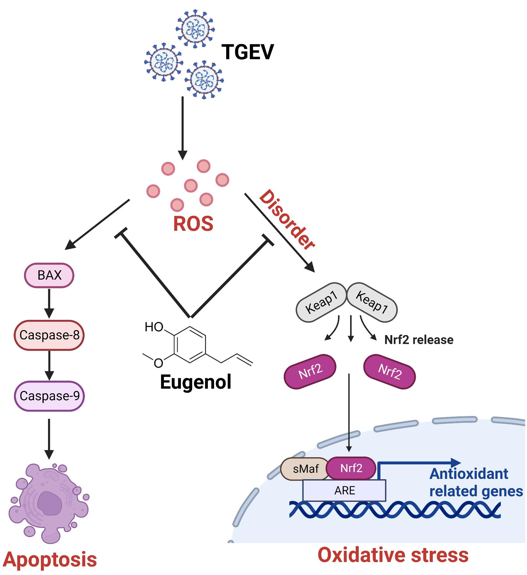
Apoptosis, a common mechanism of cell death, is currently the most extensively studied form. As shown in Figure 10A, TGEV infection increased the proportion of apoptosis (Q3), while eugenol treatment reduced TGEV-induced apoptosis. In addition, eugenol treatment significantly alleviated the TGEV-induced increase in Bax protein expression level and caspase-3 mRNA expression level, and decreased caspase-8 mRNA expression level, but had no effect on Bcl-2 mRNA expression level (Figure 10B,D).

4. Discussion

Under normal physiological conditions, ROS is produced at low levels and controlled by endogenous antioxidants [32]. T-AOC is the sum of enzymatic and non-enzymatic antioxidants. The T-AOC value increases with the increase in antioxidant capacity and decreases with the increase in lipid peroxidation. Therefore, T-AOC can reflect the antioxidant capacity and the level of oxidative damage [33]. MDA is a toxic end product of lipid peroxidation. Its expression level can directly reflect the rate and extent of lipid peroxidation, and indirectly reflects the ability to scavenge free radicals [34]. In the present study, it was observed that TGEV challenge increased the level of ROS in the jejunum of piglets and decreased T-AOC in serum. Many studies have shown that ROS levels are involved in apoptosis, and play an important role in regulating apoptosis [35,36,37]. Therefore, we hypothesized that the increase in intestinal ROS levels induced by TGEV infection might be involved in the occurrence of intestinal apoptosis. This may be the reason why TGEV induces further oxidative stress in the piglet intestine, which has not been reported before. Eugenol has excellent antioxidant capacity and free radical scavenging properties [38,39], and its addition to the diet can overcome the production of ROS caused by TGEV infection. Interestingly, we found a tendency for eugenol to reduce the percentage of apoptosis compared to the control group. This somewhat suggests that the antioxidant capacity of eugenol makes the intestine of piglets less exposed to weaning stress. This may be the direct antioxidant effect of eugenol, or it may be indirectly achieved through antioxidant pathways such as inhibiting MDA levels and increasing T-AOC. More research is needed.
In common antioxidant pathways, Nrf2 is a transcription factor that regulates the nuclear transcription of many genes encoding antioxidant proteins that are required to eliminate oxidative stress [40]. Once activated, Nrf2 dissociates from the Keap1-Nrf2 binding and translocates into the nucleus, where it transactivates genes driven by antioxidant response elements such as NQO1 and HO-1 [41,42]. In this study, TGEV infection significantly increased mRNA expressions of Keap1 in the jejunum of weaned piglets; significantly decreased HO-1 and NQO1 protein expression; and increased Keap1 protein expression in the jejunum of weaned pigs (which may be the indirect reason for the increase in ROS level induced by TGEV). In addition, under TGEV infection, the relative expression of NQO1 mRNA in the jejunum of piglets was significantly increased by dietary eugenol supplementation, which significantly alleviated the TGEV-induced decrease in HO-1 and NQO1 protein levels and the increase in Keap1 protein levels. We noted differences in mRNA and protein expression between HO-1 and NQO1 in the animal samples. The possible reasons may be as follows: (i) The homogeneity between samples is not good and the follow-up experiment can increase the sample size. (ii) Gene mRNA expression is not necessarily linearly related to protein expression because there are many factors regulating gene expression, and the regulation of transcription level is only one link. Post-transcriptional regulation and translational and post-translational regulation all have an impact on the final protein expression. (iii) Moreover, mRNA degradation, protein degradation, modification and folding may lead to inconsistency between mRNA expression and protein expression levels. In general, eugenol could prevent TGEV-induced ROS overproduction and oxidative stress-mediated intestinal apoptosis by enhancing T-AOC, NQO1 and HO-1 levels and decreasing MDA and Keap1 levels.
Disturbed redox homeostasis is a common feature of intestinal disease, characterized by uncontrolled ROS levels and impaired antioxidant defenses [8]. In general, viruses can benefit from either activating or inhibiting Nrf2 in host cells [43,44]. Our results demonstrate that TGEV infection disrupts the Keap1-Nrf2-ARE antioxidant defense system in IPEC-J2 cells. At the same time, eugenol alleviated the TGEV-induced increase in the protein expression level of Keap-1 in IPEC-J2 cells and ameliorated the TGEV-induced decrease in Nrf2, HO-1 and NQO1 protein expression in IPEC-J2 cells. We used NAC as a scavenger of ROS to gain insight into the mechanism by which eugenol alleviates TGEV-induced oxidative stress. A previous study showed that ROS levels affect the Keap1-Nrf2-ARE signaling pathway, which is consistent with our findings [45]. Our data demonstrated that the mRNA and protein expression of Nrf2, HO-1, and NQO1 were increased in IPEC-J2 cells after ROS scavenging in the presence of TGEV infection. This suggests that eugenol may improve the cellular antioxidant capacity by scavenging ROS, which is associated with its potent antioxidant capacity.
During viral infection, viruses interfere with many cellular functions by affecting various intracellular mediators, especially various major cell death pathways [46,47]. The dynamic balance of cell death mode is the functional basis for maintaining the continuous self-renewal of the body [48]. It is well known that the mitochondrial pathway is an important mechanism of apoptotic cell death. The mitochondrial pathway activates apoptotic factors Caspase-9, -8 and -3 [49], and is regulated by Bcl-2 family proteins [50]. TGEV infection simultaneously mediates PK-15 apoptosis through intrinsic and extrinsic apoptotic pathways [51]. Our results showed that eugenol supplementation alleviated TGEV-induced increases in Caspase-3 and Caspase-8 mRNA, as well as BAX protein expression in IPEC-J2 cells. In parallel, the flow cytometry results showed that eugenol downregulated the apoptotic rate of TGEV-infected IPEC-J2 cells. It is noteworthy that ROS are involved in virus-induced apoptosis [52,53,54]. In addition, porcine parvovirus infection leads to ST cell apoptosis through the activation of the ROS-mediated mitochondrial apoptosis pathway [55]. These results imply that eugenol alleviates the apoptosis of IPEC-J2 cells, which may be related to the clearance of intracellular ROS and the enhancement of antioxidant capacity.
The effect of oxidative stress is not specific to TGEV. According to relevant literature, other coronaviruses (SARS-CoV [56], SARS-CoV-2 [57,58] and MERS [59]) can also induce oxidative stress. However, there are no relevant reports showing the effect of eugenol on other coronavirus. Recent reports suggest that TGEV can provide a good model for human highly pathogenic coronaviruses [60,61]. In addition, oxidative stress is one of the pathogeneses of aging, diabetes and cancer. Therefore, we believe that eugenol, a natural product with antioxidative stress properties, has the potential to be an antiviral, antiaging, antidiabetic and anticancer medication.

5. Conclusions

In conclusion, our results reveal the potential therapeutic use of eugenol in TGEV infection. We confirmed that eugenol alleviates TGEV-induced oxidative stress in intestinal epithelial cells by regulating ROS, thereby affecting the Keap1-Nrf2-ARE signaling pathway and reducing TGEV-induced excessive cell apoptosis. This study may reveal the novel mechanisms by which eugenol alleviates oxidative stress in TGEV infection and contribute to its future application in coronavirus-related diseases.

Author Contributions

K.W. and J.L. conceived and designed the experiments. K.W., X.W., H.L. and Y.T. performed cell experiments and animal experiments and wrote the manuscript. D.C., B.Y., J.H., X.M., Z.H., H.Y., Y.L., J.Y. and P.Z. performed biochemical analysis. A.W., J.L., X.M., H.Y. and H.W. gave constructive comments for the results and discussion of the manuscript. All authors have read and agreed to the published version of the manuscript.

Funding

This work was supported by National Natural Science Foundation of China (NSFC) (31702124), and Natural Science Foundation of Sichuan Province (2022NSFSC0072).

Institutional Review Board Statement

All animal experiments were approved by the Institutional Animal Care and Use Committee of the Laboratory Animal Center at Sichuan Agricultural University (SICAU-2015-033).

Informed Consent Statement

Not applicable.

Data Availability Statement

The original contributions presented in the study are included in the article. Further inquiries can be directed to the corresponding author.

Acknowledgments

We thank Yuhan Wang and Yuankang Zhou for their assistance during the experiments. We thank members of the Institute of Animal Nutrition for helpful discussions and technical assistance.

Conflicts of Interest

The authors declare that the research was conducted in the absence of any commercial or financial relationships that could be construed as a potential conflict of interest.

References

  1. Singh, D.; Yi, S.V. On the origin and evolution of SARS-CoV-2. Exp. Mol. Med. 2021, 53, 537–547. [Google Scholar] [CrossRef] [PubMed]
  2. Song, D.; Moon, H.; Kang, B. Porcine epidemic diarrhea: A review of current epidemiology and available vaccines. Clin. Exp. Vaccine Res. 2015, 4, 166–176. [Google Scholar] [CrossRef] [PubMed]
  3. Liu, Q.; Wang, H.-Y. Porcine enteric coronaviruses: An updated overview of the pathogenesis, prevalence, and diagnosis. Vet. Res. Commun. 2021, 45, 75–86. [Google Scholar] [CrossRef] [PubMed]
  4. Wu, A.; Yu, B.; Zhang, K.; Xu, Z.; Wu, D.; He, J.; Luo, J.; Luo, Y.; Yu, J.; Zheng, P.; et al. Transmissible gastroenteritis virus targets Paneth cells to inhibit the self-renewal and differentiation of Lgr5 intestinal stem cells via Notch signaling. Cell Death Dis. 2020, 11, 40. [Google Scholar] [CrossRef] [PubMed]
  5. Pu, J.; Chen, D.; Tian, G.; He, J.; Huang, Z.; Zheng, P.; Mao, X.; Yu, J.; Luo, J.; Luo, Y.; et al. All-Trans Retinoic Acid Attenuates Transmissible Gastroenteritis Virus-Induced Inflammation in IPEC-J2 Cells via Suppressing the RLRs/NF-κB Signaling Pathway. Front. Immunol. 2022, 13, 734171. [Google Scholar] [CrossRef]
  6. Ding, L.; Li, J.; Li, W.; Fang, Z.; Li, N.; Wu, S.; Li, J.; Hong, M. p53- and ROS-mediated AIF pathway involved in TGEV-induced apoptosis. J. Vet. Med. Sci. 2018, 80, 1775–1781. [Google Scholar] [CrossRef]
  7. Ding, L.; Zhao, X.; Huang, Y.; Du, Q.; Dong, F.; Zhang, H.; Song, X.; Zhang, W.; Tong, D. Regulation of ROS in transmissible gastroenteritis virus-activated apoptotic signaling. Biochem. Biophys. Res. Commun. 2013, 442, 33–37. [Google Scholar] [CrossRef]
  8. Piechota-Polanczyk, A.; Fichna, J. Review article: The role of oxidative stress in pathogenesis and treatment of inflammatory bowel diseases. Naunyn Schmiedeberg’s Arch. Pharmacol. 2014, 387, 605–620. [Google Scholar] [CrossRef]
  9. Bhattacharyya, A.; Chattopadhyay, R.; Mitra, S.; Crowe, S.E. Oxidative stress: An essential factor in the pathogenesis of gastrointestinal mucosal diseases. Physiol. Rev. 2014, 94, 329–354. [Google Scholar] [CrossRef]
  10. Filomeni, G.; de Zio, D.; Cecconi, F. Oxidative stress and autophagy: The clash between damage and metabolic needs. Cell Death Differ. 2015, 22, 377–388. [Google Scholar] [CrossRef] [Green Version]
  11. Pan, X.; Zhou, Y.; Duan, X.; Cui, J.; Liu, J.; Song, X.; Ma, W.; Zhang, W.; Liu, Y.; Fan, Y. The inhibitory effect Polygonum Cillinerve polysaccharide on transmissible gastroenteritis virus of swine. Res. Vet. Sci. 2021, 140, 47–55. [Google Scholar] [CrossRef] [PubMed]
  12. Flores-Romero, H.; Ros, U.; Garcia-Saez, A.J. Pore formation in regulated cell death. EMBO J. 2020, 39, e105753. [Google Scholar] [CrossRef] [PubMed]
  13. Kaminskyy, V.O.; Zhivotovsky, B. Free radicals in cross talk between autophagy and apoptosis. Antioxid. Redox Signal. 2014, 21, 86–102. [Google Scholar] [CrossRef] [PubMed]
  14. Wang, X.-H.; Song, T.-Z.; Zheng, H.-Y.; Li, Y.-H.; Zheng, Y.-T. Jejunal epithelial barrier disruption triggered by reactive oxygen species in early SIV infected rhesus macaques. Free Radic. Biol. Med. 2021, 177, 143–155. [Google Scholar] [CrossRef]
  15. Pu, J.; Chen, D.; Tian, G.; He, J.; Huang, Z.; Zheng, P.; Mao, X.; Yu, J.; Luo, J.; Luo, Y.; et al. All-Trans Retinoic Acid Attenuates Transmissible Gastroenteritis Virus-Induced Apoptosis in IPEC-J2 Cells via Inhibiting ROS-Mediated P38MAPK Signaling Pathway. Antioxidants 2022, 11, 345. [Google Scholar] [CrossRef]
  16. Pluskal, T.; Weng, J.-K. Natural product modulators of human sensations and mood: Molecular mechanisms and therapeutic potential. Chem. Soc. Rev. 2018, 47, 1592–1637. [Google Scholar] [CrossRef]
  17. Ruiz-Medina, B.E.; Lerma, D.; Hwang, M.; Ross, J.A.; Skouta, R.; Aguilera, R.J.; Kirken, R.A.; Varela-Ramirez, A.; Robles-Escajeda, E. Green barley mitigates cytotoxicity in human lymphocytes undergoing aggressive oxidative stress, via activation of both the Lyn/PI3K/Akt and MAPK/ERK pathways. Sci. Rep. 2019, 9, 6005. [Google Scholar] [CrossRef]
  18. Morán-Santibañez, K.; Vasquez, A.H.; Varela-Ramirez, A.; Henderson, V.; Sweeney, J.; Odero-Marah, V.; Fenelon, K.; Skouta, R. Larrea tridentata Extract Mitigates Oxidative Stress-Induced Cytotoxicity in Human Neuroblastoma SH-SY5Y Cells. Antioxidants 2019, 8, 427. [Google Scholar] [CrossRef]
  19. Huang, T.; Che, Q.; Chen, X.; Chen, D.; Yu, B.; He, J.; Chen, H.; Yan, H.; Zheng, P.; Luo, Y.; et al. Apple Polyphenols Improve Intestinal Antioxidant Capacity and Barrier Function by Activating the Nrf2/Keap1 Signaling Pathway in a Pig Model. J. Agric. Food Chem. 2022, 70, 7576–7585. [Google Scholar] [CrossRef]
  20. Hseu, Y.-C.; Vudhya Gowrisankar, Y.; Wang, L.-W.; Zhang, Y.-Z.; Chen, X.-Z.; Huang, P.-J.; Yen, H.-R.; Yang, H.-L. The in vitro and in vivo depigmenting activity of pterostilbene through induction of autophagy in melanocytes and inhibition of UVA-irradiated α-MSH in keratinocytes via Nrf2-mediated antioxidant pathways. Redox Biol. 2021, 44, 102007. [Google Scholar] [CrossRef]
  21. Taleuzzaman, M.; Jain, P.; Verma, R.; Iqbal, Z.; Mirza, M.A. Eugenol as a Potential Drug Candidate: A Review. Curr. Top. Med. Chem. 2021, 21, 1804–1815. [Google Scholar] [CrossRef] [PubMed]
  22. Barboza, J.N.; da Silva Maia Bezerra Filho, C.; Silva, R.O.; Medeiros, J.V.R.; de Sousa, D.P. An Overview on the Anti-inflammatory Potential and Antioxidant Profile of Eugenol. Oxid. Med. Cell. Longev. 2018, 2018, 3957262. [Google Scholar] [CrossRef] [PubMed]
  23. Chen, S.; Wu, X.; Tang, S.; Yin, J.; Song, Z.; He, X.; Yin, Y. Eugenol Alleviates Dextran Sulfate Sodium-Induced Colitis Independent of Intestinal Microbiota in Mice. J. Agric. Food Chem. 2021, 69, 10506–10514. [Google Scholar] [CrossRef] [PubMed]
  24. Lu, M.-C.; Ji, J.-A.; Jiang, Z.-Y.; You, Q.-D. The Keap1-Nrf2-ARE Pathway As a Potential Preventive and Therapeutic Target: An Update. Med. Res. Rev. 2016, 36, 924–963. [Google Scholar] [CrossRef]
  25. Abed, D.A.; Goldstein, M.; Albanyan, H.; Jin, H.; Hu, L. Discovery of direct inhibitors of Keap1-Nrf2 protein-protein interaction as potential therapeutic and preventive agents. Acta Pharm. Sin. B 2015, 5, 285–299. [Google Scholar] [CrossRef] [PubMed]
  26. Du, J.; Chen, D.; Yu, B.; He, J.; Yu, J.; Mao, X.; Luo, Y.; Zheng, P.; Luo, J. L-Leucine Promotes STAT1 and ISGs Expression in TGEV-Infected IPEC-J2 Cells via mTOR Activation. Front. Immunol. 2021, 12, 656573. [Google Scholar] [CrossRef]
  27. Robles-Escajeda, E.; Lerma, D.; Nyakeriga, A.M.; Ross, J.A.; Kirken, R.A.; Aguilera, R.J.; Varela-Ramirez, A. Searching in mother nature for anti-cancer activity: Anti-proliferative and pro-apoptotic effect elicited by green barley on leukemia/lymphoma cells. PLoS ONE 2013, 8, e73508. [Google Scholar] [CrossRef]
  28. Kensler, T.W.; Wakabayashi, N.; Biswal, S. Cell survival responses to environmental stresses via the Keap1-Nrf2-ARE pathway. Annu. Rev. Pharmacol. Toxicol. 2007, 47, 89–116. [Google Scholar] [CrossRef]
  29. G Bardallo, R.; Panisello-Roselló, A.; Sanchez-Nuno, S.; Alva, N.; Roselló-Catafau, J.; Carbonell, T. Nrf2 and oxidative stress in liver ischemia/reperfusion injury. FEBS J. 2021. [Google Scholar] [CrossRef]
  30. Su, L.-J.; Zhang, J.-H.; Gomez, H.; Murugan, R.; Hong, X.; Xu, D.; Jiang, F.; Peng, Z.-Y. Reactive Oxygen Species-Induced Lipid Peroxidation in Apoptosis, Autophagy, and Ferroptosis. Oxid. Med. Cell. Longev. 2019, 2019, 5080843. [Google Scholar] [CrossRef] [Green Version]
  31. Guo, Z.; Mo, Z. Keap1-Nrf2 signaling pathway in angiogenesis and vascular diseases. J. Tissue Eng. Regen. Med. 2020, 14, 869–883. [Google Scholar] [CrossRef] [PubMed]
  32. Zheng, S.; Deng, Z.; Chen, F.; Zheng, L.; Pan, Y.; Xing, Q.; Tsao, R.; Li, H. Synergistic antioxidant effects of petunidin and lycopene in H9c2 cells submitted to hydrogen peroxide: Role of Akt/Nrf2 pathway. J. Food Sci. 2020, 85, 1752–1763. [Google Scholar] [CrossRef] [PubMed]
  33. Riedl, M.A.; Saxon, A.; Diaz-Sanchez, D. Oral sulforaphane increases Phase II antioxidant enzymes in the human upper airway. Clin. Immunol. 2009, 130, 244–251. [Google Scholar] [CrossRef] [PubMed]
  34. Tsikas, D. Assessment of lipid peroxidation by measuring malondialdehyde (MDA) and relatives in biological samples: Analytical and biological challenges. Anal. Biochem. 2017, 524, 13–30. [Google Scholar] [CrossRef] [PubMed]
  35. Kang, R.; Li, R.; Dai, P.; Li, Z.; Li, Y.; Li, C. Deoxynivalenol induced apoptosis and inflammation of IPEC-J2 cells by promoting ROS production. Environ. Pollut. 2019, 251, 689–698. [Google Scholar] [CrossRef]
  36. Ji, P.; Huang, H.; Yuan, S.; Wang, L.; Wang, S.; Chen, Y.; Feng, N.; Veroniaina, H.; Wu, Z.; Wu, Z.; et al. ROS-Mediated Apoptosis and Anticancer Effect Achieved by Artesunate and Auxiliary Fe(II) Released from Ferriferous Oxide-Containing Recombinant Apoferritin. Adv. Healthc. Mater. 2019, 8, e1900911. [Google Scholar] [CrossRef]
  37. Zhao, B.; Luo, J.; Wang, Y.; Zhou, L.; Che, J.; Wang, F.; Peng, S.; Zhang, G.; Shang, P. Metformin Suppresses Self-Renewal Ability and Tumorigenicity of Osteosarcoma Stem Cells via Reactive Oxygen Species-Mediated Apoptosis and Autophagy. Oxid. Med. Cell. Longev. 2019, 2019, 9290728. [Google Scholar] [CrossRef]
  38. Carvalho, R.P.R.; Lima, G.D.d.A.; Machado-Neves, M. Effect of eugenol treatment in hyperglycemic murine models: A meta-analysis. Pharmacol. Res. 2021, 165, 105315. [Google Scholar] [CrossRef]
  39. Zhang, L.; Wang, K.; Lei, Y.; Li, Q.; Nice, E.C.; Huang, C. Redox signaling: Potential arbitrator of autophagy and apoptosis in therapeutic response. Free Radic. Biol. Med. 2015, 89, 452–465. [Google Scholar] [CrossRef]
  40. DeNicola, G.M.; Chen, P.-H.; Mullarky, E.; Sudderth, J.A.; Hu, Z.; Wu, D.; Tang, H.; Xie, Y.; Asara, J.M.; Huffman, K.E.; et al. NRF2 regulates serine biosynthesis in non-small cell lung cancer. Nat. Genet. 2015, 47, 1475–1481. [Google Scholar] [CrossRef] [PubMed] [Green Version]
  41. Singh, A.; Misra, V.; Thimmulappa, R.K.; Lee, H.; Ames, S.; Hoque, M.O.; Herman, J.G.; Baylin, S.B.; Sidransky, D.; Gabrielson, E.; et al. Dysfunctional KEAP1-NRF2 interaction in non-small-cell lung cancer. PLoS Med. 2006, 3, e420. [Google Scholar] [CrossRef] [PubMed]
  42. Erlank, H.; Elmann, A.; Kohen, R.; Kanner, J. Polyphenols activate Nrf2 in astrocytes via H2O2, semiquinones, and quinones. Free Radic. Biol. Med. 2011, 51, 2319–2327. [Google Scholar] [CrossRef] [PubMed]
  43. Cuadrado, A.; Pajares, M.; Benito, C.; Jiménez-Villegas, J.; Escoll, M.; Fernández-Ginés, R.; Garcia Yagüe, A.J.; Lastra, D.; Manda, G.; Rojo, A.I.; et al. Can Activation of NRF2 Be a Strategy against COVID-19? Trends Pharmacol. Sci. 2020, 41, 598–610. [Google Scholar] [CrossRef] [PubMed]
  44. Olagnier, D.; Farahani, E.; Thyrsted, J.; Blay-Cadanet, J.; Herengt, A.; Idorn, M.; Hait, A.; Hernaez, B.; Knudsen, A.; Iversen, M.B.; et al. SARS-CoV2-mediated suppression of NRF2-signaling reveals potent antiviral and anti-inflammatory activity of 4-octyl-itaconate and dimethyl fumarate. Nat. Commun. 2020, 11, 4938. [Google Scholar] [CrossRef] [PubMed]
  45. Liu, H.; Zhu, S.; Han, W.; Cai, Y.; Liu, C. DMEP induces mitochondrial damage regulated by inhibiting Nrf2 and SIRT1/PGC-1α signaling pathways in HepG2 cells. Ecotoxicol. Environ. Saf. 2021, 221, 112449. [Google Scholar] [CrossRef] [PubMed]
  46. Foo, J.; Bellot, G.; Pervaiz, S.; Alonso, S. Mitochondria-mediated oxidative stress during viral infection. Trends Microbiol. 2022, 30, 679–692. [Google Scholar] [CrossRef]
  47. Tummers, B.; Green, D.R. The evolution of regulated cell death pathways in animals and their evasion by pathogens. Physiol. Rev. 2022, 102, 411–454. [Google Scholar] [CrossRef] [PubMed]
  48. Orzalli, M.H.; Kagan, J.C. Apoptosis and Necroptosis as Host Defense Strategies to Prevent Viral Infection. Trends Cell Biol. 2017, 27, 800–809. [Google Scholar] [CrossRef] [PubMed]
  49. Bhargava, P.; Schnellmann, R.G. Mitochondrial energetics in the kidney. Nat. Rev. Nephrol. 2017, 13, 629–646. [Google Scholar] [CrossRef] [PubMed]
  50. Shang, H.-S.; Shih, Y.-L.; Lee, C.-H.; Hsueh, S.-C.; Liu, J.-Y.; Liao, N.-C.; Chen, Y.-L.; Huang, Y.-P.; Lu, H.-F.; Chung, J.-G. Sulforaphane-induced apoptosis in human leukemia HL-60 cells through extrinsic and intrinsic signal pathways and altering associated genes expression assayed by cDNA microarray. Environ. Toxicol. 2017, 32, 311–328. [Google Scholar] [CrossRef] [PubMed]
  51. Ding, L.; Xu, X.; Huang, Y.; Li, Z.; Zhang, K.; Chen, G.; Yu, G.; Wang, Z.; Li, W.; Tong, D. Transmissible gastroenteritis virus infection induces apoptosis through FasL- and mitochondria-mediated pathways. Vet. Microbiol. 2012, 158, 12–22. [Google Scholar] [CrossRef] [PubMed]
  52. Li, F.; Li, J.; Wang, P.-H.; Yang, N.; Huang, J.; Ou, J.; Xu, T.; Zhao, X.; Liu, T.; Huang, X.; et al. SARS-CoV-2 spike promotes inflammation and apoptosis through autophagy by ROS-suppressed PI3K/AKT/mTOR signaling. Biochim. Biophys. Acta Mol. Basis Dis. 2021, 1867, 166260. [Google Scholar] [CrossRef] [PubMed]
  53. Pei, J.; Deng, J.; Ye, Z.; Wang, J.; Gou, H.; Liu, W.; Zhao, M.; Liao, M.; Yi, L.; Chen, J. Absence of autophagy promotes apoptosis by modulating the ROS-dependent RLR signaling pathway in classical swine fever virus-infected cells. Autophagy 2016, 12, 1738–1758. [Google Scholar] [CrossRef] [PubMed]
  54. Xu, X.; Xu, Y.; Zhang, Q.; Yang, F.; Yin, Z.; Wang, L.; Li, Q. Porcine epidemic diarrhea virus infections induce apoptosis in Vero cells via a reactive oxygen species (ROS)/p53, but not p38 MAPK and SAPK/JNK signalling pathways. Vet. Microbiol. 2019, 232, 1–12. [Google Scholar] [CrossRef] [PubMed]
  55. Zhao, X.; Xiang, H.; Bai, X.; Fei, N.; Huang, Y.; Song, X.; Zhang, H.; Zhang, L.; Tong, D. Porcine parvovirus infection activates mitochondria-mediated apoptotic signaling pathway by inducing ROS accumulation. Virol. J. 2016, 13, 26. [Google Scholar] [CrossRef]
  56. Imai, Y.; Kuba, K.; Neely, G.G.; Yaghubian-Malhami, R.; Perkmann, T.; van Loo, G.; Ermolaeva, M.; Veldhuizen, R.; Leung, Y.H.C.; Wang, H.; et al. Identification of oxidative stress and Toll-like receptor 4 signaling as a key pathway of acute lung injury. Cell 2008, 133, 235–249. [Google Scholar] [CrossRef] [PubMed]
  57. Zhang, S.; Wang, J.; Wang, L.; Aliyari, S.; Cheng, G. SARS-CoV-2 virus NSP14 Impairs NRF2/HMOX1 activation by targeting Sirtuin 1. Cell. Mol. Immunol. 2022, 19, 872–882. [Google Scholar] [CrossRef]
  58. Laforge, M.; Elbim, C.; Frère, C.; Hémadi, M.; Massaad, C.; Nuss, P.; Benoliel, J.-J.; Becker, C. Tissue damage from neutrophil-induced oxidative stress in COVID-19. Nat. Rev. Immunol. 2020, 20, 515–516. [Google Scholar] [CrossRef]
  59. Cai, Z.; Lu, C.; He, J.; Liu, L.; Zou, Y.; Zhang, Z.; Zhu, Z.; Ge, X.; Wu, A.; Jiang, T.; et al. Identification and characterization of circRNAs encoded by MERS-CoV, SARS-CoV-1 and SARS-CoV-2. Brief. Bioinform. 2021, 22, 1297–1308. [Google Scholar] [CrossRef] [PubMed]
  60. Sun, L.; Zhao, C.; Fu, Z.; Fu, Y.; Su, Z.; Li, Y.; Zhou, Y.; Tan, Y.; Li, J.; Xiang, Y.; et al. Genome-scale CRISPR screen identifies TMEM41B as a multi-function host factor required for coronavirus replication. PLoS Pathog. 2021, 17, e1010113. [Google Scholar] [CrossRef]
  61. Zhao, L.; Li, L.; Xue, M.; Liu, X.; Jiang, C.; Wang, W.; Tang, L.; Feng, L.; Liu, P. Gasdermin D Inhibits Coronavirus Infection by Promoting the Noncanonical Secretion of Beta Interferon. mBio 2022, 13, e0360021. [Google Scholar] [CrossRef] [PubMed]
Figure 1. Effects of eugenol on serum antioxidant indicators in TGEV-infected weaned piglets. (A,B) The levels of MDA and T-AOC in serum (n = 8). Data are expressed as the mean ± SEM. * p < 0.05, ** p < 0.01, *** p < 0.001.
Antioxidants 11 01838 g001
Figure 2. Effects of eugenol on jejunum antioxidation-related genes in TGEV-infected weaned piglets. (AD) Relative mRNA expressions of Keap1, Nrf2, HO-1 and NQO1 in the jejunum mucosa (n = 6–8). Data are expressed as the mean ± SEM. * p < 0.05, ** p < 0.01.
Antioxidants 11 01838 g002
Figure 3. Effects of eugenol on jejunum antioxidation-related protein in TGEV-infected weaned piglets. (A,B) Protein abundance of Keap1, Nrf2, HO-1 and NQO1 in jejunum mucosa (n = 3). Data are expressed as the mean ± SEM. * p < 0.05, ** p < 0.01.
Antioxidants 11 01838 g003
Figure 4. Eugenol decreases TGEV-induced ROS increase in jejunal epithelial cells of weaned piglets. (A,B) ROS levels in jejunum epithelial cells were analyzed by flow cytometry; SSC means side scatter (n = 6–8). Data are expressed as the mean ± SEM. * p < 0.05, *** p < 0.001.
Antioxidants 11 01838 g004
Figure 5. Eugenol alleviates TGEV-induced jejunal epithelial cell death in piglets. (A,B) The flow cytometry assays detecting the percentage of apoptosis in jejunum epithelial cells (n = 6–8). Data are expressed as the mean ± SEM. *** p < 0.001.
Antioxidants 11 01838 g005
Figure 6. TGEV damages the antioxidant capacity of IPEC-J2 cells. (A,B) Protein abundance of Keap1, Nrf2, HO-1 and NQO1 in IPEC-J2 cells (n = 3). Data are expressed as the mean ± SEM. ** p < 0.01, *** p < 0.001.
Antioxidants 11 01838 g006
Figure 7. Effect of eugenol on IPEC-J2 cells’ viability. IPEC-J2 cells were treated with eugenol for 36 h (n = 6). Data are expressed as the mean ± SEM. * p < 0.05.
Antioxidants 11 01838 g007
Figure 8. Eugenol ameliorates TGEV-induced oxidative stress in IPEC-J2 cells. (A,B) Protein abundance of Keap1, Nrf2, HO-1 and NQO1 in IPEC-J2 cells (n = 3). Data are expressed as the mean ± SEM. * p < 0.05, ** p < 0.01, *** p < 0.001.
Antioxidants 11 01838 g008
Figure 9. Eugenol relieves oxidative stress by removing ROS. IPEC-J2 cells were pretreated with eugenol (200 μM) or NAC (5 mM) for 1 h and then infected with TGEV for 36 h. (A,B) The flow cytometry assays detecting ROS levels in IPEC-J2 cells; SSC means side scatter (n = 3). (C) The expression of Keap1, Nrf2, HO-1 and NQO1 mRNA in IPEC-J2 cells were examined by RT-PCR (n = 3). (D) Protein abundance of Nrf2, HO-1 and NQO1 in IPEC-J2 cells. Data are expressed as the mean ± SEM. * p < 0.05, ** p < 0.01, *** p < 0.001.
Antioxidants 11 01838 g009
Figure 10. Effect of eugenol on TGEV-induced IPEC-J2 cells’ death pattern. IPEC-J2 cells were treated with 200 μM eugenol for 1 h and then infected with TGEV for 36 h. (A) The percentage of apoptosis in jejunum epithelial cells was analyzed by flow cytometry (n=3). (B,C) Immunoblot analysis and quantification of BAX in IPEC-J2 cells (n = 3). (D) Relative mRNA expressions of Bcl-2, Caspase-3 and Caspase-8 in IPEC-J2 cells (n = 3). Data are expressed as the mean ± SEM. * p < 0.05, ** p < 0.01, *** p < 0.001.
Antioxidants 11 01838 g010
Table 1. Composition and nutrient level of basal diet.
Ingredients%Nutrient Level 3Contents
Corn33.80Digestible energy (calculated, Mcal/kg)3.54
Extruded corn22.20Crude Protein (%)19.49
Soybean meal7.42Calcium (%)0.75
Extruded full-fat soybean8.79Available phosphorus (%)0.37
Fish meal3.94Lysine1.35
Whey powder5.00Methionine0.39
Soybean protein concentrate8.00Methionine + cysteine0.68
Soybean oil1.65Threonine0.80
Sucrose2.00Tryptophan0.22
Limestone0.62
Dicalcium phosphate0.46
NaCl0.20
L-Lysine HCl (78%)0.32
DL-Methionine0.07
L-Threonine (98.5%)0.02
Tryptophan (98%)0.01
Chloride choline0.15
Vitamin premix 10.05
Mineral premix 20.30
Total100
1 The vitamin premix provided the following per kg of diet: 6000 IU of VA, 3000 IU of VD3, 24 IU of VE, 3 mg of VK3, 1.5 mg of VB1, 6 mg of VB2, 3 mg of VB6, 0.02 mg of VB12, 14 mg of niacin, 15 mg of pantothenic acid, 0.75 mg of folic acid, and 0.1 mg of biotin. 2 The mineral premix provided the following per kg of diet: Fe(FeSO4·H2O), 100 mg; Cu(CuSO4·5H2O), 6 mg; Mn(MnSO4·H2O), 4 mg; Zn(ZnSO4·H2O), 100 mg; I(KI), 0.3 mg; Se(Na2SeO3), 0.3 mg. 3 Nutrient level values are calculated.
Table 2. Primer sequences table.
GenePrimersSequencesProduct sizeAccession Numbers
β-actinForwardGCAAATGCTTCTAGGCGGAC148XM_021086047.1
ReverseGCGTCCATCACAGCTTCTCA
Keap1ForwardTCTGCTTAGTCATGGTGACCT143NM_001114671.1
ReverseAAGGGACAACACCACCACTG
Nrf2ForwardCTACGGGATTGGGGTTTGGG124XM_013984303.2
ReverseAACTCAAACAGGGGAAGGGC
HO-1ForwardTACCGCTCCCGAATGAACAC140NM_001004027.1
ReverseTGGTCCTTAGTGTCCTGGGT
NQO1ForwardTGCTTACACATACGCTGCCA113NM_001159613.1
ReverseCGTGGATACCCTGCAGAGAG
Bcl-2ForwardAGCATGCGGCCTCTATTTGA120XM_021099593.1
ReverseGGCCCGTGGACTTCACTTAT
Caspase-3ForwardGGATTGAGACGGACAGTGGG124NM_214131.1
ReverseCCGTCCTTTGAATTTCGCCA
Caspase-8ForwardGGATCCCAGGATTTGCCTCC135NM_001031779.2
ReverseCAGGCTCAGGAACTTGAGGG
Publisher’s Note: MDPI stays neutral with regard to jurisdictional claims in published maps and institutional affiliations.

