Article

Knockout and Inhibition of Ape1: Roles of Ape1 in Base Excision DNA Repair and Modulation of Gene Expression

1
Department of Pharmacological Sciences, Renaissance School of Medicine, Stony Brook University, Stony Brook, NY 11794-8651, USA
2
Molecular and Cellular Biochemistry Program, Stony Brook University, Stony Brook, NY 11794-8651, USA
*
Author to whom correspondence should be addressed.
Antioxidants 2022, 11(9), 1817; https://doi.org/10.3390/antiox11091817
Submission received: 21 July 2022 / Revised: 31 August 2022 / Accepted: 7 September 2022 / Published: 15 September 2022

Abstract

:
Apurinic/apyrimidinic endonuclease 1/redox effector-1 (Ape1/Ref-1) is the major apurinic/apyrimidinic (AP) endonuclease in mammalian cells. It functions mainly in the base excision repair pathway to create a suitable substrate for DNA polymerases. Human Ape1 protein can activate some transcription factors to varying degrees, dependent on its N-terminal, unstructured domain, and some of the cysteines within it, apparently via a redox mechanism in some cases. Many cancer studies also suggest that Ape1 has potential for prognosis in terms of the protein level or intracellular localization. While homozygous disruption of the Ape1 structural gene APEX1 in mice causes embryonic lethality, and most studies in cell culture indicate that the expression of Ape1 is essential, some recent studies reported the isolation of viable APEX1 knockout cells with only mild phenotypes. It has not been established by what mechanism the Ape1-null cell lines cope with the endogenous DNA damage that the enzyme normally handles. We review the enzymatic and other activities of Ape1 and the recent studies of the properties of the APEX1 knockout lines. The APEX1 deletions in CH12F3 and HEK293 FT provide an opportunity to test for possible off-target effects of Ape1 inhibition. For this work, we tested the Ape1 endonuclease inhibitor Compound 3 and the redox inhibitor APX2009. Our results confirmed that both APEX1 knockout cell lines are modestly more sensitive to killing by an alkylating agent than their Ape1-proficient cells. Surprisingly, the knockout lines showed equal sensitivity to direct killing by either inhibitor, despite the lack of the target protein. Moreover, the CH12F3 APEX1 knockout was even more sensitive to Compound 3 than its APEX1+ counterpart. Thus, it appears that both Compound 3 and APX2009 have off-target effects. In cases where this issue may be important, it is advisable that more specific endpoints than cell survival be tested for establishing mechanism.

1. Introduction

Genomic DNA is under the relentless threat of molecular decay due to endogenous and exogenous damage, which threatens genetic stability. Reactive oxygen species (ROS), notably the byproducts of aerobic metabolism, produce an array of DNA lesions, including 8-oxoguanine (8-oxoG) and thymine glycol. Hydrolysis and various other reactive metabolites add many other DNA lesions to this burden [1]. Many of these lesions disrupt DNA structure only slightly or not at all, but they nonetheless can exert mutagenic or cytotoxic effects if left unrepaired. Ring-opened aldehyde apurinic/apyrimidinic (AP) sites can form a covalent bond with neighboring nucleotides or histone lysines, which can result in highly toxic DNA interstrand cross-links and histone-DNA cross-links [2,3].
Base excision DNA repair (BER) is the frontline process that deals with most of the small lesions. Following the removal of a damaged base by a DNA glycosylase, the 5′-phosphodiester bond of the resulting apurinic/apyrimidinic (AP) site is cleaved by Ape1; that enzyme also cleaves spontaneous hydrolytic AP sites. The gap-filling reaction is finished by other BER proteins. Ape1 is also reported to be involved in activating some transcription factors by maintaining their reduced forms; for this, the protein is also called Redox effector-1 (Ref-1). This role may contribute to cellular defenses against oxidative stress. For example, Ape1 can induce the tetramerization and expression of transcription factor p53, which can then upregulate the expression of superoxide dismutase 2 [4]. However, the detailed mechanism by which Ape1 modulates transcription factors remains unclear. A current model is via thiol exchange, with one Ape1 cysteine attacking a disulfide bond in the target protein, eventually resolving to yield the reduced target protein (and oxidized Ape1). Cysteine-65 of Ape1 has been implicated in this process, despite its being sequestered inside the protein structure [5].
Despite broad interest in the roles of Ape1 in BER and transcriptional control, animal studies have been limited because the APEX1 gene is essential and knockouts are embryonic-lethal. APEX1 conditional knockout mice have been generated by the Cre/lox method, but they still retain considerable amounts of Ape1 in some organs even after the knockout system is activated. Mice haploinsufficient for Ape1 are more sensitive to oxidative stress and have a higher chance of developing adenocarcinoma and lymphoma [6]. Surprisingly, a human APEX1 transgene could not compensate for the murine APEX1 knockout (APEX1-KO) during mouse embryo development, suggesting the importance of dynamic Ape1 expression regulation [7]. This review will summarize the biological function of Ape1 in the order of its activities based on knockout studies, and we will assess the prospects for Ape1 as a target for cancer therapy.

2. Ape1 Overview

2.1. Base Excision Repair and DNA End Processing

BER corrects much of the oxidatively-induced and other endogenous base damage in most organisms, including humans. The first step of BER is the removal of the damaged base by a DNA glycosylase, which results in an AP site. If the DNA glycosylase is monofunctional, in mammalian cells the AP site will first be processed by Ape1. Ape1 hydrolyzes the 5′ phosphodiester bond at the AP site, leaving a normal 3′-OH nucleotide on one side and a 5′-deoxyribose-5-phosphate (5′-dRP) on the other. Some DNA glycosylases are bifunctional, able to further process the AP site through a β-lyase activity (e.g., OGG1) or a β,δ-lyase activity (e.g., NEIL1). β-elimination produces a 3′-terminal unsaturated hydroxyaldehyde, which must be removed by Ape1 to allow DNA synthesis, and a normal 5′-phosphate. β,δ-elimination leaves a 3′-phosphate and a 5′-phosphate, bracketing a small gap. In that case, polynucleotide kinase phosphatase (PNKP) provides a DNA synthesis primer by removing the 3′-phosphate, which Ape1 does very inefficiently [1]. The next step is gap filling. During short patch BER (SP-BER), Ape1, Polβ, X-ray repair cross-complementing protein 1 (Xrcc1) in complex with DNA ligase IIIα (Lig3), and Poly(ADP-ribose) polymerase (PARP) are consecutively recruited to the lesion for repair. Polβ also removes the 5′-dRP by using a dRP lyase activity in its separable N-terminal domain, with the polymerase inserting a nucleotide to replace the damaged one. Finally, Lig3 can seal the nick. Proliferating cell nuclear antigen (PCNA), Ape1, Polβ (or DNA polymerase δ/ε in proliferating cells), DNA ligase I (Lig1), PARP, and Flap endonuclease 1 (Fen1) work together to conduct long patch BER (LP-BER), which involves strand replacement DNA synthesis. The resulting flap is cleaved by Fen1 and the nick sealed by Lig1.
Ape1 uses the same active site to conduct exonuclease and endonuclease activity. For correctly paired nucleotides, the Ape1 exonuclease activity is only ~1% of the AP endonuclease activity; for mismatched nucleotides the activity can be up to 10% of the AP endonuclease. The Ape1 exonuclease may contribute to the fidelity of BER, since Polβ lacks intrinsic or other associated 3′-exonuclease proofreading activity, with an error rate around 10−4 (vs. 10−6 or better for replication polymerases) [8]. Ape1 exonuclease activity can act on a variety of other substrates, including the nucleoside analog β-l-dioxolane-cytidine and a biotinylated nucleotide [9,10]. However, Ape1′s exonuclease activity varies on different substrate structures. For example, the Ape1 exonuclease activity is lower for blunt-end substrates than for substrates with recessed 3′-ends [10]. It is worth mentioning that Ape1 cannot excise nucleotides from substrates containing more than one mismatched 3′-residue.
Ape1 acts as an end processor in single-strand break repair. Single-strand breaks (SSB), which can result from free radical attack or spontaneous β-elimination at an AP site, is another prevalent type of lesion. Unlike the SSB generated during BER, these SSB are not sheltered by proteins and they need to be end-processed before DNA polymerases and DNA ligases can act [11]. Although most bulky adducts and double helix-distorting lesions are repaired by nucleotide excision repair, that pathway is unable to remove lesions at the end of single-strand breaks or double-strand breaks. Ape1 can excise bulky lesions on recessed 5′ ends, blunt ends, and 3′ overhangs shorter than two nucleotides [12]. In addition, Lin et al., pointed out that Ape1 can initiate 3′ end resection on SSB and is indispensable for inducing the ATR-Chk1 signaling pathway in Xenopus extracts [13]. The homologous Ape2 protein has much weaker AP endonuclease activity than does Ape1, but Ape2 operates to remove certain types of blocked DNA 3′ termini [14]

2.2. Redox Signaling and Oxidative G-Quadruplex Formation in Gene Expression

Genome instability, resistance to cell death, senescence, and angiogenesis are some of the hallmarks of cancer, and Ape1 may function in most of these processes. A meta-analysis study concluded that a high Ape1 expression level is associated with a short overall survival rate of patients with solid tumors [15]. This seems paradoxical because the primary role of Ape1 seems to be helping maintain genetic stability by repairing damaged DNA and inducing cell apoptosis by activating p53. However, some cancer cells may hijack this process to resist DNA damage induced by chemotherapy. Part of the reason for the upregulation of Ape1 could relate to the high ROS level in cancer cells [16]. For example, knockdown of Ape1 by siRNA in B-lymphoblastoid TK6 cells and colon tumor HCT116 cells dramatically sensitized the cells to killing by the antitumor drug bleomycin or to X-ray treatment [17].
Ape1 has been reported to activate various transcription factors, including AP-1, HIF-1α, and NF-κB. The mechanism of this activation is thought to be via a redox reaction [18,19,20]. According to the Ape1 co-crystal structure with DNA [21], no disulfide bond can form between any two cysteine residues. Homology analysis revealed that, except for Cys 65, which is unique to mammals, the rest of the cysteines are conserved among vertebrates. Single cysteine-to-alanine mutations revealed that only C65A abolished Ape1-induced AP-1 DNA binding [22,23]. In addition, cysteine substitution of zebrafish Ape1 Thr58, equivalent to Cys65 in mammalian Ape1, conferred redox activity on the zebrafish protein expressed in human cells [24]. Further cysteine-to-alanine substitutions showed that Ape1 C93A/C99A double mutations lost redox activity and Ape1 with all cysteines substituted except Cys65, Cys93, and Cys99 retained the redox activity [25]. Two other studies [20,26] found that Cys65 (Cys64 in murine Ape1) was unnecessary for the Ape1 redox activity.
Ape1 is also proposed to modulate transcription by stabilizing G-quadruplexes in certain promotors. Guanine is the DNA base most susceptible to oxidative damage due to its low redox potential [27]. Therefore, lesions such as 8-oxoG can form throughout the genome when ROS accumulates. Within a gene body, 8-oxoG can stall transcription [28,29]. However, 8-oxoG in a promotor can induce the formation of BER-stabilized G-quadruplexes that enhance gene expression [30]. In this scenario, Ogg1 first excises the 8-oxoG base and hands the AP repair intermediate to Ape1. The AP site and BER machinery destabilize the normal double helix structure and lower the energy barrier for forming G-quadruplexes in some sequences [31]. The AP endonuclease of Ape1 is highly attenuated at abasic sites in certain parts of a G-quadruplex structure, compared with its activity on double-strand DNA [32]. In such cases, rather than incising the phosphodiester bond, Ape1 now helps stabilize these G-quadruplexes. The natively unstructured Ape1 N-terminus is essential for this type of binding, with the acetylation of N-terminal lysines fine-tuning the residence time of bound Ape1 [33]. Recent studies found that some key oncogenes, including HIF-1α, VEGF, and c-MYC, have at least one G-quadruplex in their promoter [34,35,36]. These dual and overlapping functions (repair and gene expression) highlight the potential of Ape1 as a cancer therapy target. Ape1 inhibitors that separately target either the redox activity or the nuclease activity are available, some of them now undergoing clinical trials, which will be discussed in a later section.

2.3. RNA Processing

RNA accounts for 80–90% of a cell’s total nucleic acid and is more vulnerable to damage than is DNA [37]. All the same sources of damage mentioned earlier for DNA also act on RNA, which is further susceptible to many nucleases in the cell. Damaged gene-coding mRNA can produce truncated or misfolded proteins, while damaged non-coding mRNAs can compromise gene regulation [38,39].
AP endonuclease activity for RNA and the ribonuclease H activity were first observed for Ape1 in the context of lesion-containing DNA/RNA hybrids [40]. In addition, single-strand RNA endonuclease, weak 3′-phosphatase, and 3′-exoribonuclease activities have also been reported for Ape1, and they depend on the same active site as the DNA-cleaving nuclease [40,41]. Therefore, impairing the RNase H activity without disturbing the AP endonuclease activity is a significant barrier to determining the biological relevance of these activities.
Lee et al., first reported that Ape1 could cleave c-Myc rRNA between UA and CA in single-strand or weakly paired regions to control the amount of the transcript [42,43]. Tell et al., [44] found that Ape1, through its N-terminal domain, physically interacts with nucleophosmin (Npm1) and colocalizes in the nucleolus. This interaction is disrupted by the acetylation of some N-terminal Ape1 lysine residues, which releases additional enzyme into the rest of the nucleus, suggesting that this response allows a rapid deployment of BER in the face of DNA damage [44]. Suppression of Ape1 results in the accumulation of oxidized RNA and reduced protein synthesis [45]. Although Ape1 lacks the canonical mitochondrial targeting signal, it has been found in mitochondria [45] and protects the organelle’s DNA and RNA [46,47,48].

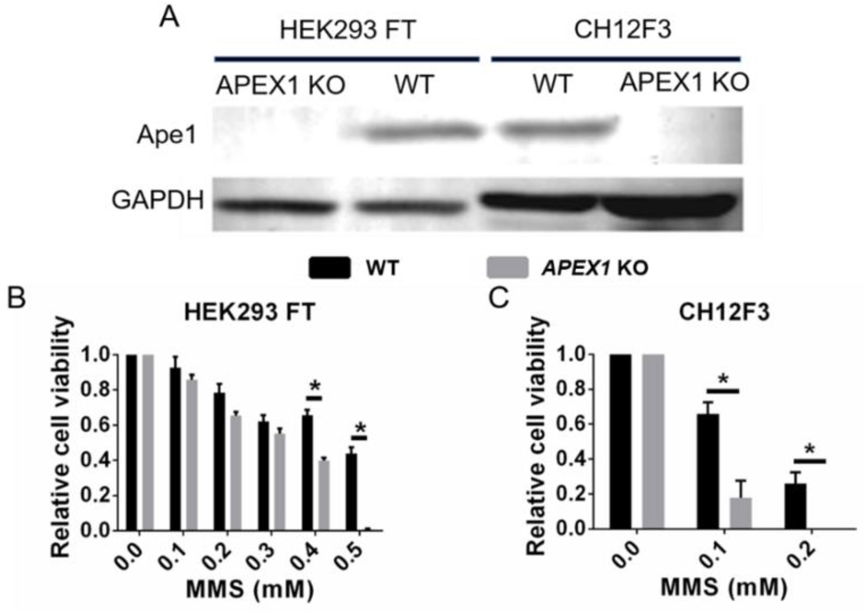
2.4. Ape1 Knockout Cell Lines: How Do They Survive?

The biological function of Ape1 has been studied using genetic knockout and RNAi-mediated knockdown approaches. Independent studies in two different mouse strains both showed that the homozygous deletion of murine APEX1 leads to embryonic degeneration after implantation, in one case by day 5.5 and in others by day 9.5 [49,50,51], with degeneration occurring throughout the embryo. However, these studies merely indicated the critical importance of Ape1 without shedding light on which of its activities might be the essential one(s).
Recently, two APEX1 conditional knockout mouse models were developed to study the relationship between Ape1 and senescence or nervous system development. A tissue-specific APEX1-KO in the central and peripheral nervous system did not discernibly affect embryonic development and pups were born in Mendelian ratios [51]. However, the rapid accumulation of DNA damage, accompanied by neuronal degeneration, was observed soon after birth, with all the animals dead by 3 weeks of age [51]. Li et al., [52] induced APEX1-KO in all tissues using an inducible Cre/lox system. Mice that lost APEX1 earlier (day 7 or day 12 after birth) exhibited severe dysfunction phenotypes, including impaired growth, reduced organ size, and premature senescence, with more than 80% of the animals dying before day 28. Mice that lost APEX1 later (6 weeks) exhibited milder defects but did show clear signs of premature aging by 8 months [52]. Although both studies failed to eliminate Ape1 protein completely, they indicated that the effects of Ape1 deficiency vary in different developmental stages and organs.
Generally, APEX1 haploinsufficiency or knockdown phenotypes are similar to but milder than those with APEX1 homozygous knockouts. Heterozygous APEX1-KO mice showed an increased sensitivity to DNA-damaging agents (e.g., ultraviolet-B radiation, 2-nitropropane, and azoxymethane) and redox dysregulation, accompanied by an increased frequency of spontaneous tumors [6,53,54,55]. Given the limitations of animal studies, efforts to identify the Ape1 activities required for survival have been addressed by manipulating cells in culture. Two independent studies confirmed the vital role of the Ape1 endonuclease in a mouse line [7] and in multiple human cell lines [56], with the cysteine-64/65 residue being unnecessary for cell survival. Suppression of Ape1 via siRNA in HCT116 (colon tumor), MCF7 (breast tumor), or TK6 (normal but immortalized human lymphoblasts) led to cell cycle arrest within 48 h, accompanied by the accumulation of abasic damage in genomic DNA and the activation of apoptosis [56]. These defects were all reversed by the expression of the non-homologous and structurally unrelated yeast Apn1 protein, providing very clear confirmation of the importance of supporting BER. Izumi et al., used vectors expressing different mutant Ape1 proteins in cells expressing an inducible APEX1-KO system. The results showed that the expression of Ape1 lacking the redox activity rescued the apoptotic phenotype, while nuclease-defective Ape1 or Ape1 mutated in Lys6 and Lys7 did not [7].
Interestingly, emerging studies reported that partial Ape1 deficiency did not detectably affect the replication of HeLa cells, CH12F3 cells, HEK293 FT cells, HCT116 cells, and HCC1937 cells [57,58,59,60]. The first report of a viable APEX1-KO was in a study addressing the role of Ape1 in AID-mediated class switch recombination in Ig genes [61], in which a rescue vector expressing Ape1 was maintained while all 3 APEX1 copies in the hypotriploid mouse line CH12F3 were deleted. Surprisingly, removal of the rescue plasmid did not affect cell proliferation, although the complete APEX1-KO did sensitize cells to the simple alkylating agent methylmethane sulfonate (MMS) [61]. Recently, a total knockout of APEX1 in (human) HEK293 FT cells (which also have three copies of the gene) gave a very similar result to that of CH12F3 cells; there was no impact on cell proliferation and only a modest increase in MMS sensitivity [59]. A severe Ape1 knockdown leads to more unrepaired AP sites in genomic DNA [56], but the mRNA levels of both monofunctional DNA glycosylases (e.g., MutyH, Ung) and multifunctional DNA glycosylases (e.g., Ogg1 and Neil2) were not significantly changed [54,59,62]. Thus, DNA glycosylase expression was not changed to compensate for the Ape1 deficiency and the extra unrepaired AP sites most likely reflect sluggish BER, with an accumulation of this intermediate. In this context, Ape1-deficient cells might have increased tolerance of AP sites in the genome. Another possibility is that other DNA repair pathways are elevated as a compensating mechanism for the loss of Ape1.

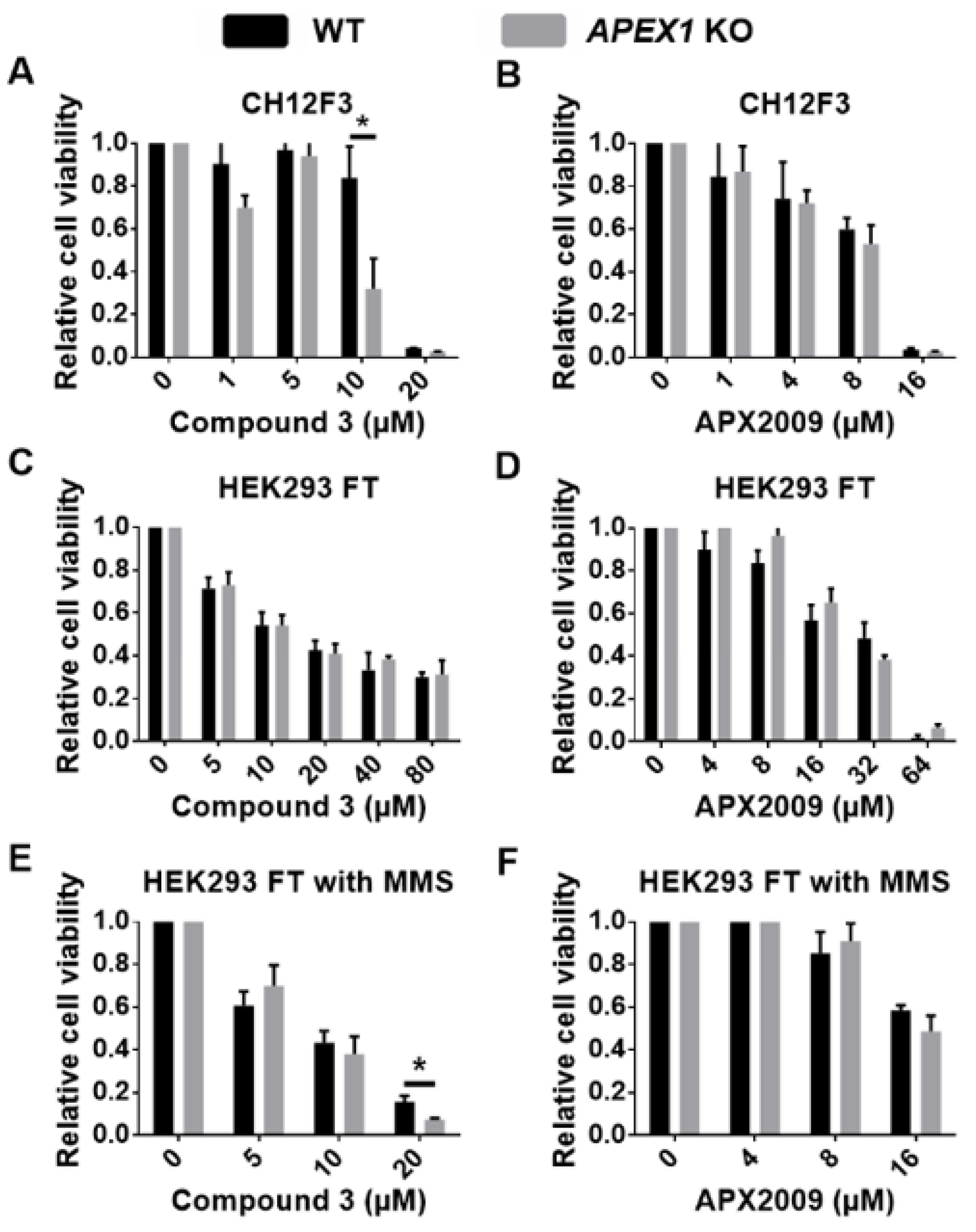
2.5. Off-Target Effects in Cell Killing by Ape1 Inhibitors

Inhibitors are simple and effective tools with which to study the biological functions of a protein, although their effectiveness can be limited by their targeting specificity. For Ape1, inhibitors have been important reagents in efforts to distinguish which of its activities underlies which phenotype. Inhibitors have been developed with specificity for either the nuclease [63,64,65,66,67] (Table 1) or the redox activity [23,68,69,70,71] (Table 2). Moreover, an inhibitor of the Ape1 redox activity is now also in clinical trials for a rare ocular disease [69]. While it may not matter for the clinical effectiveness of current applications, off-target effects of inhibitors could produce misleading conclusions during further drug development. As best we can tell from the published literature, the Ape1 inhibitors have not been thoroughly tested for possible off-target effects. Here we list some of the widely used Ape1 inhibitors and summarize the data on their possible off-target effects (Table 1 and Table 2).

3. Materials and Methods

3.1. MTT Cell Viability Assay

HEK293 FT wild-type and APEX1-KO cells were plated into 96-well plates at a density of 1.3 × 10⁴ cells/well in DMEM with 10% fetal bovine serum (FBS) and antibiotic/antimycotic mix (Sigma, A5955-100, St. Louis, MI, USA). CH12F3 wild-type and APEX1-KO cells were collected by centrifugation and resuspended in phenol red-free RPMI-1640 with L-glutamine, 10% FBS, and antibiotic/antimycotic mix (Sigma, A5955-100, St. Louis, MI, USA) and then plated on 96-well plates at a density of 2.2 × 10⁴ cells/well. The plates were incubated for 24 h at 37 °C in a 5% CO2 humidified incubator. Immediately prior to use, the Ape1 inhibitors and methylmethane sulfonate (MMS) were diluted with a culture medium containing 3% FBS. For both HEK293 FT and CH12F3 cells, the FBS in the medium was reduced from 10% to 3% before the addition of inhibitors or MMS. After 24 h of treatment, MTT (3-(4,5-dethylthiazol-2-yl)-2,5-diphenyltetrazolium bromide) was then added at a concentration of 0.5 mg/mL, followed by a 3-h incubation. Stop solution (1% SDS and 0.01 M HCl) was used to dissolve formazan crystals, and the absorbance was measured at 590 nm.

3.2. Western Blotting

Cells were collected by scraping (HEK293 FT) or centrifugation (CH12F3) and lysed in radioimmunoprecipitation assay (RIPA) buffer (150 mM NaCl, 1% NP-40, 0.5% sodium deoxycholate, 0.1% sodium dodecyl sulfate (SDS), 50 mM Tris-HCl, pH = 8.0, and complete protease inhibitor cocktail (Roche, 11697498001, Indianapolis, IN, USA). An aliquot of each protein sample was used to determine the protein concentration by Bradford assay (Bio-Rad, 5000006, Hercules, CA, USA). The remainder of the protein samples were boiled in Laemmli buffer (2% SDS, 5% 2-mercaptoethanol, 10% glycerol, 0.01% bromophenol blue, and 0.062 M Tris-HCl, pH 6.7) for 5 min and diluted to a protein concentration of 1 mg/mL. Protein samples were resolved in 10% SDS- polyacrylamide gels according to the Thermo-Fisher gel casting protocol (Thermo-Fisher Document Part 0909.INS, Waltham, MA, USA) and transferred to hydrophilic polyvinylidene fluoride membranes (Sigma, IPFL20200, St. Louis, MI, USA). The membranes were blocked in 5% non-fat milk for 1 h at room temperature and incubated with rabbit polyclonal anti-Ape1 antibody (1:800, Novus, #NB100-101SS, Centennial, CA, USA) and rabbit polyclonal anti-GAPDH antibody (1:1,000, Rockland, 600-401-A33, Limerick, PA, USA) at 4 °C overnight. The blots were then washed with blocking buffer and incubated with IRDye 800CW labeled goat anti-rabbit IgG (1:10,000, LI-COR, 926-32211, Lincoln, NE, USA) at room temperature for 30 min before imaging on a LI-COR Odyssey.

4. Evaluation of Compound 3 and APX2009 for Possible Off-Target Effects

We addressed the question of Ape1 inhibitor targeting by employing the two published APEX1-KO cell lines—mouse CH12F3 [61] and human HEK293 FT [59]—both of which appear to function as well without Ape1 protein as they do with it. We verified the APEX1-KO lines by Western blotting, which confirmed the absence of Ape1 protein (Figure 1A). We also confirmed that, compared with the parental lines, both types of APEX1-KO cells had increased sensitivity to cell killing by MMS, noting that CH12F3 was inherently more MMS-sensitive than HEK293 FT (Figure 1B,C).
Following this confirmation of their Ape1 status, we measured these cell lines for the direct toxicity of Ape1 inhibitors that target two different functions: the AP endonuclease inhibitor Compound 3 [63] and the redox inhibitor APX2009 [68]. These agents can kill Ape1-proficient cells; therefore, if they are highly targeted, the expectation was that this toxicity would be lost in the absence of Ape1. Instead, CH12F3 APEX1-KO cells exhibited even greater sensitivity to direct killing by Compound 3 than did their APEX1+ counterparts, but with a seemingly narrow window around 10 µM inhibitor treatment (Figure 2A). For the APX2009 inhibitor, we observed no APEX1-dependent difference for CH12F3 killing (Figure 2B).
For HEK293 FT cells, the survival of the APEX1+ and APEX1-KO versions was the same under all concentrations tested for both Compound 3 and APX2009 (Figure 2C,D). Thus, it appears that the direct killing of cells by either Compound 3 or APX2009 includes substantial off-target effects.
To test whether the reduction of cell viability resulted from insufficient BER, we used a combined treatment of HEK293 FT cells with 0.2 mM MMS and either of the inhibitors. MMS generates numerous base lesions that are processed by BER. The Compound 3 plus MMS treatment reduced the viability of wild-type and APEX1-KO cells to a similar extent for Compound 3 concentrations up to 10 μM (Figure 2E). That result is compatible with inhibition of Ape1 activity in vivo [67], with the Compound 3-treated APE1+ cells being a phenocopy of APE1-KO cells. Note that there was cytotoxic synergy between Compound 3 and MMS (compare Figure 2C,E). The biological significance of the difference seen at 20 μM Compound 3 combined with MMS is hard to judge, as the survival of the Ape1-expressing cells was already quite low (Figure 2E). Because APX2009 is an inhibitor of the Ape1 redox activity rather than the nuclease, we expected that it would not sensitize APEX1+ cells to MMS, which was the case, and with no additional effect in the APEX1-KO cells (compare Figure 2D,F). Kelley et al., [68] reported that APX2009 enhanced Ape1 DNA repair activity in human fibroblasts and rodent neuronal cells; however, we did not find evidence for this effect in HEK293 FT cells.

5. Discussion: Mechanism of Off-Target Effects of Ape1 Inhibitors

The Ape1 inhibitors examined in our work were developed based on their ability to suppress specific Ape1 activities [63,68]. For their potential as cancer therapy agents, cell killing was explored as a biological endpoint, although that is inherently a crude readout of toxic effects, because numerous pathways can lead to that single endpoint. While the effects of the nuclease inhibitor Compound 3 in Ape1-expressing cells were consistent with its interference with DNA repair (sensitization to MMS), it was unexpected that the agent’s direct killing effects were essentially independent of the cellular Ape1 status.
Given the verified importance of Ape1 in routine DNA maintenance [51,52,56,72], the surprising viability of the two APEX1-KO lines tested here could reflect in those cases the mobilization of other repair enzymes or pathways. For cells undergoing DNA replication, as in our experiments, the accumulation of unrepaired AP sites would lead to blocked replication forks, with a constant need for the activation of fork protection pathways to prevent the formation of double-strand breaks [73]. Thus, it is possible that, in addition to their known targeting of Ape1, Compound 3 and perhaps APX2009 also interfere with the fork protection systems. Another possibility is that the accumulation of such unrepaired AP residues leads to the formation of some sites that have other lesions positioned nearby (clustered lesions [74,75,76]), which could require the participation of additional repair systems. However, such explanations do not account very well for the lack of difference in toxicity between the APEX1+ and the APEX1-KO lines.
Clearly, future work on this issue will have to center on determining whether additional DNA repair pathways are indeed marshalled in Ape1-deficient cells to enable their survival. Alternative possibilities include the suppression of apoptosis pathways or other cell death mechanisms. Furthermore, the off-target effects of the inhibitors may actually have advantages in tumor treatment, provided that cancer cells are shown to be more sensitive to them than normal cells in the same tissues. Indeed, such off-target effects could even help identify new pathways of cell survival in the face of DNA damage.

6. Concluding Points

  • Discovered as a DNA repair enzyme, Ape1 has been associated with multiple other roles, including both redox and non-redox activation of transcription factors;
  • Ape1 can stabilize G-quadruplexes by binding but not cleaving AP sites in certain positions, which can mediate some transcriptional effects;
  • Ape1 is essential for embryonic development in mice and probably for mammals in general;
  • Genetic knockdown and knockout experiments indicate that the DNA repair function is essential in most cell types in culture;
  • Inhibitors have been developed to target either the nuclease activity of Ape1 or its redox activity;
  • Two viable cell lines have been developed with the Ape1-coding gene APEX1 deleted; these lines have mild phenotypes, the basis of which is unknown;
  • The Ape1 inhibitors show similar toxic effects in APEX1-knockout cells and their APEX1+ counterparts, indicating that the compounds have significant off-target effects.

Author Contributions

B.D. conceived the experimental approach; Z.X. planned, executed, and analyzed the experimental work. The authors jointly planned the manuscript, primarily authored by Z.X. and edited by B.D. All authors have read and agreed to the published version of the manuscript.

Funding

This work was supported by grant to B.D. from the Stony Brook University Office of the Vice-President for Research Award #93214, and NASA grant 80NSSC19M0215 (P.I. Timothy Glotch).

Institutional Review Board Statement

Not applicable.

Informed Consent Statement

Not applicable.

Data Availability Statement

Data is contained within the article.

Acknowledgments

HEK293 FT wild-type and APEX1 knockout cells were a generous gift from Dmitry O. Zharkov (Novosibirsk State University, Novosibirsk, Russia). CH12F3 wild-type and APEX1 knockout cells were obtained from Kefei Yu (Michigan State University, East Lansing, MI). APX2009 was a generous gift from Mark R. Kelley (Indiana University School of Medicine, Indianapolis, IN). We are grateful to Mark R. Kelley and David M. Wilson III (Hasselt University, Diepenbeek, Belgium) for advice on the choice and application of the inhibitors. We thank Larry W. Hardy and Hyungjin Kim for helpful comments on the manuscript. All these individuals have confirmed their listing here.

Conflicts of Interest

The authors declare no conflict of interest.

References

  1. Demple, B. Special problems for base excision repair in coping with oxidatively-induced DNA damage. In DNA Damage, DNA Repair, and Disease; Dizdaroglu, M., Lloyd, R.S., Eds.; The Royal Society of Chemistry: Cambridge, UK, 2021; Volume 1, pp. 204–219. [Google Scholar]
  2. Dutta, S.; Chowdhury, G.; Gates, K.S. Interstrand cross-links generated by abasic sites in duplex DNA. J. Am. Chem. Soc. 2007, 129, 1852–1853. [Google Scholar] [CrossRef]
  3. Pachva, M.C.; Kisselev, A.F.; Matkarimov, B.T.; Saparbaev, M.; Groisman, R. DNA-histone cross-links: Formation and repair. Front. Cell Dev. Biol. 2020, 8, 607045. [Google Scholar] [CrossRef] [PubMed]
  4. Hao, J.; Du, H.; Liu, F.; Lu, J.C.; Yang, X.C.; Cui, W. Apurinic/apyrimidinic endonuclease/redox factor 1 (Ape1) alleviates myocardial hypoxia-reoxygenation injury by inhibiting oxidative stress and ameliorating mitochondrial dysfunction. Exp. Ther. Med. 2019, 17, 2143–2151. [Google Scholar] [CrossRef] [PubMed]
  5. Park, M.S.; Kim, C.S.; Joo, H.K.; Lee, Y.R.; Kang, G.; Kim, S.J.; Choi, S.; Lee, S.D.; Park, J.B.; Jeon, B.H. Cytoplasmic localization and redox cysteine residue of Ape1/Ref-1 are associated with its anti-inflammatory activity in cultured endothelial cells. Mol. Cells 2013, 36, 439–445. [Google Scholar] [CrossRef] [PubMed]
  6. Meira, L.B.; Devaraj, S.; Kisby, G.E.; Burns, D.K.; Daniel, R.L.; Hammer, R.E.; Grundy, S.; Jialal, I.; Friedberg, E.C. Heterozygosity for the mouse APEX gene results in phenotypes associated with oxidative stress. Cancer Res. 2001, 61, 5552–5557. [Google Scholar]
  7. Izumi, T.; Brown, D.B.; Naidu, C.V.; Bhakat, K.K.; Macinnes, M.A.; Saito, H.; Chen, D.J.; Mitra, S. Two essential but distinct functions of the mammalian abasic endonuclease. Proc. Natl. Acad. Sci. USA 2005, 102, 5739–5743. [Google Scholar] [CrossRef]
  8. Osheroff, W.P.; Jung, H.K.; Beard, W.A.; Wilson, S.H.; Kunkel, T.A. The fidelity of DNA polymerase beta during distributive and processive DNA synthesis. J. Biol. Chem. 1999, 274, 3642–3650. [Google Scholar] [CrossRef]
  9. Chou, K.M.; Cheng, Y.C. An exonucleolytic activity of human apurinic/apyrimidinic endonuclease on 3′ mispaired DNA. Nature 2002, 415, 655–659. [Google Scholar] [CrossRef]
  10. Liu, T.C.; Lin, C.T.; Chang, K.C.; Guo, K.W.; Wang, S.; Chu, J.W.; Hsiao, Y.Y. Ape1 distinguishes DNA substrates in exonucleolytic cleavage by induced space-filling. Nat. Commun. 2021, 12, 601. [Google Scholar] [CrossRef]
  11. Caldecott, K.W. Single-strand break repair and genetic disease. Nat. Rev. Genet. 2008, 9, 619–631. [Google Scholar] [CrossRef]
  12. Liu, T.C.; Guo, K.W.; Chu, J.W.; Hsiao, Y.Y. Understanding Ape1 cellular functions by the structural preference of exonuclease activities. Comput. Struct. Biotechnol. J. 2021, 19, 3682–3691. [Google Scholar] [CrossRef] [PubMed]
  13. Lin, Y.; Raj, J.; Li, J.; Ha, A.; Hossain, M.A.; Richardson, C.; Mukherjee, P.; Yan, S. Ape1 senses DNA single-strand breaks for repair and signaling. Nucleic Acids Res. 2020, 48, 1925–1940. [Google Scholar] [CrossRef] [PubMed]
  14. Alvarez-Quilon, A.; Wojtaszek, J.L.; Mathieu, M.C.; Patel, T.; Appel, C.D.; Hustedt, N.; Rossi, S.E.; Wallace, B.D.; Setiaputra, D.; Adam, S.; et al. Endogenous DNA 3′ blocks are vulnerabilities for BRCA1 and BRCA2 deficiency and are reversed by the Ape2 nuclease. Mol. Cell 2020, 78, 1152–1165.e8. [Google Scholar] [CrossRef]
  15. Yuan, C.L.; He, F.; Ye, J.Z.; Wu, H.N.; Zhang, J.Y.; Liu, Z.H.; Li, Y.Q.; Luo, X.L.; Lin, Y.; Liang, R. Ape1 overexpression is associated with poor survival in patients with solid tumors: A meta-analysis. Oncotarget 2017, 8, 59720–59728. [Google Scholar] [CrossRef] [PubMed]
  16. Liou, G.Y.; Storz, P. Reactive oxygen species in cancer. Free Radic. Res. 2010, 44, 479–496. [Google Scholar] [CrossRef] [PubMed]
  17. Fung, H.; Demple, B. Distinct roles of Ape1 protein in the repair of DNA damage induced by ionizing radiation or bleomycin. J. Biol. Chem. 2011, 286, 4968–4977. [Google Scholar] [CrossRef]
  18. Bhakat, K.K.; Mantha, A.K.; Mitra, S. Transcriptional regulatory functions of mammalian AP-endonuclease (Ape1/Ref-1), an essential multifunctional protein. Antioxid. Redox Signal. 2009, 11, 621–638. [Google Scholar] [CrossRef]
  19. Logsdon, D.P.; Grimard, M.; Luo, M.; Shahda, S.; Jiang, Y.; Tong, Y.; Yu, Z.; Zyromski, N.; Schipani, E.; Carta, F.; et al. Regulation of HIF1alpha under hypoxia by Ape1/Ref-1 impacts Ca9 expression: Dual targeting in patient-derived 3d pancreatic cancer models. Mol. Cancer Ther. 2016, 15, 2722–2732. [Google Scholar] [CrossRef]
  20. Ando, K.; Hirao, S.; Kabe, Y.; Ogura, Y.; Sato, I.; Yamaguchi, Y.; Wada, T.; Handa, H. A new Ape1/Ref-1-dependent pathway leading to reduction of Nf-kappaB and AP-1, and activation of their DNA-binding activity. Nucleic Acids Res. 2008, 36, 4327–4336. [Google Scholar] [CrossRef]
  21. Whitaker, A.M.; Flynn, T.S.; Freudenthal, B.D. Molecular snapshots of Ape1 proofreading mismatches and removing DNA damage. Nat. Commun. 2018, 9, 399. [Google Scholar] [CrossRef]
  22. Walker, L.J.; Robson, C.N.; Black, E.; Gillespie, D.; Hickson, I.D. Identification of residues in the human DNA repair enzyme Hap1 (Ref-1) that are essential for redox regulation of jun DNA binding. Mol. Cell. Biol. 1993, 13, 5370–5376. [Google Scholar] [CrossRef] [PubMed]
  23. Luo, M.; Delaplane, S.; Jiang, A.; Reed, A.; He, Y.; Fishel, M.; Nyland, R.L., II; Borch, R.F.; Qiao, X.; Georgiadis, M.M.; et al. Role of the multifunctional DNA repair and redox signaling protein Ape1/Ref-1 in cancer and endothelial cells: Small-molecule inhibition of the redox function of Ape1. Antioxid. Redox Signal. 2008, 10, 1853–1867. [Google Scholar] [CrossRef] [PubMed]
  24. Georgiadis, M.M.; Luo, M.; Gaur, R.K.; Delaplane, S.; Li, X.; Kelley, M.R. Evolution of the redox function in mammalian apurinic/apyrimidinic endonuclease. Mutat. Res. 2008, 643, 54–63. [Google Scholar] [CrossRef] [PubMed]
  25. Luo, M.; Zhang, J.; He, H.; Su, D.; Chen, Q.; Gross, M.L.; Kelley, M.R.; Georgiadis, M.M. Characterization of the redox activity and disulfide bond formation in apurinic/apyrimidinic endonuclease. Biochemistry 2012, 51, 695–705. [Google Scholar] [CrossRef] [PubMed]
  26. Ordway, J.M.; Eberhart, D.; Curran, T. Cysteine 64 of Ref-1 is not essential for redox regulation of AP-1 DNA binding. Mol. Cell Biol. 2003, 23, 4257–4266. [Google Scholar] [CrossRef] [PubMed]
  27. Crespo-Hernandez, C.E.; Close, D.M.; Gorb, L.; Leszczynski, J. Determination of redox potentials for the watson-crick base pairs, DNA nucleosides, and relevant nucleoside analogues. J. Phys. Chem. B 2007, 111, 5386–5395. [Google Scholar] [CrossRef]
  28. Kuraoka, I.; Endou, M.; Yamaguchi, Y.; Wada, T.; Handa, H.; Tanaka, K. Effects of endogenous DNA base lesions on transcription elongation by mammalian RNA polymerase II. Implications for transcription-coupled DNA repair and transcriptional mutagenesis. J. Biol. Chem. 2003, 278, 7294–7299. [Google Scholar] [CrossRef]
  29. Tolentino, J.H.; Burke, T.J.; Mukhopadhyay, S.; McGregor, W.G.; Basu, A.K. Inhibition of DNA replication fork progression and mutagenic potential of 1, n6-ethenoadenine and 8-oxoguanine in human cell extracts. Nucleic Acids Res. 2008, 36, 1300–1308. [Google Scholar] [CrossRef]
  30. Fleming, A.M.; Ding, Y.; Burrows, C.J. Oxidative DNA damage is epigenetic by regulating gene transcription via base excision repair. Proc. Natl. Acad. Sci. USA 2017, 114, 2604–2609. [Google Scholar] [CrossRef]
  31. Fleming, A.M.; Burrows, C.J. Oxidative stress-mediated epigenetic regulation by G-quadruplexes. NAR Cancer 2021, 3, zcab038. [Google Scholar] [CrossRef]
  32. Fleming, A.M.; Manage, S.A.H.; Burrows, J.C. Binding of AP endonuclease-1 to G-quadruplex DNA depends on the N-terminal domain, Mg(2+) and ionic strength. ACS Bio Med Chem Au 2021, 1, 44–56. [Google Scholar] [CrossRef] [PubMed]
  33. Roychoudhury, S.; Pramanik, S.; Harris, H.L.; Tarpley, M.; Sarkar, A.; Spagnol, G.; Sorgen, P.L.; Chowdhury, D.; Band, V.; Klinkebiel, D.; et al. Endogenous oxidized DNA bases and Ape1 regulate the formation of G-quadruplex structures in the genome. Proc. Natl. Acad. Sci. USA 2020, 117, 11409–11420. [Google Scholar] [CrossRef] [PubMed]
  34. Welsh, S.J.; Dale, A.G.; Lombardo, C.M.; Valentine, H.; de la Fuente, M.; Schatzlein, A.; Neidle, S. Inhibition of the hypoxia-inducible factor pathway by a G-quadruplex binding small molecule. Sci. Rep. 2013, 3, 2799. [Google Scholar] [CrossRef]
  35. Agrawal, P.; Hatzakis, E.; Guo, K.; Carver, M.; Yang, D. Solution structure of the major G-quadruplex formed in the human VEGF promoter in K+: Insights into loop interactions of the parallel G-quadruplexes. Nucleic Acids Res. 2013, 41, 10584–10592. [Google Scholar] [CrossRef]
  36. Chaudhuri, R.; Bhattacharya, S.; Dash, J.; Bhattacharya, S. Recent update on targeting c-MYC G-quadruplexes by small molecules for anticancer therapeutics. J. Med. Chem. 2021, 64, 42–70. [Google Scholar] [CrossRef] [PubMed]
  37. Li, Z.; Chen, X.; Liu, Z.; Ye, W.; Li, L.; Qian, L.; Ding, H.; Li, P.; Aung, L.H.H. Recent advances: Molecular mechanism of RNA oxidation and its role in various diseases. Front. Mol. Biosci. 2020, 7, 184. [Google Scholar] [CrossRef]
  38. Shan, X.; Chang, Y.; Lin, C.L. Messenger RNA oxidation is an early event preceding cell death and causes reduced protein expression. FASEB J. 2007, 21, 2753–2764. [Google Scholar] [CrossRef]
  39. Tanaka, M.; Chock, P.B.; Stadtman, E.R. Oxidized messenger RNA induces translation errors. Proc. Natl. Acad. Sci. USA 2007, 104, 66–71. [Google Scholar] [CrossRef] [PubMed]
  40. Barzilay, G.; Walker, L.J.; Robson, C.N.; Hickson, I.D. Site-directed mutagenesis of the human DNA repair enzyme Hap1: Identification of residues important for ap endonuclease and RNase H activity. Nucleic Acids Res. 1995, 23, 1544–1550. [Google Scholar] [CrossRef]
  41. Chohan, M.; Mackedenski, S.; Li, W.M.; Lee, C.H. Human apurinic/apyrimidinic endonuclease 1 (Ape1) has 3′ RNA phosphatase and 3′ exoribonuclease activities. J. Mol. Biol. 2015, 427, 298–311. [Google Scholar] [CrossRef]
  42. Barnes, T.; Kim, W.C.; Mantha, A.K.; Kim, S.E.; Izumi, T.; Mitra, S.; Lee, C.H. Identification of apurinic/apyrimidinic endonuclease 1 (Ape1) as the endoribonuclease that cleaves c-MYC mRNA. Nucleic Acids Res. 2009, 37, 3946–3958. [Google Scholar] [CrossRef] [PubMed] [Green Version]
  43. Kim, W.C.; King, D.; Lee, C.H. RNA-cleaving properties of human apurinic/apyrimidinic endonuclease 1 (Ape1). Int. J. Biochem. Mol. Biol. 2010, 1, 12–25. [Google Scholar]
  44. Fantini, D.; Vascotto, C.; Marasco, D.; D′Ambrosio, C.; Romanello, M.; Vitagliano, L.; Pedone, C.; Poletto, M.; Cesaratto, L.; Quadrifoglio, F.; et al. Critical lysine residues within the overlooked N-terminal domain of human Ape1 regulate its biological functions. Nucleic Acids Res. 2010, 38, 8239–8256. [Google Scholar] [CrossRef]
  45. Vascotto, C.; Fantini, D.; Romanello, M.; Cesaratto, L.; Deganuto, M.; Leonardi, A.; Radicella, J.P.; Kelley, M.R.; D′Ambrosio, C.; Scaloni, A.; et al. Ape1/Ref-1 interacts with NPM1 within nucleoli and plays a role in the rRNA quality control process. Mol. Cell Biol. 2009, 29, 1834–1854. [Google Scholar] [CrossRef] [PubMed]
  46. Barchiesi, A.; Bazzani, V.; Jabczynska, A.; Borowski, L.S.; Oeljeklaus, S.; Warscheid, B.; Chacinska, A.; Szczesny, R.J.; Vascotto, C. DNA repair protein Ape1 degrades dysfunctional abasic mRNA in mitochondria affecting oxidative phosphorylation. J. Mol. Biol. 2021, 433, 167125. [Google Scholar] [CrossRef] [PubMed]
  47. Chattopadhyay, R.; Wiederhold, L.; Szczesny, B.; Boldogh, I.; Hazra, T.K.; Izumi, T.; Mitra, S. Identification and characterization of mitochondrial abasic (AP)-endonuclease in mammalian cells. Nucleic Acids Res. 2006, 34, 2067–2076. [Google Scholar] [CrossRef]
  48. Tomkinson, A.E.; Bonk, R.T.; Linn, S. Mitochondrial endonuclease activities specific for apurinic/apyrimidinic sites in DNA from mouse cells. J. Biol. Chem. 1988, 263, 12532–12537. [Google Scholar] [CrossRef]
  49. Xanthoudakis, S.; Smeyne, R.J.; Wallace, J.D.; Curran, T. The redox/DNA repair protein, Ref-1, is essential for early embryonic development in mice. Proc. Natl. Acad. Sci. USA 1996, 93, 8919–8923. [Google Scholar] [CrossRef] [PubMed]
  50. Ludwig, D.L.; MacInnes, M.A.; Takiguchi, Y.; Purtymun, P.E.; Henrie, M.; Flannery, M.; Meneses, J.; Pedersen, R.A.; Chen, D.J. A murine AP-endonuclease gene-targeted deficiency with post-implantation embryonic progression and ionizing radiation sensitivity. Mutat. Res./DNA Repair 1998, 409, 17–29. [Google Scholar] [CrossRef]
  51. Dumitrache, L.C.; Shimada, M.; Downing, S.M.; Kwak, Y.D.; Li, Y.; Illuzzi, J.L.; Russell, H.R.; Wilson, D.M., III; McKinnon, P.J. Apurinic endonuclease-1 preserves neural genome integrity to maintain homeostasis and thermoregulation and prevent brain tumors. Proc. Natl. Acad. Sci. USA 2018, 115, E12285–E12294. [Google Scholar] [CrossRef] [PubMed]
  52. Li, M.; Yang, X.; Lu, X.; Dai, N.; Zhang, S.; Cheng, Y.; Zhang, L.; Yang, Y.; Liu, Y.; Yang, Z.; et al. Ape1 deficiency promotes cellular senescence and premature aging features. Nucleic Acids Res. 2018, 46, 5664–5677. [Google Scholar] [CrossRef] [PubMed]
  53. Cheo, D.L.; Meira, L.B.; Burns, D.K.; Reis, A.M.; Issac, T.; Friedberg, E.C. Ultraviolet B radiation-induced skin cancer in mice defective in the xpc, trp53, and APEX (HAP1) genes: Genotype-specific effects on cancer predisposition and pathology of tumors. Cancer Res. 2000, 60, 1580–1584. [Google Scholar] [PubMed]
  54. Unnikrishnan, A.; Raffoul, J.J.; Patel, H.V.; Prychitko, T.M.; Anyangwe, N.; Meira, L.B.; Friedberg, E.C.; Cabelof, D.C.; Heydari, A.R. Oxidative stress alters base excision repair pathway and increases apoptotic response in apurinic/apyrimidinic endonuclease 1/redox factor-1 haploinsufficient mice. Free Radic. Biol. Med. 2009, 46, 1488–1499. [Google Scholar] [CrossRef] [PubMed]
  55. Ballista-Hernandez, J.; Martinez-Ferrer, M.; Velez, R.; Climent, C.; Sanchez-Vazquez, M.M.; Torres, C.; Rodriguez-Munoz, A.; Ayala-Pena, S.; Torres-Ramos, C.A. Mitochondrial DNA integrity is maintained by Ape1 in carcinogen-induced colorectal cancer. Mol. Cancer Res. 2017, 15, 831–841. [Google Scholar] [CrossRef]
  56. Fung, H.; Demple, B. A vital role for Ape1/Ref1 protein in repairing spontaneous DNA damage in human cells. Mol. Cell 2005, 17, 463–470. [Google Scholar] [CrossRef]
  57. Chen, T.; Liu, C.; Lu, H.; Yin, M.; Shao, C.; Hu, X.; Wu, J.; Wang, Y. The expression of Ape1 in triple-negative breast cancer and its effect on drug sensitivity of olaparib. Tumour Biol. 2017, 39, 1010428317713390. [Google Scholar] [CrossRef] [PubMed]
  58. Illuzzi, J.L.; McNeill, D.R.; Bastian, P.; Brenerman, B.; Wersto, R.; Russell, H.R.; Bunz, F.; McKinnon, P.J.; Becker, K.G.; Wilson, D.M., III. Tumor-associated Ape1 variant exhibits reduced complementation efficiency but does not promote cancer cell phenotypes. Environ. Mol. Mutagen. 2017, 58, 84–98. [Google Scholar] [CrossRef]
  59. Kim, D.V.; Kulishova, L.M.; Torgasheva, N.A.; Melentyev, V.S.; Dianov, G.L.; Medvedev, S.P.; Zakian, S.M.; Zharkov, D.O. Mild phenotype of knockouts of the major apurinic/apyrimidinic endonuclease Apex1 in a non-cancer human cell line. PLoS ONE 2021, 16, e0257473. [Google Scholar] [CrossRef] [PubMed]
  60. Walker, L.J.; Craig, R.B.; Harris, A.L.; Hickson, I.D. A role for the human DNA repair enzyme Hap1 in cellular protection against DNA damaging agents and hypoxic stress. Nucleic Acids Res. 1994, 22, 4884–4889. [Google Scholar] [CrossRef]
  61. Masani, S.; Han, L.; Yu, K. Apurinic/apyrimidinic endonuclease 1 is the essential nuclease during immunoglobulin class switch recombination. Mol. Cell. Biol. 2013, 33, 1468–1473. [Google Scholar] [CrossRef]
  62. Raffoul, J.J.; Cabelof, D.C.; Nakamura, J.; Meira, L.B.; Friedberg, E.C.; Heydari, A.R. Apurinic/apyrimidinic endonuclease (Ape/Ref-1) haploinsufficient mice display tissue-specific differences in DNA polymerase beta-dependent base excision repair. J. Biol. Chem. 2004, 279, 18425–18433. [Google Scholar] [CrossRef]
  63. Rai, G.; Vyjayanti, V.N.; Dorjsuren, D.; Simeonov, A.; Jadhav, A.; Wilson, D.M., III; Maloney, D.J. Synthesis, biological evaluation, and structure-activity relationships of a novel class of apurinic/apyrimidinic endonuclease 1 inhibitors. J. Med. Chem. 2012, 55, 3101–3112. [Google Scholar] [CrossRef] [Green Version]
  64. Naidu, M.D.; Agarwal, R.; Pena, L.A.; Cunha, L.; Mezei, M.; Shen, M.; Wilson, D.M., III; Liu, Y.; Sanchez, Z.; Chaudhary, P.; et al. Lucanthone and its derivative hycanthone inhibit apurinic endonuclease-1 (Ape1) by direct protein binding. PLoS ONE 2011, 6, e23679. [Google Scholar] [CrossRef] [PubMed]
  65. Luo, M.; Kelley, M.R. Inhibition of the human apurinic/apyrimidinic endonuclease (Ape1) repair activity and sensitization of breast cancer cells to DNA alkylating agents with lucanthone. Anticancer Res. 2004, 24, 2127–2134. [Google Scholar] [PubMed]
  66. Madhusudan, S.; Smart, F.; Shrimpton, P.; Parsons, J.L.; Gardiner, L.; Houlbrook, S.; Talbot, D.C.; Hammonds, T.; Freemont, P.A.; Sternberg, M.J.; et al. Isolation of a small molecule inhibitor of DNA base excision repair. Nucleic Acids Res. 2005, 33, 4711–4724. [Google Scholar] [CrossRef]
  67. Bapat, A.; Glass, L.S.; Luo, M.; Fishel, M.L.; Long, E.C.; Georgiadis, M.M.; Kelley, M.R. Novel small-molecule inhibitor of apurinic/apyrimidinic endonuclease 1 blocks proliferation and reduces viability of glioblastoma cells. J. Pharmacol. Exp. Ther. 2010, 334, 988–998. [Google Scholar] [CrossRef] [PubMed]
  68. Kelley, M.R.; Wikel, J.H.; Guo, C.; Pollok, K.E.; Bailey, B.J.; Wireman, R.; Fishel, M.L.; Vasko, M.R. Identification and characterization of new chemical entities targeting apurinic/apyrimidinic endonuclease 1 for the prevention of chemotherapy-induced peripheral neuropathy. J. Pharmacol. Exp. Ther. 2016, 359, 300–309. [Google Scholar] [CrossRef]
  69. Sardar Pasha, S.P.B.; Sishtla, K.; Sulaiman, R.S.; Park, B.; Shetty, T.; Shah, F.; Fishel, M.L.; Wikel, J.H.; Kelley, M.R.; Corson, T.W. Ref-1/Ape1 inhibition with novel small molecules blocks ocular neovascularization. J. Pharmacol. Exp. Ther. 2018, 367, 108–118. [Google Scholar] [CrossRef]
  70. Shimizu, N.; Sugimoto, K.; Tang, J.; Nishi, T.; Sato, I.; Hiramoto, M.; Aizawa, S.; Hatakeyama, M.; Ohba, R.; Hatori, H.; et al. High-performance affinity beads for identifying drug receptors. Nat. Biotechnol. 2000, 18, 877–881. [Google Scholar] [CrossRef]
  71. Zhong, C.; Xu, M.; Wang, Y.; Xu, J.; Yuan, Y. An Ape1 inhibitor reveals critical roles of the redox function of Ape1 in KSHV replication and pathogenic phenotypes. PLoS Pathog. 2017, 13, e1006289. [Google Scholar] [CrossRef]
  72. Xu, J.; Husain, A.; Hu, W.; Honjo, T.; Kobayashi, M. Ape1 is dispensable for S-region cleavage but required for its repair in class switch recombination. Proc. Natl. Acad. Sci. USA 2014, 111, 17242–17247. [Google Scholar] [CrossRef] [PubMed]
  73. Kharat, S.S.; Ding, X.; Swaminathan, D.; Suresh, A.; Singh, M.; Sengodan, S.K.; Burkett, S.; Marks, H.; Pamala, C.; He, Y.; et al. Degradation of 5hmC-marked stalled replication forks by Ape1 causes genomic instability. Sci. Signal. 2020, 13, eaba8091. [Google Scholar] [CrossRef]
  74. Cunniffe, S.M.; Lomax, M.E.; O′Neill, P. An ap site can protect against the mutagenic potential of 8-oxoG when present within a tandem clustered site in E. coli. DNA Repair 2007, 6, 1839–1849. [Google Scholar] [CrossRef] [PubMed]
  75. Yang, N.; Galick, H.; Wallace, S.S. Attempted base excision repair of ionizing radiation damage in human lymphoblastoid cells produces lethal and mutagenic double strand breaks. DNA Repair 2004, 3, 1323–1334. [Google Scholar] [CrossRef] [PubMed]
  76. Georgakilas, A.G.; Bennett, P.V.; Wilson, D.M., III; Sutherland, B.M. Processing of bistranded abasic DNA clusters in gamma-irradiated human hematopoietic cells. Nucleic Acids Res. 2004, 32, 5609–5620. [Google Scholar] [CrossRef] [PubMed]
Figure 1. Ape1 deficiency sensitizes HEK293 FT and CH12F3 cells to methylmethane sulfonate (MMS). (A) Ape1 protein levels in HEK293 FT wild-type, HEK293 FT APEX1-KO, CH12F3 wild-type, and CH12F3 APEX1-KO cells. Cell viability of HEK293 FT wild-type and HEK293 FT APEX1-KO (B) and CH12F3 wild-type and CH12F3 APEX1-KO (C) cells after 24 h treatment with the indicated amount of MMS. Cell viability was determined using the MTT assay. * p < 0.01. Data are presented as the mean ± SD, with n = 4 per group, and analyzed by Student t-test.
Antioxidants 11 01817 g001
Figure 2. Direct cell killing and off-target effects of Compound 3 or APX2009. CH12F3 APEX1+ cells and APEX1-KO cells were treated for 24 h with the indicated amounts of Compound 3 (A) or APX2009 (B). HEK293 FT cells were treated with compound 3 (C) or APX2009 (D) alone or combined with 0.2 mM MMS (E,F). Cell viability was determined using the MTT assay. * p < 0.01. Data are presented as mean ± SD, with n = 4 per group, and analyzed by Student t-test.
Antioxidants 11 01817 g002
Table 1. Major Ape1 endonuclease inhibitors and their characteristics.
Inhibitor NameApe1 AP Endonuclease AssayAP Site Reactivity of CompoundApe1 Redox ActivityOther Ape1 ActivityOther DNA Repair Pathways
Compound 3 [63]HeLa WCE * incision assayNA *NAApe1 AP site binding not affectedNA
Lucanthone [64,65]U251-MG glioblastoma multiforme cell WCE incision assayEnzyme digestion assay; no bindingNo effectDid not affect exonuclease activityNA
CRT0044876 [66]Recombinant Ape1 incision assayEnzyme digestion assay; no bindingNA3′-phosphatase and 3′-phosphoglycolate diesterase activities not affectedDid not potentiate the cytotoxicity of ionizing radiation or UV light
AR03 (Synonym: BMH-23) [67]SF767 cell WCE incision assayFluorescence intercalation displacement assay; no bindingDid not affect AP-1 DNA binding in vitroNANA
* Note: WCE—whole cell extract; NA—data not available.
Table 2. Major Ape1 redox inhibitors and their characteristics.
Inhibitor NameTranscription Factor TargetApe1 Endo
Activity
NF-kBAP-1HIF-1a
APX2009 [68,69]Transactivation in a cell-based reporter assay systemElectrophoretic mobility shift assay (EMSA *)NA *In vitro AP site cleavage increased
E3330 (APX3330) [23,70]Transactivation in a cell-based reporter assay system and EMSATransactivation in a cell-based reporter assay system and EMSAEMSAIn vitro AP site digestion; no effect
C10 [71]NAEMSA; InhibitedNAIn vitro AP site digestion; no effect
* Note: EMSA—electrophoretic mobility shift assay; NA—data not available.
Publisher’s Note: MDPI stays neutral with regard to jurisdictional claims in published maps and institutional affiliations.

