Next Issue
Volume 10, June
Previous Issue
Volume 10, April


Brain Sci., Volume 10, Issue 5 (May 2020) – 68 articles

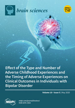
Cover Story (view full-size image): We aimed to clarify the effect of the type and number of adverse childhood experiences (ACE) and the timing of adverse experiences on clinical outcomes in 2,675 patients with bipolar disorders. We found that ACE have robust negative effects on clinical outcomes, including earlier age at onset, presence of psychotic episodes, suicide attempts, mixed symptoms, substance misuse comorbidity, and worse life functioning. Specifically, the number of ACE had the most significant effect on clinical outcomes; however, specific ACE, such as physical abuse, had a considerable influence. Moreover, post-childhood adverse experiences had a weaker effect on clinical outcomes than ACE.View this paper.
  • Issues are regarded as officially published after their release is announced to the table of contents alert mailing list.
  • You may sign up for e-mail alerts to receive table of contents of newly released issues.
  • PDF is the official format for papers published in both, html and pdf forms. To view the papers in pdf format, click on the "PDF Full-text" link, and use the free Adobe Reader to open them.
Order results
Result details
Section
Select all
Export citation of selected articles as:
8 pages, 1722 KiB  
Case Report
Intracranial Carotid Artery Aneurysm Treatment: First Reported Case of DERIVO®Flow-Diverter Placement by Direct Carotid Artery Puncture
by Giuseppe Guzzardi, Bruno Del Sette, Carmelo Stanca, Andrea Paladini, Andrea Galbiati, Marco Spinetta, Massimiliano Cernigliaro, Massimiliano Leigheb and Alessandro Carriero
Brain Sci. 2020, 10(5), 320; https://doi.org/10.3390/brainsci10050320 - 25 May 2020
Cited by 4 | Viewed by 3191
Abstract
Brain health may be threatened by aneurysm ruptures, and early recognition of these vascular malformations allows for neuroradiological intervention. Neurointerventional procedures are usually performed with femoral artery access. In patients with severe anatomical complexity of the supra-aortic vessels, however, treatment by this approach [...] Read more.
Brain health may be threatened by aneurysm ruptures, and early recognition of these vascular malformations allows for neuroradiological intervention. Neurointerventional procedures are usually performed with femoral artery access. In patients with severe anatomical complexity of the supra-aortic vessels, however, treatment by this approach could be hindered or impossible. Flow-diverter stent deployment is an effective and safe treatment for large, wide necked intracranial aneurysms, but it requires a complete and firm stability of the coaxial system to achieve a correct and precise deployment of the device. We present the first reported Italian case of a patient with an intracranial aneurysm which was treated with Flow-diverter stent (DERIVO®; AcandisGmbH & Co. KG; Pforzheim; Germany) by direct common carotid artery puncture due to severe tortuosity of supra-aortic trunks. Full article
(This article belongs to the Special Issue Imaging of Neuronal Environments)
Show Figures

Figure 1

18 pages, 4946 KiB  
Article
Logistic Regression Algorithm Differentiates Gulf War Illness (GWI) Functional Magnetic Resonance Imaging (fMRI) Data from a Sedentary Control
by Destie Provenzano, Stuart D. Washington, Yuan J. Rao, Murray Loew and James N. Baraniuk
Brain Sci. 2020, 10(5), 319; https://doi.org/10.3390/brainsci10050319 - 25 May 2020
Cited by 5 | Viewed by 4096
Abstract
Gulf War Illness (GWI) is a debilitating condition characterized by dysfunction of cognition, pain, fatigue, sleep, and diverse somatic symptoms with no known underlying pathology. As such, uncovering objective biomarkers such as differential regions of activity within a Functional Magnetic Resonance Imaging (fMRI) [...] Read more.
Gulf War Illness (GWI) is a debilitating condition characterized by dysfunction of cognition, pain, fatigue, sleep, and diverse somatic symptoms with no known underlying pathology. As such, uncovering objective biomarkers such as differential regions of activity within a Functional Magnetic Resonance Imaging (fMRI) scan is important to enhance validity of the criteria for diagnosis. Symptoms are exacerbated by mild activity, and exertional exhaustion is a key complaint amongst sufferers. We modeled this exertional exhaustion by having GWI (n = 80) and sedentary control (n = 31) subjects perform submaximal exercise stress tests on two consecutive days. Cognitive differences were assessed by comparing fMRI scans performed during 2-Back working memory tasks before and after the exercise. Machine learning algorithms were used to identify differences in brain activation patterns between the two groups on Day 1 (before exercise) and Day 2 (after exercise). The numbers of voxels with t > 3.17 (corresponding to p < 0.001 uncorrected) were determined for brain regions defined by the Automated Anatomical Labeling (AAL) atlas. Data were divided 70:30 into training and test sets. Recursive feature selection identified twenty-nine regions of interest (ROIs) that significantly distinguished GWI from control on Day 1 and 28 ROIs on Day 2. Ten regions were present in both models between the two days, including right anterior insula, orbital frontal cortex, thalamus, bilateral temporal poles, and left supramarginal gyrus and cerebellar Crus 1. The models had 70% accuracy before exercise on Day 1 and 85% accuracy after exercise on Day 2, indicating the logistic regression model significantly differentiated subjects with GWI from the sedentary control group. Exercise caused changes in these patterns that may indicate the cognitive differences caused by exertional exhaustion. A second set of predictive models was able to classify previously identified GWI exercise subgroups START, STOPP, and POTS for both Days 1 and Days 2 with 67% and 69% accuracy respectively. This study was the first of its kind to differentiate GWI and the three sub-phenotypes START, STOPP, and POTS from a sedentary control using a logistic regression estimation method. Full article
Show Figures

Graphical abstract

11 pages, 647 KiB  
Article
Promoting the Social Inclusion of Children with ASD: A Family-Centred Intervention
by Roy McConkey, Marie-Therese Cassin and Rosie McNaughton
Brain Sci. 2020, 10(5), 318; https://doi.org/10.3390/brainsci10050318 - 25 May 2020
Cited by 15 | Viewed by 5142
Abstract
The social isolation of children with autism spectrum disorder (ASD) is well documented. Their dearth of friends outside of the family and their lack of engagement in community activities places extra strains on the family. A project in Northern Ireland provided post-diagnostic support [...] Read more.
The social isolation of children with autism spectrum disorder (ASD) is well documented. Their dearth of friends outside of the family and their lack of engagement in community activities places extra strains on the family. A project in Northern Ireland provided post-diagnostic support to nearly 100 families and children aged from 3 to 11 years. An experienced ASD practitioner visited the child and family at home fortnightly in the late afternoon into the evening over a 12-month period. Most children had difficulty in relating to other children, coping with change, awareness of dangers, and joining in community activities. Likewise, up to two-thirds of parents identified managing the child’s behaviour, having time to spend with other children, and taking the child out of the house as further issues of concern to them. The project worker implemented a family-centred plan that introduced the child to various community activities in line with their learning targets and wishes. Quantitative and qualitative data showed improvements in the children’s social and communication skills, their personal safety, and participation in community activities. Likewise, the practical and emotional support provided to parents boosted their confidence and reduced stress within the family. The opportunities for parents and siblings to join in fun activities with the child with ASD strengthened their relationships. This project underscores the need for, and the success of family-based, post-diagnostic support to address the social isolation of children with ASD and their families. Full article
Show Figures

Graphical abstract

16 pages, 946 KiB  
Article
The Left Posterior Parietal Cortex Contributes to the Selection Process for the Initial Swing Leg in Gait Initiation
by Koichi Hiraoka, Shintaro Gonno and Ryota Inomoto
Brain Sci. 2020, 10(5), 317; https://doi.org/10.3390/brainsci10050317 - 22 May 2020
Cited by 2 | Viewed by 2355
Abstract
The present study examined whether the left posterior parietal cortex contributes to the selection process for the initial swing leg in gait initiation. Healthy humans initiated the gait in response to an auditory start cue. Transcranial magnetic stimulation (TMS) was given over P3, [...] Read more.
The present study examined whether the left posterior parietal cortex contributes to the selection process for the initial swing leg in gait initiation. Healthy humans initiated the gait in response to an auditory start cue. Transcranial magnetic stimulation (TMS) was given over P3, P4, F3 or F4 simultaneously, with the auditory start cue, in the on-TMS condition. A coil was placed over one of the four TMS sites, but TMS was not given in the off-TMS condition. The probability of right leg selection in the on-TMS condition was significantly lower than in the off-TMS condition when the coil was placed over P3, indicating that the left posterior parietal cortex contributes to the selection process of the initial swing leg of gait initiation. The latency of the anticipatory postural adjustment for gait initiation with the left leg was shortened by TMS over F4 or P4, but with the right leg was shortened by TMS over P3 or P4. Thus, the cortical process affecting the time taken to execute the motor process of gait initiation with the right leg may be related to the selection process of the initial swing leg of gait initiation. Full article
Show Figures

Figure 1

17 pages, 414 KiB  
Article
Evaluation of Psychological Stress Parameters in Coronary Patients by Three Different Questionnaires as Pre-Requisite for Comprehensive Rehabilitation
by Ana Maria Pah, Nicoleta Florina Buleu, Anca Tudor, Ruxandra Christodorescu, Dana Velimirovici, Stela Iurciuc, Maria Rada, Gheorghe Stoichescu-Hogea, Marius Badalica-Petrescu, Doina Georgescu, Dorina Nutiu, Mircea Iurciuc and Simona Dragan
Brain Sci. 2020, 10(5), 316; https://doi.org/10.3390/brainsci10050316 - 22 May 2020
Cited by 5 | Viewed by 2821
Abstract
Background: Negative psychological conditions are common in patients with cardiovascular diseases. Although depression has been scrutinized over the years in these patients, only recently has anxiety emerged as another important risk factor. The purpose of this study was to compare the parameters of [...] Read more.
Background: Negative psychological conditions are common in patients with cardiovascular diseases. Although depression has been scrutinized over the years in these patients, only recently has anxiety emerged as another important risk factor. The purpose of this study was to compare the parameters of psychological stress in a population of coronary patients with and without myocardial revascularization procedures and to analyze lifestyle and socio-economic contributors to the state of health of these patients before inclusion in a comprehensive individualized rehabilitation program. Methods: This study included 500 patients with coronary artery disease (CAD) in stable condition divided in 2 groups: 200 patients who underwent coronary artery bypass grafting (CABG) or percutaneous transluminal coronary angioplasty (PTCA) (Group 1) and 300 patients without myocardial revascularization (Group 2) with stable angina or thrombolyzed myocardial infarction. The protocol included screening for anxiety/depression after procedure using three different scales: Duke Anxiety-Depression Scale, Hospital Anxiety and Depression Scale (HADS) and the Type D Personality Scale (DS-14) scale that evaluates negative affectivity (NA) and social inhibition (SI). Results: Significant differences between groups were observed for HAD-A (9.1 ± 4.18 for Group 1 vs. 7.8 ± 4.03 for Group 2, p = 0.002) and DUKE scores (30.2 ± 12.25 for Group 1 vs. 22.7 ± 12.13 for Group 2, p < 0.001). HAD-A scores (p = 0.01) and DUKE scores (p = 0.04) were significantly higher in patients who underwent PTCA vs. CABG. CAD patients without myocardial revascularization (Group 2, n = 300) presented anxiety in proportion of 72.3% (n = 217) out of which 10.7% (n = 32) had severe anxiety, and 180 patients had depression (a proportion of 60%) out of which 1.3% (n = 4) presented severe depression. The correlation between the presence of type 2 diabetes mellitus (T2DM) and type D personality in revascularized patients (n = 200) was significant (Chi2 test, p = 0.010). By applying multinomial regression according to the Cox and Snell R-square model and multivariate linear regression by the Enter method, we demonstrated that male gender, age and marital status proved significant predictors for psychological stress in our study population. Conclusions: The results obtained in this study provide a framework for monitoring anxiety, depression and type D personality in coronary patients before inclusion in comprehensive rehabilitation programs. Behavioral and psychological stress responses in patients with CAD significantly correlate with risk factors, and could influence the evolution of the disease. Moreover, other factors like gender, income and marital status also seem to play a decisive role. Evaluation of psychological stress parameters contributes to a better individualization at the start of these programs, because it allows adjusting of all potential factors that may influence positive outcomes. Full article
Show Figures

Figure 1

20 pages, 7648 KiB  
Article
Liver Growth Factor Induces Glia-Associated Neuroprotection in an In Vitro Model of Parkinson´s Disease
by Rafael Gonzalo-Gobernado, Diana Reimers, María José Casarejos, Lucía Calatrava Ferreras, Manuela Vallejo-Muñoz, Adriano Jiménez-Escrig, Juan José Diaz-Gil, Gonzalo M. Ulzurrun de Asanza and Eulalia Bazán
Brain Sci. 2020, 10(5), 315; https://doi.org/10.3390/brainsci10050315 - 22 May 2020
Cited by 3 | Viewed by 2513
Abstract
Parkinson’s disease is a neurodegenerative disorder characterized by the progressive death of dopaminergic (DA) neurons in the substantia nigra (SN), which leads to a loss of the neurotransmitter dopamine in the basal ganglia. Current treatments relieve the symptoms of the disease, but none [...] Read more.
Parkinson’s disease is a neurodegenerative disorder characterized by the progressive death of dopaminergic (DA) neurons in the substantia nigra (SN), which leads to a loss of the neurotransmitter dopamine in the basal ganglia. Current treatments relieve the symptoms of the disease, but none stop or delay neuronal degeneration. Liver growth factor (LGF) is an albumin–bilirubin complex that stimulates axonal growth in the striatum and protects DA neurons in the SN of 6-hydroxydopamine-lesioned rats. Our previous results suggested that these effects observed in vivo are mediated by microglia and/or astrocytes. To determine if these cells are LGF targets, E14 (embryos from Sprague Dawley rats of 14 days) rat mesencephalic glial cultures were used. Treatment with 100 pg/mL of LGF up-regulated the mitogen-activated protein kinases (MAPKs) extracellular signal-regulated kinases 1/2 (ERK1/2) and the cyclic AMP response element binding protein (CREB) phosphorylation in glial cultures, and it increased the microglia marker Iba1 and tumor necrosis factor alpha (TNF-alpha) protein levels. The treatment of E14 midbrain neurons with a glial-conditioned medium from LGF-treated glial cultures (GCM-LGF) prevented the loss of DA neurons caused by 6-hydroxy-dopamine. This neuroprotective effect was not observed when GCM-LGF was applied in the presence of a blocking antibody of TNF-alpha activity. Altogether, our findings strongly suggest the involvement of microglia and TNF-alpha in the neuroprotective action of LGF on DA neurons observed in vitro. Full article
(This article belongs to the Section Neuroglia)
Show Figures

Figure 1

15 pages, 3171 KiB  
Article
Massive Data Management and Sharing Module for Connectome Reconstruction
by Jingbin Yuan, Jing Zhang, Lijun Shen, Dandan Zhang, Wenhuan Yu and Hua Han
Brain Sci. 2020, 10(5), 314; https://doi.org/10.3390/brainsci10050314 - 22 May 2020
Cited by 2 | Viewed by 3042
Abstract
Recently, with the rapid development of electron microscopy (EM) technology and the increasing demand of neuron circuit reconstruction, the scale of reconstruction data grows significantly. This brings many challenges, one of which is how to effectively manage large-scale data so that researchers can [...] Read more.
Recently, with the rapid development of electron microscopy (EM) technology and the increasing demand of neuron circuit reconstruction, the scale of reconstruction data grows significantly. This brings many challenges, one of which is how to effectively manage large-scale data so that researchers can mine valuable information. For this purpose, we developed a data management module equipped with two parts, a storage and retrieval module on the server-side and an image cache module on the client-side. On the server-side, Hadoop and HBase are introduced to resolve massive data storage and retrieval. The pyramid model is adopted to store electron microscope images, which represent multiresolution data of the image. A block storage method is proposed to store volume segmentation results. We design a spatial location-based retrieval method for fast obtaining images and segments by layers rapidly, which achieves a constant time complexity. On the client-side, a three-level image cache module is designed to reduce latency when acquiring data. Through theoretical analysis and practical tests, our tool shows excellent real-time performance when handling large-scale data. Additionally, the server-side can be used as a backend of other similar software or a public database to manage shared datasets, showing strong scalability. Full article
(This article belongs to the Special Issue Human Brain Dynamics: Latest Advances and Prospects)
Show Figures

Figure 1

2 pages, 175 KiB  
Erratum
Erratum: Pagonabarraga, J.; et al. A Spanish Consensus on the Use of Safinamide for Parkinson’s Disease in Clinical Practice. Brain Sci. 2020, 10, 176
by Javier Pagonabarraga, José Matías Arbelo, Francisco Grandas, Maria-Rosario Luquin, Pablo Martínez Martín, Maria Cruz Rodriguez-Oroz, Francesc Valldeoriola and Jaime Kulisevsky
Brain Sci. 2020, 10(5), 313; https://doi.org/10.3390/brainsci10050313 - 22 May 2020
Cited by 2 | Viewed by 2121
Abstract
We would like to submit the following erratum to our recently published paper [...] Full article
(This article belongs to the Special Issue Frontiers in Parkinson’s Disease (PD))
16 pages, 312 KiB  
Article
Parental Education and Youth Inhibitory Control in the Adolescent Brain Cognitive Development (ABCD) Study: Blacks’ Diminished Returns
by Shervin Assari
Brain Sci. 2020, 10(5), 312; https://doi.org/10.3390/brainsci10050312 - 21 May 2020
Cited by 25 | Viewed by 5661
Abstract
Background: Non-Hispanic Black (NHB) youth are at a higher risk of high-risk behaviors compared to non-Hispanic White (NHW) youth. Some of this racial gap is shown to be due to weaker effects of parental educational attainment on reducing the prevalence of behavioral risk [...] Read more.
Background: Non-Hispanic Black (NHB) youth are at a higher risk of high-risk behaviors compared to non-Hispanic White (NHW) youth. Some of this racial gap is shown to be due to weaker effects of parental educational attainment on reducing the prevalence of behavioral risk factors such as impulsivity, substance use, aggression, obesity, and poor school performance for NHBs, a pattern called Minorities’ Diminished Returns. These diminishing returns may be due to lower than expected effects of parental education on inhibitory control. Aim: We compared NHW and NHB youth for the effect of parental educational attainment on youth inhibitory control, a psychological and cognitive construct that closely predicts high-risk behaviors such as the use of drugs, alcohol, and tobacco. Methods: This was a cross-sectional analysis that included 4188 youth from the Adolescent Brain Cognitive Development (ABCD) study. The independent variable was parental educational attainment. The main outcome was youth inhibitory control measured by the stop-signal task (SST), which was validated by parent reports on the Child Behavior Checklist (CBCL). Results: In race/ethnicity-stratified models, high parental educational attainment was associated with a higher level of inhibitory control for NHB than NHW youth. In the pooled sample, race/ethnicity showed a statistically significant interaction with parental educational attainment on youth inhibitory control suggesting that high parental educational attainment has a smaller boosting effect on inhibitory control for NHB than NHW youth. Conclusion: Parental educational attainment boosts inhibitory control for NHW but not NHB youth. To minimize the racial gap in youth brain development, we need to address societal barriers that diminish the returns of family economic and human resources, particularly parental educational attainment, for racial and ethnic minority youth. Social and public policies should address structural and societal barriers such as social stratification, segregation, racism, and discrimination that hinder NHB parents’ abilities to effectively mobilize their human resources and secure tangible outcomes for their developing youth. Full article
15 pages, 4630 KiB  
Article
Glutamatergic Fate of Neural Progenitor Cells of Rats with Inherited Audiogenic Epilepsy
by Alexandra A. Naumova, Ekaterina A. Oleynik, Elena V. Chernigovskaya and Margarita V. Glazova
Brain Sci. 2020, 10(5), 311; https://doi.org/10.3390/brainsci10050311 - 21 May 2020
Cited by 5 | Viewed by 2544
Abstract
Epilepsy is associated with aberrant neurogenesis in the hippocampus and may underlie the development of hereditary epilepsy. In the present study, we analyzed the differentiation fate of neural progenitor cells (NPC), which were isolated from the hippocampus of embryos of Krushinsky-Molodkina (KM) rats [...] Read more.
Epilepsy is associated with aberrant neurogenesis in the hippocampus and may underlie the development of hereditary epilepsy. In the present study, we analyzed the differentiation fate of neural progenitor cells (NPC), which were isolated from the hippocampus of embryos of Krushinsky-Molodkina (KM) rats genetically prone to audiogenic epilepsy. NPCs from embryos of Wistar rats were used as the control. We found principal differences between Wistar and KM NPC in unstimulated controls: Wistar NPC culture contained both gamma-aminobutyric acid (GABA) and glutamatergic neurons; KM NPC culture was mainly represented by glutamatergic cells. The stimulation of glutamatergic differentiation of Wistar NPC resulted in a significant increase in glutamatergic cell number that was accompanied by the activation of protein kinase A. The stimulation of KM NPC led to a decrease in immature glutamatergic cell number and was associated with the activation of extracellular signal-regulated kinases 1 and 2 (ERK1/2) and protein kinase B/ glycogen synthase kinase 3 beta (Akt/GSK3β), which indicates the activation of glutamatergic cell maturation. These results suggest genetically programmed abnormalities in KM rats that determine the glutamatergic fate of NPC and contribute to the development of audiogenic epilepsy. Full article
(This article belongs to the Special Issue The Molecular and Cellular Mechanisms of Epilepsy)
Show Figures

Figure 1

14 pages, 1236 KiB  
Review
Transcranial Direct Current Stimulation to Facilitate Lower Limb Recovery Following Stroke: Current Evidence and Future Directions
by Samuel Gowan and Brenton Hordacre
Brain Sci. 2020, 10(5), 310; https://doi.org/10.3390/brainsci10050310 - 21 May 2020
Cited by 13 | Viewed by 6459
Abstract
Stroke remains a global leading cause of disability. Novel treatment approaches are required to alleviate impairment and promote greater functional recovery. One potential candidate is transcranial direct current stimulation (tDCS), which is thought to non-invasively promote neuroplasticity within the human cortex by transiently [...] Read more.
Stroke remains a global leading cause of disability. Novel treatment approaches are required to alleviate impairment and promote greater functional recovery. One potential candidate is transcranial direct current stimulation (tDCS), which is thought to non-invasively promote neuroplasticity within the human cortex by transiently altering the resting membrane potential of cortical neurons. To date, much work involving tDCS has focused on upper limb recovery following stroke. However, lower limb rehabilitation is important for regaining mobility, balance, and independence and could equally benefit from tDCS. The purpose of this review is to discuss tDCS as a technique to modulate brain activity and promote recovery of lower limb function following stroke. Preliminary evidence from both healthy adults and stroke survivors indicates that tDCS is a promising intervention to support recovery of lower limb function. Studies provide some indication of both behavioral and physiological changes in brain activity following tDCS. However, much work still remains to be performed to demonstrate the clinical potential of this neuromodulatory intervention. Future studies should consider treatment targets based on individual lesion characteristics, stage of recovery (acute vs. chronic), and residual white matter integrity while accounting for known determinants and biomarkers of tDCS response. Full article
Show Figures

Figure 1

20 pages, 341 KiB  
Review
Update on Atypicalities of Central Nervous System in Autism Spectrum Disorder
by Ahmad Naqib Shuid, Putri Ayu Jayusman, Nazrun Shuid, Juriza Ismail, Norazlin Kamal Nor and Isa Naina Mohamed
Brain Sci. 2020, 10(5), 309; https://doi.org/10.3390/brainsci10050309 - 20 May 2020
Cited by 9 | Viewed by 3717
Abstract
Autism spectrum disorder (ASD) is a heterogeneous, behaviorally defined, neurodevelopmental disorder that has been modeled as a brain-based disease. The behavioral and cognitive features of ASD are associated with pervasive atypicalities in the central nervous system (CNS). To date, the exact mechanisms underlying [...] Read more.
Autism spectrum disorder (ASD) is a heterogeneous, behaviorally defined, neurodevelopmental disorder that has been modeled as a brain-based disease. The behavioral and cognitive features of ASD are associated with pervasive atypicalities in the central nervous system (CNS). To date, the exact mechanisms underlying the pathophysiology of ASD still remain unknown and there is currently no cure or effective treatment for this disorder. Many publications implicated the association of ASD with inflammation, immune dysregulation, neurotransmission dysfunction, mitochondrial impairment and cell signaling dysregulation. This review attempts to highlight evidence of the major pathophysiology of ASD including abnormalities in the brain structure and function, neuroglial activation and neuroinflammation, glutamatergic neurotransmission, mitochondrial dysfunction and mechanistic target of rapamycin (mTOR) signaling pathway dysregulation. Molecular and cellular factors that contributed to the pathogenesis of ASD and how they may affect the development and function of CNS are compiled in this review. However, findings of published studies have been complicated by the fact that autism is a very heterogeneous disorder; hence, we addressed the limitations that led to discrepancies in the reported findings. This review emphasizes the need for future studies to control study variables such as sample size, gender, age range and intelligence quotient (IQ), all of which that could affect the study measurements. Neuroinflammation or immune dysregulation, microglial activation, genetically linked neurotransmission, mitochondrial dysfunctions and mTOR signaling pathway could be the primary targets for treating and preventing ASD. Further research is required to better understand the molecular causes and how they may contribute to the pathophysiology of ASD. Full article
(This article belongs to the Section Neuroglia)
13 pages, 1194 KiB  
Article
Disentangling Restrictive and Repetitive Behaviors and Social Impairments in Children and Adolescents with Gilles de la Tourette Syndrome and Autism Spectrum Disorder
by Mariangela Gulisano, Rita Barone, Salvatore Alaimo, Alfredo Ferro, Alfredo Pulvirenti, Lara Cirnigliaro, Selena Di Silvestre, Serena Martellino, Nicoletta Maugeri, Maria Chiara Milana, Miriam Scerbo and Renata Rizzo
Brain Sci. 2020, 10(5), 308; https://doi.org/10.3390/brainsci10050308 - 18 May 2020
Cited by 10 | Viewed by 3758
Abstract
Gilles de la Tourette syndrome (GTS) and autism spectrum disorder (ASD) are two neurodevelopmental disorders with male predominance, frequently comorbid, that share clinical and behavioral features. The incidence of ASD in patients affected by GTS was reported to be between 2.9% and 22.8%. [...] Read more.
Gilles de la Tourette syndrome (GTS) and autism spectrum disorder (ASD) are two neurodevelopmental disorders with male predominance, frequently comorbid, that share clinical and behavioral features. The incidence of ASD in patients affected by GTS was reported to be between 2.9% and 22.8%. We hypothesized that higher ASD rates among children affected by GTS previously reported may be due to difficulty in discriminating GTS sub-phenotypes from ASD, and the higher scores in the restrictive and repetitive behaviors in particular may represent at least a “false comorbidity”. We studied a large population of 720 children and adolescents affected by GTS (n = 400) and ASD (n = 320), recruited from a single center. Patients were all assessed with The Yale Global Tic Severity Rating Scale (YGTSS), The Autism Diagnostic Observation Schedule (ADOS), The Autism Diagnostic Interview Revised (ADI-R), The Children’s Yale–Brown Obsessive–Compulsive Scale (CY-BOCS), and The Children’s Yale–Brown Obsessive–Compulsive Scale for autism spectrum disorder (CY-BOCS ASD). Our results showed statistically significant differences in ADOS scores for social aspects between GTS with comorbid attention deficit hyperactivity disorder (ADHD) and obsessive–compulsive disorder (OCD) sub-phenotypes and ASD. No differences were present when we compared GTS with comorbid ASD sub-phenotype to ASD, while repetitive and restrictive behavior scores in ASD did not present statistical differences in the comparison with GTS and comorbid OCD and ASD sub-phenotypes. We also showed that CY-BOCS ASD could be a useful instrument to correctly identify OCD from ASD symptoms. Full article
(This article belongs to the Special Issue Advances in Autism Research)
Show Figures

Figure 1

24 pages, 4698 KiB  
Article
Auditory and Visual Response Inhibition in Children with Bilateral Hearing Aids and Children with ADHD
by Laura Bell, Wolfgang Scharke, Vanessa Reindl, Janina Fels, Christiane Neuschaefer-Rube and Kerstin Konrad
Brain Sci. 2020, 10(5), 307; https://doi.org/10.3390/brainsci10050307 - 18 May 2020
Cited by 13 | Viewed by 4954
Abstract
Children fitted with hearing aids (HAs) and children with attention deficit/hyperactivity disorder (ADHD) often have marked difficulties concentrating in noisy environments. However, little is known about the underlying neural mechanism of auditory and visual attention deficits in a direct comparison of both groups. [...] Read more.
Children fitted with hearing aids (HAs) and children with attention deficit/hyperactivity disorder (ADHD) often have marked difficulties concentrating in noisy environments. However, little is known about the underlying neural mechanism of auditory and visual attention deficits in a direct comparison of both groups. The current functional near-infrared spectroscopy (fNIRS) study was the first to investigate the behavioral performance and neural activation during an auditory and a visual go/nogo paradigm in children fitted with bilateral HAs, children with ADHD and typically developing children (TDC). All children reacted faster, but less accurately, to visual than auditory stimuli, indicating a sensory-specific response inhibition efficiency. Independent of modality, children with ADHD and children with HAs reacted faster and tended to show more false alarms than TDC. On a neural level, however, children with ADHD showed supra-modal neural alterations, particularly in frontal regions. On the contrary, children with HAs exhibited modality-dependent alterations in the right temporopolar cortex. Higher activation was observed in the auditory than in the visual condition. Thus, while children with ADHD and children with HAs showed similar behavioral alterations, different neural mechanisms might underlie these behavioral changes. Future studies are warranted to confirm the current findings with larger samples. To this end, fNIRS provided a promising tool to differentiate the neural mechanisms underlying response inhibition deficits between groups and modalities. Full article
(This article belongs to the Special Issue New Advances in Attention Deficit Hyperactivity Disorder (ADHD))
Show Figures

Graphical abstract

14 pages, 1420 KiB  
Article
Anhedonia to Gentle Touch in Fibromyalgia: Normal Sensory Processing but Abnormal Evaluation
by Rebecca Boehme, Helene van Ettinger-Veenstra, Håkan Olausson, Björn Gerdle and Saad S. Nagi
Brain Sci. 2020, 10(5), 306; https://doi.org/10.3390/brainsci10050306 - 18 May 2020
Cited by 21 | Viewed by 5263
Abstract
Social touch is important for interpersonal interaction. Gentle touch and slow brushing are typically perceived as pleasant, the degree of pleasantness is linked to the activity of the C-tactile (CT) fibers, a class of unmyelinated nerves in the skin. The inability to experience [...] Read more.
Social touch is important for interpersonal interaction. Gentle touch and slow brushing are typically perceived as pleasant, the degree of pleasantness is linked to the activity of the C-tactile (CT) fibers, a class of unmyelinated nerves in the skin. The inability to experience pleasure in general is called anhedonia, a common phenomenon in the chronic pain condition fibromyalgia. Here, we studied the perception and cortical processing of gentle touch in a well-characterized cohort of fibromyalgia. Patients and controls participated in functional brain imaging while receiving tactile stimuli (brushing) on the forearm. They were asked to provide ratings of pleasantness of the tactile stimulus and ongoing pain. We found high distress, pain catastrophizing, and insomnia, and a low perceived state of health in fibromyalgia. Further, patients rated both slow (CT-optimal) and fast (CT-suboptimal) brushing as less pleasant than healthy participants. While there was no difference in brain activity during touch, patients showed deactivation in the right posterior insula (contralateral to the stimulated arm) during pleasantness rating and activation during pain rating. The opposite pattern was observed in healthy participants. Voxel-based morphometry analysis revealed reduced grey matter density in patients, in the bilateral hippocampus and anterior insula. Our results suggest anhedonia to gentle touch in fibromyalgia with intact early-stage sensory processing but dysfunctional evaluative processing. These findings contribute to our understanding of the mechanisms underlying anhedonia in fibromyalgia. Full article
Show Figures

Figure 1

12 pages, 855 KiB  
Review
Prevalence and Incidence of Multiple Sclerosis in Russian Federation: 30 Years of Studies
by Alexey Boyko and Mikhail Melnikov
Brain Sci. 2020, 10(5), 305; https://doi.org/10.3390/brainsci10050305 - 18 May 2020
Cited by 28 | Viewed by 3851
Abstract
In the Russian Federation, multiple sclerosis prevalence rates vary from 10 to 80 cases per 100,000, depending on region and the nationality of the population. The main characteristics of multiple sclerosis epidemiology in the XX century in this big territory are: (1) steady [...] Read more.
In the Russian Federation, multiple sclerosis prevalence rates vary from 10 to 80 cases per 100,000, depending on region and the nationality of the population. The main characteristics of multiple sclerosis epidemiology in the XX century in this big territory are: (1) steady increase in multiple sclerosis prevalence and incidence rates, maybe because of better diagnosis and treatment, but also changes in environmental/epigenetic risk profile and/or lifestyle factors; (2) increase of the female to male ratio, increase in multiple sclerosis incidence mainly in females; (3) appearance and increasing frequency of multiple sclerosis in ethnic groups, previously free of multiple sclerosis (Northern Tribes, Yakuts and others). The latest data show that in European Russia, the multiple sclerosis prevalence varies from 30 to 80 cases, in Siberia—from 20 to 70 cases, with steady increases, especially in women. Full article
(This article belongs to the Special Issue A Decade of Brain Sciences)
Show Figures

Figure 1

11 pages, 994 KiB  
Article
Optimising Cognitive Enhancement: Systematic Assessment of the Effects of tDCS Duration in Older Adults
by Claire J. Hanley, Sophie L. Alderman and Elinor Clemence
Brain Sci. 2020, 10(5), 304; https://doi.org/10.3390/brainsci10050304 - 16 May 2020
Cited by 7 | Viewed by 3455
Abstract
Transcranial direct current stimulation (tDCS) has been shown to support cognition and brain function in older adults. However, there is an absence of research specifically designed to determine optimal stimulation protocols, and much of what is known about subtle distinctions in tDCS parameters [...] Read more.
Transcranial direct current stimulation (tDCS) has been shown to support cognition and brain function in older adults. However, there is an absence of research specifically designed to determine optimal stimulation protocols, and much of what is known about subtle distinctions in tDCS parameters is based on young adult data. As the first systematic exploration targeting older adults, this study aimed to provide insight into the effects of variations in stimulation duration. Anodal stimulation of 10 and 20 min, as well as a sham-control variant, was administered to dorsolateral prefrontal cortex. Stimulation effects were assessed in relation to a novel attentional control task. Ten minutes of anodal stimulation significantly improved task-switching speed from baseline, contrary to the sham-control and 20 min variants. The findings represent a crucial step forwards for methods development, and the refinement of stimulation to enhance executive function in the ageing population. Full article
Show Figures

Figure 1

16 pages, 902 KiB  
Article
Somatic Marker Production Deficits Do Not Explain the Relationship between Psychopathic Traits and Utilitarian Moral Decision Making
by Shawn E. Fagan, Liat Kofler, Sarah Riccio and Yu Gao
Brain Sci. 2020, 10(5), 303; https://doi.org/10.3390/brainsci10050303 - 15 May 2020
Cited by 1 | Viewed by 3266
Abstract
In moral dilemma tasks, high levels of psychopathic traits often predict increased utilitarian responding—specifically, endorsing sacrificing one person to save many. Research suggests that increased arousal (i.e., somatic marker production) underlies lower rates of utilitarian responding during moral dilemmas. Though deficient somatic marker [...] Read more.
In moral dilemma tasks, high levels of psychopathic traits often predict increased utilitarian responding—specifically, endorsing sacrificing one person to save many. Research suggests that increased arousal (i.e., somatic marker production) underlies lower rates of utilitarian responding during moral dilemmas. Though deficient somatic marker production is characteristic of psychopathy, how this deficit affects the psychopathy–utilitarian connection remains unknown. We assessed psychopathic traits in undergraduates, as well as behavioral performance and skin conductance level reactivity (SCL-R; a measure of somatic marker production) during a moral dilemma task. High psychopathic traits and low SCL-R were associated with increased utilitarian decisions in dilemmas involving direct personal harm. Psychopathic traits were unrelated to SCL-R, nor did SCL-R mediate the relationship between psychopathy and utilitarianism. The present study did not find evidence that somatic marker production explains the connection between utilitarianism and psychopathy in a college population. Further research is necessary to identify the neural mechanisms relating psychopathy and moral decision-making in nonclinical samples. Full article
(This article belongs to the Special Issue CNS-Arousal—Transdiagnostic Relevance and Therapeutic Implications)
Show Figures

Figure 1

12 pages, 6348 KiB  
Article
Modulation of Fibers to Motor Cortex during Thalamic DBS in Tourette Patients Correlates with Tic Reduction
by Pablo Andrade, Petra Heiden, Moritz Hoevels, Marc Schlamann, Juan C. Baldermann, Daniel Huys and Veerle Visser-Vandewalle
Brain Sci. 2020, 10(5), 302; https://doi.org/10.3390/brainsci10050302 - 15 May 2020
Cited by 16 | Viewed by 3099
Abstract
Probabilistic tractography in Tourette syndrome (TS) patients have shown an alteration in the connectivity of the primary motor cortex and supplementary motor area with the striatum and thalamus, suggesting an abnormal connectivity of the cortico-striatum-thalamocortical-pathways in TS. Deep brain stimulation (DBS) of the [...] Read more.
Probabilistic tractography in Tourette syndrome (TS) patients have shown an alteration in the connectivity of the primary motor cortex and supplementary motor area with the striatum and thalamus, suggesting an abnormal connectivity of the cortico-striatum-thalamocortical-pathways in TS. Deep brain stimulation (DBS) of the centromedian nucleus–nucleus ventrooralis internus (CM-Voi complex) in the thalamus is an effective treatment for refractory TS patients. We investigated the connectivity of activated fibers from CM-Voi to the motor cortex and its correlation between these projections and their clinical outcome. Seven patients with TS underwent CM-Voi-DBS surgery and were clinically evaluated preoperatively and six months postoperatively. We performed diffusion tensor imaging to display the activated fibers projecting from the CM-Voi to the different motor cortex regions of interest. These analyses showed that the extent of tic reduction during DBS is associated with the degree of stimulation-dependent connectivity between CM-Voi and the motor cortex, and in particular, an increased density of projections to the presupplementary motor area (preSMA). Non-responder patients displayed the largest amount of active fibers projecting into cortical areas other than motor cortex compared to responder patients. These findings support the notion that an abnormal connectivity of thalamocortical pathways underlies TS, and that modulation of these circuits through DBS could restore the function and reduce symptoms. Full article
(This article belongs to the Special Issue Deep Brain Stimulation and Tourette Syndrome)
Show Figures

Figure 1

8 pages, 931 KiB  
Article
Deep Brain Stimulation for Gilles de la Tourette Syndrome: Toward Limbic Targets
by Domenico Servello, Tommaso Francesco Galbiati, Roberta Balestrino, Guglielmo Iess, Edvin Zekaj, Sara De Michele and Mauro Porta
Brain Sci. 2020, 10(5), 301; https://doi.org/10.3390/brainsci10050301 - 15 May 2020
Cited by 14 | Viewed by 3237
Abstract
Gilles de la Tourette syndrome (GTS) is a complex neurodevelopmental disorder characterized by tics and, frequently, psychiatric and behavioral comorbidities. Above all, obsessive compulsive disorder/behavior (OCD/OCB) influences the clinical picture and has a severe impact on quality of life, eventually more than the [...] Read more.
Gilles de la Tourette syndrome (GTS) is a complex neurodevelopmental disorder characterized by tics and, frequently, psychiatric and behavioral comorbidities. Above all, obsessive compulsive disorder/behavior (OCD/OCB) influences the clinical picture and has a severe impact on quality of life, eventually more than the tics themselves. Deep brain stimulation (DBS) is an effective therapy in selected, refractory cases. Clinical response to DBS may vary according to the clinical picture, comorbidities, and to the anatomical target. This retrospective study compares the results obtained from DBS in the ventralis oralis/centromedian-parascicular nucleus of the thalamus (Voi-Cm/Pf) (41 patients) and antero-medial Globus Pallidus internus (am-GPi) (14 patients), evaluating clinical response over time by means of Yale Global Tic Severity Scale (YGTSS) and Yale–Brown Obsessive-Compulsive Scale (YBOCS) scores over a period of 48 months. A significant and stable improvement in the YGTSS and YBOCS has been obtained in both groups (p < 0.001). There was a significant difference in YBOCS improvement over time between the am-GPi group and the Voi-Cm/Pf group, indicating a better and faster control of OCD/OCB symptoms in the former group. The ratio of hardware removal was 23% and limited to 13 patients in the Voi-Cm/Pf group. These results confirm that DBS is an effective therapy in treating GTS and suggest that the am-GPi might be superior to Voi-Cm/Pf in alleviating comorbid OCD/OCB. Full article
(This article belongs to the Special Issue Deep Brain Stimulation and Tourette Syndrome)
Show Figures

Figure 1

17 pages, 351 KiB  
Review
Boosting Slow Oscillations during Sleep to Improve Memory Function in Elderly People: A Review of the Literature
by Federico Salfi, Aurora D’Atri, Daniela Tempesta, Luigi De Gennaro and Michele Ferrara
Brain Sci. 2020, 10(5), 300; https://doi.org/10.3390/brainsci10050300 - 15 May 2020
Cited by 19 | Viewed by 4876
Abstract
Sleep represents a crucial time window for the consolidation of memory traces. In this view, some brain rhythms play a pivotal role, first of all the sleep slow waves. In particular, the neocortical slow oscillations (SOs), in coordination with the hippocampal ripples and [...] Read more.
Sleep represents a crucial time window for the consolidation of memory traces. In this view, some brain rhythms play a pivotal role, first of all the sleep slow waves. In particular, the neocortical slow oscillations (SOs), in coordination with the hippocampal ripples and the thalamocortical spindles, support the long-term storage of the declarative memories. The aging brain is characterized by a disruption of this complex system with outcomes on the related cognitive functions. In recent years, the advancement of the comprehension of the sleep-dependent memory consolidation mechanisms has encouraged the development of techniques of SO enhancement during sleep to induce cognitive benefits. In this review, we focused on the studies reporting on the application of acoustic or electric stimulation procedures in order to improve sleep-dependent memory consolidation in older subjects. Although the current literature is limited and presents inconsistencies, there is promising evidence supporting the perspective to non-invasively manipulate the sleeping brain electrophysiology to improve cognition in the elderly, also shedding light on the mechanisms underlying the sleep-memory relations during healthy and pathological aging. Full article
(This article belongs to the Special Issue Disturbances of Sleep Among Older People)
20 pages, 764 KiB  
Review
Molecular Interventions towards Multiple Sclerosis Treatment
by Athanasios Metaxakis, Dionysia Petratou and Nektarios Tavernarakis
Brain Sci. 2020, 10(5), 299; https://doi.org/10.3390/brainsci10050299 - 15 May 2020
Cited by 12 | Viewed by 4875
Abstract
Multiple sclerosis (MS) is an autoimmune life-threatening disease, afflicting millions of people worldwide. Although the disease is non-curable, considerable therapeutic advances have been achieved through molecular immunotherapeutic approaches, such as peptides vaccination, administration of monoclonal antibodies, and immunogenic copolymers. The main aims of [...] Read more.
Multiple sclerosis (MS) is an autoimmune life-threatening disease, afflicting millions of people worldwide. Although the disease is non-curable, considerable therapeutic advances have been achieved through molecular immunotherapeutic approaches, such as peptides vaccination, administration of monoclonal antibodies, and immunogenic copolymers. The main aims of these therapeutic strategies are to shift the MS-related autoimmune response towards a non-inflammatory T helper 2 (Th2) cells response, inactivate or ameliorate cytotoxic autoreactive T cells, induce secretion of anti-inflammatory cytokines, and inhibit recruitment of autoreactive lymphocytes to the central nervous system (CNS). These approaches can efficiently treat autoimmune encephalomyelitis (EAE), an essential system to study MS in animals, but they can only partially inhibit disease progress in humans. Nevertheless, modern immunotherapeutic techniques remain the most promising tools for the development of safe MS treatments, specifically targeting the cellular factors that trigger the initiation of the disease. Full article
(This article belongs to the Special Issue Advances in Multiple Sclerosis Research—Series I)
Show Figures

Graphical abstract

8 pages, 875 KiB  
Case Report
Gaze Palsy as a Manifestation of Todd’s Phenomenon: Case Report and Review of the Literature
by Karmele Olaciregui Dague, Manuel Dafotakis, Jörg B. Schulz and Rainer Surges
Brain Sci. 2020, 10(5), 298; https://doi.org/10.3390/brainsci10050298 - 15 May 2020
Cited by 3 | Viewed by 3317
Abstract
Background: Though Todd’s phenomenon (TP) is a relatively rare occurrence, its correct identification is of key diagnostic and therapeutic importance as a stroke mimic. Here we describe a case of isolated gaze palsy as a manifestation of TP, discuss periictal gaze abnormalities [...] Read more.
Background: Though Todd’s phenomenon (TP) is a relatively rare occurrence, its correct identification is of key diagnostic and therapeutic importance as a stroke mimic. Here we describe a case of isolated gaze palsy as a manifestation of TP, discuss periictal gaze abnormalities as lateralizing sign involving the frontal eye field (FEF), and present a narrative literature review. Methods: We reviewed the main features of the case and conducted a structured literature search of TP and gaze palsy using PubMed. We restricted the search to publications in English, Spanish, French, and German. Case presentation: A 71-year-old male with a history of right frontotemporal subarachnoid hemorrhage was admitted to the Emergency Department of our institution after suffering a first unprovoked focal to bilateral tonic-clonic seizure with ictal gaze deviation to the left. Cranial imaging showed no signs of ischemia, intracerebral hemorrhage, or tumor. The patient presented the following postictal features: involuntary eye deviation to the right due to left-sided gaze palsy and disorientation in time with preserved responsiveness. Eye movements were normal three days later. We concluded that the patient suffered from new-onset epilepsy due to sequelae following the right frontotemporal subarachnoid hemorrhage, affecting the FEF with contralateral ictal gaze deviation, and postictal gaze palsy with ipsilateral eye deviation as an unusual Todd’s phenomenon. Conclusion: Unusual manifestations of TP are uncommon but clinically highly relevant, as they can mimic stroke or epileptic status and are decisive in the diagnostic and therapeutic decision-making process. Though postictal gaze palsy has been reported associated with other deficits, this constitutes, to our knowledge, the first report of isolated gaze palsy as a form of TP. Further research into the underlying causes is needed. Ictal contralateral gaze and head deviation, and probably postictal ipsilateral gaze deviation if present, are very helpful for the lateralization of the seizure-onset zone. Full article
Show Figures

Figure 1

9 pages, 558 KiB  
Article
Stimulus Intensity Affects Variability of Motor Evoked Responses of the Non-Paretic, but Not Paretic Tibialis Anterior Muscle in Stroke
by Anjali Sivaramakrishnan and Sangeetha Madhavan
Brain Sci. 2020, 10(5), 297; https://doi.org/10.3390/brainsci10050297 - 15 May 2020
Cited by 5 | Viewed by 2493
Abstract
Background: Transcranial magnetic stimulus induced motor evoked potentials (MEPs) are quantified either with a single suprathreshold stimulus or using a stimulus response curve. Here, we explored variability in MEPs influenced by different stimulus intensities for the tibialis anterior muscle in stroke. Methods: MEPs [...] Read more.
Background: Transcranial magnetic stimulus induced motor evoked potentials (MEPs) are quantified either with a single suprathreshold stimulus or using a stimulus response curve. Here, we explored variability in MEPs influenced by different stimulus intensities for the tibialis anterior muscle in stroke. Methods: MEPs for the paretic and non-paretic tibialis anterior (TA) muscle representations were collected from 26 participants with stroke at seven intensities. Variability of MEP parameters was examined with coefficients of variation (CV). Results: CV for the non-paretic TA MEP amplitude and area was significantly lower at 130% and 140% active motor threshold (AMT). CV for the paretic TA MEP amplitude and area did not vary with intensity. CV of MEP latency decreased with higher intensities for both muscles. CV of the silent period decreased with higher intensity for the non-paretic TA, but was in reverse for the paretic TA. Conclusion: We recommend a stimulus intensity of greater than 130% AMT to reduce variability for the non-paretic TA. The stimulus intensity did not affect the MEP variability of the paretic TA. Variability of MEPs is affected by intensity and side tested (paretic and non-paretic), suggesting careful selection of experimental parameters for testing. Full article
Show Figures

Figure 1

13 pages, 271 KiB  
Case Report
Immersive Virtual Reality in Stroke Patients as a New Approach for Reducing Postural Disabilities and Falls Risk: A Case Series
by Irene Cortés-Pérez, Francisco Antonio Nieto-Escamez and Esteban Obrero-Gaitán
Brain Sci. 2020, 10(5), 296; https://doi.org/10.3390/brainsci10050296 - 15 May 2020
Cited by 31 | Viewed by 7932
Abstract
Stroke is a neurologic disorder considered the first cause of disability worldwide due to motor, cognitive, and sensorial sequels. Balance dysfunctions in stroke survivors increase the risk of falls and physiotherapeutic rehabilitation is essential to reduce it. Virtual reality (VR) seems to be [...] Read more.
Stroke is a neurologic disorder considered the first cause of disability worldwide due to motor, cognitive, and sensorial sequels. Balance dysfunctions in stroke survivors increase the risk of falls and physiotherapeutic rehabilitation is essential to reduce it. Virtual reality (VR) seems to be an alternative to conventional physiotherapy (CT), providing virtual environments and multisensorial inputs to train balance in stroke patients. The aim of this study was to assess if immersive VR treatment is more effective than CT to improve balance after stroke. This study got the approval from the Ethics Committee of the University of Almeria. Three chronic ischemic stroke patients were selected. One patient who received 25 sessions of immersive VR intervention for two months was compared with another patient who received equivalent CT and a third patient with no intervention. Balance, gait, risk of falling, and vestibular and visual implications in the equilibrium were assessed. After the interventions, the two patients receiving any of the treatments showed an improvement in balance compared to the untreated patient. In comparison to CT, our results suggest a higher effect of immersive VR in the improvement of balance and a reduction of falls risk due to the active upright work during the VR intervention. Full article
(This article belongs to the Special Issue Stroke Treatments and Therapies)
Show Figures

Graphical abstract

10 pages, 1996 KiB  
Article
Comparison Analysis between the Medication Efficacy of the Milnacipran and Functional Connectivity of Neural Networks in Fibromyalgia Patients
by Seong-Ho Kim, Min-Woo Lee, Min-Jae Kang, Sung Gun Lee, Jung-Goo Lee and Chi-Woong Mun
Brain Sci. 2020, 10(5), 295; https://doi.org/10.3390/brainsci10050295 - 15 May 2020
Cited by 2 | Viewed by 2554
Abstract
Milnacipran is a reuptake inhibitor of both serotonin and noradrenaline, used in the treatment of fibromyalgia with severe depression. However, few studies have been conducted on the efficacies of milnacipran drug on the functional connectivity of the neural network. The authors aimed to [...] Read more.
Milnacipran is a reuptake inhibitor of both serotonin and noradrenaline, used in the treatment of fibromyalgia with severe depression. However, few studies have been conducted on the efficacies of milnacipran drug on the functional connectivity of the neural network. The authors aimed to find the correlation between the drug efficacy and the changes in neural network in fibromyalgia patients. Resting-state-functional magnetic resonance imaging (rs-fMRI) were obtained before and after milnacipran drug administration. Graph theory indexes and small-worldness were calculated using preprocessed blood-oxygen-level-dependent signals from the rs-fMRI scans of 14 brain regions-of-interest. Statistical analyses were conducted to compare the topological network parameters. Significant changes in the neural network indexes appeared in three of the 14 brain regions-of-interest. In the pain network, the average path length on the left side of Brodmann area 32 was shortened. In the default mode network, functional connectivity changes were observed in the left lateral parietal cortex and medial prefrontal cortex. In the left lateral parietal cortex, the degree and betweenness centrality increased, whereas the clustering coefficient decreased. In the medial prefrontal cortex, local efficiency decreased. The small-worldness declined after milnacipran medication. The present results demonstrate that functional connectivity indexes in the brains of female fibromyalgia patients obtained from rs-fMRI data can be used as potential prognosis markers of milnacipran drug treatment. Full article
Show Figures

Figure 1

11 pages, 1158 KiB  
Case Report
Dynamic Relationship between Sense of Agency and Post-Stroke Sensorimotor Deficits: A Longitudinal Case Study
by Yu Miyawaki, Takeshi Otani and Shu Morioka
Brain Sci. 2020, 10(5), 294; https://doi.org/10.3390/brainsci10050294 - 15 May 2020
Cited by 7 | Viewed by 4598
Abstract
Post-stroke sensorimotor deficits impair voluntary movements. This impairment may alter a person’s sense of agency, which is the awareness of controlling one’s actions. A previous study showed that post-stroke patients incorrectly aligned themselves with others’ movements and proposed that their misattributions might be [...] Read more.
Post-stroke sensorimotor deficits impair voluntary movements. This impairment may alter a person’s sense of agency, which is the awareness of controlling one’s actions. A previous study showed that post-stroke patients incorrectly aligned themselves with others’ movements and proposed that their misattributions might be associated with their sensorimotor deficits. To investigate this hypothesis, the present study compared the agency dynamics in a post-stroke patient A (PA) with sensorimotor deficits, who rarely used her paretic upper limbs in her daily life to patient B (PB), who had a paretic upper limb with almost normal functions and activity. At the second, fourth, and eighth weeks following their strokes, PA and PB completed experiments where they performed horizontal movements while receiving visual feedback, and analyzed if the visual feedback represented their own or another’s movements. Consequently, PB made no misattributions each week; whereas, PA made incorrect self-attributions of other’s movements at the fourth week. Interestingly, this misattribution noticeably decreased at the eighth week, where PA, with an improved paretic upper limb, used her limb almost as much as before her stroke. These results suggest that the sense of agency alters according to the sensorimotor deficit severity and paretic upper limb activity. Full article
(This article belongs to the Special Issue The Role of Body in Brain Plasticity)
Show Figures

Figure 1

10 pages, 261 KiB  
Article
Preconception Risk Factors for Autism Spectrum Disorder—A Pilot Study
by Hankus Magdalena, Kazek Beata, Paprocka Justyna, Kapinos-Gorczyca Agnieszka, Magdalena Szczepara-Fabian, Agata Buczek and Emich-Widera Ewa
Brain Sci. 2020, 10(5), 293; https://doi.org/10.3390/brainsci10050293 - 14 May 2020
Cited by 7 | Viewed by 5508
Abstract
Autism spectrum disorder (ASD) is a neurodevelopmental disorder of multifactorial etiology. Preconception risk factors are still poorly understood. A survey on preconception risk factors for ASD was conducted among parents of 121 ASD patients aged 3–12 years and parents of 100 healthy children [...] Read more.
Autism spectrum disorder (ASD) is a neurodevelopmental disorder of multifactorial etiology. Preconception risk factors are still poorly understood. A survey on preconception risk factors for ASD was conducted among parents of 121 ASD patients aged 3–12 years and parents of 100 healthy children aged 3–12 years. The exclusion criteria were as follows: the presence of associated problems such as intellectual disability, epilepsy or other genetic and neurological diseases. Thirteen parameters were considered, a few among which were conception problems, conception with assisted reproductive techniques, the use and duration of oral contraception, the number of previous pregnancies and miscarriages, time since the previous pregnancy (in months), the history of mental illness in the family (including ASD), other chronic diseases in the mother or father and maternal and paternal treatment in specialist outpatient clinics. Three factors statistically significantly increased the risk of developing ASD: mental illness in the mother/mother’s family (35.54% vs. 16.0%, p = 0.0002), maternal thyroid disease (16.67% vs. 5.0%, p = 0.009) and maternal oral contraception (46.28% vs. 29.0%, p = 0.01). Children of mothers with thyroid disorders or with mental illness in relatives should be closely monitored for ASD. Further studies are warranted to assess a potential effect of oral contraception on the development of offspring. Full article
(This article belongs to the Special Issue Advances in Autism Research)
28 pages, 370 KiB  
Review
Attention-Deficit/Hyperactivity Disorder Predominantly Inattentive Subtype/Presentation: Research Progress and Translational Studies
by Ike C. de la Peña, Michael C. Pan, Chau Giang Thai and Tamara Alisso
Brain Sci. 2020, 10(5), 292; https://doi.org/10.3390/brainsci10050292 - 14 May 2020
Cited by 13 | Viewed by 8516
Abstract
Research on the predominantly inattentive attention-deficit/hyperactivity disorder (ADHD-PI) subtype/presentation is important given its high prevalence, but paradoxically it is under-recognized and undertreated. The temporal stability of the inattention symptom could impact the high worldwide prevalence of ADHD-PI. Some evidence suggests differences in the [...] Read more.
Research on the predominantly inattentive attention-deficit/hyperactivity disorder (ADHD-PI) subtype/presentation is important given its high prevalence, but paradoxically it is under-recognized and undertreated. The temporal stability of the inattention symptom could impact the high worldwide prevalence of ADHD-PI. Some evidence suggests differences in the nature of attentional deficit in ADHD-PI vs. that in other subtypes. Impairments in neuropsychological, neurocognitive, and social functioning are also evident in ADHD-PI, which could be specific to the subtype (e.g., processing speed, social perception, and skills), or differ from others in severity. Neuroimaging studies have also revealed ADHD-PI-specific neuropathological abnormalities and those that are shared with other subtypes. ADHD-PI is highly comorbid with learning and internalizing (e.g., anxiety and depression) disorders. There is no solid evidence for ADHD-PI-specific genetic etiologies and differential responses of subtypes to ADHD medications. Translational studies have used the Wistar Kyoto/NCrl substrain which requires further characterizations as an ADHD-PI model. Overall, ADHD-PI research has been conducted in the context of the Diagnostic and Statistical Manual, which arguably does not conform to the widely recognized “dimensional” view of ADHD. The Research Domain Criteria has been proposed to provide a novel framework for understanding the nature of neuropsychiatric illnesses and ultimately improve their diagnosis and treatment. Full article
(This article belongs to the Special Issue Recent Advances in Neurodevelopmental Disorders)
14 pages, 587 KiB  
Article
Self-Reported Autistic Traits Using the AQ: A Comparison between Individuals with ASD, Psychosis, and Non-Clinical Controls
by Laura Fusar-Poli, Alessia Ciancio, Alberto Gabbiadini, Valeria Meo, Federica Patania, Alessandro Rodolico, Giulia Saitta, Lucia Vozza, Antonino Petralia, Maria Salvina Signorelli and Eugenio Aguglia
Brain Sci. 2020, 10(5), 291; https://doi.org/10.3390/brainsci10050291 - 14 May 2020
Cited by 21 | Viewed by 4342
Abstract
The term “autism” was originally coined by Eugen Bleuler to describe one of the core symptoms of schizophrenia. Even if autism spectrum disorder (ASD) and schizophrenia spectrum disorders (SSD) are now considered two distinct conditions, they share some clinical features. The present study [...] Read more.
The term “autism” was originally coined by Eugen Bleuler to describe one of the core symptoms of schizophrenia. Even if autism spectrum disorder (ASD) and schizophrenia spectrum disorders (SSD) are now considered two distinct conditions, they share some clinical features. The present study aimed to investigate self-reported autistic traits in individuals with ASD, SSD, and non-clinical controls (NCC), using the Autism-Spectrum Quotient (AQ), a 50-item questionnaire. The study was conducted in the Psychiatry Unit of Policlinico “G. Rodolico”, Catania, Italy. The AQ was administered to 35 adults with ASD, 64 with SSD, and 198 NCC. Overall, our data showed that the ASD sample scored significantly higher than NCC. However, no significant differences were detected between individuals with ASD and SSD. Notably, the three groups scored similarly in the subscale “attention to detail”. AQ showed good accuracy in differentiating ASD from NCC (AUC = 0.84), while discriminant ability was poor in the clinical sample (AUC = 0.63). Finally, AQ did not correlate with clinician-rated ADOS-2 scores in the ASD sample. Our study confirms that symptoms are partially overlapping in adults with ASD and psychosis. Moreover, they raise concerns regarding the usefulness of AQ as a screening tool in clinical populations. Full article
(This article belongs to the Special Issue Advances in Autism Research)
Show Figures

Figure 1

Previous Issue
Next Issue
Back to TopTop