Article

Aerosol OT Quantity Impacts on Calcium Nitrate Self-Healing Microcapsule Properties Used for Sustainable Construction Applications

1
Department of Civil Engineering, Munib and Angela Masri Faculty of Engineering, Aqaba University of Technology, Aqaba 11947, Jordan
2
Department of Construction Management, University of Houston, Houston, TX 77204-4020, USA
3
Department of Architecture, Al-Balqa’ Applied University, Al-Salt 19117, Jordan
4
Department of Civil and Architectural Engineering, Qatar University, Doha 2713, Qatar
*
Author to whom correspondence should be addressed.
Buildings 2022, 12(12), 2121; https://doi.org/10.3390/buildings12122121
Submission received: 6 November 2022 / Revised: 25 November 2022 / Accepted: 29 November 2022 / Published: 2 December 2022
(This article belongs to the Collection Sustainable and Green Construction Materials)

Abstract

:
This paper is a continuation of a previously published paper on this issue that studied the microencapsulation of calcium nitrate in urea-formaldehyde shell using Aerosol OT (AOT) in hexane solution. The aim of this paper is to determine the quantity of AOT that optimizes microcapsule distribution, diameter, and shell thickness. Different quantities of AOT, namely 0.25 g, 0.50 g, 1.5 g, and 2.5 g were dissolved in 180 g of hexane solution to prepare the continuous phase. A Scanning Electron Microscopy (SEM) was used to characterize the distribution and the diameters of the prepared microcapsules. A Transmission Electron Microscopy (TEM) was used to investigate the microcapsule shell thicknesses. The SEM images have shown that using 0.25 g of AOT may be insufficient to totally polymerize the whole quantity of the core materials into fully independent capsules. On the other hand, using 0.50 g of AOT has shown a uniform distribution and almost complete polymerization of the core material components into distinct microcapsules. Higher quantities of AOT (i.e., 1.50 g and 2.5 g) have resulted in agglomerated microcapsules and nonuniform distributions. The results have also demonstrated that the quantity of AOT does not have a significant impact on the microcapsule diameter. Microcapsule average shell thicknesses were found to decrease by increasing AOT amount up to 0.50 g and to increase again due to the agglomeration witnessed for increased AOT quantity. Accordingly, 0.50 g of AOT was recommended for the preparation of calcium nitrate microcapsules in future research work.

1. Introduction

Recently, microencapsulation has become a promising technology to protect a core material inside a specific shell using in situ polymerization technique. The produced microcapsules have been successfully used in fields such as electronics, catalysts, pharmaceuticals, [1,2,3,4].
Many researchers have recently utilized microencapsulation technology to produce self-healing microcapsules for concrete structure repair and crack prevention [5,6,7,8,9,10,11,12,13,14,15,16,17]. When cracks develop in concrete elements, their tips rupture the microcapsules shells and, consequently, the released microcapsule healing agents react with a catalyst to form calcium–silicate–hydrate gels (C-S-H) to fill the cracks and hinder their propagation.
Many materials have been investigated as healing agents for construction applications. Sodium silicate and polyurethane have been successfully encapsulated as healing agents, which were used in cementitious mixes [11,15,18]. Due to their high costs, alternative low-cost healing agents have been investigated by researchers. Hassan et al. [8] were the first to encapsulate low-cost calcium nitrate material into a urea–formaldehyde shell. However, these microcapsules have reduced the compressive and flexural strengths of cementitious mixes despite their satisfactory healing efficacy [7]. Therefore, Hassan et al. [8] have proposed modifications to the original microencapsulation procedure to alleviate strength reductions. The modifications included partial replacement of the sulfonic acid catalyst with low Hydrophilic–Lipophilic (HLB) emulsifiers, namely Span 85 (Sorbitane trioleate) or Sorbitan monostearate (Span 60) surfactant to stabilize the emulsion [5,6]. Span 85 microcapsules have caused a 40% elastic modulus reduction for the mortar samples incorporating 0.75%, by cement weight, of these microcapsules [6]. It is worth noting that the prepared microcapsules and those used in the original procedure had average diameters of 70 µm and 51 µm, respectively [6]. On the other hand, Span 60 [5] has generated microcapsules with an average diameter of 22 µm using an agitation rate of 1500 rpm. However, after incorporating them into steel fiber concrete mixes, their healing efficiencies were found to be less than those prepared using the original procedure [8].
He and Shi [19] have evaluated the cement mortar early-age durability improvement of a self-healing system consisting of calcium nitrate in urea–formaldehyde shell microcapsules and polyvinyl alcohol (PVA) microfibers. The reported results showed that the system has eliminated 25% of the total shrinkage and decreased the gas permeability of the mortar samples by more than 75%. However, the system has decreased the compressive strength and chloride migration coefficient of the tested samples.
Recently, AbuTaqa et al. [20] have prepared submicron calcium nitrate refined microcapsules using the anionic surfactant Aerosol OT (AOT) in hexane solution. The idea of using AOT as a stabilizer for the synthesis of calcium nitrate microcapsules arse from the fact that AOT has been successfully and extensively used to prepare oil and water emulsions for the synthesis of different nano particles and also used to prepare pharmaceutical formulations due to its biocompatibility [21,22,23,24,25,26,27,28,29,30].
The modification proposed by Abu Taqa et al. [20] consisted of (1) keeping the aqueous phase components in the original encapsulation procedure unaltered and (2) using 5.8 g of AOT as a stabilizer in hexane solution for the polymerization. The amount of AOT has been considered a preliminary one given that a water to AOT ratio (w) of 10 has been used. Scanning Electron Microscopy (SEM) has been used to characterize the produced microcapsules. The examination of the SEM images of the produced microcapsules has helped to prepare refined calcium nitrate microcapsules with an average diameter of 2.5 µm and approximately 0.58 µm average shell thickness. It is worth noting that the values of diameter and shell thickness were smaller than those reported by other authors [5,6,8,19]. The literature has reported that adding higher percentages of smaller size microcapsules to cementitious mixes may lead to an optimum self-healing efficiency [6,31]. Abu Taqa et al. [20] have suggested that refined and small-sized self-healing microcapsules enhance the healing efficiency because they are uniformly distributed within the matrix and fill the macro and microcracks as also reported by other authors [32]. Moreover, Abu Taqa et al. [20] have incorporated 75% of microcapsules (by weight of cement) into mortar samples, which have shown insignificant reduction in their mechanical properties due to the addition of these microcapsules.
This study is an extension to the work of Abu Taqa et al. [20] on polymerization of calcium nitrate microcapsules using Aerosol OT (AOT) in hexane solution. Using the same encapsulation procedure, the study will optimize AOT amount in the hexane solution to fully polymerize the calcium nitrate core material into perfectly separated self-healing microcapsules. The preliminary AOT amount proposed by Abu Taqa et al. [20] (i.e., 5.8 g) will be reduced in the study because the constituent cost may be relatively high. In the study, different reduced amounts of AOT (i.e., 0.25 g, 0.50 g, 1.5 g and 2.5 g AOT amounts) will be dissolved in the hexane solution to prepare the continuous phase. It is worth noting that the aqueous phase in the original encapsulation procedure will not be altered. The distribution and the diameters of the prepared microcapsules will be characterized using Scanning Electron Microscopy (SEM). On the other hand, the Transmission Electron Microscopy (TEM) will be used to investigate the microcapsule shell thicknesses.
The relevance of this study arises from the fact that repairing cracks in concrete structures by the conventional methods became a tedious work that wastes a lot of time and cost; hence, using self-healing microcapsules to repair the cracks in structures may be considered a viable solution to achieve sustainable concrete structures. Accordingly, the main objective of this study is to determine the amount of AOT for the preparation of the calcium nitrate microcapsules to achieve the most optimum uniform distribution, diameter and shell thickness of the produced microcapsules. Additionally, this research may demonstrate results that fill the gap between the industrial/construction sectors and research incomes by introducing the prepared microcapsules to be incorporated into mortar and/or concrete samples. Future work will be directed towards investigating the mechanical properties of such samples and the self-healing efficiency of the prepared self-healing microcapsules.

2. Experimental Program

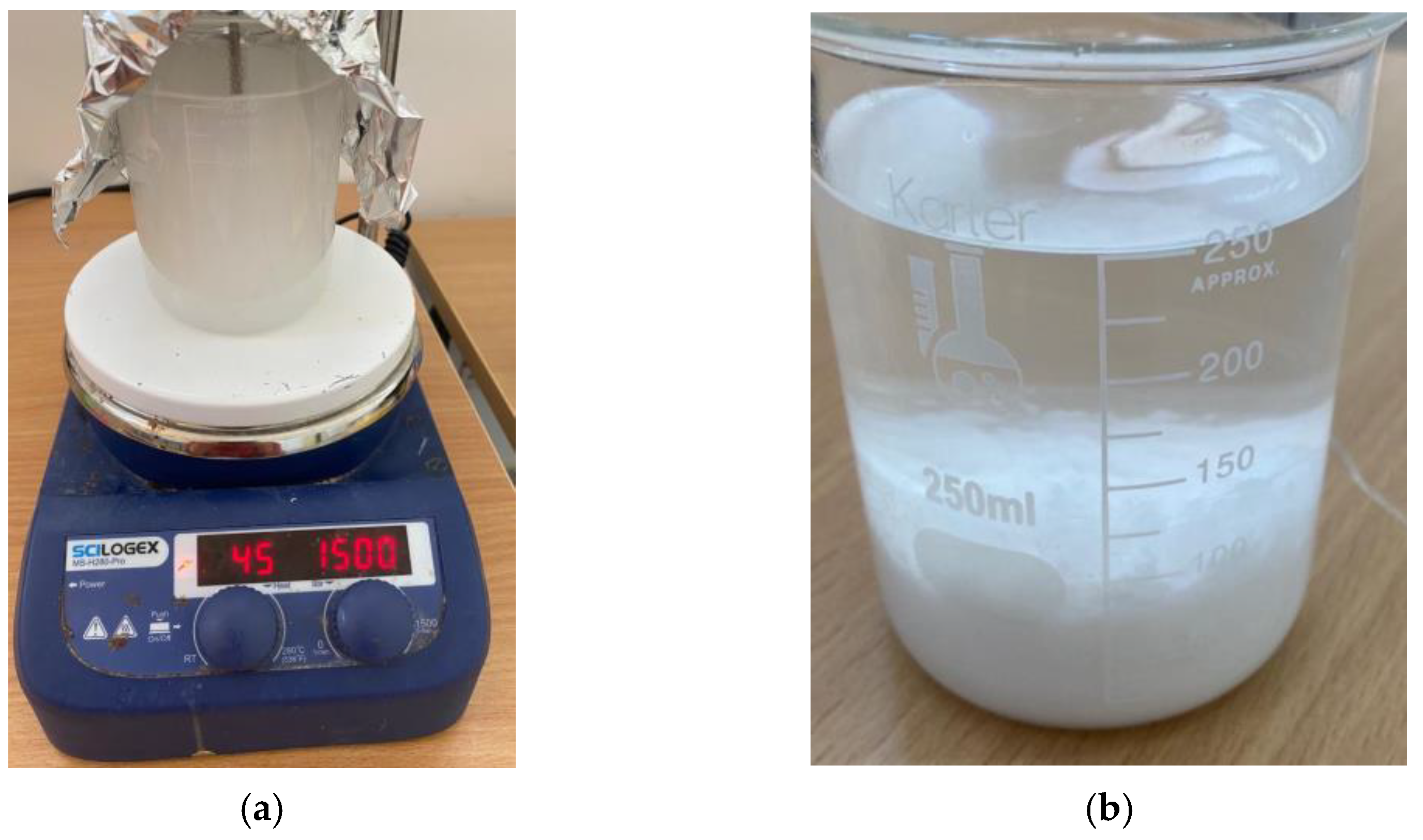
2.1. Microcapsule Preparation

2.1.1. Synthesis

This study uses the preparation procedure of self-healing submicron calcium nitrate microcapsules using Aerosol-OT in hexane solution developed by AbuTaqa et al. [20]. In the procedure, the calcium nitrate healing agent is polymerized in urea–formaldehyde shell using water-in-oil emulsion. In this study, the amounts of the aqueous phase components (urea, formaldehyde, resorcinol, ammonium chloride, calcium nitrate and the distilled water) were kept the same as in the previous study [20]. For the continuous phase, the amount of the organic solvent (Hexane) was unaltered. On the other hand, the amount of the anionic surfactant (AOT) was changed to investigate the effect of its dosage on the distribution, diameter, and shell thickness of the prepared microcapsules. Instead of using 5.8 g AOT in the continuous phase (water to AOT ratio (w)), different reduced amounts of 0.25 g, 0.50 g, 1.5 g and 2.5 g were investigated. The trial compositions are summarized in Table 1. Each trial has been repeated 3 times and the prepared microcapsules of the three repetitions have been mixed to obtain a representative sample.

2.1.2. Emulsification and Polymerization

An agitation rate of 1500 RPM and a temperature of 40−45 °C were used herein ([7,8,20]). A dropwise addition of the aqueous phase into the AOT-in-hexane continuous phase was also adopted. It is worth noting that the polymerization time increased for low-concentration AOT. The agitation has been completed for all trials by visually observing that the stirring stopped working indicating that the whole aqueous phase amount has been polymerized. Finally, the solution was let to settle for some time, and the excess hexane was decanted. The settled microcapsules were air-dried in a wide pan. Figure 1a,b shows the stirring setup and the settled microcapsules, respectively.

2.2. Scanning Electron Microscopy (SEM)

A Scanning Electron Microscopy (SEM) was used for the characterization of the microcapsule diameters. A Nova NanoSEM model was used to analyze the prepared microcapsules, which were scattered on the top of a double-sided tape attached to a pin stub specimen mount. The samples were coated with platinum for four minutes. An accelerating voltage of 3 kV secondary electron mode was used to image them. The images were captured for the scales of 10 µm and 30 µm while the average diameters of all microcapsules shown in the images were considered.

2.3. Transmission Electron Microscopy (TEM)

The Transmission Electron Microscopy (TEM) was used to characterize the microcapsule shell thickness because their shell thicknesses were smaller than their diameters. TEM may be considered as a high-resolution imaging technique in which a beam of electrons passes through a thin sample to produce a contrast in the resulting image depending on the sample thickness, density, and crystallinity. However, SEM only gathers the net intensity of secondary electrons in each point of the scan. Accordingly, and due to the small wavelength of the transmitted electrons, TEM microscopy can collect sub-nanometer scale images. Thus, TEM is considered to be more precise than SEM, especially when small wavelengths are to be measured.
FEI Tecnai G2 F20 X-TWIN Transmission Electron Microscope with 200 kV operating voltage was used in this study. The prepared microcapsules were dispersed in isopropanol using sonicator for 15 min and then drop-casted on a carbon—200 copper mesh. The mesh was then properly dried and put under the TEM microscope. The shell thicknesses were considered as the average of those in all images taken at scales of 500 nm and 1µm in each trial.

3. Results and Discussion

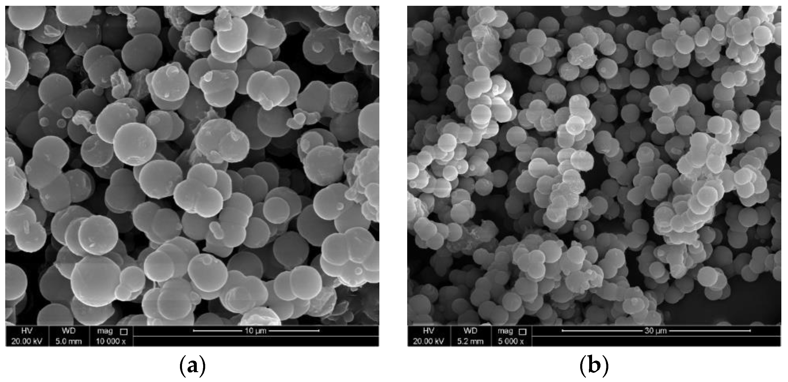
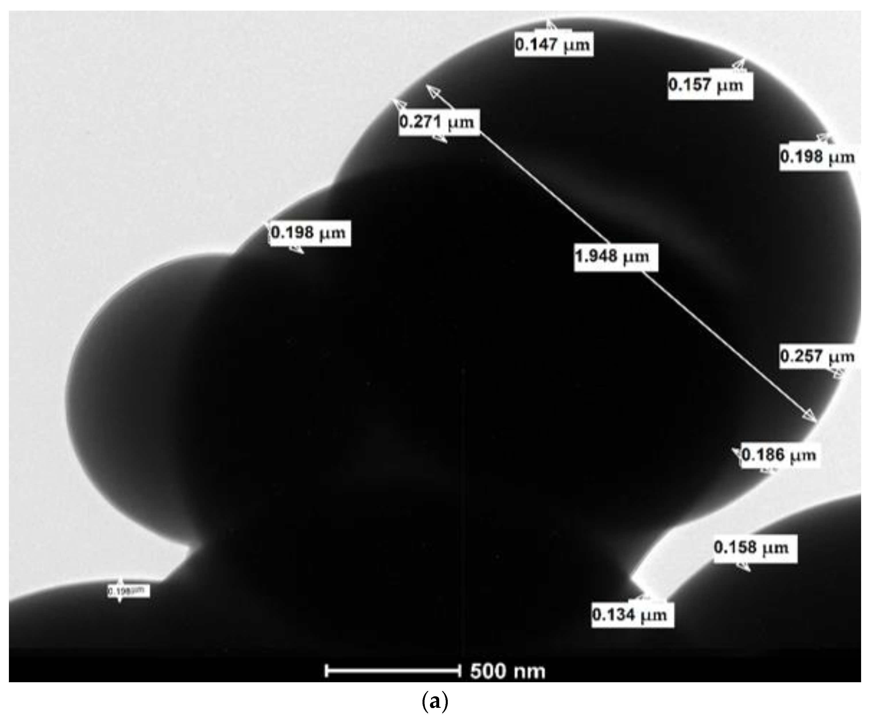
3.1. Microcapsule Diameter (Scanning Electron Microscopy)

Figure 2a,b, Figure 3a,b, Figure 4a,b and Figure 5a,b show the microcapsule diameters for all the trials prepared using AOT in Hexane solution using SEM images at 10 µm and 30 µm scales, respectively. The study investigates the effect of AOT dosage on the distribution, diameter, and shell thickness of the prepared microcapsules. Because of their large numbers, the microcapsules were randomly selected from the SEM images [7,8]. Table 2 summarizes the microcapsule minimum, maximum, and average diameter in each trial for the image scales of 10 µm and 30 µm.
The SEM images in Figure 2 of the microcapsules produced using only 0.25 g AOT show that the encapsulated component shapes are spherical and large parts of the products are agglomerated or partially polymerized. This observation is more visible in Figure 2b due to larger image scale. This may indicate that the amount of AOT used (i.e., 0.25 g) is not sufficient to totally polymerize the whole core materials into full independent capsules.
Figure 3 shows that the shape of the produced microcapsules using larger amount of AOT (i.e., 0.50 g) is uniform and the shape of the microcapsules is perfectly spherical. The SEM images in Figure 3 show that almost all of the core material components have been polymerized into single distinct microcapsules. These observations suggest that using 0.50 g of AOT-in-hexane solution is sufficient to fully polymerize the whole component amounts of the aqueous phase presented in Table 1.
The SEM images in Figure 4 and Figure 5 show the distribution, shape, and diameters of the microcapsules produced with higher AOT amounts of 1.50 g and 2.5 g, respectively. They also show that (1) the shape of the produced microcapsules is spherical and (2) almost all the components have been polymerized. However, the produced microcapsules are agglomerated or stuck to each other resulting in a nonuniform distribution. This suggests that increasing the amount of AOT above 0.50 g is not feasible and does not produce the promising self-healing efficiency for single separated microcapsules.
Table 2 shows that the minimum microcapsule diameter is reduced by increasing the AOT amount. On the other hand, the maximum microcapsule diameter increases by increasing the amount of AOT for polymerization. Moreover, the average microcapsule diameter increases for higher AOT concentrations. The average diameters are 2.13–2.39 µm, 2.54–2.57 µm, 2.63–2.77 µm, 2.78–2.88 µm for AOT amounts of 0.25, 0.50, 1.50 and 2.5 g, respectively. These values are very close to each other and hence the AOT amount may not have a significant effect on the produced microcapsules.
Considering all above observations, using 0.50 g of AOT along with the other component amounts listed in Table 1 is recommended in terms of microcapsule distribution, shape, and average diameter. Testing the healing efficiency of the microcapsules produced using 0.50 g of AOT-in-hexane solution should be investigated thoroughly in future work.

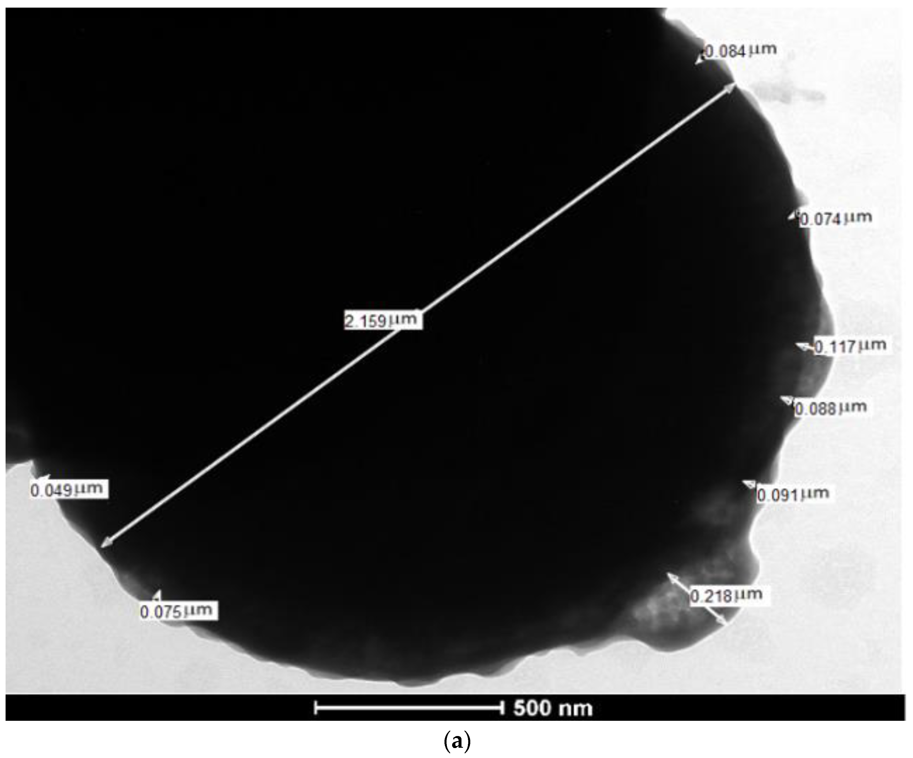
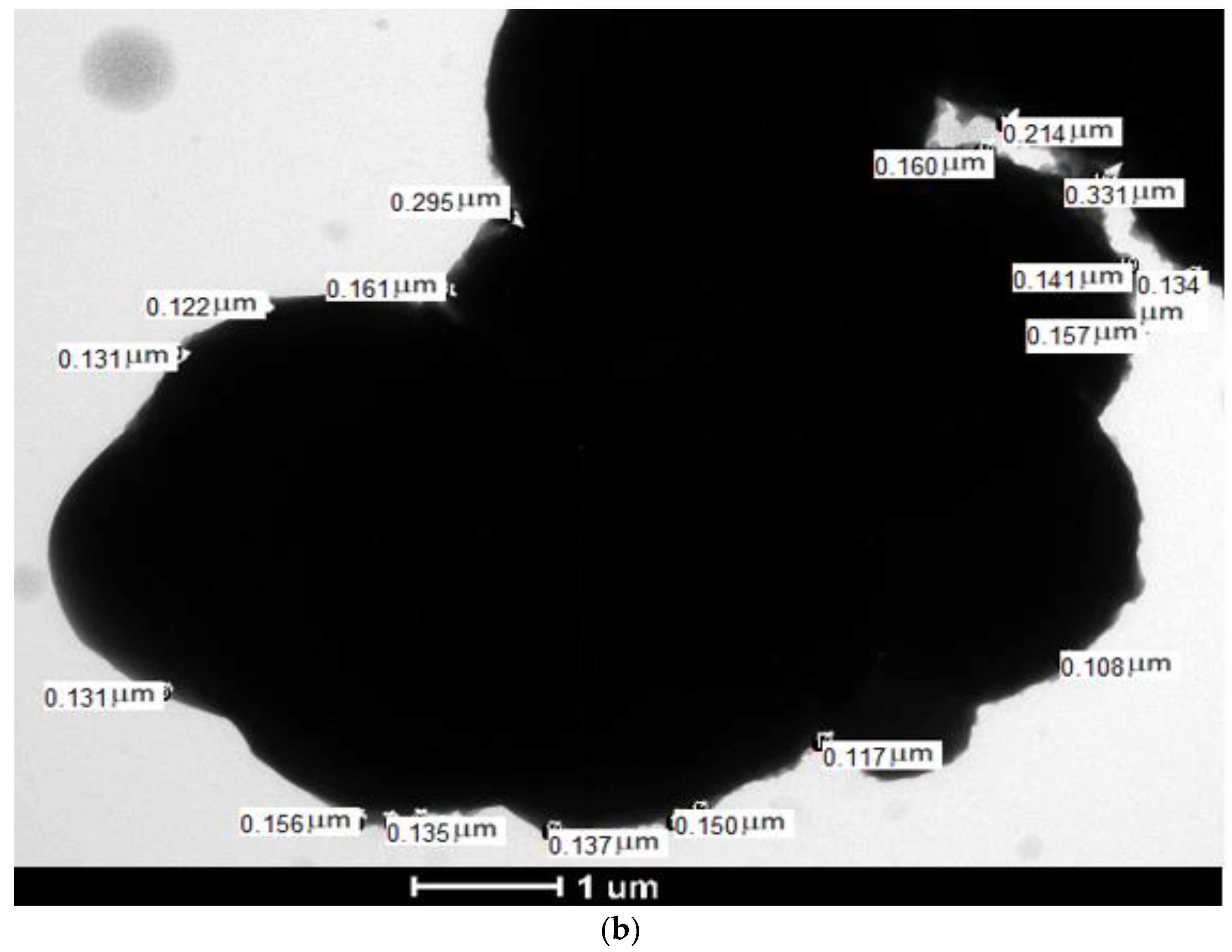
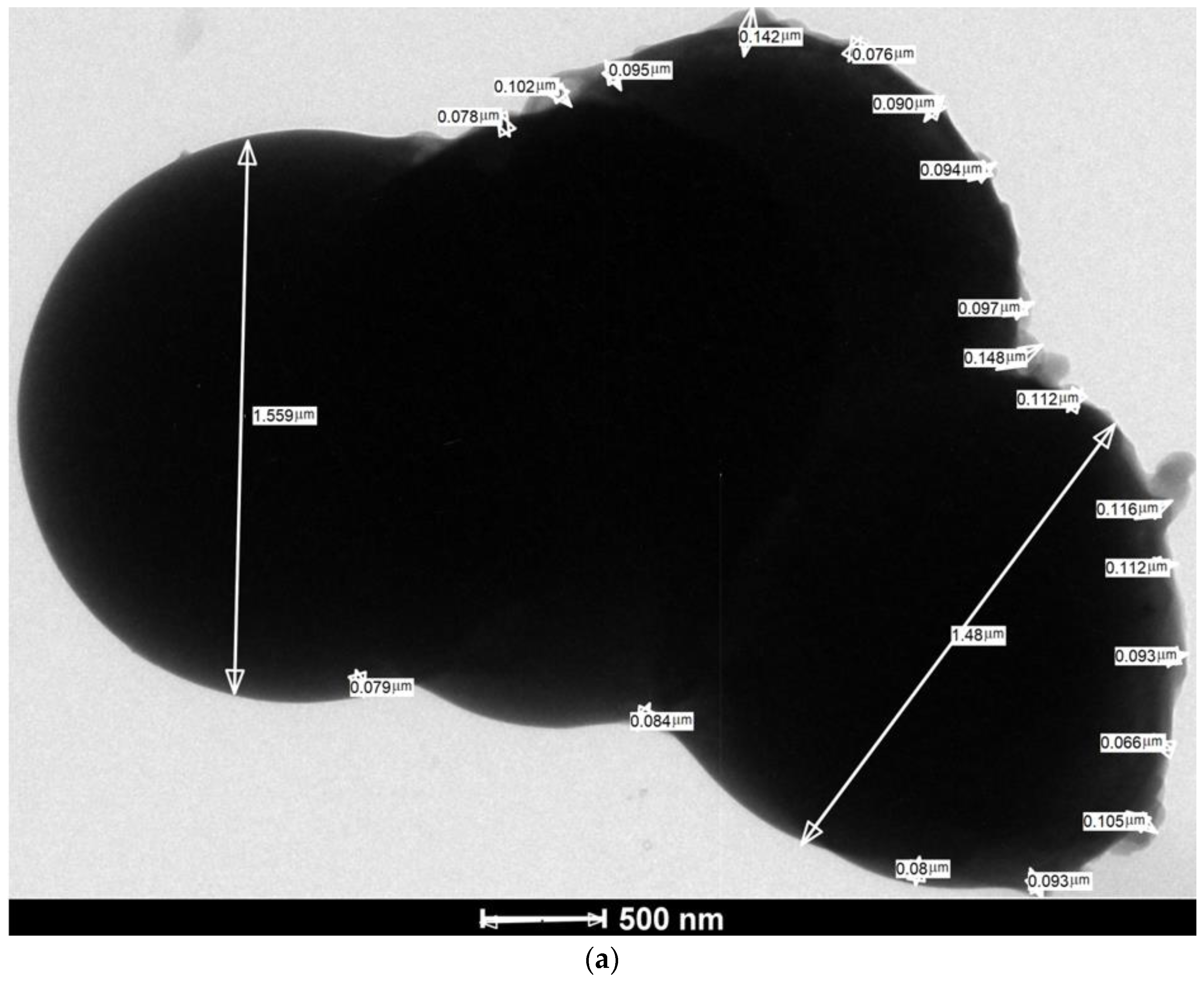
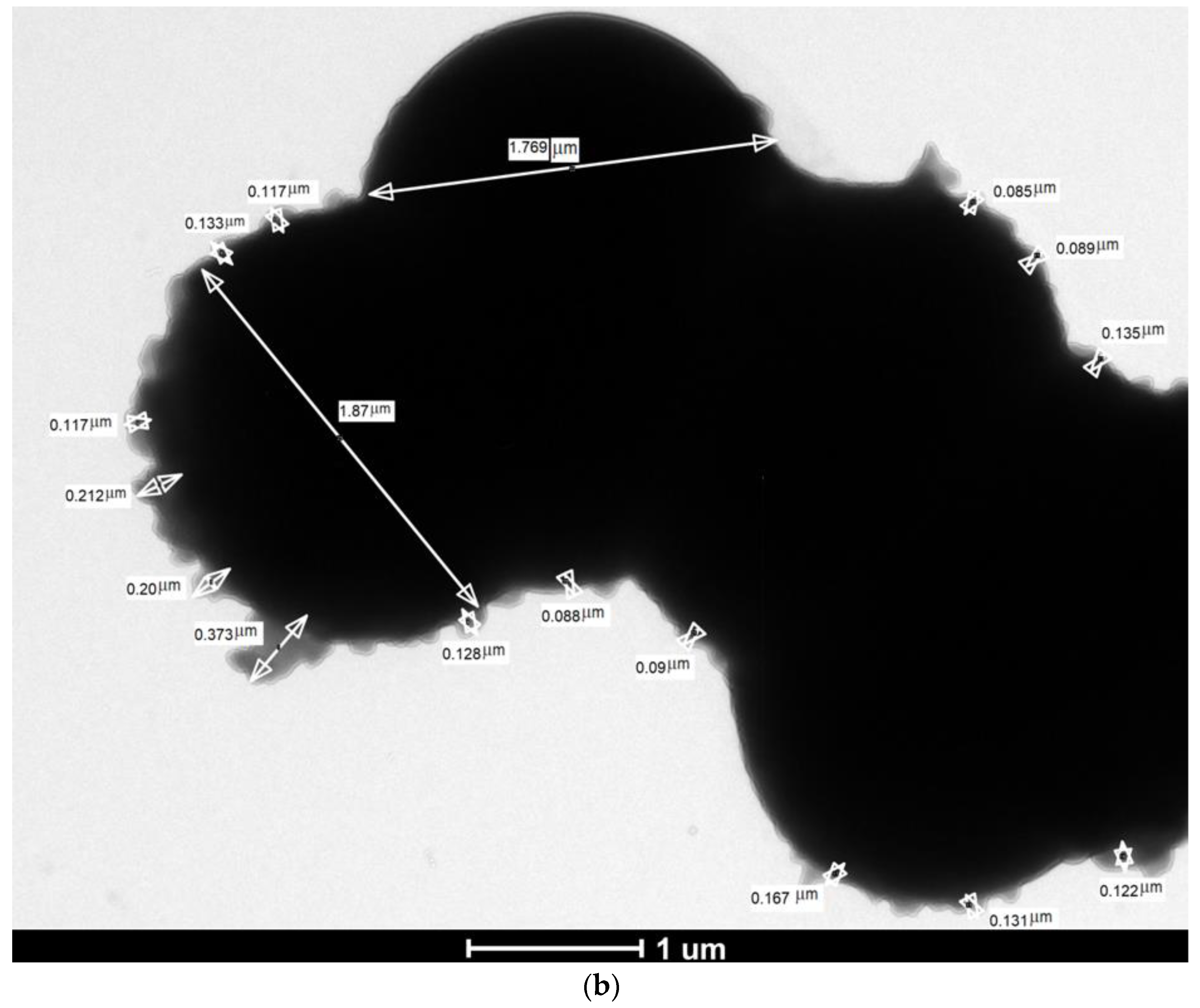
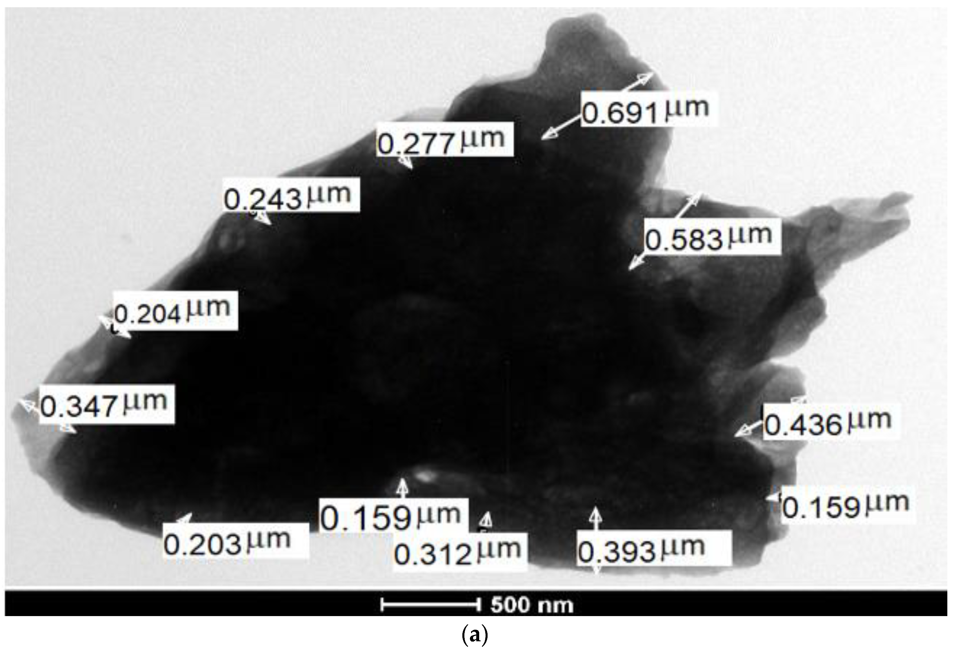
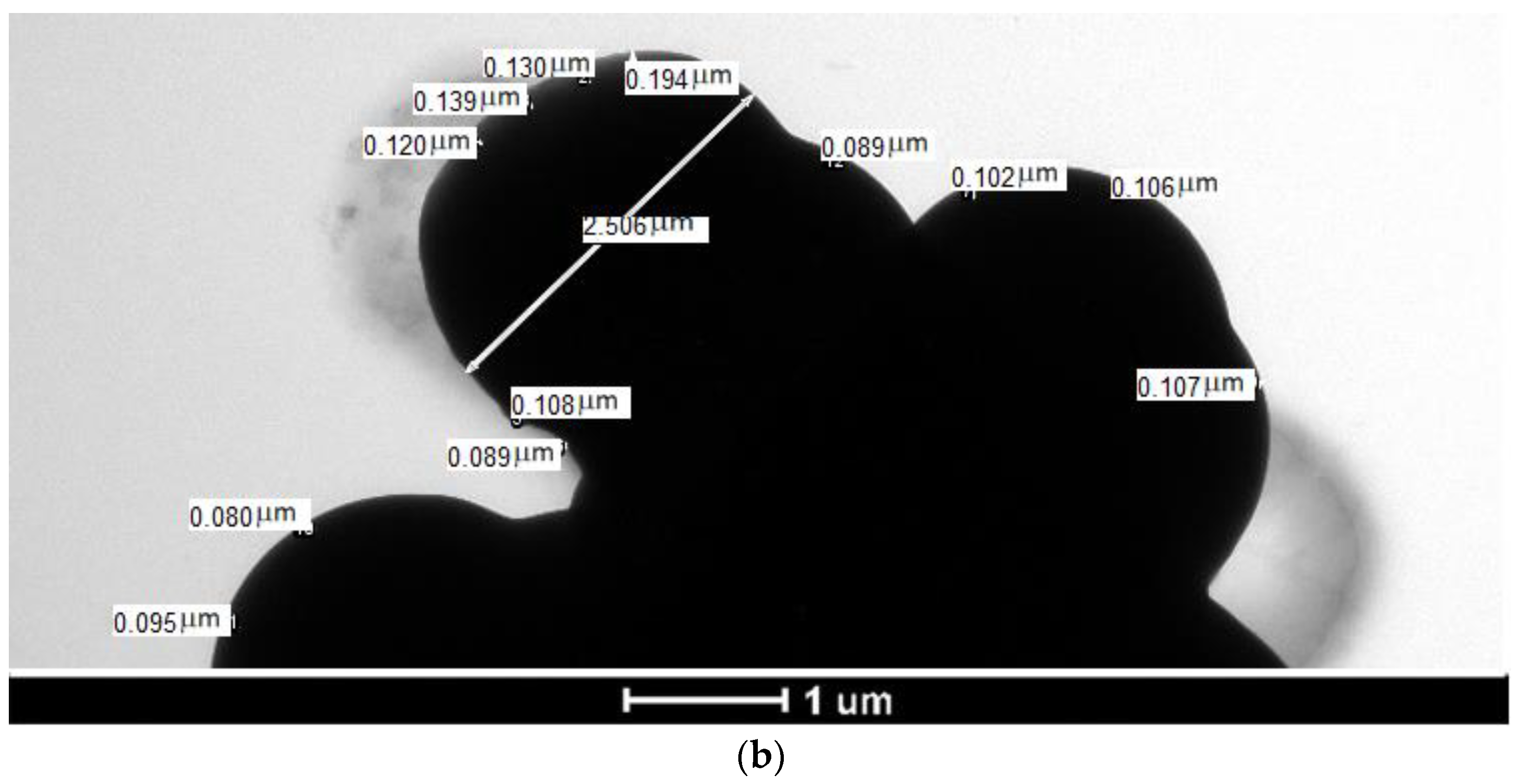
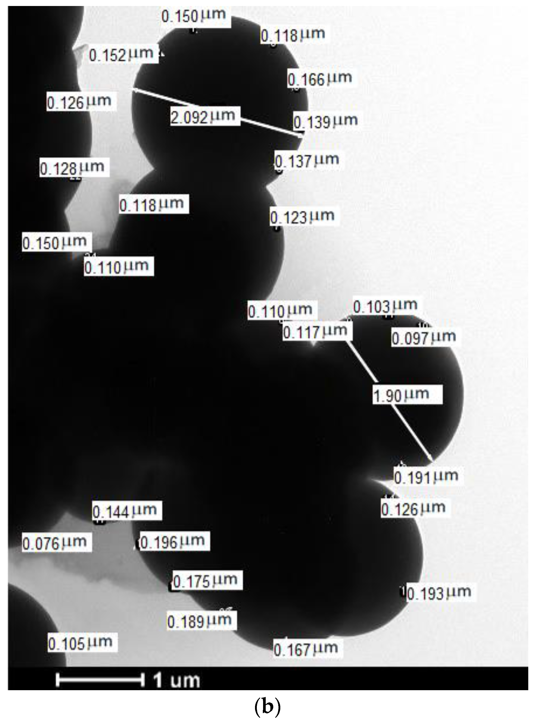
3.2. Microcapsule Shell Thickness (Transmission Electron Microscopy)

Figure 6a,b, Figure 7a,b, Figure 8a,b and Figure 9a,b show the TEM images of the microcapsules prepared using AOT in Hexane solution using scales of 500 nm and 1 µm, respectively. Table 3 summarizes the shell thickness averages of randomly selected microcapsules from the TEM images of each trial using scales of 500 nm and 1 µm, respectively.
The TEM images in Figure 6, Figure 7, Figure 8 and Figure 9 show the microcapsule shell thicknesses as the lighter parts surrounding the core. The shell thicknesses have been measured at random locations of each image for two scales (500 nm and 1µm) and their average values are summarized in Table 3.
Table 3 shows that the average shell thickness of the produced microcapsules decreased by increasing the AOT amount up to 0.50 g. However, for higher AOT concentrations (1.5 g and 2.5 g), the shell thicknesses increased again. This may be due to the agglomeration effect witnessed due to AOT increased amount as explained in Section 3.1. The microcapsules with thinner shell thicknesses are more easily ruptured by the initiated cracks. Therefore, incorporating microcapsules with smaller shell thickness into cementitious materials may improve their self-healing efficiency. Incorporating microcapsules prepared with 0.50 g AOT-in-hexane solution may lead to the optimum healing efficiency and this should be investigated in a subsequent study.

4. Conclusions

The experimental study results lead to the following conclusions:
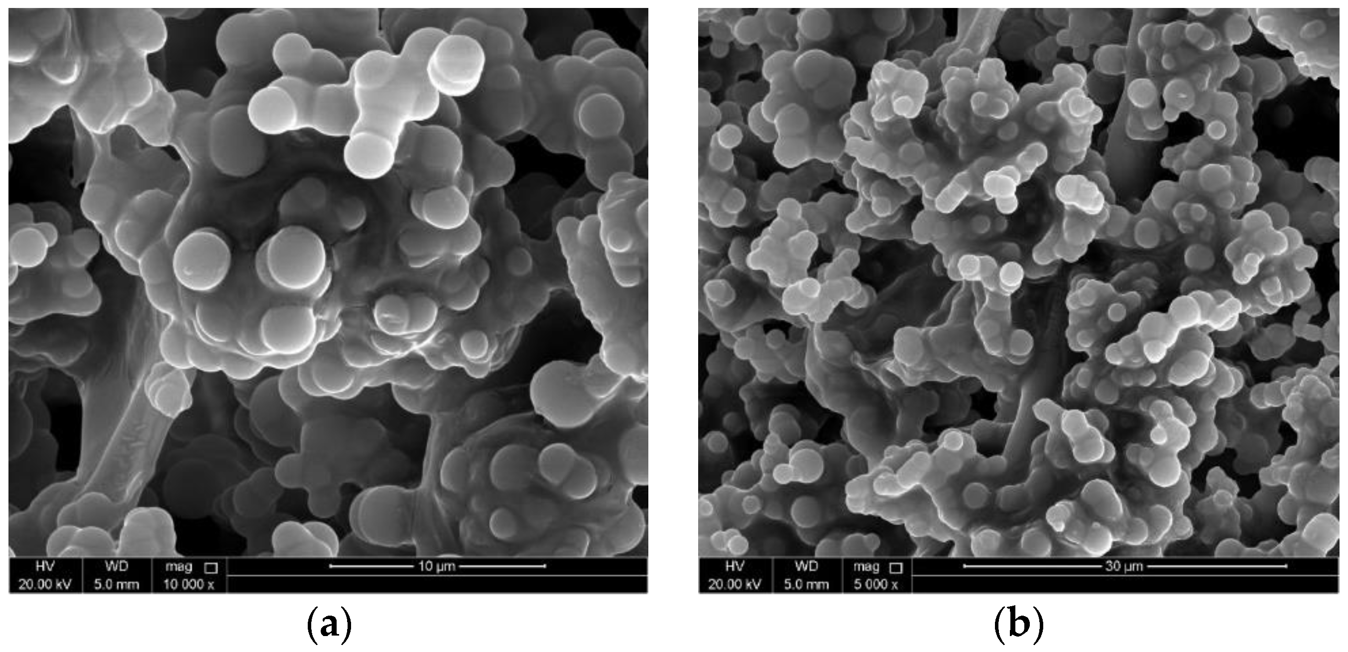
  • SEM images showed that spherically shaped microcapsules could be produced using only 0.25 g of AOT; however, they showed that considerable product parts are partially polymerized. This may indicate that using 0.25 g of AOT may not be sufficient to totally polymerize the whole core material amount.
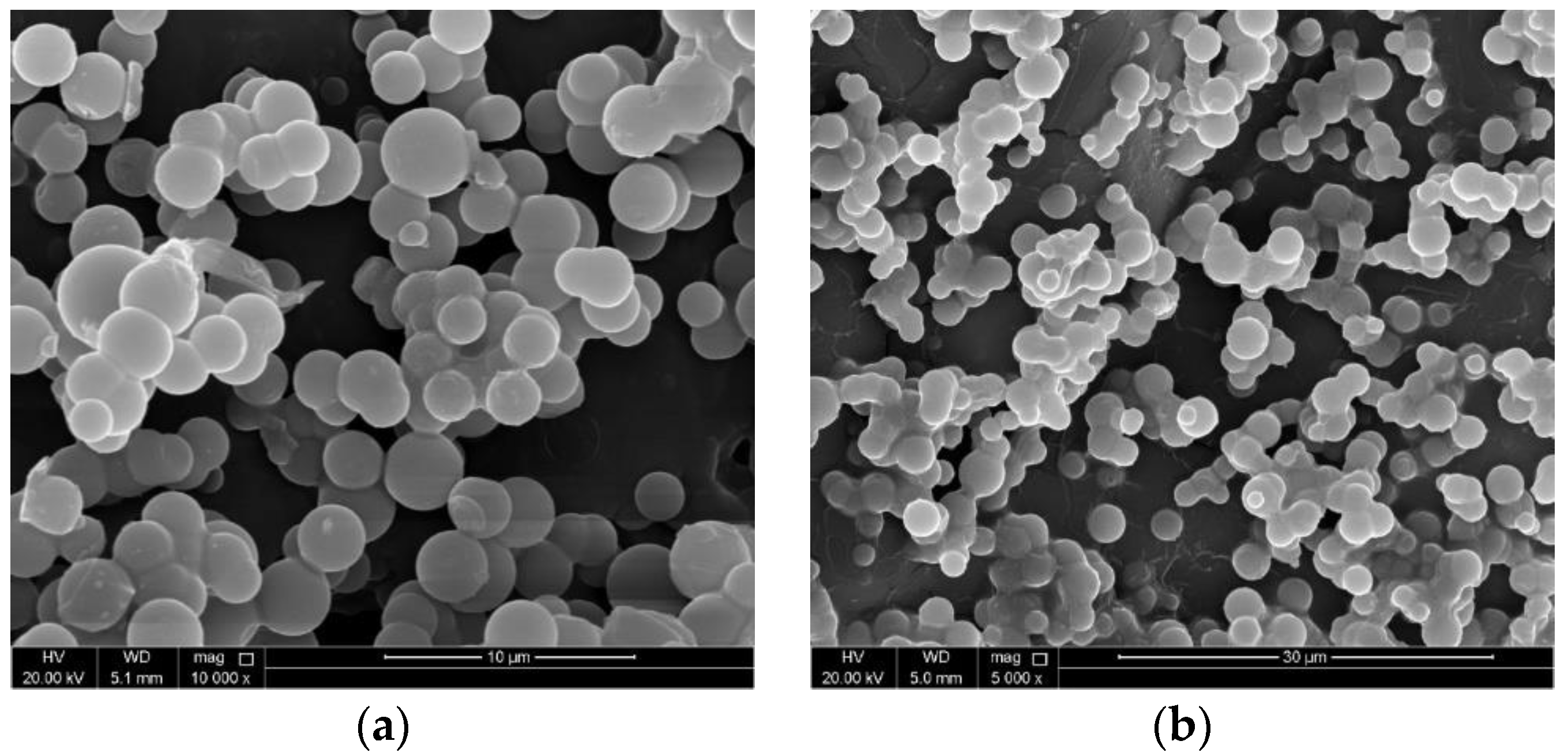
  • The SEM images of the microcapsules prepared using 0.50 g of AOT showed a uniform distribution of the produced microcapsules with perfectly spherical shape. Moreover, almost all core material components have been polymerized into distinct microcapsules. These observations suggest that using 0.50 g of AOT is sufficient to fully polymerize the whole components of the aqueous phase.
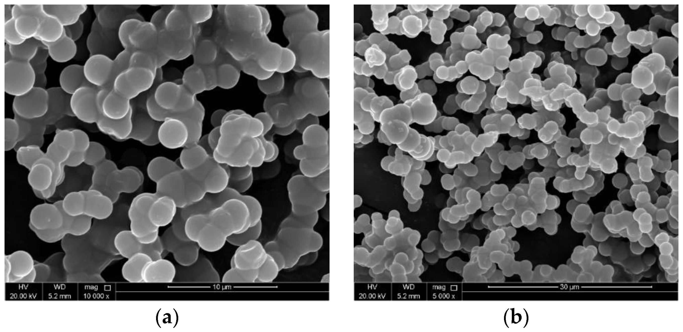
  • The shape of the microcapsules produced using 1.50 g and 2.5 g of AOT is still spherical and almost all the components have been polymerized. However, the SEM images show that the produced microcapsules are agglomerated resulting in a nonuniform distribution. This might indicate that increasing the amount of AOT above 0.50 g may not facilitate the promising self-healing efficiency single separated microcapsules.
  • The average diameters of all trials of various AOT concentrations are very close to each other which suggests that the amount of AOT used does not have a significant effect on the diameter of the produced microcapsules. In all cases, the diameters were found within the range of 2.13–2.88 µm. A value of 2.5 µm may be considered in future studies.
  • TEM was used to characterize microcapsule shell thicknesses because they are smaller than microcapsule diameters. It could be noted that the average shell thickness of the produced microcapsules decreased by increasing AOT amount up to 0.50 g. However, shell thicknesses increased again for higher AOT concentrations (1.5 g and 2.5 g). This may be due to the agglomeration effect resulting from increased AOT amount as shown in the SEM images.
  • For the preparation of calcium nitrate microcapsules, 0.50 g of AOT may be recommended using the methodology proposed by the authors in previous work [20].
  • In order to assess the practical aspect from the research carried out, a successive future study will be directed towards incorporating self-healing microcapsule prepared using 0.5 g of AOT in hexane solution into mortar and/or concrete samples with different microcapsule concentrations to investigate the mechanical properties of such samples and the healing efficiency of the prepared microcapsules.

5. Future Work

The mechanical properties and healing efficiency of mortar and/or concrete samples incorporating self-healing microcapsule prepared using 0.5 g of AOT in hexane solution with different microcapsules concentrations should be investigated in future work. Determining the appropriate percentages of the microcapsules to be incorporated into the concrete samples may be challenging in such future work; however, such percentages may be determined preliminarily based on previous similar studies on incorporating other types of self-healing microcapsules into concrete samples. The limitations of the percentages of the microcapsules will be demonstrated in the future work and will be considered as the basis for other successive works.

Author Contributions

The authors have contributed to this research article as follows: literature review, A.A.T., G.S., M.O.M. and M.A.-H.; experimental work and testing, A.A.T., D.O.A.-M., M.O.M. and M.A.-A.; investigation, A.A.T., M.O.M. and D.O.A.-M.; writing—original draft preparation, A.A.T.; writing—review and editing, G.S., A.S. and M.A.-H.; supervision, M.A.-H., M.O.M. and A.S.; project administration, G.S., A.S, M.A.-A., M.O.M. and A.A.T.; funding acquisition, A.A.T. and G.S. All authors have read and agreed to the published version of the manuscript.

Funding

This research was funded by the deanship of scientific research at Aqaba University of Technology.

Institutional Review Board Statement

Not applicable.

Informed Consent Statement

Not applicable.

Data Availability Statement

The data used to support the findings of this study are available upon request from the corresponding author.

Acknowledgments

This work was made possible by the support and deanship of scientific research at Aqaba University of Technology as well as the collaboration of the University of Houston. The statements made herein are solely the responsibility of the authors.

Conflicts of Interest

The authors declare that they have no known competing financial interest or personal relationships that could have appeared to influence the work reported in this paper.

References

  1. Patil, R.; Bendre, R. Preparation, characterization and controlled release study of poly(urea–formaldehyde) microcapsules enclosing Pretilachlor herbicide. Iran. Polym. J. 2022, 31, 691–704. [Google Scholar] [CrossRef]
  2. Gao, P.P.; Zhou, Z.H.; Yang, B.; Ji, X.; Pan, M.; Tang, J.H.; Lin, H.; Zhong, G.J.; Li, Z.M. Structural regulation of poly(urea-formaldehyde) microcapsules containing lube base oil and their thermal properties. Prog. Org. Coat. 2021, 150, 105990. [Google Scholar] [CrossRef]
  3. Vintila, I.S.; Iovu, H.; Alcea, A.; Cucuruz, A.; Mandoc, A.C.; Vasile, B.S. The Synthetization and Analysis of Dicyclopentadiene and Ethylidene-Norbornene Microcapsule Systems. Polymers 2020, 12, 1052. [Google Scholar] [CrossRef]
  4. Arefin, P.; Habib, M.S.; Chakraborty, D.; Bhattacharjee, S.C.; Das, S. An overview of microcapsule dosage form. Int. J. Pharm. Chem. Anal. 2020, 7, 155–160. [Google Scholar] [CrossRef]
  5. Milla, J.; Hassan, M.M.; Rupnow, T.; Daly, W.H. Measuring the crack-repair efficiency of steel fiber reinforced concrete beams with microencapsulated calcium nitrate. Constr. Build. Mater. 2019, 201, 526–538. [Google Scholar] [CrossRef]
  6. Al-Ansari, M.; Taqa, A.A.; Hassan, M.; Senouci, A.; Milla, J. Performance of modified self-healing concrete with calcium nitrate microencapsulation. Constr. Build. Mater. 2017, 149, 525–534. [Google Scholar] [CrossRef]
  7. Milla, J.; Hassan, M.; Rupnow, T.; Al-Ansari, M.; Arce, G. Evaluation of the Effect of Self-Healing Calcium Nitrate Microcapsules on Concrete Properties. In Proceedings of the Paper # 16-0422, Submitted to the 95th Transportation Research Board Annual Meeting, Washington, DC, USA, 10–14 January 2016. [Google Scholar]
  8. Hassan, M.; Milla, J.; Rupnow, T.; Al-Ansari, M.; Daly, B. Microencapsulation of Calcium Nitrate for Concrete Applications. Transp. Res. Rec. 2016, 2577, 8–16. [Google Scholar] [CrossRef]
  9. Fayyad, E.M.; Almaadeed, M.A.; Jones, A. Preparation and characterization of urea–formaldehyde microcapsules filled with paraffin oil. Polym. Bull. 2016, 73, 631–646. [Google Scholar] [CrossRef]
  10. Mostavi, E.; Asadi, S.; Hassan, M.; Alansari, M. Evaluation of Self-Healing Mechanisms in Concrete with Double-Walled Sodium Silicate Microcapsules. J. Mater. Civ. Eng. 2015, 27, 04015035. [Google Scholar] [CrossRef]
  11. Kaes, M.; Van-Tittelboom, K.; De-Belie, N. The efficiency of self-healing cementitious materials by means of encapsulated polyurethane in chloride containing environments. Constr. Build. Mater. 2014, 71, 528–537. [Google Scholar] [CrossRef]
  12. Van-Tittelboom, K.; Belie, N.D. Self-Healing in Cementitious Materials-a Review. Materials 2013, 6, 2182–2217. [Google Scholar] [CrossRef] [PubMed] [Green Version]
  13. Schlangen, E.; Sangadji, S. Addressing Infrastructure Durability and Sustainability by Self-Healing Mechanisms—Recent Advances in Self-HealingConcrete and Asphalt. Procedia Eng. 2013, 54, 39–57. [Google Scholar] [CrossRef] [Green Version]
  14. Yang, Z.; Hollar, J.; He, X.; Shi, X. A Self-Healing Cementitious Composite Using Oil Core/silica Gel Shell Microcapsules. Cem. Concr. Compos. 2011, 33, 506–512. [Google Scholar] [CrossRef]
  15. Van-Tittelboom, K.; De-Belie, N.; Van-Loo, D.; Jacobs, P. Self-Healing Efficiency of Cementitious Materials Containing Tubular Capsules Filled with Healing Agent. Cem. Concr. Compos. 2011, 33, 497–505. [Google Scholar] [CrossRef]
  16. Huang, H.; Ye, G. Application of Sodium Silicate Solution as Self-Healing Agent in Cementitious Materials. In Proceedings of the International RILEM Conference on Advances in Construction Materials through Science and Engineering, Hong Kong, China, 5–7 September 2011; pp. 530–536. [Google Scholar]
  17. Podgornik, B.B.; Šumiga, B. Microencapsulation technology and its applications in building construction materials. RMZ-Mater. Geoenviron. 2008, 55, 329–344. [Google Scholar]
  18. Pelletier, M.; Brown, R.; Shukla, A.; Bose, A. Self-Healing Concrete with a Microencapsulated Healing Agent. Available online: http://energetics.chm.uri.edu/system/files/Self%20healing%20concrete%20-7-11.pdf (accessed on 31 March 2022).
  19. He, J.; Shi, X. Laboratory assessment of early-age durability benefits of a self-healing system to cementitious composites. J. Build. Eng. 2021, 44, 102602. [Google Scholar] [CrossRef]
  20. AbuTaqa, A.; Suleiman, G.; Senouci, A.; Mohsen, M.O. Using Aerosol OT in Hexane Solution to Synthesize Calcium Nitrate Self-Healing Refined Microcapsules for Construction Applications. Buildings 2022, 12, 751. [Google Scholar] [CrossRef]
  21. Kalyanram, P.; Valenzuela, P.; Gupta, A.R. Aerosol-Ot Stabilized Micro and Nano-Scale Emulsions for Pharmaceutical Formulations. Org. Med. Chem. Int. J. 2017, 4, 88–90. [Google Scholar] [CrossRef]
  22. Hensel, J.K.; Carpenter, A.P.; Ciszewski, R.K.; Schabes, B.K.; Kittredge, C.T.; Moore, F.G.; Richmond, G.L. Molecular characterization of water and surfactant AOT at nanoemulsion surfaces. Proc. Natl. Acad. Sci. USA 2017, 114, 13351–13356. [Google Scholar] [CrossRef] [Green Version]
  23. Srivastava, S.; Sharma, S.K.; Sharma, R.K. Synthesis of gold nanorods using concentrated aerosol OT in hexane and its application as catalyst for the reduction of eosin. Colloid Surf. Phys. Eng Asp. 2011, 373, 61–65. [Google Scholar] [CrossRef]
  24. Gupta, M.; Gupta, A.K. In vitro cytotoxicity studies of hydrogel pullulan nanoparticles prepared by AOT/N-hexane micellar system. J. Pharm. Pharm. Sci. Publ. Can. Soc. Pharm. Sci. Soc. Can. Des Sci. Pharm. 2004, 7, 38–46. [Google Scholar]
  25. Kizling, J.; Kronberg, B. On the formation of concentrated stable w/o emulsions. Adv. Colloid Interface Sci. 2001, 89–90, 395–399. [Google Scholar] [CrossRef]
  26. La Mesa, C.; Coppola, L.; Ranieri, G.A.; Terenzi, M.; Chidichimo, G. Phase diagram and phase properties of the system water-hexane-Aerosol OT. Langmuir 1992, 8, 2616–2622. [Google Scholar] [CrossRef]
  27. Eastoe, J.; Fragneto, G.; Robinson, B.H.; Towey, T.F.; Heenan, R.K.; Leng, F.J. Variation of surfactant counterion and its effect on the structure and properties of Aerosol-OT-based water-in-oil microemulsions. J. Chem. Soc. Faraday Trans. 1992, 88, 461–471. [Google Scholar] [CrossRef]
  28. Eastoe, J.; Robinson, B.H.; Steytler, D.C.; Thorn-Leeson, D. Structural studies of microemulsions stabilised by aerosol-OT. Adv. Colloid Interface Sci. 1991, 36, 1–31. [Google Scholar] [CrossRef]
  29. Aveyard, R.; Binks, B.P.; Clark, S.; Mead, J. Interfacial tension minima in oil–water–surfactant systems. Behaviour of alkane–aqueous NaCl systems containing aerosol OT. J. Chem. Soc. Faraday Trans. 1 1986, 82, 125–142. [Google Scholar] [CrossRef]
  30. Leong, Y.S.; Candau, F. Inverse microemulsion polymerization. J. Phys. Chem. 1982, 86, 2269–2271. [Google Scholar] [CrossRef]
  31. Aïssa, B.; Therriault, D.; Haddad, E.; Jamroz, W. Self-Healing Materials Systems: Overview of Major Approaches and Recent Developed Technologies. Adv. Mater. Sci. Eng. 2012, 2012, 854203. [Google Scholar] [CrossRef] [Green Version]
  32. Blaiszik, B.J.; Sottos, N.R.; White, S.R. Nanocapsules for self-healing materials. Compos. Sci. Technol. 2008, 68, 978–986. [Google Scholar] [CrossRef]
Figure 1. (a) Stirring setup and (b) settled microcapsules.
Buildings 12 02121 g001
Figure 2. Trial-1 (0.25 g AOT) microcapsule SEM images for scales of (a) 10 µm and (b) 30 µm.
Buildings 12 02121 g002
Figure 3. Trial-2 (0.50 g AOT) microcapsule SEM images for scales of (a) 10 µm and (b) 30 µm.
Buildings 12 02121 g003
Figure 4. Trial-3 (1.50 g AOT) microcapsule SEM images for scales of (a) 10 µm and (b) 30 µm.
Buildings 12 02121 g004
Figure 5. Trial-4 (2.50 g AOT) microcapsule SEM images for scales of (a) 10 µm and (b) 30 µm.
Buildings 12 02121 g005
Figure 6. Trial-1 microcapsule TEM images at scales of (a) 500 nm and (b) 1µm.
Buildings 12 02121 g006aBuildings 12 02121 g006b
Figure 7. Trial-2 microcapsule TEM images at scales of (a) 500 nm and (b) 1µm.
Buildings 12 02121 g007aBuildings 12 02121 g007b
Figure 8. Trial-3 microcapsule TEM images at scales of (a) 500 nm and (b) 1µm.
Buildings 12 02121 g008aBuildings 12 02121 g008b
Figure 9. Trial-4 microcapsule TEM images at scales of (a) 500 nm and (b) 1µm.
Buildings 12 02121 g009aBuildings 12 02121 g009b
Table 1. The compositions of all microcapsule preparation trials.
Trial-1Trial-2Trial-3Trial-4
Aqueous PhaseConstituentAmount (g)
Urea5.00
Formaldehyde (37% solution)12.67
Resorcinol0.50
Ammonium Chloride0.50
Calcium Nitrate10.00
Distilled Water50.00
Continuous PhaseOrganic Solvent (Hexane)180.0
Dioctyl Sodium Sulfosuccinate (AOT)0.250.501.52.5
Table 2. Microcapsule minimum, maximum, and average diameters at different image scales.
Image Scale10 µm30 µm
TrialAOT Amount (g)Min. Dia. (µm)Max. Dia. (µm)Average Dia. (µm)Min. Dia. (µm)Max. Dia. (µm)Average Dia. (µm)
Trial-10.251.423.402.131.483.732.39
Trial-20.501.244.212.541.394.512.57
Trial-31.501.144.082.631.344.032.77
Trial-42.500.604.482.781.205.052.88
Table 3. Microcapsule average shell thicknesses for all trials at different image scales.
Image Scale500 nm1 µm
Trial AOT Amount (g)Average Dia. (µm)Average Dia. (µm)
Trial-10.250.0100.166
Trial-20.500.0980.146
Trial-31.500.1300.154
Trial-42.500.1910.192
Publisher’s Note: MDPI stays neutral with regard to jurisdictional claims in published maps and institutional affiliations.

