Article

Second Natural Occurrence of KFeS2 (Hanswilkeite): An Inclusion in Diamond from the Udachnaya Kimberlite Pipe (Siberian Craton, Yakutia)

by
Alla M. Logvinova
1,* and
Igor S. Sharygin
1,2
1
V. S. Sobolev Institute of Geology and Mineralogy, Siberian Branch of the Russian Academy of Sciences, Koptyuga Pr. 3, Novosibirsk 630090, Russia
2
Institute of the Earth’s Crust, Siberian Branch of the Russian Academy of Sciences, Lermontova St. 138, Irkutsk 664033, Russia
*
Author to whom correspondence should be addressed.
Minerals 2023, 13(7), 874; https://doi.org/10.3390/min13070874
Submission received: 24 May 2023 / Revised: 21 June 2023 / Accepted: 23 June 2023 / Published: 28 June 2023

Abstract

:
Potassium sulfide KFeS2 (hanswilkeite) has been identified in polymineralic inclusions in a diamond from the Udachnaya kimberlite pipe (Siberian craton, Yakutia). This is the second occurrence of hanswilkeite in nature and the first one in mantle-derived samples. Sulfide KFeS2 is monoclinic, the space group—C 2/c. Its crystal structure consists of chains with K in the interstices. The tetrahedra are centered by iron ions and linked by edges, thus forming chains of [FeS2] frameworks. The strongest lines of the electron diffraction powder pattern are 7.05 Å—(200); 5.34 Å (0 2 ¯ 0); and 3.05 Å (2 2 ¯ 0), and the angles between directions are <2 2 ¯ 0/0 2 ¯ 0>—60° and <2 2 ¯ 0/200>—30°. KFeS2 has been found as a discrete phase within polymineralic inclusions consisting of apatite, ilmenite, chondrodite, phlogopite, dolomite, and a fluid phase. The data obtained from the composition of the hanswilkeite (KFeS2) inclusion and other rare minerals (chondrodite, Mg-apatite, Cr-ilmenite) in primary inclusions in a diamond from the Udachnaya kimberlite testify to the important role of metasomatic processes in diamond formation.

1. Introduction

Sulfides are widespread mineral inclusions in diamonds [1,2,3,4]. They provide significant information about Earth’s mantle and have been the subject of intensive studies during many years [1,2,3,4,5,6,7]. The abundance of sulfide as inclusions in diamonds and their intergrowth with syn-/protogenetic mantle silicates (olivine, orthopyroxene, clinopyroxene, and pyrope) in diamonds suggest that inclusions of sulfide are also syn-/protogenetic. Sulfides in diamond inclusions are represented by pyrrhotite, pentlandite, troilite, chalcopyrite, pyrite, djerfisherite, and monosulfide solid solutions [8]. Similar to silicate assemblages in diamonds, sulfide inclusions also belong to two types of mantle paragenesis: eclogitic (Ni-poor, 0–12 wt% Ni) and peridotitic (Ni-rich, 22–36 wt% Ni) [1,4]. Thus, sulfides are thought to be present in growth environments of diamonds.
Sulfides have also been described in nanometer-sized (submicron) polymineral inclusions in fibrous diamonds [9,10]. These inclusions represent well-preserved samples of diamond-forming media and appear to represent originally homogeneous phases (melts or high-density fluids) entrapped by the growth of the fibrous diamonds at depths > 4 GPa. Subsequently, these trapped melts or fluids crystallized into a range of daughter minerals and fluid phase(s). Sulfides in such inclusions are represented by chalcopyrite CuFeS2, pentlandite (Fe,Ni)9S8, heazlewoodite Ni3S2, pyrrhotite Fe1-xS, and troilite FeS, as well as unidentified sulfides of Cu and Ni. Moreover, energy-dispersive spectra of some Fe-rich sulfide phases in nanometer-sized polymineralic inclusions showed a stable presence of potassium [9]. However, K-containing sulfides were not reliably identified.
Currently, five potassium-bearing sulfides are approved as mineral species by the International Mineralogical Association (IMA): murunskite K2(Cu,Fe)4S4, rasvumite KFe2S3, djerfisherite K6(Fe,Cu,Ni)25S26Cl, bartonite K6Fe20S26S, chlorbartonite K6Fe24S26Cl, and hanswilkeite KFeS2 [11,12,13,14,15,16,17,18,19]. KFeS2 has been known since 1869. Crystals of KFeS2 were obtained from the reaction of a mixture of potassium carbonate, iron, and sulfur, followed by extracting the solidified melt with water. The structure was first determined in 1942 by Boon and MacGillavry [20,21]. KFeS2 is not oxidized in air and it is resistant to water, and thus it is a stable phase in laboratory experiments [22]; however, until recently, it has not been known as a naturally occurring mineral. In 2022, KFeS2 was recognized by the IMA as a new mineral—hanswilkeite (IMA no. 2022-041) [19]. Hanswilkeite was found in fine-grained tilleite–calcite marble, often intergrown with rasvumite, in association with pyrite and oldgamite [S.N. Britvin, personal communication]. The size of its discharge is up to 1 mm.
In this paper, we, for the first time, identified potassium sulfide KFeS2 (potassium thioferrite) in nanometer-sized polymineralic inclusions in a diamond from the Udachnaya kimberlite pipe (Siberian craton, Yakutia).

2. Materials and Methods

The studied diamond (sample no. Ud-45) is a cubic crystal with a fibrous inner structure and dark cloud of numerous nanometer-sized polymineralic inclusions in its central part (Figure 1a).
Microstructures and compositional features of phases in polymineralic inclusions in diamond Ud-45 were identified with the transmission electron microscope (TEM) Tecnai G2 F20 X-Twin operated at 200 kV with a Schottky emitter as an electron source (FEG) at GeoForschungsZentrum (GFZ), Potsdam, Germany. The diamond crystal was polished parallel to (110) on both sides to make a 1 mm thick plate (Figure 1a). Electron-transparent foils with typical dimensions of 15 μm × 8 μm × 0.15 μm were prepared, applying focused ion beam (FIB) sample preparation. Details of the foil preparation are given in [23,24]. The TEM was equipped with an EDAX X-ray analyzer with an ultra-thin window, a Fishione high-angle annular dark-field (HAADF) detector, and a Gatan imaging filter (Tridiem, Richmond, VA, USA) for electron energy-loss spectroscopy and energy filtered imaging. HAADF images were collected with a camera length of 330 mm displaying a diffraction contrast plus a Z-contrast or with a camera length of 75 mm, which shows a Z-contrast only. Nanophases were identified by acquiring high-resolution lattice fringe images with a short acquisition time (0.6 s) to avoid decomposition during exposure to the electron beam [24]. The calculated diffraction patterns (using the Fast Fourier Transform (TEM)) from high-resolution images were used to measure d-spacing and angles between the adjacent lattice planes. A comparison of the observed data with calculated data from known phases (literature data) allowed for identifying the phases present. The chemical composition of the phases was always measured in the scanning transmission mode (STEM), thus avoiding a significant mass loss during data acquisition. The acquisition time was 60 or 120 s.

3. Results and Discussion

Twelve separate polymineralic inclusions ranging from 100 to 700 nm in size were studied in the diamond Ud-45 (Figure 2). The inclusions show negative crystal shapes with parallel facets (Figure 3). They are not related to any healed cracks. Therefore, they may be considered as primary/fluid inclusions (i.e., those that form while crystals are growing).
The daughter phase assemblages are represented by silicate, carbonate, phosphate, oxide, and chloride minerals. There are voids inside inclusions that likely are filled by the fluid phase. The main minerals are dolomite, chondrodite (a member of the humite group: (Mg, Fe2+)5(SiO4)2(F,OH)2)), phlogopite, Mg-rich apatite, Cr-bearing ilmenite, magnetite, K-Fe-sulfide, and KCl (Figure 2 and Figure 3). Dolomite was recognized as the dominant phase in the inclusions. The next most common phases are silicates: phlogopite and chondrodite.
Chondrodite has not yet been reported as an inclusion in a diamond. This silicate was identified using diffraction patterns. All measured hkl values of the studied chondrodite were compared with the calculated ones for interplanar distances of the reference Mg5(SiO4)2(F,OH)2 (Table 1). The chondrodite phases within polymineralic inclusions are characterized by Ti impurity (Figure 3A,E).
The porous microstructure of the polymineralic inclusions requires the presence of a fluid phase during the formation of the inclusion, which has been partly released during FIB sample preparation (Figure 2). For simplicity, these decrepitated fluid inclusions are labeled “fluid” in the figures.
A total of 21 phases in 12 individual polymineralic nanoinclusions were analyzed. For the smallest phases after imaging with high-resolution electron microscopy (HREM), the compositional measurements were carried out using a defocused beam. It turned out to be extremely important to defocus the beam (the size of the focusing spot is about 0.1 nm) in order to avoid a mass loss during the measurement. This was achieved by expanding the beam to the diameter of nanocrystals (about 20–30 nm). Crystal structure data of the minerals within nanoinclusions were derived from selected area diffraction patterns (Figure 3) and from Fast Fourier Transforms (FFT), which were calculated from energy-filtered high-resolution images using the Gatan Digital Micrograph software package version 3.5. The main emphasis was placed on the study of the FeKS2 phase.
The largest sulfide grain of a potassium iron sulfide was found in inclusion #6 that abuts a void, formerly filed by a fluid phase (Figure 4). Its diameter is about 50 nm. The nanoinclusion of sulfide is mainly composed of potassium, iron, and sulfur. It was identified by EDX spectra with a very intense peak of K, Fe, and S (Figure 4E). The weak X-ray intensities of Cu–Kα in the spectrum are due to the copper grid.
Further identification of the K-Fe-sulfide was performed with diffraction measurements (Figure 4B,C). The dhkl values obtained for the studied K-Fe-sulfide match well with those of the synthetic reference KFeS2 [20,21].
According to [21], the crystal structure of the synthetic phase KFeS2 is monoclinic with the cell parameters ao = 7.05 Å; bo = 11.28 Å; and co = 5.39 Å. The space group of KFeS2 is C 2/c.
All measured dhkl distances obtained from the HREM images and from the electron diffraction patterns of the selected areas, compared with the synthetic FeKS2 data, are presented in Table 1. This table also lists data on the silicate phase of chondrodite, which is present in these polymineralic inclusions.
The first column lists the Miller indices (hkl), which are used to label the planes. The second column displays the dhkl values observed from the electron diffraction pattern. The third column shows the data dhkl calculated from HREM results. The lattice parameters are very close. All calculations of distances and angles between planes (φ) were carried out using the Desktop Microscopy software package version 2.1. [21].
The angles between the planes, indicated in Table 1, in the case of KFeS2 were also recalculated one by one, applying the program “Single Crystal” using the zone axis [002] derived from the diffraction pattern. The angles are the same.
The diffraction data and the cell constants of the KFeS2 obtained are shown on the projection of the crystal lattice along the z-axis (Figure 5).
The crystal structure of the KFeS2 phase in the inclusions was plotted using the Crystal Maker 2.7.3 program and is shown in Figure 6. It consists of chains with K in the interstices. Iron occupies the centers of the tetrahedra and is surrounded by S. These tetrahedra are centered by iron ions and linked by edges, thus forming chains of L[FeS2] (Figure 6). Such an arrangement fits the symmetry conditions of the space group C 2/c very well. Placing the eight S atoms in general positions, we can calculate the parameter values, which lead to the arrangement illustrated in Figure 6.
Submicron polymineralic inclusions in fibrous diamonds (including cuboid, coated, and cloudy varieties) are snapshots of (metasomatic) fluids or melts from which the diamonds grew [9,10,25,26]. The described multiphase assemblage of such inclusions represents a set of daughter phases of an originally homogenous liquid phase called high-density fluid (HDF), which is similar to supercritical fluids or melts enriched with volatile components [26]. The bulk compositions of submicron inclusions in fibrous diamonds widely vary between three general end-members: (i) a silicic end-member rich in Si, Al, water, and alkalis; (ii) a saline end-member rich in Cl, water, and alkalis; and (iii) a carbonatitic end-member rich in CO2, Mg, Ca, and alkalis [26,27,28,29,30,31,32,33]. The daughter phase assemblage of the inclusions in the diamond Ud-45 is dolomite > chondrodite and phlogopite > Mg-rich apatite, Cr-bearing ilmenite, magnetite, K-Fe-sulfide (KFeS2) and sylvite. This indicates that a melt/fluid entrapped in the studied diamond Ud-45 is carbonatitic or silicate–carbonate in composition with high potassium and volatile contents.
The KFeS2 compound has previously been reported as a naturally occurring mineral at low pressures. KFeS2 reported here as an inclusion in a diamond is the first discovery in high-pressure natural materials. Experimental studies of a complex K-Fe-S system are limited by 300–600 °C and ambient pressure [34]. This study predicted that the formation of KFeS2 in nature is possible, but it requires either extremely high potassium activity or high sulfur fugacity (Figure 7) [34]. Such conditions could be realized inside the inclusions in the studied diamond Ud-45 during the freezing of the melt/fluid.

4. Conclusions

(1)
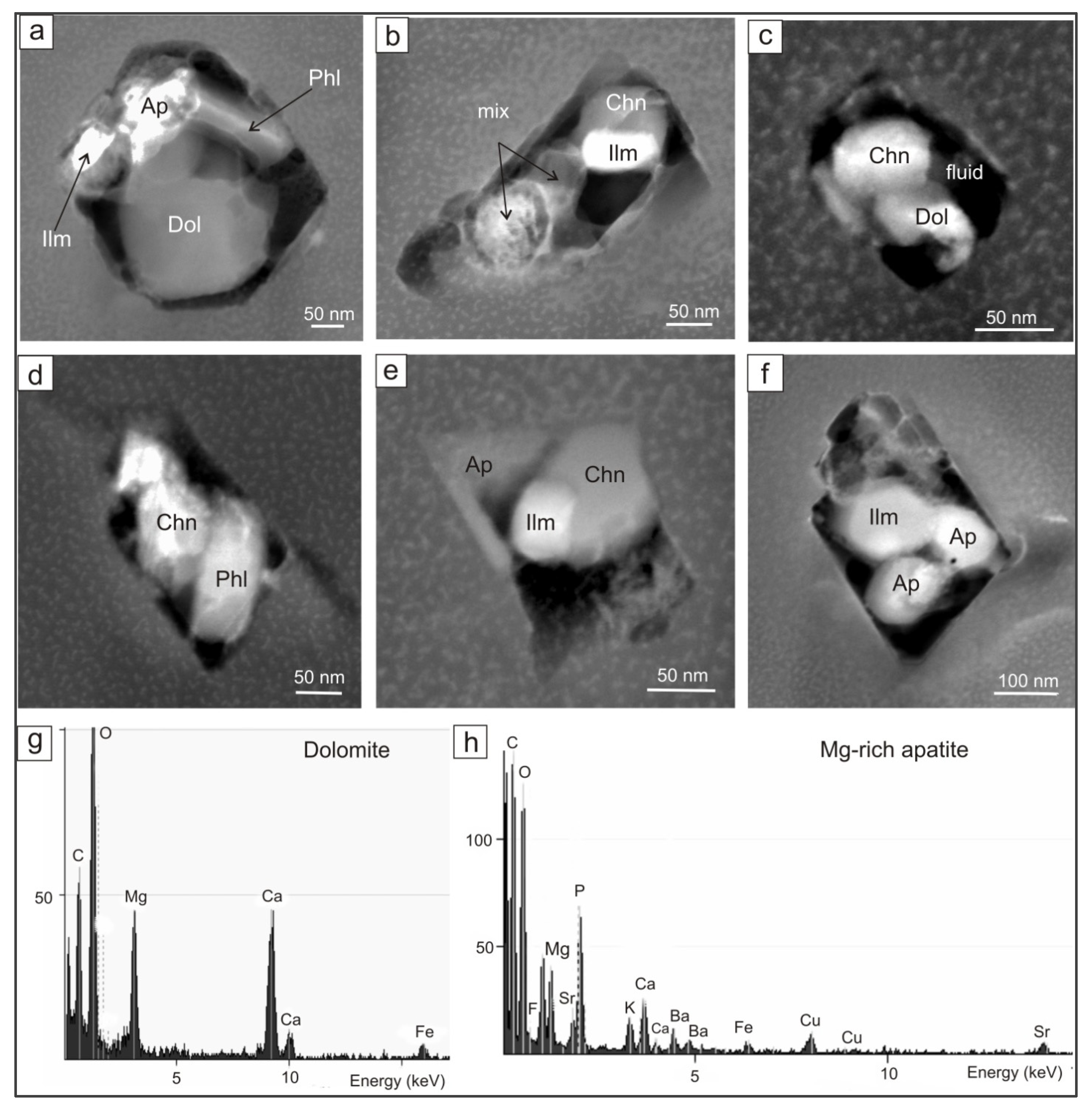
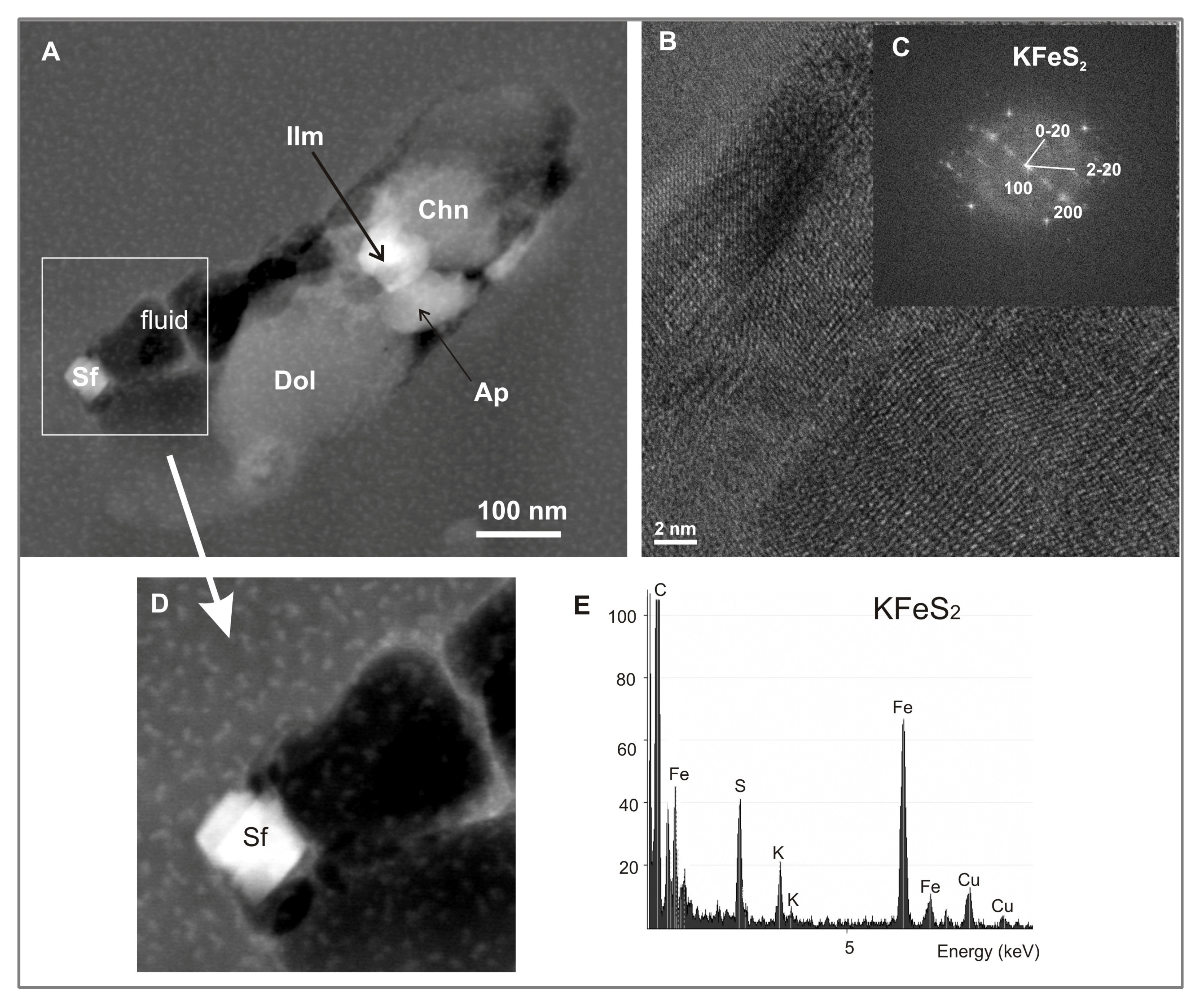
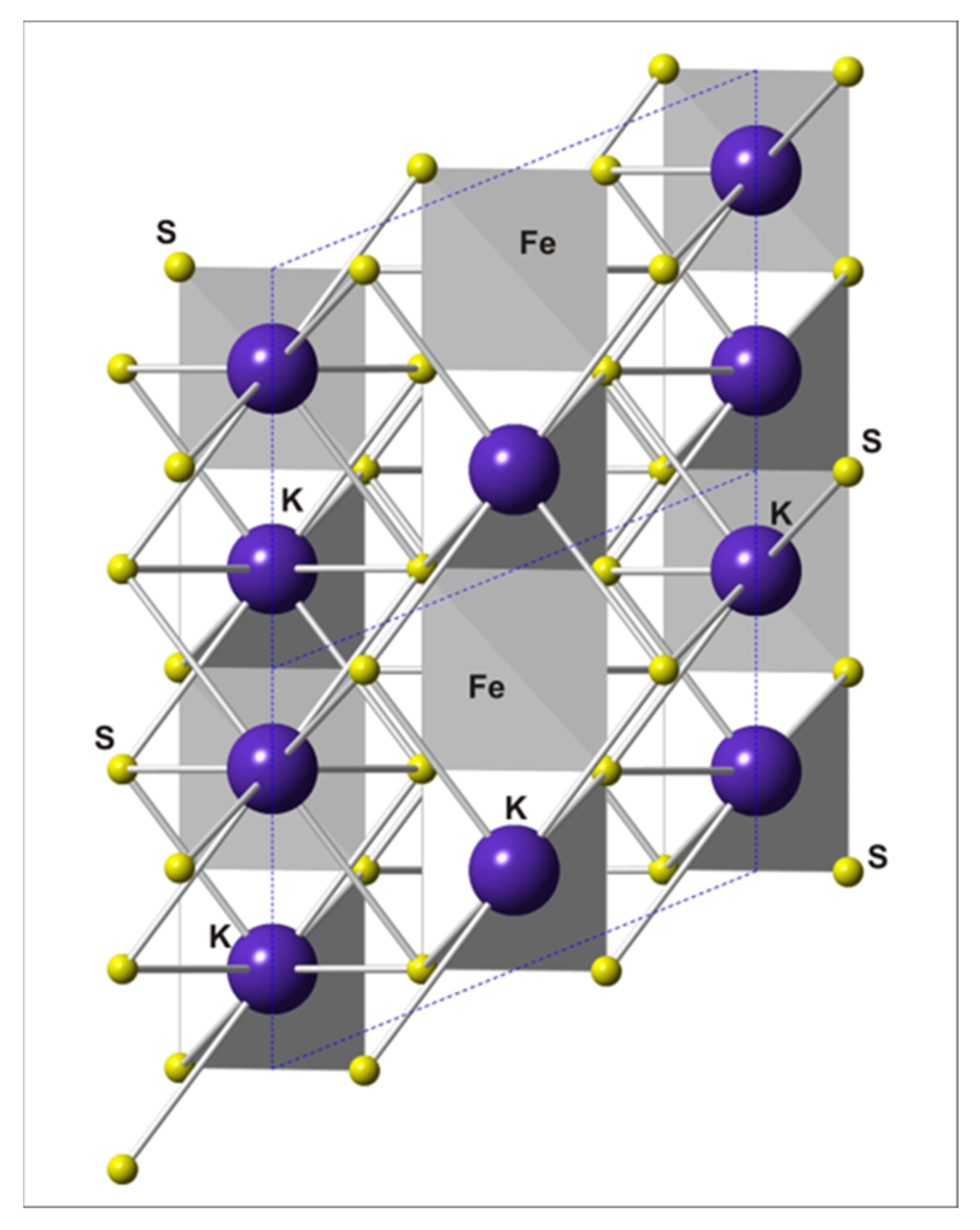
The daughter phase assemblage of the polymineralic inclusions (i.e., the crystallized mantle melt/fluid) in the cubic fibrous diamond Ud-45 from the Udachnaya kimberlite pipe is represented by dolomite, chondrodite, phlogopite, Mg-rich apatite, Cr-bearing ilmenite, magnetite, hanswilkeite (KFeS2), KCl, and fluid. The melt/fluid is likely carbonatitic or silicate–carbonate in composition with high potassium and volatile contents.
(2)
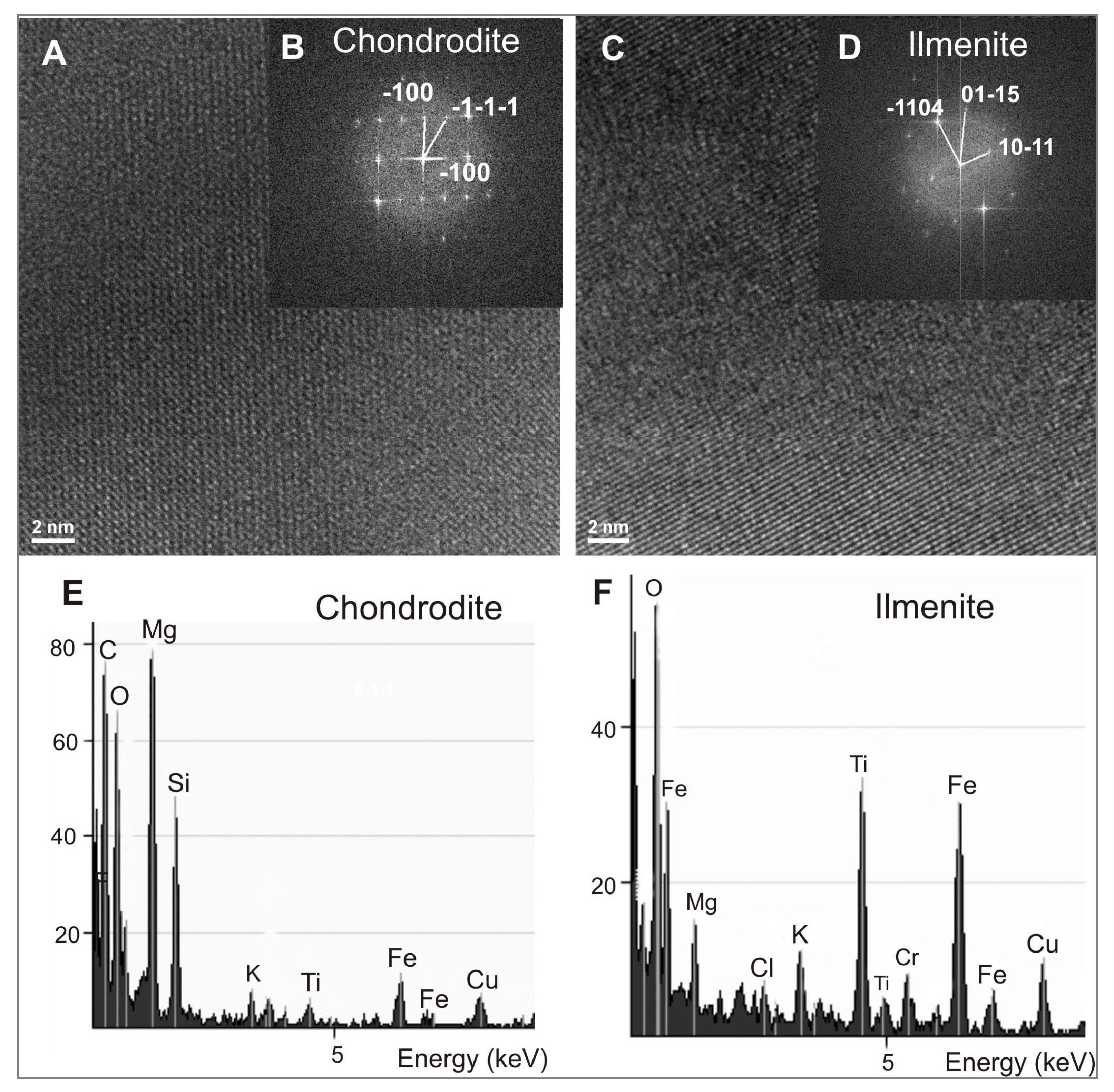
For the first time, potassium sulfide hanswilkeite KFeS2 and chondrodite have been found in a diamond as a mineral reflecting mantle substrates.

Author Contributions

Conceptualization, A.M.L. and I.S.S.; investigation, A.M.L.; data curation, A.M.L. and I.S.S.; writing—original draft preparation, A.M.L.; writing—review and editing, I.S.S.; visualization, A.M.L. and I.S.S.; funding acquisition, I.S.S. All authors have read and agreed to the published version of the manuscript.

Funding

This work was financially supported by the Russian Science Foundation (grant no. 18-77-10062-P) and state assignment of IGM SB RAS (no. 122041400157-9).

Data Availability Statement

Not applicable.

Acknowledgments

The authors are very grateful to Richard Wirth for the research carried out on the TEM at the Helmholtz Center Potsdam GFZ German Research Center for Geosciences (Germany) with unique results. We also express our gratitude to Yu.V. Seretkin (IGM SB RAS, Novosibirsk, Russia) for help in constructing projection images using special software.

Conflicts of Interest

The authors declare no conflict of interest.

References

  1. Bulanova, G.P.; Shestakova, O.E.; Leskova, N.V. Djerfisherite from Sulfide Inclusions in Diamond. Yakutia. Dokl. Earth Sci. 1980, 255, 430–433. [Google Scholar]
  2. Sharp, W.E. Pyrrhotite, a common inclusion in South African diamonds. Nature 1966, 211, 402–403. [Google Scholar] [CrossRef]
  3. Sobolev, N.V.; Yefimova, E.S.; Logvinova, A.M.; Sukhodol’skaya, O.P.; Solodova, Y.P. Abundance and composition of mineral inclusions in large diamonds from Yakutia. Dokl. Earth Sci. 2001, 376, 34–38. [Google Scholar]
  4. Yefimova, E.S.; Sobolev, N.V.; Pospelova, L.N. Sulfide inclusions in diamonds and features of their parageneses. Zap. Vses. Mineral. Ova. 1983, 112, 300–310. [Google Scholar]
  5. Deines, P.; Harris, J.W. Sulfide inclusion chemistry and carbon isotopes of African diamonds. Geochim. Cosmochim. Acta 1995, 59, 3173–3188. [Google Scholar] [CrossRef]
  6. Harris, J.W. Black material on mineral inclusions and in Internal fracture planes in diamond. Contrib. Mineral. Petrol. 1972, 35, 22–33. [Google Scholar] [CrossRef]
  7. Pearson, D.G.; Shirey, S.B.; Harris, J.W.; Carlson, R.W. Sulphide inclusions in diamonds from the Koffiefontein kimberlite, S Africa: Constraints on diamond ages and mantle Re-Os systematics. Earth Planet. Sci. Lett. 1998, 160, 311–326. [Google Scholar] [CrossRef]
  8. Taylor, L.A.; Liu, Y. Diamond sulfide inclusions are not Mss: Exsolution of pyrrhotite, pentlandite and chalcopyrite. Russ. Geol. Geophys. 2009, 50, 1201–1211. [Google Scholar] [CrossRef]
  9. Logvinova, A.M.; Wirth, R.; Fedorova, E.N.; Sobolev, N.V. Nanometer-sized mineral and fluid inclusions in cloudy Siberian diamonds: New insights on diamond formation. Eur. J. Miner. 2008, 20, 317–331. [Google Scholar] [CrossRef]
  10. Logvinova, A.M.; Wirth, R. Black cluster microinclusions in the core of Yakutian diamonds: Implications for diamond nucleation. In Proceedings of the 10th International Workshop “Deep Seated Magmatism, Its Sources and Plumes”, Irkutsk, Russia; 2010; pp. 93–103. [Google Scholar]
  11. Dobrovol’Skaya, M.G. Murunskite, K2Cu3FeS4, a new sulfide of potassium, copper, and iron. Int. Geol. Rev. 1982, 24, 1109–1114. [Google Scholar] [CrossRef]
  12. Pekov, I.V.; Zubkova, N.V.; Lisitsyn, D.V.; Pushcharovsky, D.Y. Crystal chemistry of murunskite. Dokl. Earth Sci. 2009, 424, 139–141. [Google Scholar] [CrossRef]
  13. Sokolova, M.N.; Dobrovol’skaya, M.G.; Organova, N.I.; Dimitrik, A.L. A sulfide of iron and potassium, the new mineral rasvumite. Zap. Vsesoyuznogo Mineral. Obs. 1970, 99, 712–720. [Google Scholar]
  14. Clark, J.R.; Brown, G.E. Crystal structure of rasvumite, KFe2S3. Am. Mineral. 1980, 65, 477–482. [Google Scholar]
  15. Fuchs, L.H. Djerfisherite, alkali copper-iron sulfide: A new mineral from enstatite chondrites. Science 1966, 153, 166–167. [Google Scholar] [CrossRef]
  16. Zaccarini, F.; Thalhammer, O.A.; Princivalle, F.; Lenaz, D.; Stanley, C.J.; Garuti, G. Djerfisherite in the Guli dunite complex, Polar Siberia: A primary or metasomatic phase? Can. Mineral. 2007, 45, 1201–1211. [Google Scholar] [CrossRef]
  17. Czamanske, G.K.; Erd, R.C.; Leonard, B.F.; Clark, J.R. Bartonite, a new potassium iron sulfide mineral. Am. Mineral. 1981, 66, 369–375. [Google Scholar]
  18. Yakovenchuk, V.N.; Pakhomovsky, Y.A.; Men’shikov, Y.P.; Ivanyuk, G.Y.; Krivovichev, S.V.; Burns, P.C. Chlorbartonite, K6Fe24S26(Cl,S), a new mineral species from a hydrothermal vein in the Khibina massif, Kola Peninsula, Russia: Description and crystal structure. Can. Mineral. 2003, 41, 503–511. [Google Scholar] [CrossRef]
  19. Murashko, M.N.; Britvin, S.N.; Krzhizhanoskaya, M.G.; Vereshchagin, O.S.; Vapnik, Y.; Vlasenko, N.S.; Shelukhina, Y.S.; Bocharov, V.N. Hanswilkeite, IMA-2022-041, in: CNMNC Newsletter 69. Eur. J. Mineral. 2022, 34, 463–468. [Google Scholar] [CrossRef]
  20. Boon, J.W.; MacGillavry, C.H. The crystal structure of potassium thioferrite KFeS2 and sodium thiochromite NaCrS2. Recl. Des Trav. Chim. Des Trav. Pays-Bas 1942, 61, 910–920. [Google Scholar] [CrossRef]
  21. Bronger, W.; Kyas, A.; Muller, P.J. The Antiferromagnetic Structures of KFeS2, RbFeS2, KFeSe2, and RbFeSe2 and the Correlation between Magnetic Moments and Crystal Field Calculations. Solid State Chem. 1987, 70, 262. [Google Scholar] [CrossRef]
  22. Boller, H. On the synthesis, crystal chemistry and magnetic properties of rasvumite and related compound. Acta Cryst. 2004, 60, s47. [Google Scholar] [CrossRef] [Green Version]
  23. Wirth, R. Focused ion beam (FIB): A novel technology for advanced application of micro- and nanoanalysis in geosciences and applied mineralogy. Eur. J. Mineral. 2004, 16, 863–877. [Google Scholar] [CrossRef] [Green Version]
  24. Wirth, R. Focused Ion Beam (FIB) combined with SEM and TEM: Advanced analytical tools for studies of chemical composition, microstructure and crystal structure in geomaterials on a nanometre scale. Chem. Geol. 2009, 261, 217–229. [Google Scholar] [CrossRef]
  25. Navon, O.; Hutcheon, I.D.; Rossman, G.R.; Wasserburg, G.J. Mantle-derived fluids in diamond micro-inclusions. Nature 1988, 335, 784–789. [Google Scholar] [CrossRef]
  26. Gubanov, N.; Zedgenizov, D.; Sharygin, I.; Ragozin, A. Origin and Evolution of High-Mg Carbonatitic and Low-Mg Carbonatitic to Silicic High-Density Fluids in Coated Diamonds from Udachnaya Kimberlite Pipe. Minerals 2019, 9, 734. [Google Scholar] [CrossRef] [Green Version]
  27. Schrauder, M.; Navon, O. Hydrous and carbonatitic mantle fluids in fibrous diamonds from Jwaneng, Botswana. Geoch. Cosmochim. Acta 1994, 58, 761–771. [Google Scholar] [CrossRef]
  28. Klein-BenDavid, O.; Izraeli, E.S.; Hauri, E.; Navon, O. Fluid inclusions in diamonds from the Diavik mine, Canada and the evolution of diamond-forming fluids. Geochim. Cosmochim. Acta 2007, 71, 723–744. [Google Scholar] [CrossRef]
  29. Klein-BenDavid, O.; Logvinova, A.M.; Schrauder, M.; Spetius, Z.V.; Weiss, Y.; Hauri, E.H.; Kaminsky, F.V.; Sobolev, N.V.; Navon, O. High-Mg carbonatitic microinclusions in some Yakutian diamonds—A new type of diamond-forming fluid. Lithos 2009, 112, 648–659. [Google Scholar] [CrossRef]
  30. Zedgenizov, D.A.; Ragozin, A.L.; Shatsky, V.S. Compositional features of diamond growth medium: From the study of microinclusions in natural diamonds. Proc. Russ. Miner. Soc. 2007, 7, 159–172. [Google Scholar]
  31. Zedgenizov, D.A.; Ragozin, A.L.; Shatsky, V.S.; Araujo, D.; Griffin, W.L.; Kagi, H. Mg and Fe-rich carbonate–silicate high-density fluids in cuboid diamonds from the Internationalnaya kimberlite pipe (Yakutia). Lithos 2009, 112, 638–647. [Google Scholar] [CrossRef]
  32. Skuzovatov, S.Y.; Zedgenizov, D.A.; Ragozin, A.L.; Shatsky, V.S. Growth medium composition of coated diamonds from the Sytykanskaya kimberlite pipe (Yakutia). Russ. Geol. Geophys. 2012, 53, 1197–1208. [Google Scholar] [CrossRef]
  33. Logvinova, A.; Zedgenizov, D.; Wirth, R. Specific Multiphase Assemblages of Carbonatitic and Al-Rich Silicic Diamond-Forming Fluids/Melts: TEM Observation of Microinclusions in Cuboid Diamonds from the Placers of Northeastern Siberian Craton. Minerals 2019, 9, 50. [Google Scholar] [CrossRef] [Green Version]
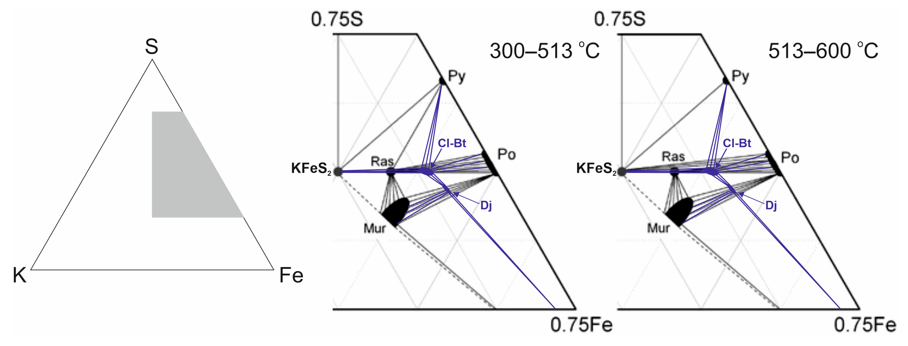
  34. Osadchii, V.O.; Voronin, M.V.; Baranov, A.V. Phase equilibria in the KFeS2–Fe–S system at 300–600 °C and bartonite stability. Contrib. Mineral. Petrol. 2018, 173, 44. [Google Scholar] [CrossRef]
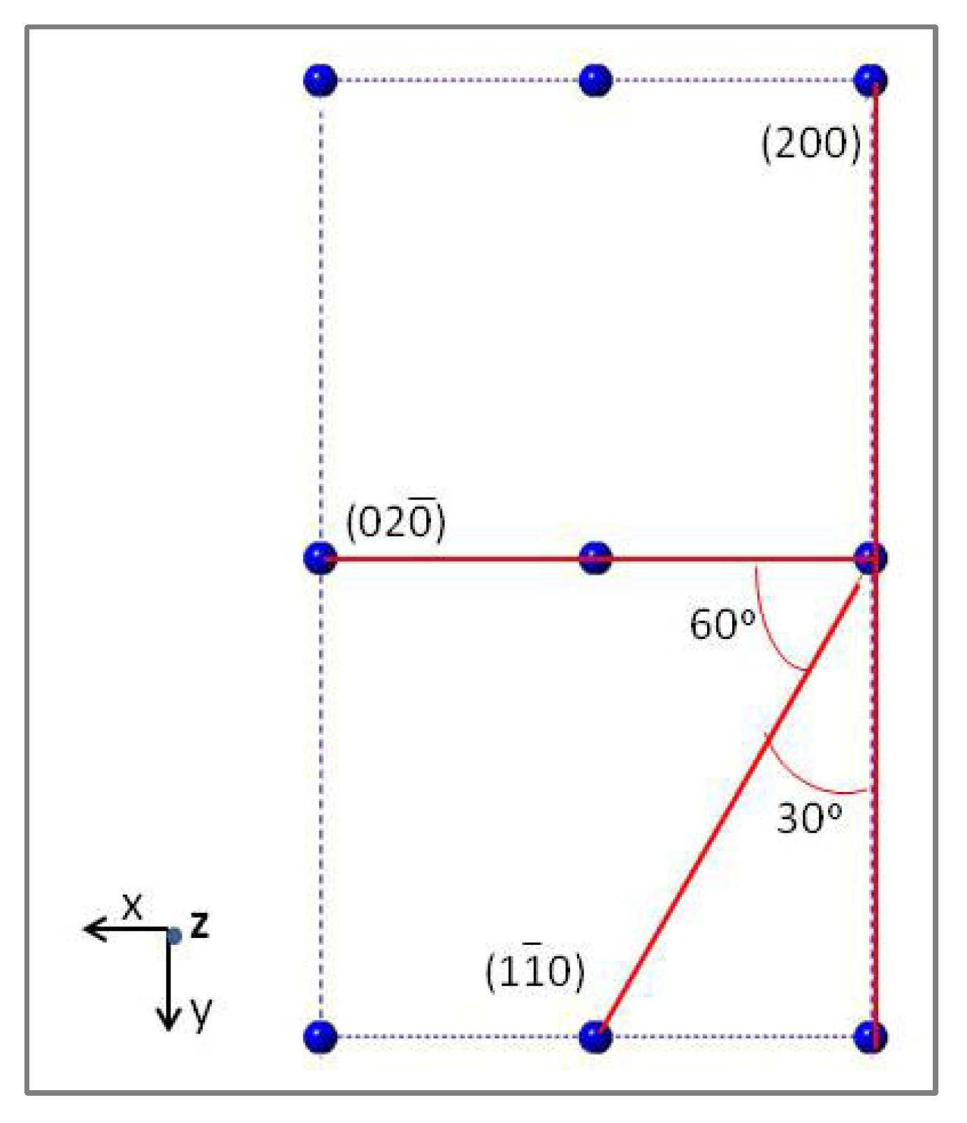
Figure 1. Optical micrograph of the cubic diamond Ud-45 from Udachnaya kimberlite pipe (a); TEM bright field image of nanometer-sized inclusions in diamond (b). The numbers indicate the location of the studied inclusions.
Minerals 13 00874 g001
Figure 2. TEM images of nanometer-sized polymineralic inclusions in diamond Ud-45 (af): Ap—apatite, Ilm—ilmenite, Chn—chondrodite, Dol—dolomite, Phl—phlogopite; (g,h)—EDX spectra of dolomite and apatite (Cu intensity comes from the copper grid).
Minerals 13 00874 g002
Figure 3. Energy filtered lattice fringe images and diffraction patterns (FFT) from the framed area of two nanometric phases in polymineral inclusion #6 (see Figure 1b) from the diamond Ud-45: (A,B)—chondrodite; (C,D)—ilmenite. (E,F) X-ray intensity spectrum of nanometric phases: (E) chondrodite and (F) ilmenite. Cu intensity comes from the copper grid.
Minerals 13 00874 g003
Figure 4. TEM bright field image of nanometer-sized inclusion #6 (see Figure 1b) from the diamond Ud-45 (A): Ap—apatite, Ilm—ilmenite, Chn—chondrodite, Dol—dolomite, Sf—sulfide KFeS2; (B)—energy filtered high-resolution (HREM) lattice fringe image of sulfide KFeS2. (C) Diffraction pattern (FFT) from framed area in KfeS2. The spacing between the fringes is used for phase identification. (D)—Enlarged fragment of TEM image of sulfide inclusion KFeS2. (E)—Energy-dispersive X-ray (EDX) spectra of sulfide KFeS2. Cu intensity comes from the copper grid.
Minerals 13 00874 g004
Figure 5. Projection of the crystal lattice of KFeS2 along the z-axis.
Minerals 13 00874 g005
Figure 6. Crystal structure of mineral phase KFeS2 in the diamond Ud-45. Projection is along the [010] direction. Interatomic distances: K-S—3.38–3.45 Å (eight-fold coordination) and Fe-S—2.18–2.19 Å (tetrahedral coordination).
Minerals 13 00874 g006
Figure 7. K–Fe–S–Cl phase diagram presented by Osadchii et al. (2018) [34]. Black color shows the phase relations experimentally obtained in the K–Fe–S system at 300–600 °C [34]. Blue color shows the phase relations including chlorbartonite and djerfisherite. Py—pyrite, Po—pyrrhotite, Ras—rasvumite, Mur—murunskite, Ch-Bt—chlorbartonite, Dj—djerfisherite.
Minerals 13 00874 g007
Table 1. Interplanar distances and angles between planes with electron diffraction of nanometer-sized phases of chondrodite and KFeS2 in diamond Ud-45.
hkldhkl Observed, Ådhkl Calculated, Åφ *φobserved,
Degrees
φcalculated,
Degrees
Chondrodite
1 ¯ 007.377.432
0 1 ¯ 1 ¯ 4.184.251
1 ¯ 1 ¯ 1 ¯ 3.583.481
< 1 ¯ 1 ¯ 1 ¯ /0 1 ¯ 1 ¯ > 28.527
< 1 ¯ 1 ¯ 1 ¯ / 1 ¯ 00>5754.1
KFeS2
200 (100)7.05 (3.535)7.022 (3.511)
0 2 ¯ 05.345.644
2 2 ¯ 03.052.816
<2 2 ¯ 0/0 2 ¯ 0>6061
<2 2 ¯ 0/200>3030
Note: * φ—angle between planes.
Disclaimer/Publisher’s Note: The statements, opinions and data contained in all publications are solely those of the individual author(s) and contributor(s) and not of MDPI and/or the editor(s). MDPI and/or the editor(s) disclaim responsibility for any injury to people or property resulting from any ideas, methods, instructions or products referred to in the content.

