
Shortwave UV Blue Luminescence of Some Minerals and Gems Due to Titanate Groups

Abstract
:1. Introduction
2. Materials and Methods
3. Results
3.1. Inductively-Coupled Plasma Mass Spectrometry by Laser Ablation (LA-ICP-MS)
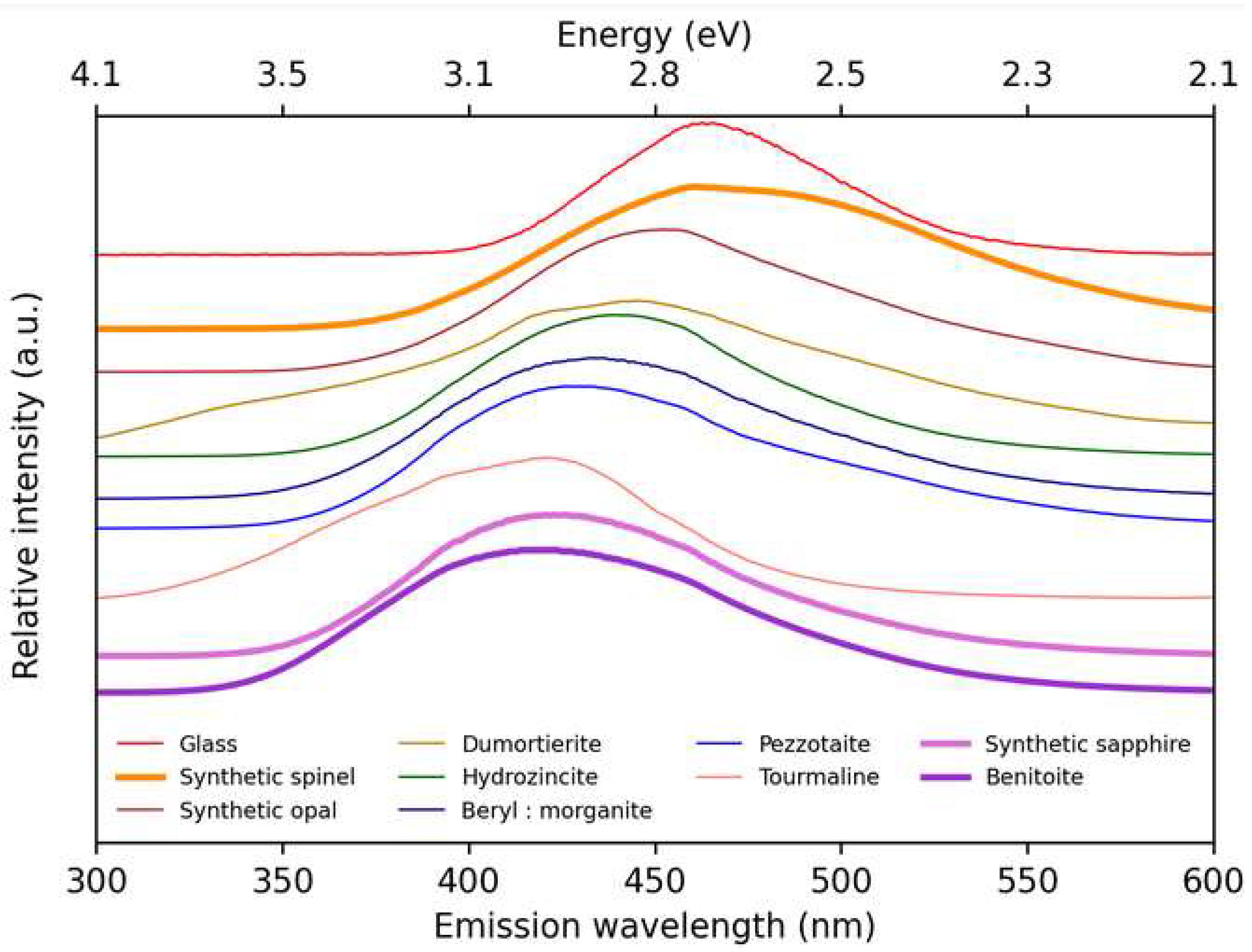
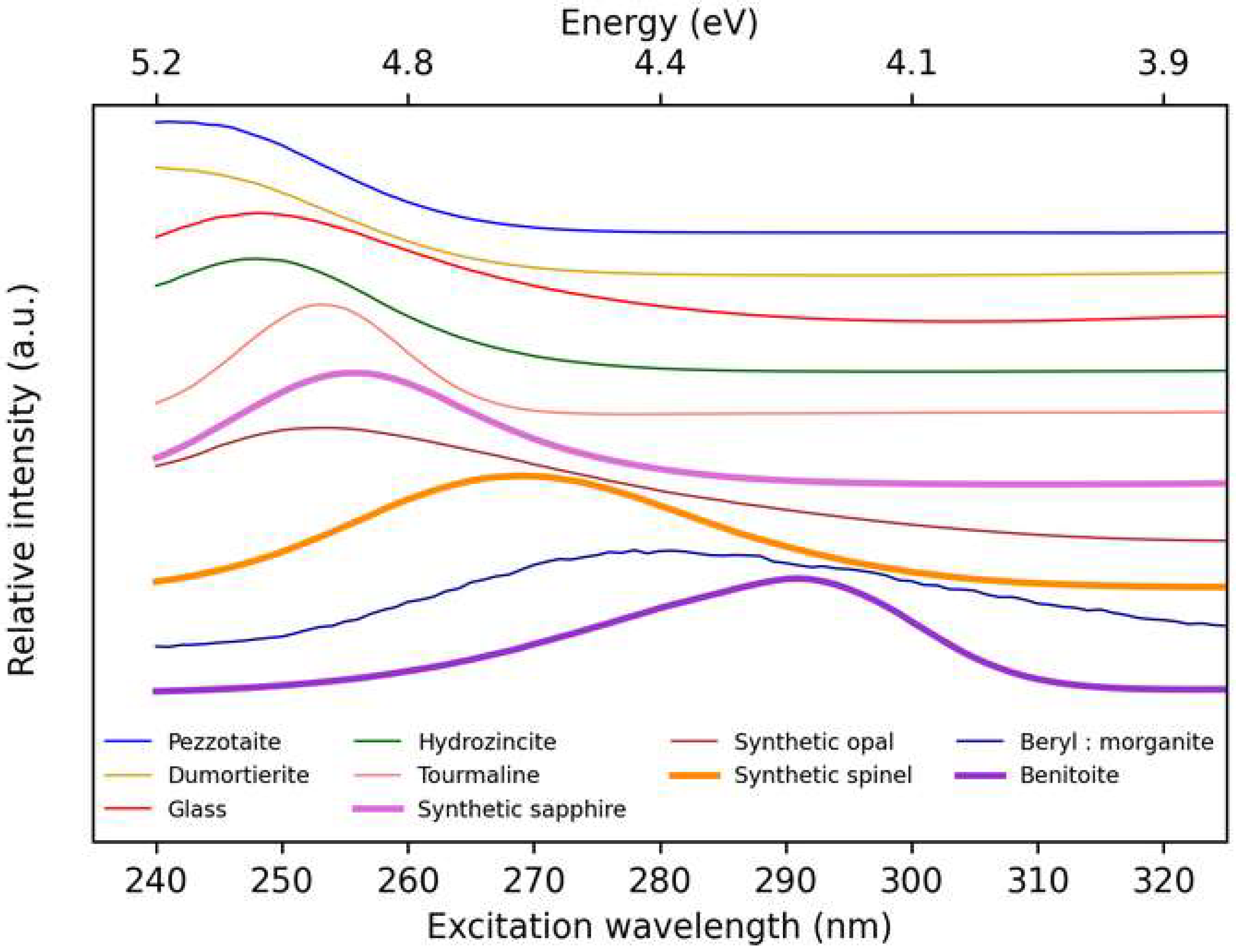
3.2. Photoluminescence Properties
3.2.1. Emission Spectra
3.2.2. Excitation Spectra
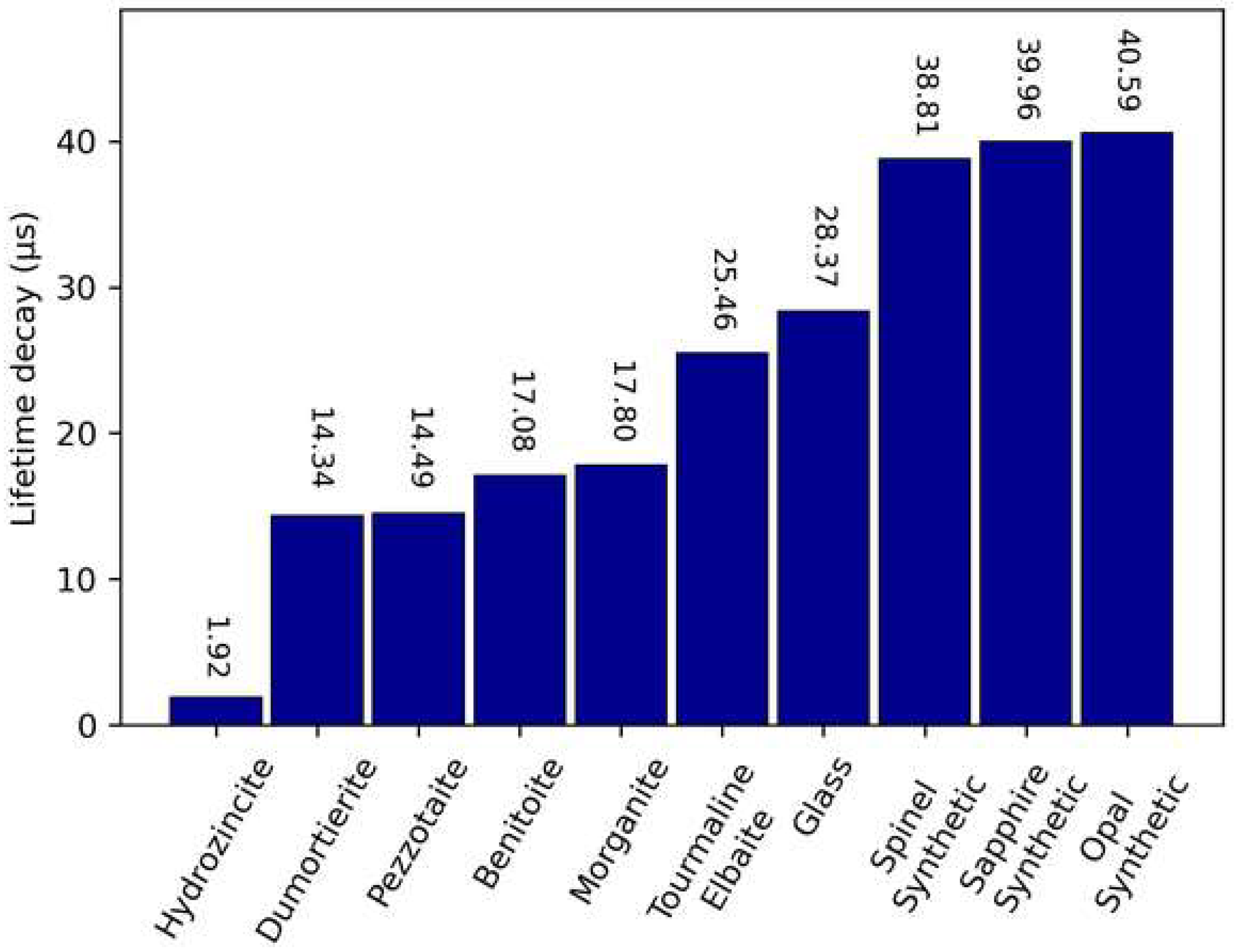
3.2.3. Lifetime Decay of Luminescence
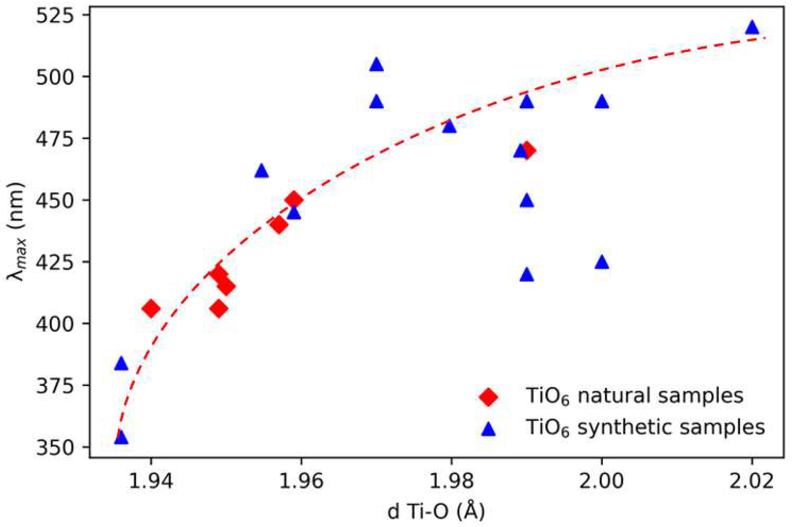
4. Discussion
5. Conclusions
Supplementary Materials
Author Contributions
Funding
Data Availability Statement
Acknowledgments
Conflicts of Interest
References
- Tang, H.; Berger, H.; Schmid, P.E.; Lévy, F.; Burri, G. Photoluminescence in TiO2 anatase single crystals. Solid State Commun. 1993, 87, 847–850. [Google Scholar] [CrossRef]
- Marshall, D.J. Cathodoluminescence of Geological Materials; Unwin-Hyman: Boston, MA, USA, 1988; 146p. [Google Scholar]
- Garcia-Guinea, J.; Garrido, F.; Lopez-Arce, P.; Correcher, V.; de la Figuera, J. Spectral Green cathodoluminescence emission from surfaces of insulators with metal-hydroxyl bonds. J. Lumin. 2017, 190, 128–135. [Google Scholar] [CrossRef]
- Takahashi, N.; Tsujimori, K.; Kayam, M.; Nishido, H. Cathodoluminescence petrography of P-type jadeitites from the New Idria serpentinite body, California. J. Mineral. Petrol. Sci. 2017, 112, 291–299. [Google Scholar] [CrossRef] [Green Version]
- Blasse, G. The Luminescence of Closed-Shell Transition-Metal Complexes. New Developments. In Luminescence and Energy Transfer; Springer: Berlin/Heidelberg, Germany, 1980; pp. 1–41. [Google Scholar]
- Hemphill, W.R.; Tyson, R.M.; Theisen, A.F. Spectral luminescence properties of natural specimens in the scheelite-powellite series, and an assessment of their detectivity with an airborne Fraunhofer line discriminator. Econ. Geol. 1988, 83, 637–646. [Google Scholar] [CrossRef]
- Tyson, R.M.; Hemphill, W.R.; Theisen, A.F. Effect of the W:Mo ratio on the shift of excitation and emission spectra in the scheelite-powellite series. Am. Mineral. 1988, 73, 1145–1154. [Google Scholar]
- Bode, J.H.G.; Van Oosterhout, A.B. Defect luminescence of ordered perovskites A2BWO6. J. Lumin. 1975, 10, 237–242. [Google Scholar] [CrossRef] [Green Version]
- Gaft, M.; Nagli, l.; Waychunas, G.; Weiss, D. The nature of blue luminescence from natural benitoite BaTiSi3O9. Phys. Chem. Miner. 2004, 31, 365–373. [Google Scholar] [CrossRef]
- Waychunas, G.A. Apatite luminescence. Rev. Mineral. Geochem. 2002, 48, 701–742. [Google Scholar] [CrossRef]
- Nikbakht, T.; Kakuee, O.; Lamehi-rachti, M. An efficient ionoluminescence analysis of turquoise gemstone as a weakly luminescent mineral. Spectrochim. Acta Part A Mol. Biomol. Spectrosc. 2017, 179, 171–177. [Google Scholar] [CrossRef]
- Gaft, M.; Seigel, H.; Panczer, G.; Reisfeld, R. Laser-induced time-resolved luminescence spectroscopy of Pb2+ in minerals. Eur. J. Mineral. 2002, 14, 1041–1048. [Google Scholar] [CrossRef]
- Panczer, G.; Gaft, M.; De Ligny, D.; Boudeulle, M.; Champagnon, B. Luminescent centres in pezzottaite, CsBe2LiAl2Si6O18. Eur. J. Mineral. 2010, 22, 605–612. [Google Scholar] [CrossRef]
- Chang, I.F.; Onton, A. Optical properties of photochromatic sulfur-doped chlorosodalite. J. Electron. Mater. 1973, 2, 17–46. [Google Scholar] [CrossRef]
- Carvalho, J.M.; Rodrigues, L.C.; Hölsä, J.; Lastusaari, M.; Nunes, L.A.; Felinto, M.C.; Brito, H.F. Influence of titanium and lutetium on the persistent luminescence of ZrO 2. Opt. Mater. Express 2012, 2, 331–340. [Google Scholar] [CrossRef] [Green Version]
- Sidike, A.; Kobayashi, S.; Zhu, H.-J.; Kusachi, I.; Yamashita, N. Photoluminescence of baratovite and katayamalite. Phys. Chem. Miner. 2010, 37, 705–710. [Google Scholar] [CrossRef]
- Lupei, A.; Lupei, V.; Ionescu, C.; Tang, H.G.; Chen, M.L. Spectroscopy of Ti3+: α-Al2O3. Opt. Commun. 1986, 59, 36–38. [Google Scholar] [CrossRef]
- Lafargue-dit-Hauret, W.; Schira, R.; Latouche, C.; Jobic, S. Theoretical calculations meet experiment to explain the luminescence properties and the presence of defects in m-ZrO2. Chem. Mater. 2021, 33, 2984–2992. [Google Scholar] [CrossRef]
- Takahashi, Y.; Kitamura, K.; Iyi, N.; Inoue, S.; Fujiwara, T. Blue photoluminescence of germania-stabilized benitoite. J. Ceram. Soc. Jpn. 2008, 116, 1143–1146. [Google Scholar] [CrossRef] [Green Version]
- Pathak, N.; Ghosh, P.S.; Gupta, S.K.; Mukherjee, S.; Kadam, R.; Arya, A. An insight into the various defects-induced emission in MgAl2O4 and their tunability with phase behavior: Combined experimental and theoretical approach. J. Phys. Chem. C 2016, 120, 4016–4031. [Google Scholar] [CrossRef]
- Sawai, S.; Uchino, T. Visible photoluminescence from MgAl2O4 spinel with cation disorder and oxygen vacancy. J. Appl. Phys. 2012, 112, 103523. [Google Scholar] [CrossRef] [Green Version]
- Raj, S.S.; Gupta, S.K.; Grover, V.; Muthe, K.P.; Natarajan, V.; Tyagi, A.K. MgAl2O4 spinel: Synthesis, carbon incorporation and defect-induced luminescence. J. Mol. Struct. 2015, 1089, 81–85. [Google Scholar] [CrossRef]
- Mikhailik, V.B.; Kraus, H.; Balcerzyk, M.; Czarnacki, W.; Moszynski, M.; Mykhaylyk, M.S.; Wahl, D. Low-temperature spectroscopic and scintillation characterisation of Ti-doped Al2O3. Nucl. Instrum. Methods Phys. Res. Sect. A Accel. Spectrometers Detect. Assoc. Equip. 2005, 546, 523–534. [Google Scholar] [CrossRef]
- Chen, W.; Tang, H.; Shi, C.; Deng, J.; Shi, J.; Zhou, Y.; Yin, S. Investigation on the origin of the blue emission in titanium doped sapphire: Is F+ color center the blue emission center ? Appl. Phys. Lett. 1995, 67, 317–319. [Google Scholar] [CrossRef]
- Mikhailik, V.B.; Di Stefano, P.C.F.; Henry, S.; Kraus, H.; Lynch, A.; Tsybulskyi, V.; Verdier, M.A. Studies of concentration dependences in the luminescence of Ti-doped Al2O3. J. Appl. Phys. 2011, 109, 053116. [Google Scholar] [CrossRef] [Green Version]
- Pallotti, D.K.; Passoni, L.; Maddalena, P.; Di Fonzo, F.; Lettieri, S. Photoluminescence mechanisms in anatase and rutile TiO2. J. Phys. Chem. C 2017, 121, 9011–9021. [Google Scholar] [CrossRef]
- Norrbo, I.; Gluchowski, P.; Hyppanen, I.; Laihinen, T.; Laukkanen, P.; Makela, J.; Lastusaari, M. Mechanisms of tenebrescence and persistent luminescence in synthetic hackmanite Na8Al6Si6O24(Cl,S)2. ACS Appl. Mater. Interfaces 2016, 8, 11592–11602. [Google Scholar] [CrossRef]
- Andrade LH, C.; Lima, S.M.; Novatski, A.; Neto, A.M.; Bento, A.C.; Baesso, M.L.; Boulon, G. Spectroscopic assignments of Ti3+ and Ti4+ in titanium-doped OH− free low-silica calcium aluminosilicate glass and role of structural defects on the observed long lifetime and high fluorescence of Ti3+ ions. Phys. Rev. B 2008, 78, 224202. [Google Scholar] [CrossRef] [Green Version]
- Gorobets, B.S.; Rogojine, A.A. Luminescent Spectra of Minerals: Reference-Book; All-Russia Institute of Mineral Resources (VIMS): Moscow, Russia, 2002; 302p. [Google Scholar]
- Taher, M.A.; Asadollahzadeh, H.; Fazelirad, H. Determination of trace amounts of iron by a simple fluorescence quenching method. Anal. Methods 2015, 7, 6726–6731. [Google Scholar] [CrossRef]
- Al-kady, A.S.; Gaber, M.; Hussein, M.M.; Ebeid, E.Z.M. Structural and fluorescence quenching characterization of hematite nanoparticles. Spectrochim. Acta Part A Mol. Biomol. Spectrosc. 2011, 83, 398–405. [Google Scholar] [CrossRef]
- Amthauer, G.; Rossman, G.R. Mixed valence of iron in minerals with cation clusters. Phys. Chem. Miner. 1984, 11, 37–51. [Google Scholar] [CrossRef]
- Fritsch, E.; Waychunas, G.A. Gemstones. M. Robbins, Fluorescence; Geoscience Press: Phoenix, AZ, USA, 1994; pp. 163–165. [Google Scholar]
- Macke, A.J.H. Luminescence and energy transfer in the ordered perovskite system La2MgSn1–xTixO6. Phys. Status Solidi 1977, 39, 117–123. [Google Scholar] [CrossRef]
- Macke, A.J.H. Investigations on the luminescence of titanium-activated stannates and zirconates. J. Solid State Chem. 1976, 18, 337–346. [Google Scholar] [CrossRef]
- Wu, Q.; Hu, Z. Na2TiGeO5—A self-light-emitting phosphor with the stable structure and tunable emission resulted from Cr3+-doped for FEDs. J. Am. Ceram. Soc. 2019, 102, 2727–2736. [Google Scholar]
- Takahashi, Y.; Masai, h.; Fujiwara, T.; Kitamura, K.; Inoue, S. Afterglow in synthetic bazirite, BaZrSi3O9. J. Ceram. Soc. Jpn. 2008, 116, 357–360. [Google Scholar] [CrossRef] [Green Version]
- Smith, A.L. Some new complex silicate phosphors containing calcium, magnesium, and beryllium. J. Electrochem. Soc. 1949, 96, 287. [Google Scholar] [CrossRef]
- Evans, B.D.; Pogatshnik, G.J.; Chen, Y. Optical properties of lattice defects in α-Al2O3. Nucl. Instrum. Methods Phys. Res. Sect. B: Beam Interact. Mater. At. 1994, 91, 258–262. [Google Scholar] [CrossRef]
- Bausa, L.E.; Vergara, I.; Jaque, F.; Sole, J.G. Ultraviolet laser excited luminescence of Ti-sapphire. J. Phys. Condens. Matter 1990, 2, 9919. [Google Scholar] [CrossRef]
- Marques, C.; Santos, L.; Falcao, A.N.; Silva, R.C.; Alves, E. Luminescence studies in colour centres produced in natural topaz. J. Lumin. 2000, 87, 583–585. [Google Scholar] [CrossRef]
- Iida, Y.; Sawamura, K.; Iwasaki, K.; Nakanishi, T.; Iwakura, F.; Nakajima, Y.; Yasumori, A. Persistent Luminescence Properties of Ti4+-doped K2ZrSi3O9 Wadeite. Sens. Mater. 2020, 32, 1427–1433. [Google Scholar] [CrossRef] [Green Version]
- Moon, C.; Nishi, M.; Miura, K.; Hirao, K. Blue long-lasting phosphorescence of Ti-doped BaZrO3 perovskites. J. Lumin. 2009, 129, 817–819. [Google Scholar] [CrossRef]
- Cavignac, T.; Jobic, S.; Latouche, C. Modeling Luminescence Spectrum of BaZrO3: Ti Including Vibronic Coupling from First Principles Calculations. J. Chem. Theory Comput. 2022, 18, 7714–7721. [Google Scholar] [CrossRef]
- Konijnendijk, W.L. Luminescence of BaSnSi3O9: Ti4+ compared to BaZrSi3O9: Ti4+. Inorg. Nucl. Chem. Lett. 1981, 17, 129–132. [Google Scholar] [CrossRef]
- Zeng, G.; Dong, Q.; Bao, W. Preparation and Photoluminescence Properties of Ti-Doped Lu2O3 Powder. J. Appl. Spectrosc. 2016, 83, 460–465. [Google Scholar] [CrossRef]
- Kawano, T.; Yamane, H. Synthesis, crystal structure analysis, and photoluminescence of Ti4+-doped Mg5SnB2O10. Chem. Mater. 2010, 22, 5937–5944. [Google Scholar] [CrossRef]
- Konijnendijk, W.L.; Blasse, G. On the luminescence of Ti4+ in Mg5SnB2O10 and Mg3ZrB2O8. Mater. Chem. Phys. 1985, 12, 591–599. [Google Scholar] [CrossRef] [Green Version]
- Cong, Y.; Li, B.; Yue, S.; Fan, D.; Wang, X.J. Effect of oxygen vacancy on phase transition and photoluminescence properties of nanocrystalline zirconia synthesized by the one-pot reaction. J. Phys. Chem. C 2009, 113, 13974–13978. [Google Scholar] [CrossRef]
- Jochum, K.P.; Weis, U.; Stoll, B.; Kuzmin, D.; Yang, Q.; Raczek, I.; Enzweiler, J. Determination of reference values for NIST SRM 610–617 glasses following ISO guidelines. Geostand. Geoanalytical Res. 2011, 35, 397–429. [Google Scholar] [CrossRef]
- Van Achterbergh, E.; Ryan, C.G.; Griffin, W.L. Glitter: On-line interactive data reduction for the laser ablation inductively coupled plasma mass spectrometry microprobe. In Proceedings of the Ninth Annual VM Goldschmidt Conference, Cambridge, MA, USA, 22–27 August 1999; p. 7215. [Google Scholar]
- Sun, R.; Ooi, Y.K.; Dickens, P.T.; Lynn, K.G.; Scarpulla, M.A. On the origin of red luminescence from iron-doped β-Ga2O3 bulk crystals. Appl. Phys. Lett. 2020, 117, 052101. [Google Scholar] [CrossRef]
- Gaillou, E.; Fritsch, E.; Massuyeau, F. Luminescence of gem opals: A review of intrinsic an extrinsic emission. Aust. Gemmol. 2012, 24, 365–373. [Google Scholar]
- Gaft, M.; Reisfeld, R.; Panczer, G. Modern Luminescence Spectroscopy of Minerals and Materials; Springer: Berlin/Heidelberg, Germany, 2015; 606p. [Google Scholar]
- Curtice, R.E.; Scott, A.B. The luminescence of thallium (I) halo complexes. Inorg. Chem. 1964, 3, 1383–1387. [Google Scholar] [CrossRef]
- Ogorodnikov, I.N.; Pustovarov, V.A.; Puzikov, V.M.; Salo, V.I.; Voronov, A.P. A luminescence and absorption spectroscopy study of KH2PO4 crystals doped with Tl+ ions. Opt. Mater. 2012, 34, 1522–1528. [Google Scholar] [CrossRef] [Green Version]
- Kirk, R.D. Role of sulfur in the luminescence and coloration of some aluminosilicates. J. Electrochem. Soc. 1954, 101, 461. [Google Scholar] [CrossRef]
- Stoliaroff, A.; Schira, R.; Blumentritt, F.; Fritsch, E.; Jobic, S.; Latouche, C. Point Defects Modeling Explains Multiple Sulfur Species in Sulfur-Doped Na4(Al3Si3O12)Cl Sodalite. J. Phys. Chem. C 2021, 125, 16674–16680. [Google Scholar] [CrossRef]
- Folkerts, H.F.; Blasse, G. Two types of luminescence from Pb2+ in alkaline-earth carbonates with the aragonite structure. J. Phys. Chem. Solids 1996, 57, 303–306. [Google Scholar] [CrossRef]
- Lin, J.; Su, Q. Luminescence of Pb2+ and energy transfer from Pb2+ to rare earth ions in silicate oxyapatites. Phys. Status Solidi 1996, 196, 261–267. [Google Scholar] [CrossRef]
- Folkerts, H.F.; Blasse, G. Luminescence of Pb2+ in several calcium borates. J. Mater. Chem. 1995, 5, 273–276. [Google Scholar] [CrossRef]
- Folkerts, H.F.; Hamstra, M.A.; Blasse, G. The luminescence of Pb2+ in alkaline earth sulfates. Chem. Phys. Lett. 1995, 246, 135–138. [Google Scholar] [CrossRef]
- Asano, S.; Yamashita, N. Effet du champ magnétique sur la luminescence de l’ion Pb2+ dans les luminophores CaO, CaS, CaSe et MgS. Phys. Status Solidi 1981, 108, 549–558. [Google Scholar] [CrossRef]
- Jary, V.; Nikl, M.; Mihokova, E.; Bohacek, P.; Trunda, B.; Polak, K.; Studnicka, V.; Mucka, V. Photoluminescence of Pb2+-doped SrHfO3. Radiat. Meas. 2010, 45, 406–408. [Google Scholar] [CrossRef]
- Tsuboi, T. Temporal Evolution of Luminescence by Pb2+-Doped KBr and KCl Phosphors. Electrochem. Solid-State Lett. 2000, 3, 200. [Google Scholar] [CrossRef]
- Folkerts, H.F.; Blasse, G. Luminescence of Pb2+ in SrTiO3. Chem. Mater. 1994, 6, 969–972. [Google Scholar] [CrossRef]
- Skuja, L. Optically active oxygen-deficiency-related centers in amorphous silicon dioxide. J. NON-Cryst. Solids 1998, 239, 16–48. [Google Scholar] [CrossRef]
- Yu, D.P.; Hang, Q.L.; Ding, Y.; Zhang, Z.G.; Bai, Z.G.; Wang, J.J.; Zou, Y.H.; Qian, Q.; Xiong, G.C.; Feng, S.Q. Amorphous silica nanowires: Intensive blue light emitters. Appl. Phys. Lett. 1998, 73, 3076–3078. [Google Scholar] [CrossRef]
- Nassau, K. Gems Made by Man, 1st ed.; Chilton Book Co.: Radnor, PA, USA, 1980; p. 364. [Google Scholar]
- Gauthier, J.P. Observation directe par microscopie électronique à transmission de diverses variétés d’opale. II. Opale synthétique. J. De Microsc. Et De Spectrosc. Electron. 1986, 11, 37–52. [Google Scholar]
- Mason, B. Principle of Geochemistry, 3rd ed.; John Wiley and Sons: New York, NY, USA, 1952; p. 276. [Google Scholar]
- Waychunas, G.A. Synchrotron radiation XANES spectroscopy of Ti in minerals; effects of Ti bonding distances, Ti valence, and site geometry on absorption edge structure. Am. Mineral. 1987, 72, 89–101. [Google Scholar]
- Morrison, G.; Christian, M.S.; Besmann, T.M.; Zur Loye, H.C. Flux Growth of Uranyl Titanates: Rare Examples of TiO4 tetrahedra and TiO5 square bipyramids. J. Phys. Chem. A 2020, 124, 9487–9495. [Google Scholar] [CrossRef]
- Jiang, N.; Su, D.; Spence, J.C.H. Determination of Ti coordination from pre-edge peaks in Ti K-edge XANES. Phys. Rev. B 2007, 76, 214117. [Google Scholar] [CrossRef]
- Ichihashi, Y.; Yamashita, H.; Anpo, M.; Souma, Y.; Matsumura, Y. Photoluminescence properties of tetrahedral titanium oxide species in zeolitic materials. Catal. Lett. 1998, 53, 107–109. [Google Scholar] [CrossRef]
- Nichols, E.L. The luminescence of titanium oxide. Phys. Rev. 1923, 22, 420. [Google Scholar] [CrossRef]
- Satoh, Y.; Takemoto, M. Charge transfer-type fluorescence of Ti-doped Ca14Al10Zn6O35. J. Lumin. 2017, 185, 141–144. [Google Scholar] [CrossRef]
- Keil, K.; Fuchs, L.H. Hibonite [Ca2(Al,Ti)24O38] from the Leoville and Allende chondritic meteorites. Earth Planet. Sci. Lett. 1971, 12, 184–190. [Google Scholar] [CrossRef]
- Hall, M.R.; Ribbe, P.H. An electron microprobe study of luminescence centers in cassiterite. Am. Mineral. J. Earth Planet. Mater. 1971, 56, 31–45. [Google Scholar]
- Bloise, A.; Pingitore, V.; Miriello, D.; Apollaro, C.; Armentano, D.; Barrese, E.; Oliva, A. Flux growth and characterization of Ti-and Ni-doped enstatite single crystals. J. Cryst. Growth 2011, 329, 86–91. [Google Scholar] [CrossRef]
- Zhang, S.; Zhang, P.; Liu, X.; Yang, Z.; Huang, Y.; Seo, H.J. Synthesis, structure, and luminescence of Eu3+-activated La4Ti3O12 nanoparticles with layered perovskite structure. J. Am. Ceram. Soc. 2019, 102, 1784–1793. [Google Scholar] [CrossRef]
- Freitas, G.F.G.; Nasar, R.S.; Cerqueira, M.; Melo, M.; Longo, E.; Varela, J.A. Luminescence in semi-crystalline zirconium titanate doped with lanthanum. Mater. Sci. Eng. A 2006, 434, 19–22. [Google Scholar] [CrossRef]
- Blasse, G.; Dirksen, G.J. The luminescence of barium titanium phosphate, BaTi(PO4)2. Chem. Phys. Lett. 1979, 62, 19–20. [Google Scholar] [CrossRef]
- Li, Y.; Zhou, Y.; Xiao, J.; Yang, D.; Dai, L.; Yang, Y.; Zhao, L. A rare-earth-free self-activated phosphor: Li2TiSiO5 with TiO5 square pyramids. N. J. Chem. 2020, 44, 5828–5833. [Google Scholar] [CrossRef]
- Ding, J.; Li, Y.; Wu, Q.; Long, Q.; Wang, Y.; Wang, Y. A novel self-activated white-light-emitting phosphor of Na2TiSiO5 with two Ti sites of TiO5 and TiO6. RSC Adv. 2016, 6, 8605–8611. [Google Scholar] [CrossRef]
- Takahashi, Y.; Kitamura, K.; Iyi, N.; Inoue, S. Visible orange photoluminescence in a barium titanosilicate BaTiSi2O7. Appl. Phys. Lett. 2006, 88, 151903. [Google Scholar] [CrossRef]
- De Haart, L.G.J.; De Vries, A.J.; Blasse, G. On the photoluminescence of semiconducting titanates applied in photoelectrochemical cells. J. Solid State Chem. 1985, 59, 291–300. [Google Scholar] [CrossRef]
- Huang, Y.; Tsuboi, T.; Seo, H.J. A high efficient Ba2TiP2O9 phosphor for X-ray and UV excitation. Ceram. Int. 2013, 39, 861–864. [Google Scholar] [CrossRef]
- Bouma, B.; Blasse, G. Dependence of luminescence of titanates on their crystal structure. J. Phys. Chem. Solids 1995, 56, 261–265. [Google Scholar] [CrossRef]
- De Figueiredo, A.T.; Longo, V.M.; de Lazaro, S.; Mastelaro, V.R.; De Vicente, F.S.; Hernandes, A.C.; Longo, E. Blue-green and red photoluminescence in CaTiO3: Sm. J. Lumin. 2007, 126, 403–407. [Google Scholar] [CrossRef]
- Ma, Q.; Zhang, A.; Lue, M.; Zhou, Y.; Qiu, Z.; Zhou, G. Novel class of aeschynite structure LaNbTiO6-based orange-red phosphors via a modified combustion approach. J. Phys. Chem. B 2007, 111, 12693–12699. [Google Scholar] [CrossRef]
- Lin, Y.; Nan, C.W.; Wang, J.; He, H.; Zhai, J.; Jiang, L. Photoluminescence of nanosized Na0.5Bi0.5TiO3 synthesized by a sol–gel process. Mater. Lett. 2004, 58, 829–832. [Google Scholar] [CrossRef]
- Parsons, I.; Steele, D.A.; Lee, M.R.; Magee, C.W. Titanium as a cathodoluminescence activator in alkali feldspars. Am. Mineral. 2008, 93, 875–879. [Google Scholar] [CrossRef]
- Cathelineau, T. Catapleiite from Mont Saint-Hilaire, Québec, Canada. J. Gemmol. 2020, 37, 237–239. [Google Scholar] [CrossRef]
- Silva Santos, H.; Norrbo, I.; Laihinen, T.; Sinkkonen, J.; Mäkilä, E.; MCarvalho, J.; Lastusaari, M. Synthesis and Features of Luminescent Bromo-and Iodohectorite Nanoclay Materials. Appl. Sci. 2017, 7, 1243. [Google Scholar] [CrossRef] [Green Version]
- Yamashita, T.; Ueda, K. Blue photoluminescence in Ti-doped alkaline-earth stannates. J. Solid State Chem. 2007, 180, 1410–1413. [Google Scholar] [CrossRef]
- Xia, M.; Zhang, Y.; Li, M.; Zhong, Y.; Gu, S.; Zhou, N.; Zhou, Z. High thermal stability and blue-violet emitting phosphor CaYAlO4: Ti4+ with enhanced emission by Ca2+ vacancies. J. Rare Earths 2020, 38, 227–233. [Google Scholar] [CrossRef]
- Wong, W.C.; Danger, T.; Huber, G.; Petermann, K. Spectroscopy and excited-state absorption of Ti4+: Li4Ge5O12 and Ti4+: Y2SiO5. J. Lumin. 1997, 72, 208–210. [Google Scholar] [CrossRef]
- Lin, C.M.; Lai, Y.-S.; Chen, J.S. Photoluminescence behavior of Ti-doped Zn2SiO4 thin film phosphors. ECS Trans. 2006, 1, 1. [Google Scholar] [CrossRef] [Green Version]
- Haldar, P.K.; Dey, S.; Mukhopadhyay, S.; Mukhopahyay, S.; Parya, T.K. Structural and optical properties of Ti4+ doped sintered ZnAl2O4 ceramics. Interceram-Int. Ceram. Rev. 2014, 63, 382–385. [Google Scholar] [CrossRef]
- Alajerami, Y.S.M.; Hashim, S.; Hassan, W.M.S.W.; Ramli, A. The effect of titanium oxide on the optical properties of lithium potassium borate glass. J. Mol. Struct. 2012, 1026, 159–167. [Google Scholar] [CrossRef]
- Fischer, K. Verfeinerung der Kristallstruktur von Benitoit BaTi [Si309]. Z. Für Krist.-Cryst. Mater. 1969, 129, 222–243. [Google Scholar] [CrossRef]
- Sandomirskii, P.A.; Simonov, M.A.; Belov, N.V. Crystal structure of baratovite, KLi3Ca7Ti2(Si6O18)2F2. In Doklady Akademii Nauk; Russian Academy of Sciences: Moscow, Russia, 1976; pp. 615–618. [Google Scholar]
- Kato, T.; Murakami, N. The crystal structure of katayamalite. Mineral. J. 1985, 12, 206–217. [Google Scholar] [CrossRef] [Green Version]
- Sundberg, M.R.; Lehtinen, M.; Kivekas, R. Refinement of the crystal structure of ramsayite (lorenzenite). Am. Mineral. 1987, 72, 173–177. [Google Scholar]
- Speer, J.A.; Gibbs, G.V. The crystal structure of synthetic titanite, CaTiOSiO4, and the domain textures of natural titanites. Am. Mineral. 1976, 61, 238–247. [Google Scholar]
- Merlino, S.; Pasero, M.; Artioli, G.; Khomyakov, A.P. Penkvilksite, a new kind of silicate structure: OD character, X-ray single-crystal (1 M), and powder Rietveld (2O) refinements of two MDO polytypes. Am. Mineral. 1994, 79, 1185–1193. [Google Scholar]
- Horn, M.S.C.F.; Schwebdtfeger, C.F.; Meagher, E.P. Refinement of the structure of anatase at several temperatures. Z. Krist.-Cryst. Mater. 1972, 136, 273–281. [Google Scholar]
- Monarumit, N.; Wongkokua, W.; Satitkune, S. Oxidation state of Ti atoms and Ti-O bond length on natural sapphire gem-materials probed by X-ray absorption spectroscopy. In Key Engineering Materials; Trans Tech Publications Ltd.: Wollerau, Switzerland, 2017; pp. 585–589. [Google Scholar]
- Sasaki, S.; Prewitt, C.T.; Bass, J.D.; Schulze, W.A. Orthorhombic perovskite CaTiO3 and CdTiO3: Structure and space group. Acta Crystallogr. Sect. C: Cryst. Struct. Commun. 1987, 43, 1668–1674. [Google Scholar] [CrossRef]
- Yakubovich, O.V.; Kireev, V.V. Refinement of the crystal structure of Na2Ti3O7. Crystallogr. Rep. 2003, 48, 24–28. [Google Scholar] [CrossRef]
- Ziadi, A.; Thiele, G.; Elouadi, B. The crystal structure of Li2TiSiO5. J. Solid State Chem. 1994, 109, 112–115. [Google Scholar] [CrossRef]
- Nyman, H.; O’Keeffe, M.; Bovin, J.O. Sodium titanium silicate, Na2TiSiO5. Acta Crystallogr. Sect. B: Struct. Crystallogr. Cryst. Chem. 1978, 34, 905–906. [Google Scholar] [CrossRef]
- Longo, J.M.; Kierkegaard, P. The crystal structure of NbOPO4. Acta Chem. Scand. 1966, 20, 72–78. [Google Scholar] [CrossRef]
- Robertson, A.; Fletcher, J.G.; Skakle, J.M.S.; West, A.R. Synthesis of LiTiPO5 and LiTiAsO5 with the α-Fe2PO5 structure. J. Solid State Chem. 1994, 109, 53–59. [Google Scholar] [CrossRef]
- Culbertson, C.M.; Flak, A.T.; Yatskin, M.; Cheong, P.H.Y.; Can, D.P.; Dolgos, M.R. Neutron total scattering studies of group ii titanates (ATiO3, A2+= Mg, Ca, Sr, Ba). Sci. Rep. 2020, 10, 1–10. [Google Scholar] [CrossRef] [Green Version]
- Tordjman, P.I.; Masse, E.; Guitel, J.C. Structure cristalline du monophosphate KTiPO5. Z. Krist. 1974, 139, 103–115. [Google Scholar] [CrossRef]
- Golobič, A.; Škapin, S.D.; Suvorov, D.; Meden, A. Solving structural problems of ceramic materials. Croat. Chem. Acta 2004, 77, 435–446. [Google Scholar]
- Yoneda, Y.; Noguchi, Y. Nanoscale structural analysis of Bi0.5Na0.5TiO3. Jpn. J. Appl. Phys. 2020, 59, SPPA01. [Google Scholar] [CrossRef]


| Minerals | Emission nm (FWHM eV) | Excitation nm (FWHM eV) | Lifetime Decay | Reference |
|---|---|---|---|---|
| Baratovite KCa7(Ti,Zr)2Li3Si12O36F2 | 406 (1.00) | 250 (1.63) | – | [16] |
| Benitoite BaTiSi3O9 | 420 (0.57) | 240 (1.77) and 280 (1.29) | 2.6 µs | [9] |
| Berezanskite Ti2□2KLi3(Si12O30) | 480 | 290 | – | [29] |
| Katayamalite KLi3Ca7Ti2(SiO3)12(OH)2 | 406 (1.00) | 250 (1.62) | – | [16] |
| Natisite Na2TiO[SiO4] | 450 (0.94) | 250 (1.00) | – | [29] |
| Penkvilksite Na4Ti2Si8O22·4H2O | 440 (0.99) | 250 (1.00) | – | [29] |
| Crystals | Emission nm | Excitation nm | Lifetime Decay | Proposed Cause | Reference |
|---|---|---|---|---|---|
| LaMgSn-xTixO8 | 464–480 | 265 | Ti4+ | [34] | |
| Mg2TiO4 | 441 | 266 | [35] | ||
| Na2TiGeO5 | 447 | 254 | Ti4+ | [36] |
| Minerals | Emission nm (FWHM eV) | Excitation nm (FWHM eV) | Lifetime Decay | Proposed Cause | Reference |
|---|---|---|---|---|---|
| Aluminosilicate glass | 490 | 270 | 0.7 and 5.6 µs | TiO6 | [28] |
| Bazirite BaZrSi3O9 | 460 (1.23) | 250 (1.00) | Ti4+ | [37] | |
| Diopside CaMgSi2O6 | 415 (1.53) | “Shortwaves” | [38] | ||
| Hydrozincite Zn5(CO3)2 (OH)2 | 430 (0.68) | 240 (1.54) | 0.7 µs | Pb2+ | [12] |
| Pezzotaite CsAl₂Si₆O₁₈ | 425 (0.84) | 266 | 2–8.3 µs | Tl+ | [13] |
| Corundum: Sapphire Al2O3 | 425 (1.45) | 250 | 42 µs | [39,40] | |
| Spinel MgAl2O4 | 465 (0.88) | 233 (1.64) and 260 (1.11) | 3.5; 9.3; 46.3 µs | Ti4+ F-centers | [21] |
| Topaz Al2SiO4(F,OH)2 | 460 (1.23) | 320 | “10−3” ms | Heavy elements | [41] |
| Wadeite K2ZrSi3O9 | 480 (1.13) | 235 (1.14), 300 (0.69) | TiO6 | [29,42] |
| Crystals | Emission nm (FWHM eV) | Excitation nm (FWHM eV) | Lifetime Decay | Proposed Cause | Reference |
|---|---|---|---|---|---|
| Ti:BaZrO3 | 408 (0.76) | 274 (0.83) | Ti4+ | [43,44] | |
| Ti:BaSnSi3O9 | 425 (0.70) | 245 (1.04) | Ti4+ | [45] | |
| Ti:CaZrO3 | 427 | 260 | Ti | [35] | |
| Ti:Ca3Al4ZnO10 | 370 (1.45) | 265 (0.89) | Ti4+ | [36] | |
| Ti:La2Sn2O7 | 434 | 265 | Ti | [35] | |
| Ti:Lu2O3 | 380 (0.43) | 240 (1.65) | Ti4+ | [46] | |
| Ti:Mg5SnB2O10 | 430 (1.42) | 260 (1.12) | TiO6 | [47,48] | |
| Ti:MgSnO4 | 473 | 265 | 2.5 µs | Ti | [35] |
| ZrO2 monoclinic | 470 | 235, 290, and 375 | 2.46 µs | Ti4+ | [18,49] |
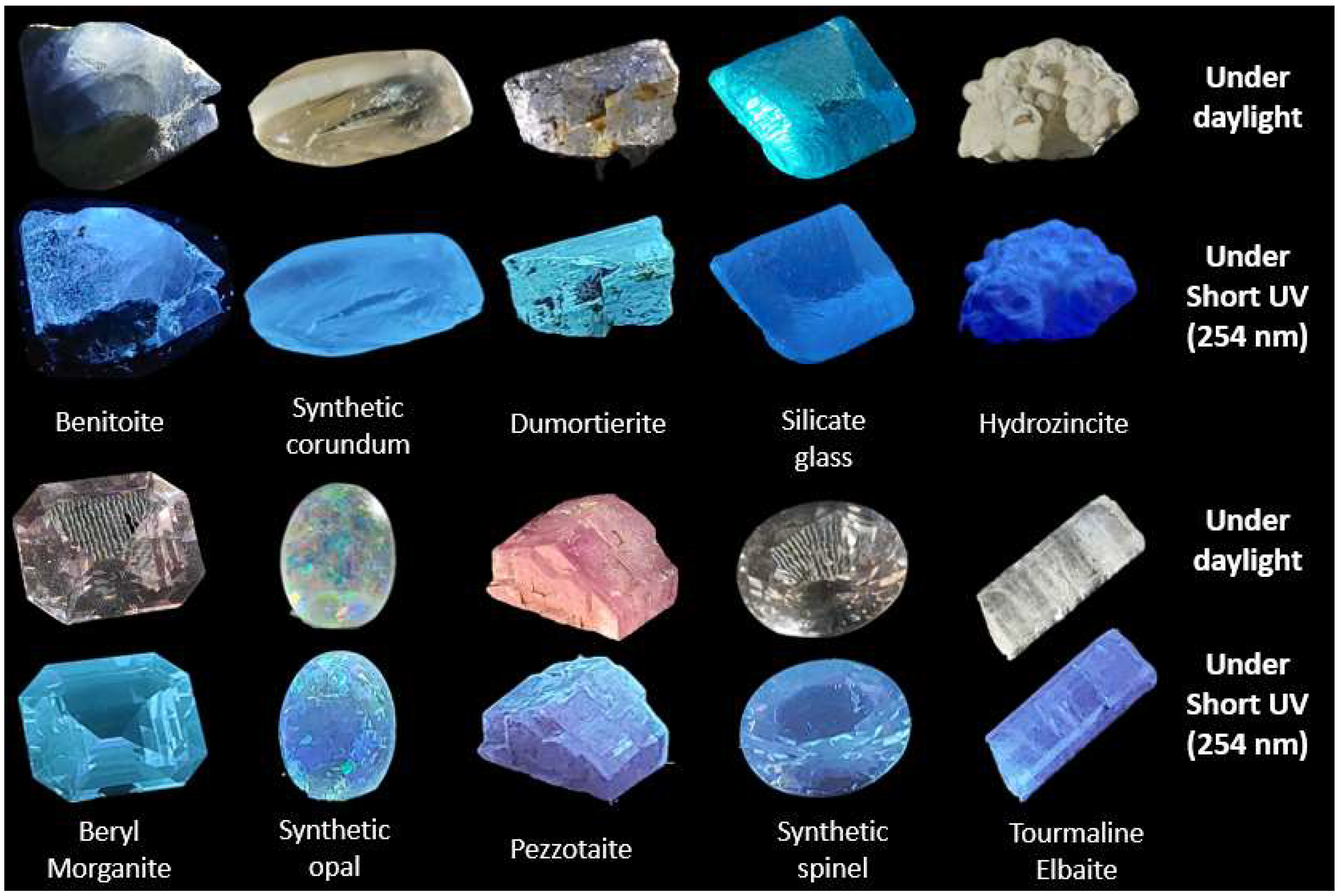
| Minerals | Chemical Formula | Nature | Absorption Color | Sample |
|---|---|---|---|---|
| Benitoite | BaTiSi3O9 | Natural | Blue | 3684 |
| Corundum: synthetic sapphire | Al2O3 | Synthetic | Colorless | 3697 |
| Dumortierite | Al7BO3Si3O18 | Natural | Blue | 3977 |
| Silicate glass: “Fondu du Jura” | 65.7%w SiO2, 9.1%w Na2O, 25.2%w Al2O3 | Synthetic | Blue | 1846 |
| Hydrozincite | Zn5(CO3)2(OH)6 | Natural | White | 4101 |
| Beryl Morganite | Be₃Al₂(SiO₃)₆ | Natural | Colorless | 1865 |
| Synthetic Opal | SiO2, nH2O | Synthetic | White with play of color | 2029 |
| Pezzotaite | CsAl₂Si₆O₁₈ | Natural | Purple-Pink | 966 |
| Flame fusion synthetic Spinel | MgAl2O4 | Synthetic | Colorless | 1386 |
| Tourmaline: Elbaite | Na(Li,Al)1.5Al6(Si6O18)(BO3)3 (OH)3 | Natural | Colorless | 3056 |
| Benitoite | Synthetic Sapphire | Dumortierite | Glass | Hydrozincite | |
| avg-SD | avg-SD | avg-SD | avg-SD | avg-SD | |
| 33S | 201.072–36.852 | nd | 1064.6–341.6 | 2136.8–624.5 | 10.1–1.0 |
| 47Ti | Constituant | 23.5–1.5 | 3658.2–127.2 | 1274.2–46.5 | 31.2–3.3 |
| 205Tl | nd | nd | 0.074–0.014 | 0.08–0.01 | nd |
| 208Pb | 0.354–0.021 | 1.3–0.1 | 11.6–0.9 | 2895.0–236.4 | 438.2–115.2 |
| Beryl (Morganite) | Synthetic opal | Pezzotaite | synthetic spinel | Tourmaline (Elbaite) | |
| avg-SD | avg-SD | avg-SD | avg-SD | avg-SD | |
| 33S | 703.216–225.848 | 254.6–36.5 | 396.3–131.5 | nd | 630.5–195.8 |
| 47Ti | 23.374–1.104 | 28.5–1.1 | 508.4–23.8 | 23.5–1.5 | 38.9–1.5 |
| 205Tl | 7.100–0.576 | nd | 2.59–0.14 | nd | 0.074–0.009 |
| 208Pb | 5.230–0.361 | 1.27–0.06 | 8.4–0.5 | 1.27–0.09 | 108.2–5.9 |
| Mineral | Emission Maxima (nm–eV) | Excitation Maxima (nm–eV) | Lifetime Decay (µs) |
|---|---|---|---|
| Benitoite | 414–2.99 | 291–4.26 | 17.08 |
| Corundum: Synthetic Sapphire | 425–2.92 | 256–4.84 | 39.96 |
| Dumortierite | 446–2.78 | Near 240–Near 5.16 | 14.34 |
| Pb silicate glass | 463–2.68 | 248–5.00 | 28.37 |
| Hydrozincite | 439–2.82 | 248–5.00 | 1.92 |
| Beryl Morganite | 433–2.86 | 278–4.46 | 17.8 |
| Synthetic Opal | 452–2.74 | 253–4.90 | 40.59 |
| Pezzotaite | 426–2.91 | Near 240–Near 5.16 | 14.49 |
| Flame fusion Synthetic Spinel | 461–2.69 | 269–4.61 | 38.81 |
| Tourmaline: Elbaite | 420–2.95 | 253–4.90 | 25.46 |
Disclaimer/Publisher’s Note: The statements, opinions and data contained in all publications are solely those of the individual author(s) and contributor(s) and not of MDPI and/or the editor(s). MDPI and/or the editor(s) disclaim responsibility for any injury to people or property resulting from any ideas, methods, instructions or products referred to in the content. |
© 2023 by the authors. Licensee MDPI, Basel, Switzerland. This article is an open access article distributed under the terms and conditions of the Creative Commons Attribution (CC BY) license (https://creativecommons.org/licenses/by/4.0/).
Share and Cite
Vigier, M.; Fritsch, E.; Cavignac, T.; Latouche, C.; Jobic, S. Shortwave UV Blue Luminescence of Some Minerals and Gems Due to Titanate Groups. Minerals 2023, 13, 104. https://doi.org/10.3390/min13010104
Vigier M, Fritsch E, Cavignac T, Latouche C, Jobic S. Shortwave UV Blue Luminescence of Some Minerals and Gems Due to Titanate Groups. Minerals. 2023; 13(1):104. https://doi.org/10.3390/min13010104
Chicago/Turabian StyleVigier, Maxence, Emmanuel Fritsch, Théo Cavignac, Camille Latouche, and Stéphane Jobic. 2023. "Shortwave UV Blue Luminescence of Some Minerals and Gems Due to Titanate Groups" Minerals 13, no. 1: 104. https://doi.org/10.3390/min13010104