
Origin of the Dolomitic Ooids Formed in the Pliocene Shizigou Formation in the Qaidam Basin, Northern Tibet Plateau and Implications for Climate Change
Abstract
:1. Introduction
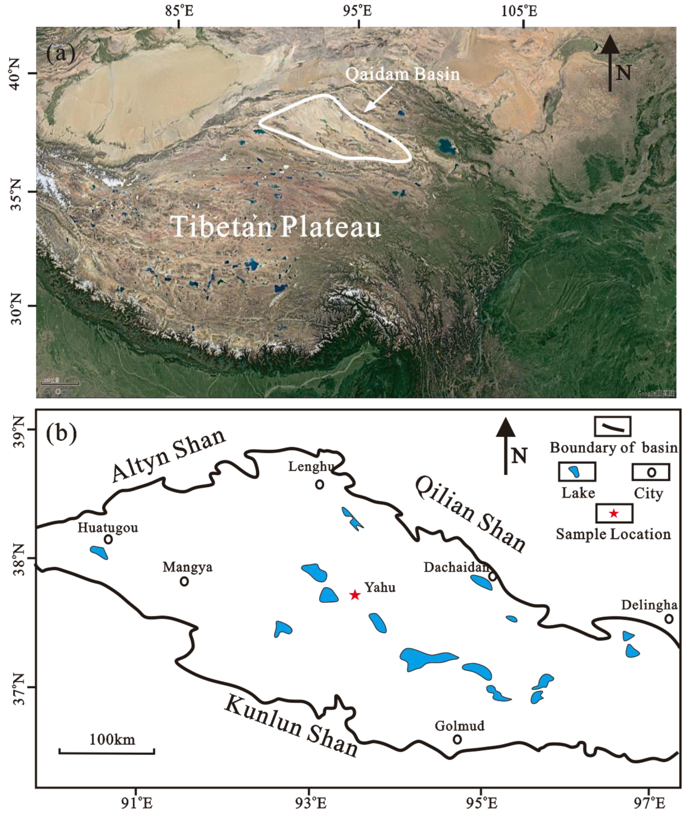
2. Study Area
3. Materials and Methods
4. Results
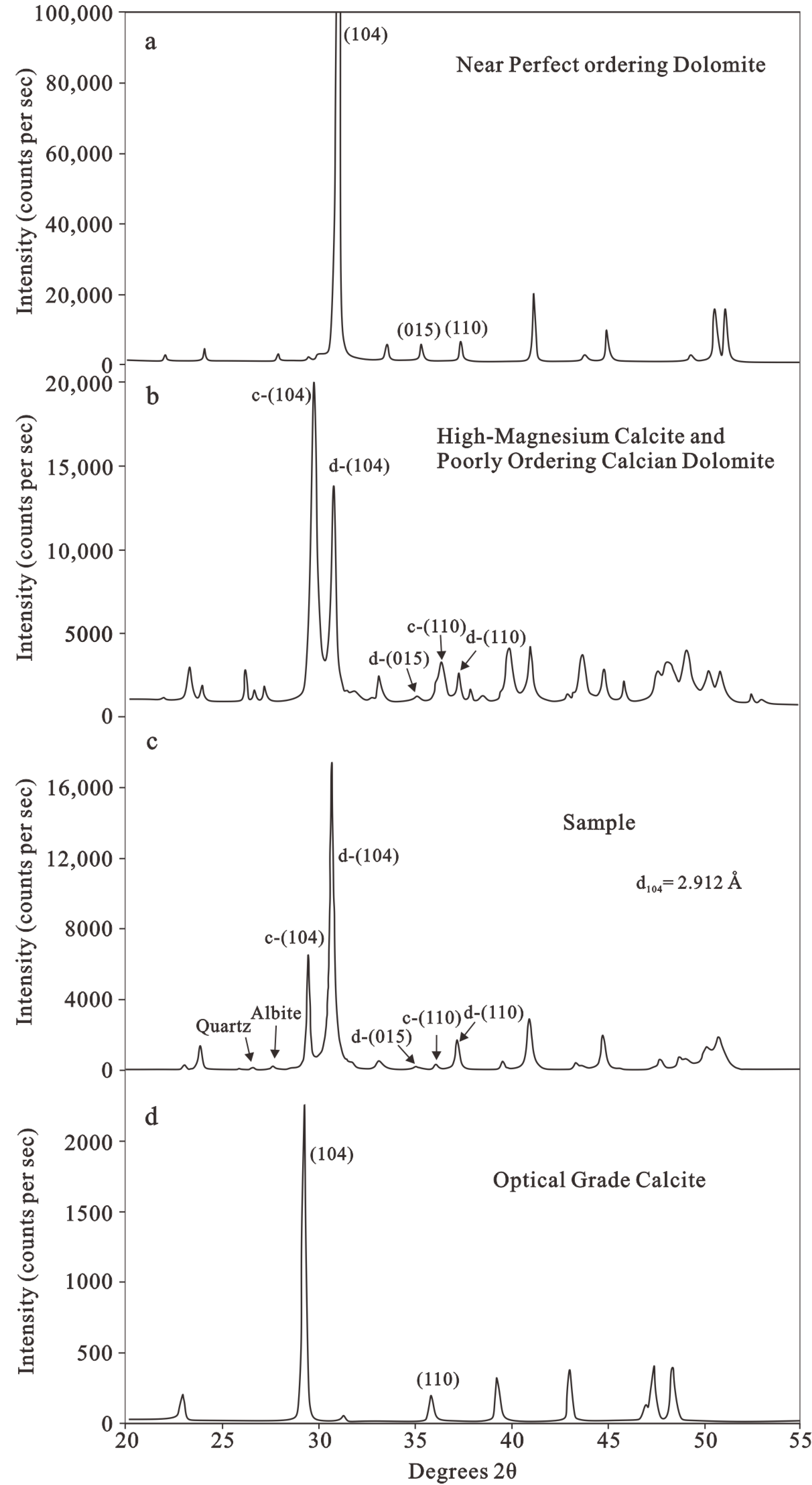
4.1. Mineralogy
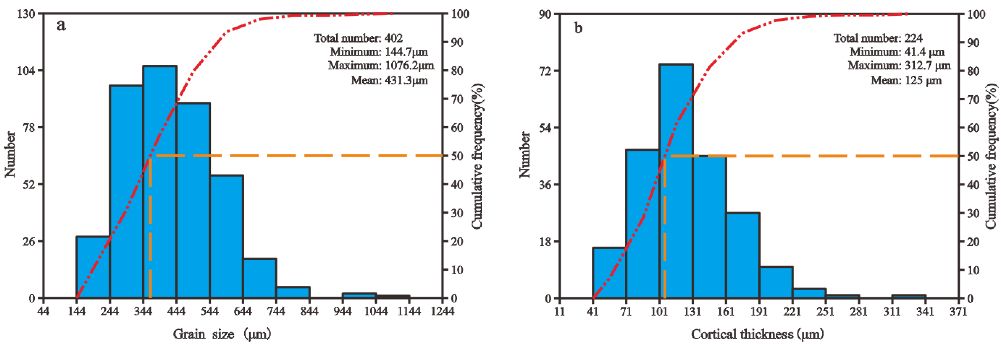
4.2. Characteristics of Ooids and Stable Isotopic Compositions
5. Discussion
5.1. Primary Mineral of Ooids
5.2. Origin of Ooids
5.3. Implications for the Paleoenvironment
6. Conclusions
- (1)
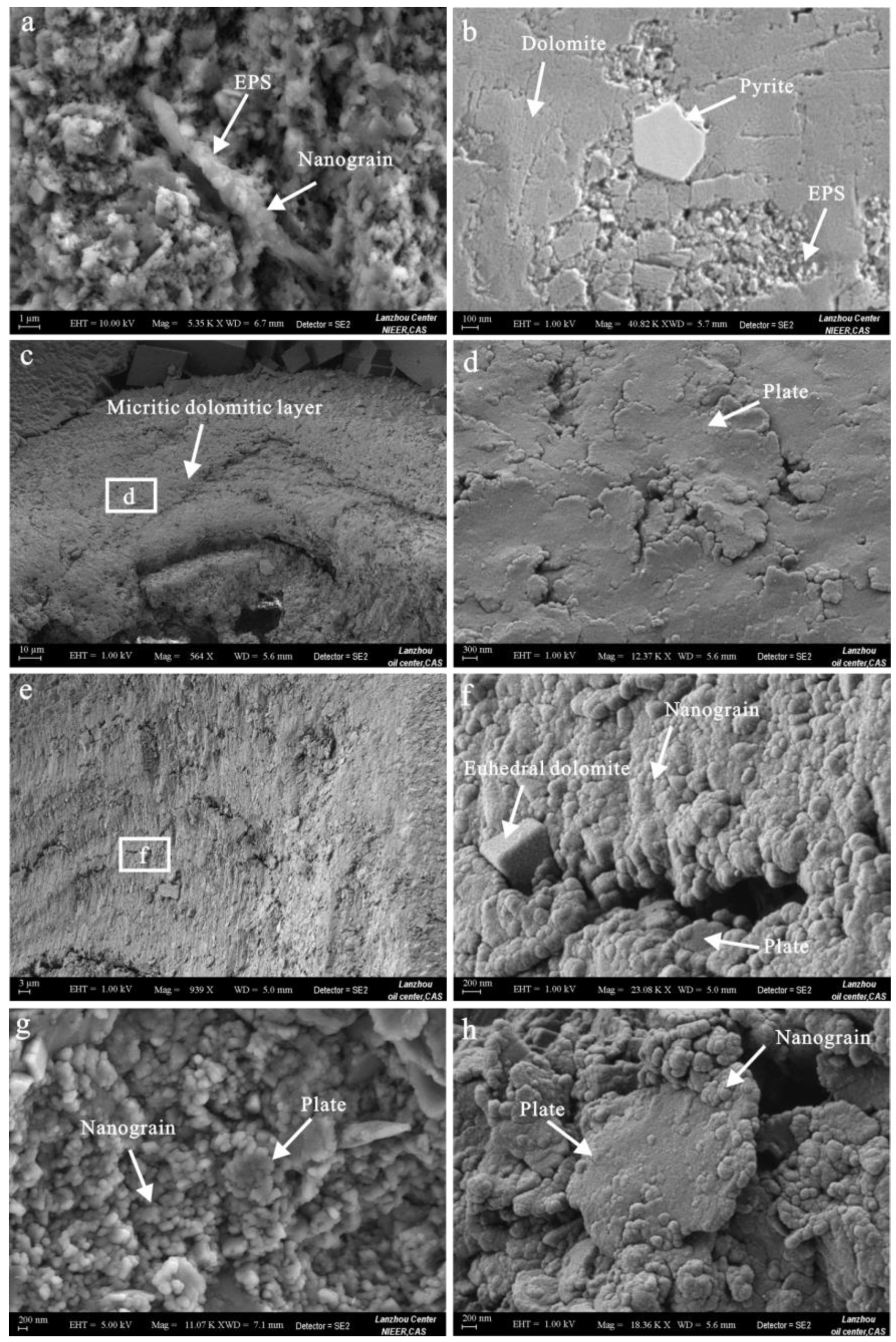
- The ooids formed in the Pliocene Shizigou Formation in the Qaidam Basin mainly consist of micritic dolomite that is poorly ordered. The nuclear minerals are mostly micritic dolomite with a minor clay mineral and pyrite. The thin-section and SEM observations combined with CL indicate that the primary mineral of the ooid cortices is dolomite. The micritic dolomite was formed through a reducing condition, and its formation was related to the presence of sulfate-reducing bacteria and dissolved silica. The euhedral dolomite crystals were formed in the pore water after the ooids were deposited.
- (2)
- The formation of the dolomitic ooids in the Shizigou Formation occurred according to the “conveyor belt” model. The ooids grew on the offshore lake floor, where nanominerals were precipitated on the EPS due to the involvement of sulfate-reducing bacteria and other microbes. Then, the ooids were reformed under strong hydrodynamic surf zones, where the random nanograin minerals were abraded to form flattened plates as a new polished layer. The sediment transport was repeated periodically until the ooids exceeded a threshold size.
- (3)
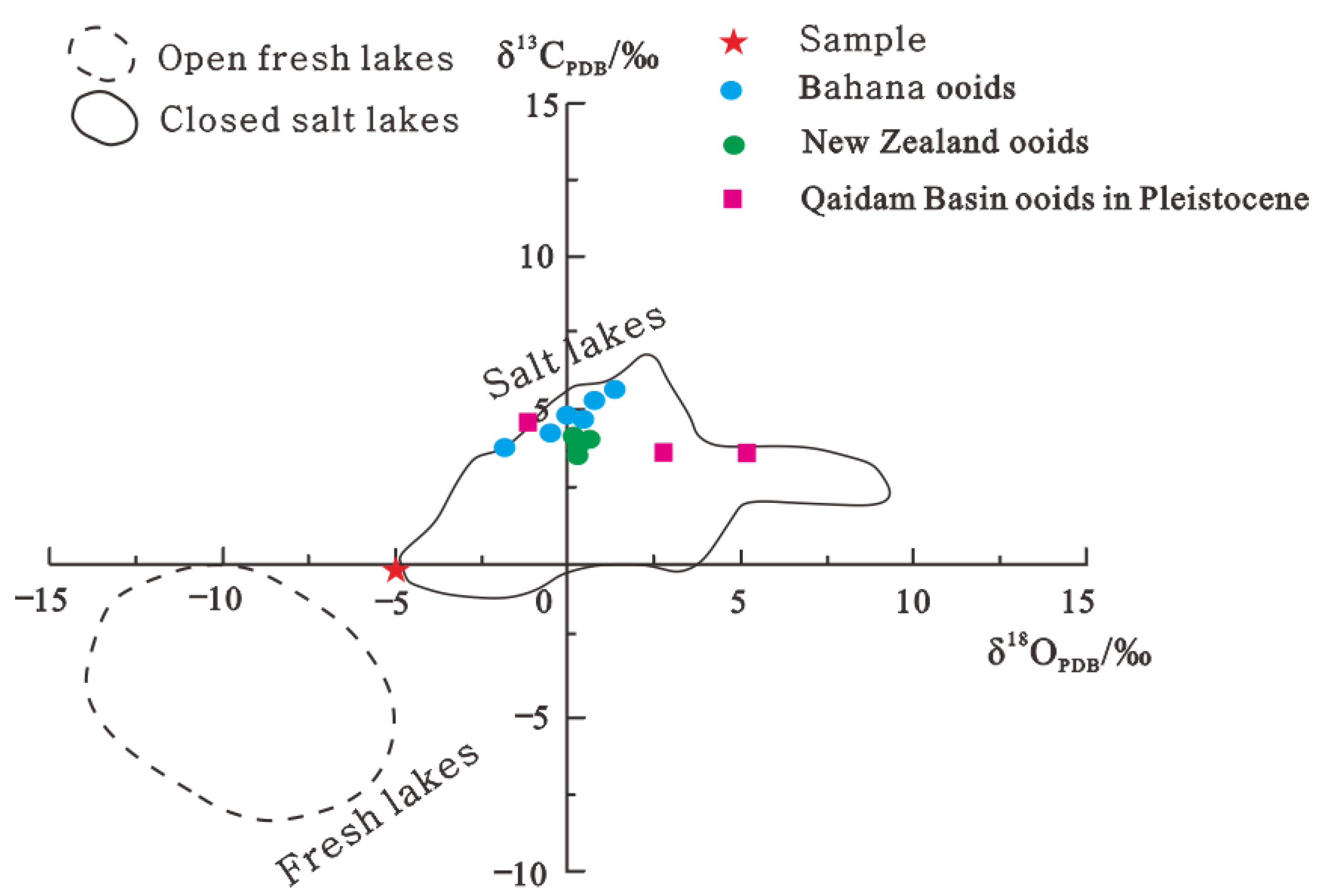
- Ooids from the different uplift stages of the Qaidam Basin (in the Pliocene and Pleistocene) have variations in mineral compositions and the carbon and oxygen isotopes. The minerals of the ooid cortices changed from Pliocene dolomite to Pleistocene aragonite. The δ13C and δ18O values of the Pleistocene carbonates are higher than those of the Pliocene. These differences indicate that the Pliocene lakes had a lower salinity and were more humid than Pleistocene lakes. Ooids may be an effective proxy for reflecting the climatic change and uplift history of the Tibet Plateau.
Supplementary Materials
Author Contributions
Funding
Data Availability Statement
Conflicts of Interest
References
- Diaz, M.R.; Eberli, G.P. Decoding the mechanism of formation in marine ooids: A review. Earth-Sci. Rev. 2019, 190, 536–556. [Google Scholar] [CrossRef]
- Ball, M.M. Carbonate sand bodies of florida and the bahamas. J. Sediment. Petrol. 1967, 37, 556–591. [Google Scholar]
- Harris, P.M.; Halley, R.B.; Lukas, K.J. Endolith microborings and their preservation in Holocene-Pleistocene (Bahamas-Florida) ooids. Geology 1979, 7, 216–220. [Google Scholar] [CrossRef]
- Li, F.; Yan, J.X.; Algeo, T.; Wu, X. Paleoceanographic conditions following the end Permian mass extinction recorded by giant ooids (Moyang, South China). Glob. Planet. Chang. 2013, 105, 102–120. [Google Scholar] [CrossRef]
- Liu, X.; Chen, X.; Tostevin, R.; Yao, H.; Han, K.; Guo, H.; Jafarian, A. Post-depositional modification of carbonate ooids by sulfate-reducing bacteria: Evidence from the Lower–Middle Jurassic, Tethyan Himalayas of southern Tibet. Sed. Geol. 2021, 426, 106027. [Google Scholar] [CrossRef]
- Davies, P.J.; Bubela, B.; Ferguson, J. The formation of ooids. Sedimentology 1978, 25, 703–730. [Google Scholar] [CrossRef]
- Rankey, E.C.; Reeder, S.L. Holocene ooids of Aitutaki Atoll, Cook Islands, South Pacific. Geology 2009, 37, 971–974. [Google Scholar] [CrossRef]
- Diaz, M.R.; Swart, P.K.; Eberli, G.P.; Oehlert, A.M.; Devlin, Q.; Saeid, A.; Altabet, M.A. Geochemical evidence of microbial activity within ooids. Sedimentology 2015, 62, 2090–2112. [Google Scholar] [CrossRef]
- Diaz, M.R.; Eberli, G.P.; Blackwelder, P.; Phillips, B.; Swart, P.K. Microbially mediated organomineralization in the formation of ooids. Geology 2017, 45, 771–774. [Google Scholar] [CrossRef]
- Li, F.; Yan, J.; Burne, R.V.; Chen, Z.Q.; Algeo, T.J.; Zhang, W.; Tian, L.; Gan, Y.; Liu, K.; Xie, S. Paleo-seawater REE compositions and microbial signatures preserved in laminae of lower Triassic ooids. Palaeogeogr. Palaeoclimatol. Palaeoecol. 2017, 486, 96–107. [Google Scholar] [CrossRef]
- O’Reilly, S.S.; Mariotti, G.; Winter, A.R.; Newman, S.A.; Matys, E.D.; McDermott, F.; Pruss, S.B.; Bosak, T.; Summons, R.E.; Klepac-Ceraj, V. Molecular biosignatures reveal common benthic microbial sources of organic matter in ooids and grapestones from Pigeon Cay, the Bahamas. Geobiology 2017, 15, 112–130. [Google Scholar] [CrossRef] [PubMed]
- Batchelor, M.T.; Burne, R.V.; Henry, B.I.; Li, F.; Paul, J. A biofilm and organomineralisation model for the growth and limiting size of ooids. Sci. Rep. 2018, 8, 559. [Google Scholar] [CrossRef] [PubMed] [Green Version]
- Opdyke, B.N.; Wilkinson, B.H. Paleolatitude distribution of Phanerozoic marine ooids and cements. Palaeogeogr. Palaeoclimatol. Palaeoecol. 1990, 78, 135–148. [Google Scholar] [CrossRef] [Green Version]
- Sumner, D.Y.; Grotzinger, J.P. Numerical modeling of ooid size and the problem of Neoproterozoic giant ooids. J. Sediment. Res. 1993, 63, 974–982. [Google Scholar]
- Thorie, A.; Mukhopadhyay, A.; Banerjee, T.; Mazumdar, P. Giant ooids in a Neoproterozoic carbonate shelf, Simla Group, Lesser Himalaya, India: An analogue related to Neoproterozoic glacial deposits. Mar. Pet. Geol. 2018, 98, 582–606. [Google Scholar] [CrossRef]
- Lu, C.; Li, F.; Oehlert, A.M.; Li, J.; Zou, H. Reconstructing paleoceanographic conditions during the middle Ediacaran: Evidence from giant ooids in South China. Precambrian Res. 2020, 351, 105945. [Google Scholar] [CrossRef]
- Li, F.; Wu, S.; Liu, K. Identification of Ooid Primary Mineralogy: A clue for understanding the variation in paleo-oceanic chemistry. Acta Sedimentol. Sin. 2015, 33, 500–511. [Google Scholar]
- Land, L.S.; Behrens, E.W.; Frishman, S.A. The ooids of Baffin Bay, Texas. J. Sediment. Petrol. 1979, 49, 1269–1278. [Google Scholar]
- Fang, X.M.; Zhang, W.L.; Meng, Q.Q.; Gao, J.P.; Wang, X.M.; King, J.; Song, C.H.; Dai, S.; Miao, Y.F. High-resolution magneto stratigraphy of the Neogene Huaitoutala section in the eastern Qaidam Basin on the NE Tibetan Plateau, Qinghai Province, China and its implication on tectonic uplift of the NE Tibetan Plateau. Earth Planet. Sci. Lett. 2007, 258, 293–306. [Google Scholar] [CrossRef]
- Fang, X.; Li, M.; Wang, Z.; Wang, J.; Li, J.; Liu, X. Oscillation of mineral compositions in Core SG-1b, western Qaidam Basin, NE Tibetan Plateau. Sci. Rep. 2016, 6, 32848. [Google Scholar] [CrossRef] [Green Version]
- Wu, F.L.; Fang, X.M.; Herrmann, M.; Mosbrugger, V.; Miao, Y.F. Extended drought in the interior of Central Asia since the Pliocene reconstructed fromsporopollen records. Glob. Planet. Chang. 2011, 76, 16–21. [Google Scholar] [CrossRef]
- Zhang, W.; Appel, E.; Fang, X.; Yan, M.; Song, C.; Cao, L. Paleoclimatic implications of magnetic susceptibility in late Pliocene–Quaternary sediments from deep drilling core SG-1 in the western Qaidam Basin (NE Tibetan Plateau). J. Geophys. Res. 2012, 117, B06101. [Google Scholar] [CrossRef] [Green Version]
- Lu, Y.; Fang, X.; Appel, E.; Wang, J.; Herb, C.; Han, W.; Wu, F.; Song, C. A 7.3–1.6 Ma grain size record of interaction between anticline uplift and climate change in the western Qaidam Basin, NE Tibetan Plateau. Sed. Geol. 2015, 319, 40–51. [Google Scholar] [CrossRef]
- Sun, Y.; Li, Y.; Li, L.; He, H. Preservation of cyanobacterial UVR-shielding pigment scytonemin in carbonate ooids formed in Pleistocene salt lakes in the Qaidam Basin, Tibetan Plateau. Geophys. Res. Lett. 2019, 46, 10375–10383. [Google Scholar] [CrossRef]
- Han, W.; Fang, X.; Ye, C.; Teng, X.; Zhang, T. Tibet forcing Quaternary stepwise enhancement of westerly jet and central Asian aridification: Carbonate isotope recordsfrom deep drilling inthe Qaidam salt playa, NETibet. Glob. Planet. Chang. 2014, 116, 68–75. [Google Scholar] [CrossRef]
- Cheng, F.; Jolivet, M.; Guo, Z.; Wang, L.; Zhang, C.; Li, X. Cenozoic evolution of the Qaidam basin and implications for the growth of the northern Tibetan plateau: A review. Earth-Sci. Rev. 2021, 220, 103730. [Google Scholar] [CrossRef]
- Zhu, L.; Wang, C.; Zheng, H.; Xiang, F.; Yi, H.; Liu, D. Tectonic and sedimentary evolution of basins in the northeast of Qinghai-Tibet Plateau and their implication for the northward growth of the Plateau. Palaeogeogr. Palaeoclimatol. Palaeoecol. 2006, 241, 49–60. [Google Scholar] [CrossRef]
- Jian, X.; Guan, P.; Zhang, D.W.; Zhang, W.; Feng, F.; Liu, R.J.; Lin, S.D. Provenance of Tertiary sandstone in the northern Qaidam basin, northeastern Tibetan Plateau: Integration of framework petrography, heavy mineral analysis and mineral chemistry. Sed. Geol. 2013, 290, 109–125. [Google Scholar] [CrossRef]
- Sun, G.; Wang, M.; Guo, J.; Wang, Y.; Yang, Y. Geochemical Significance of Clay Minerals and Elements in Paleogene Sandstones in the Center of the Northern Margin of the Qaidam Basin, China. Minerals 2020, 10, 505. [Google Scholar] [CrossRef]
- Heermance, R.V.; Pullen, A.; Kapp, P.; Garzione, C.N.; Bogue, S.; Ding, L.; Song, P. Climatic and tectonic controls on sedimentation and erosion during the Pliocene–Quaternary in the Qaidam Basin (China). Geol. Soc. Am. Bull. 2013, 125, 833–856. [Google Scholar] [CrossRef] [Green Version]
- Goldsmith, J.R.; Graf, D.L. Structural and compositional variations in some natural dolomites. J. Geol. 1958, 66, 678–693. [Google Scholar] [CrossRef]
- Fang, Y.; Xu, H. A New Approach to Quantify the Ordering State of Protodolomite Using XRD, TEM, and Z-Contrast Imaging. J. Sediment. Res. 2019, 89, 537–551. [Google Scholar] [CrossRef]
- Graf, D.L.; Goldsmith, J.R. Some hydrothermal syntheses of dolomite and protodolomite. J. Geol. 1956, 64, 173–186. [Google Scholar] [CrossRef]
- Chave, K.E. Factors influencing the mineralogy of carbonate sediments. Limnol. Oceanogr. 1962, 7, 218–223. [Google Scholar] [CrossRef]
- Chave, K.E. A solid solution between calcite and dolomite. J. Geol. 1952, 60, 190–192. [Google Scholar] [CrossRef]
- Gregg, J.M.; Bish, D.L.; Kaczmarek, S.E.; Machel, H.G. Mineralogy, nucleation and growth of dolomite in the laboratory and sedimentary environment: A review. Sedimentology 2015, 62, 1749–1769. [Google Scholar] [CrossRef]
- Marshall, J.F.; Davies, P.J. High-magnesium calcite ooids from the Great Barrier Reef. J. Sediment. Res. 1975, 45, 285–291. [Google Scholar]
- Milliman, J.D.; Barreto, H.T. Relict magnesian calcite oolite and subsidence of the Amazon shelf. Sedimentology 1975, 22, 137–145. [Google Scholar] [CrossRef]
- Flügel, E. Microfacies of Carbonate Rock: Analysis, Interpretation and Application, 2nd ed.; Springer: Berlin, German, 2010; p. 984. [Google Scholar]
- Trower, E.J.; Grotzinger, J.P. Sedimentology, diagenesis, and stratigraphic occurrence of giant ooids in the Ediacaran Rainstorm Member, Johnnie Formation, Death Valley region, California. Precambrian Res. 2010, 80, 113–124. [Google Scholar] [CrossRef]
- Banerjee, A.; Słowakiewicz, M.; Majumder, T.; Khan, S.; Patranabis-Deb, S.; Tucker, M.E.; Saha, D. A Palaeoproterozoic dolomite (Vempalle Formation, Cuddapah Basin, India) showing Phanerozoic-type dolomitisation. Precambrian Res. 2019, 328, 9–26. [Google Scholar] [CrossRef] [Green Version]
- Guo, Q.; Jin, Z.; Zhu, X.; Shi, S.; Wang, J.; Wang, J.; Li, Y.; Li, S. Characteristics and mechanism of dolomitization in the ooids of the Cambrian Zhangxia Formation, Xiaweidian, China. Carbonates Evaporites 2020, 35, 7. [Google Scholar] [CrossRef]
- Land, L.S. The isotopic and trace element geochemistry of dolomite: The state of the art. SEPM Spec. Publ. 1980, 28, 87–110. [Google Scholar]
- Lippmann, F. Sedimentary Carbonate Minerals, Rocks and Inorganic Materials. In Monograph Series of Theoretical and Experimental Studies 4; Springer: Berlin, German, 1973; p. 228. [Google Scholar]
- Warren, J. Dolomite: Occurrence evolution and economically important associations. Earth Sci. Rev. 2000, 52, 1–81. [Google Scholar] [CrossRef]
- Rodriguez-Blanco, J.D.; Shaw, S.; Benning, L.G. A route for the direct crystallization of dolomite. Am. Mineral. 2015, 100, 1172–1181. [Google Scholar] [CrossRef] [Green Version]
- Vasconcelos, C.; McKenzie, J.A.; Warthmann, R.; Bernasconi, S.M. Microbial mediation as a possible mechanism for natural dolomite formation at low temperatures. Nature 1995, 377, 220–222. [Google Scholar] [CrossRef]
- Wright, D.T.; Oren, A. Nonphotosynthetic bacteria and the formation of carbonates and evaporites through time. Geomicrobiol. J. 2005, 22, 27–53. [Google Scholar] [CrossRef]
- Qiu, X.; Wang, H.; Yao, Y.; Duan, Y. High salinity facilitates dolomite precipitation mediated by Haloferax volcanii DS52. Earth Planet. Sci. Lett. 2017, 472, 197–205. [Google Scholar] [CrossRef]
- Zhang, F.; Xu, H.; Konishi, H.; Shelobolina, E.S.; Roden, E.E. Polysaccharide-catalyzed nucleation and growth of disordered dolomite: A potential precursor of sedimentary dolomite. Am. Miner. 2012, 97, 556–567. [Google Scholar] [CrossRef]
- Zhang, F.; Xu, H.; Konishi, H.; Kemp, J.M.; Roden, E.E.; Shen, Z. Dissolved sulfide-catalyzed precipitation of disordered dolomite: Implications for the formation mechanism of sedimentary dolomite. Geochim. Cosmochim. Acta 2012, 97, 148–165. [Google Scholar] [CrossRef]
- Zhang, F.; Xu, H.; Shelobolina, E.S.; Konishi, H.; Converse, B.; Shen, Z.; Roden, E.E. The catalytic effect of bound extracellular polymeric substances excreted by anaerobic microorganisms on Ca-Mg carbonate precipitation: Implications for the “dolomite problem”. Am. Miner. 2015, 100, 483–494. [Google Scholar] [CrossRef]
- Xu, H.; Zhou, M.; Fang, Y.; Teng, H.H. Effect of Mica and Hematite (001) Surfaces on the Precipitation of Calcite. Minerals 2018, 8, 17. [Google Scholar] [CrossRef] [Green Version]
- Liu, D.; Xu, Y.; Papineau, D.; Yu, N.; Fan, Q.; Qiu, X.; Wang, H. Experimental evidence for abiotic formation of low-temperature proto-dolomite facilitated by clay minerals. Geochim. Cosmochim. Acta 2019, 247, 83–95. [Google Scholar] [CrossRef] [Green Version]
- Fang, Y.; Xu, H. Dissolved silica-catalyzed disordered dolomite precipitation. Am. Miner. 2022, 107, 443–452. [Google Scholar] [CrossRef]
- Last, W.M. Lacustrine dolomite-an overview of modern, Holocene, and Pleistocene occurrences. Earth-Science Rev. 1990, 27, 221–263. [Google Scholar] [CrossRef]
- Yu, B.; Dong, H.; Jiang, H.; Li, S.; Liu, Y. Discovery of Spheric Dolomite Aggregations in Sediments from the Bottom of Qinghai Lake and Its Significance for Dolomite Problem. Geoscience 2007, 21, 66–70. [Google Scholar]
- Bruhn, F.; Bruckschen, P.; Richter, D.K.; Meijer, J.; Stephan, A.; Veizer, J. Diagenetic history of sedimentary carbonates: Constraints from combined cathodoluminescence and trace element analyses by micro-PIXE. Nucl Instr Meth Phys Res 1995, B104, 409–414. [Google Scholar] [CrossRef]
- Mariano, A.N. Some further geological applications of cathodoluminescence. In Cathodoluminescence of Geological Materials; Marshall, J.D., Ed.; Unwin Hyman: Boston, MA, USA, 1988; pp. 94–123. [Google Scholar]
- Pagel, M.; Barbin, V.; Blanc, P.; Ohnenstetter, D. Cathodoluminescence in Geosciences; Springer: Berlin, German, 2000; p. 514. [Google Scholar]
- Kaczmarek, S.E.; Thornton, B.P. The effect of temperature on stoichiometry, cation ordering, and reaction rate in high-temperature dolomitization experiments. Chem. Geol. 2017, 468, 32–41. [Google Scholar] [CrossRef]
- Mariotti, G.; Pruss, S.B.; Klepac-Ceraj, V.; Summons, R.E.; Newman, S.A.; Bosak, T. Where Is the Ooid Factory? In Proceedings of the American Geophysical Union Fall Meeting, San Francisco, CA, USA, 15–19 December 2014. [Google Scholar]
- Duguid, S.M.A.; Kyser, T.K.; James, N.P.; Rankey, E.C. Microbes and ooids. J. Sediment. Res. 2010, 80, 236–251. [Google Scholar] [CrossRef]
- Rankey, E.C.; Reeder, S.L. Holocene oolitic marine sand complexes of the Bahamas. J. Sediment. Res. 2011, 81, 97–117. [Google Scholar] [CrossRef]
- Talbot, M.R. A review of the palaeohydrological interpretation of carbon and oxygen isotopic ratios in primary lacustrine carbonates. Chem. Geol. Isot. Geosci. Sect. 1990, 80, 261–279. [Google Scholar] [CrossRef]
- Leng, M.J.; Marshall, J.D. Palaeoclimate interpretation of stable isotope data from lake sediment archives. Quat. Sci. Rev. 2004, 23, 811–831. [Google Scholar] [CrossRef] [Green Version]
- Fang, Y.; Xu, H. Study of an Ordovician Carbonate with alternating dolomite–calcite laminations and its implication for catalytic effects of microbes on the formation of Sedimentary dolomite. J. Sediment. Res. 2018, 88, 679–695. [Google Scholar] [CrossRef]
- Fang, Y.; Xu, H. Coupled dolomite and silica precipitation from continental weathering during deglaciation of the Marinoan Snowball Earth. Precambrian Res. 2022, 380, 106824. [Google Scholar] [CrossRef]
- Li, J.J.; Fang, X.M. Uplift of Tibetan Plateau and environmental changes. Chinese Sci. Bull. 1999, 44, 2117–2124. [Google Scholar] [CrossRef]
- Sun, J.M.; Zhu, R.X.; Bowler, J. Timing of the Tianshan Mountains uplift constrained by magnetostratigraphic analysis of molasses deposits. Earth Planet. Sci. Lett. 2004, 219, 239–253. [Google Scholar] [CrossRef]
- Li, J.; Li, M.; Fang, X.; Zhang, G.; Zhang, W.; Liu, X. Isotopic composition of gypsum hydration water in deep Core SG-1, western Qaidam basin (NE Tibetan Plateau), implications for paleoclimatic evolution. Glob. Planet. Chang. 2017, 155, 70–77. [Google Scholar] [CrossRef]


Publisher’s Note: MDPI stays neutral with regard to jurisdictional claims in published maps and institutional affiliations. |
© 2022 by the authors. Licensee MDPI, Basel, Switzerland. This article is an open access article distributed under the terms and conditions of the Creative Commons Attribution (CC BY) license (https://creativecommons.org/licenses/by/4.0/).
Share and Cite
Hao, L.; Jia, J.; Tao, H.; Chen, J.; Ma, X.; Li, S.; Qiu, J. Origin of the Dolomitic Ooids Formed in the Pliocene Shizigou Formation in the Qaidam Basin, Northern Tibet Plateau and Implications for Climate Change. Minerals 2022, 12, 1586. https://doi.org/10.3390/min12121586
Hao L, Jia J, Tao H, Chen J, Ma X, Li S, Qiu J. Origin of the Dolomitic Ooids Formed in the Pliocene Shizigou Formation in the Qaidam Basin, Northern Tibet Plateau and Implications for Climate Change. Minerals. 2022; 12(12):1586. https://doi.org/10.3390/min12121586
Chicago/Turabian StyleHao, Lewei, Jiantuan Jia, Huifei Tao, Jinniu Chen, Xiaofeng Ma, Shutong Li, and Junli Qiu. 2022. "Origin of the Dolomitic Ooids Formed in the Pliocene Shizigou Formation in the Qaidam Basin, Northern Tibet Plateau and Implications for Climate Change" Minerals 12, no. 12: 1586. https://doi.org/10.3390/min12121586