Article

Endophytic Candida membranifaciens from Euphorbia milii L. Alleviate Salt Stress Damages in Maize

1
Department of Botany, Garden Campus, Abdul Wali Khan University, Mardan 23200, Pakistan
2
Department of Horticulture and Life Science, Yeungnam University, Gyeongsan 38541, Korea
3
Center of Biotechnology and Microbiology, University of Peshawar, Peshawar 25120, Pakistan
4
Department of Applied Biosciences, Kyungpook National University, Daegu 41566, Korea
*
Authors to whom correspondence should be addressed.
Agronomy 2022, 12(10), 2263; https://doi.org/10.3390/agronomy12102263
Submission received: 6 September 2022 / Revised: 19 September 2022 / Accepted: 20 September 2022 / Published: 21 September 2022

Abstract

:
Fungal endophytes are not widely known for their role in bioactive metabolite production and salinity stress alleviation in different crop plants. Presently, we investigated the salt stress (NaCl, KCl, and H2SO4) mitigation capabilities of fungal endophyte Candida membranifaciens (FH15) isolated from Euphorbia milii L. The pure culture filtrate (CF) of C. membranifaciens revealed siderophore production and solubilization of phosphate, with high levels of indoleacetic acid (IAA: 35.8µg/mL), phenolics (70 µg/mL), and flavonoids (50 µg/mL) by using a UV spectrophotometer. The LC/MS analysis of the CF showed different phenols and flavonoids that were identified as Salicylic acid, Baicalein, Aconitic acid, Feruloylquinic acid, Coniferyl aldehyde hexoside, Pentose, Chlorogenic acid, Myricetin, Propoxyphene, and Amino-flunitrazepam. Inoculation of maize seedlings with C. membranifaciens significantly (p = 0.05) enhanced the fresh and dry biomass, carotenoid, and chlorophyll contents under 100 mM salt stress conditions. Similarly, the catalase, peroxidase activity, phenols, proline flavonoids and relative water contents (RWC) of the maize plants were enhanced. More interestingly, the inoculation of C. membranifaciens on maize revealed a higher endogenous IAA level as compared to non-inoculated control plants. Endophyte C. membranifaciens inoculation on maize seedlings under salt stress revealed a 20.87% and 16.60% increase in fresh and dry biomass, as well as significantly enhanced root shoot length and allied growth attributes, in addition to an alleviation of the adverse effects of salinity stress. Conclusively, endophytic C. membranifaciens significantly enhanced the growth attributes of maize and mitigated the adverse effects of salinity stress. Such endophytic fungal strain could be used for further field trails to enhance agricultural productivity and facilitate sustainable agricultural practices.

1. Introduction

Salt stress can reduce crop productivity by triggering an osmotic and ionic imbalance inside plant cells; salinity is one of the main stressors impacting agricultural crops around the world. Plants’ growth and development are slowed down by salinity stress, which also causes toxicity, a reduction in water availability, the immobilization of reserves in storage, and alterations to the organization of structural proteins [1,2,3]. In fact, to address such stresses and to ensure sustainable agriculture, robust measures must be taken. To combat salinity, a number of strategies are recommended, such as the production of salt-resistant crops; however, the techniques are either time-consuming or extremely expensive. It has been revealed that using fungal endophytes to deal with salinity stress is affordable, effective, and environmentally beneficial [1,2,4]. Plant-associated microorganisms fulfill pivotal functions and influence the growth and development of their host plant by various mechanisms, whereas the integration of beneficial microorganisms is pivotal for augmenting plant growth, nutrient uptake, and stress-tolerance in the context of modern agricultural systems.
The pragmatic role of beneficial fungal endophytes has attracted more interest recently, and fungal endophytes have received heightened attention from plant biologists due to their role in phytostimulation under environmental stress to develop the most appropriate biofertilizers [5]. Fungal endophytes contribute to the robust life cycle of their host plant through a variety of mechanisms, and confer stress tolerance. Endophytic fungi greatly contribute through the availability of macro- and micro-nutrients to their host plant [6], and release phytohormones such as IAA and gibberellic acids (Waqas et al., 2012). Additionally, endophytic fungi improve mycorrhizal colonization and hyphae production, provide bio-fixed nitrogen, produce siderophores, etc. [7,8]. Fungal endophytes are also known for their ability to solubilize zinc, phosphate, and potassium, etc. [9], and have a higher phosphorus solubilization efficiency than bacteria [10].
Endophytes are asymptomatically occurring beneficial microorganisms living inside a host plant that greatly contribute to the growth and development of the host plant, enhance nutrient uptake, reduce disease severity, and enhance host plant tolerance to environmental stresses. Plants in saline environments face physiological drought, and the lack of water available to the roots causes osmotic stress and induces ionic and nutrient imbalance, while fungal endophytes support plants and confer stress tolerance, and ultimately, promote plant growth. Besides being highly diverse in nature, these endophytes are a novel source of bioactive secondary metabolites. Endophytic symbiosis decreases Na+ toxicity and ROS generation, and enhances the plants as compared to un-inoculated plants [11,12,13]. The mutualistic interaction mitigates stress by compromising the activities of catalase, polyphenol, oxidase, and peroxidase. Endophytes ameliorate the stressful conditions by regulating hormonal levels, such as altering jasmonic acid, enhancing salicylic acid, and down regulating ABA, compared to control plants [13]. Fungal endophytes enhance plant growth and development by influencing the main characteristics of plant physiology and host defense against stressful conditions [14]. Endophytic fungi related to higher plants have recently been discovered to be a good source of potent antioxidants [15]. Antioxidants significantly alleviate harmful effects by deactivating free radicals before they can attack the cells and inhibit damage to proteins, enzymes, lipids, carbohydrates, and DNA [16]. Endophytes encode for plant hormones [17] that can influence the synthesis of secondary metabolites to help plants escape stressful conditions [18]. Phytohormones such as ABA, jasmonic acid (JA), and salicylic acid (SA) respond to abiotic stress stimuli and perform as defense-signaling components [19]. Gibberellins, along with other phytohormones such as IAA, secreted by endophytic fungi, can enhance crop production [20]. Production of IAA by plant-associated endophytes is shown to be a key character allowing the fungal endophytes to stimulate plant growth under abiotic stress [21]. Conversely, host plants without fungal endophytes are devastated by different environmental stresses.
After wheat and rice, maize is the third leading cereal crop in the world. Maize is economically important due to the minerals, vitamins, fiber, and oil it contains. The dry weight of maize consists of starch (71%), protein (9%), and oil (4%); 80% of maize is consumed by humans and animals, and the remaining 20% is utilized by different industrial processes. Maize provides several nutrients to humans and animals and helps as a basic raw material for the production of oils, starches, food sweeteners, alcoholic beverages, proteins, and more recently, fuel. The maize plant is pharmacologically useful for its anti-inflammatory, hypoglycemic, diuretic, and antioxidant properties [22,23]. In the present study, we aimed to isolate, screen, and identify the most competent fungal endophyte, which would not only improve plant growth by producing bioactive secondary metabolites but also extend greater salt stress tolerance to maize plants.

2. Materials and Methods

2.1. Isolation of Fungal Endophyte

The samples of Euphorbia milii L. were carefully washed with tape water and then surface sterilized. Tape water was used to remove any dust materials that were attached to the plant samples, which were then washed with 70% ethanol and 4% sodium hypochlorite for 30 s to remove any attached microorganisms. Finally, the plant parts were washed with dual purified water to eliminate sterilizing agents, and double filter papers were used to dry the plant materials, which were then surface sterilized and cut into small pieces (5–6 mm) with a sterilized blade and subjected on PDA plates by following the methods of Photita, et al. [24]. The fungal isolates were sub-cultured until pure colonies were developed, which were then stored at 4 °C for further processing.

2.2. Fungal Isolates Screening for PGP Traits

The methods of Jan, et al. [25] were followed for the screening of endophytic fungal isolates for their PGP traits. Accordingly, fungal spore suspension was used on waito-c rice to evaluate the growth-augmenting capabilities of the fungal isolates. Each plant in the pot was treated with fungal spore (10 mL) suspension. Under natural conditions for fungal plant symbiosis, the seedlings were grown in pots for about two weeks. The growth characteristics of the seedlings were measured after two weeks.

2.3. Halo-Tolerance Screening of Fungal Isolates

The fungus isolate was tested for halo tolerance at different salt concentrations (NaCl). In the Czapek broth medium, the fungal endophyte was grown, then treated with NaCl (0, 100, 150 and 200 mM) to monitor the potential of the isolated strain to treat salt stress conditions. For 7 days, at 27 °C at 120 rpm, the flask was incubated in a shaking incubator. The mycelia were filtered and checked for their fresh weight and dry weight after 7 days of incubation [26].

2.4. Fungus Evaluation for the Production IAA, Siderophore and Phosphate Solubilization

The endophytic fungi with robust results was used for further screening. For the production of IAA, endophytic fungi were grown in a 100 mL flask in 50 mL Czapek liquid media at 120 rpm for 6 days at 30 °C. The method of Chadha, et al. [27] was followed for IAA in the CF. For phosphate solubilization and siderophore production, endophytic fungi were inoculated in Pikovskaya ‘s media and Chrome–AzurolS (CAS) media, respectively, and incubated for five days of incubation at 27 °C, according to the detailed methods of Chadha, Prasad, and Varma [27], and Schwyn and Neilands (1987).

2.5. Estimation of Phenol, Flavonoid in the CF of Endophytic Fungi

The detailed method of Bhalodia, et al. [28] was followed for phenol estimation and checked at 750 nm. The aluminum chloride colorimetric procedure was employed to determine the flavonoid contents according to the detailed method of Akbay, et al. [29], and measured at 416 nm. The creamy white appearance was a clear indication of the presence of flavonoids. A Quercetin calibration curve was used for flavonoid quantification.

2.6. Molecular Identification of the Fungal Isolate

The competent fungal endophyte was subjected for molecular identification according to the detailed method of [13,17]. The fungal endophyte was inoculated in Czapek-broth media for one week and kept on a shaking incubator at 120 rpm, 28 °C. Fungal mycelium was collected for the extraction of genomic DNA according to manufacturers protocols by using a Solgent Kit (SGD-S120). The sequences were obtained by using specific primers. The BLASTn search program was used to compare sequence similarity, and closely related sequences were aligned through CLUSTAL W using MEGA (Version 6.0) software, by Thompson and Higgins, Uinversity College Dublin, Ireland [17].

2.7. Evaluation of Fungi Effect on Maize Growth under Salt Stress

The seeds were surface-sterilized by soaking In 3% sodium hypochlorite for 90 s, then ethanol (70%) was applied for 90 s, and finally, the seeds were washed three times with sterilized distilled water. Uniform seedlings were selected after germination of the sterilized seeds at 27 °C for 3 days. The uniform seedlings were transferred to pots with autoclaved soil. Treatments consisted of control (100 mM KCl, NaCl, K2SO4) salinity and control without salinity, endophytic fungi with salt stress (100 mM KCl, NaCl, K2SO4), and endophytic fungi without salt stress. After the maize plants had been established in pots, each maize plant in the pot was supplied with 10 mL fungal spore suspension. The seedlings were inoculated with fungal endophytes for about two weeks, in pots. Salt stress was induced on each plant in the pot after two weeks of fungal symbiosis. A 20 mL (100 mM) quantity of salt solution was applied to the pots after each third day for 20 days. The growth characteristics, such as biomass and length of harvested plants, were measured after 20 days of salt stress. The methods of Khan, et al. [30] were adopted for carotenoids and chlorophyll contents. Similarly, for the estimation of the relative water content (RWC) in maize, the method of Bagheri, et al. [31] was followed by using the following equation:
RWC (maize) = FW − DW/TW − DW × (100%)

2.8. Estimation of Proline, Catalase and Peroxidase Activities

For proline determination in the leaves, the detailed method of Bagheri, Saadatmand, Niknam, Nejadsatari, and Babaeizad [31] was used. Absorbance was checked at 520 nm. For catalase, the detailed method of Sherameti, et al. [32] was used. There was an absorption decrease at 240 mm and the activity of the enzyme with the use of a formula was determined. Catalase activity was measured as a reduction in absorbance at 240 nm and represented as a number of units in which one unit of catalase was defined as g of H2O2 released/mg protein/minute. For the activity of peroxidase (POD), the protocol of Ikram, Ali, Jan, Iqbal, Hamayun, Jan, Hussain, and Lee [1] was used by incubating the reaction mixture at 25 °C for 5 min, while 5% 0.5 mL H2SO4 was used to stop the reaction at 420 nm. The proline content, catalase, and peroxidase activities were investigated for all treatments in triplicate.

2.9. Total Phenolics, Flavonoids and Phytohormones Estimation in Maize

According to the detailed methods of Gurupavithra and Jayachitra [33], the Folin–Ciocalteau reagent procedure was used to determine the total phenolic content. As provided by Zhishen, et al. [34], the aluminum chloride colorimetric procedure was used to quantify flavonoids. Salkowski reagent, as proposed by Zhishen, Mengcheng, and Jianming [34], determined a quantitative estimate of IAA. At 540 nm, UV absorbance was checked using a UV spectrophotometer.

2.10. LCMS Data Analysis of Endophytic Fungal Culture Filtrate

Cultural filtrate with prominent results in antioxidant activities were subjected for bioactive compounds analysis by using LC MS/MS (LTQ XL, Thermo Electron Corporation, California, USA analysis, as described earlier by Khan, et al. [35]. A positive-mode Electron Spray Ionization (ESI) probe was used for the direct injection mode detection. While the sample flow rate was set to 8 L/min, the capillary temperature was held constant at 280 °C. A range of 50 to 1000 m/z was chosen as the mass range. Depending on the kind of parent molecule ion, the collision-induced dissociation energy (CID) during MS/MS was maintained in the range of 10–45. For the HPLC fractions, acetonitrile and methanol were mixed at a mobile phase ratio of 80:20 (v/v). By manually adjusting the settings and infusing the analytes, the MS parameters for each compound were adjusted to provide the best ionization and ion transfer, and achieved the best signal for both the precursor and fragment ions. Similarly, the source parameters were identical for all of the analytes.

2.11. Statistical Analysis

ANOVA and Ducan’s Multiple Range Test (DMRT) were used to examine the data using SPSS statistical software. Graph Pad Prism was used for plotting the graphs. The experiment was repeated in triplicate.

3. Results

3.1. Isolation of FH15 from Euphorbia Milii

Currently, 08 endophytic fungi were isolated from Euphorbia milii. Among the isolated endophytes, the cultural filtrate (CF) of FH15 revealed 30 ± 1.7 µg mL−1, 60 ± 1.8 µg mL−1, and 50 ± 1.5 µg mL−1 of IAA, phenols, and flavonoids, respectively. Moreover, the fungal endophyte (FH15) revealed resistance to different salts (NaCl, KCl and H2SO4) at a higher concentration of 100 mM (Data not shown). Based on the initial screening, FH15 was selected for further detailed study.

3.2. Physiochemical Traits of FH15 and Salt Tolerance

The fungal strain was assessed on waito-C rice for its PGP characteristics. Due to the plant growth-stimulating ability of fungal strains, FH15 was selected for halotolerance. The growth of fungal endophytes was supplemented with varying NaCl concentrations in the Czapek broth medium to investigate the halotolerance potential of the selected strain. The results showed that the current weight and dry weight of FH15 were not significantly affected by salt stress at a 100 mM NaCl concentration, which showed its potential to relieve NaCl stress. However, with an increase in NaCl concentration above 100 mM, the fresh and dry weight of FH15 was reduced, compared to control (Table 1). Based on its halotolerance and marvelous growth-improvement performance for waito-c rice, FH15 was chosen for molecular identification and further study. Likewise, FH15 encoded for IAA, phenol, and flavonoids in prominent quantities, and showed robust growth and tolerance to NaCl (100 mM), whereas other isolates could not tolerate the higher concentration of NaCl (Table 1).

3.3. Molecular Identification and Phylogenetic Analysis of FH15 Isolate

The ITS region of the isolate FH15 was compared to related sequences present in the NCBI GenBank database (https://blast.ncbi.nlm.nih.gov/Blast.cgi, accessed on 23 November 2021) to identify the isolate. The ITS region includes partial sequences of 18S rDNA, ITS1, and ITS2 complete sequences, complete sequences of 5.8S rDNA, and partial sequences of 28S rDNA genes. The isolate FH15 sequencing displayed 98% homology and 99.9% query with E values (0.0) with 99% homology to Candida membranifaciens. The isolate was grouped with Candida membranifaciens, having 98 bootstrap supports. Based on molecular analysis, the isolate FH15 was named as Candida membranifaciens FH15 (Figure 1).

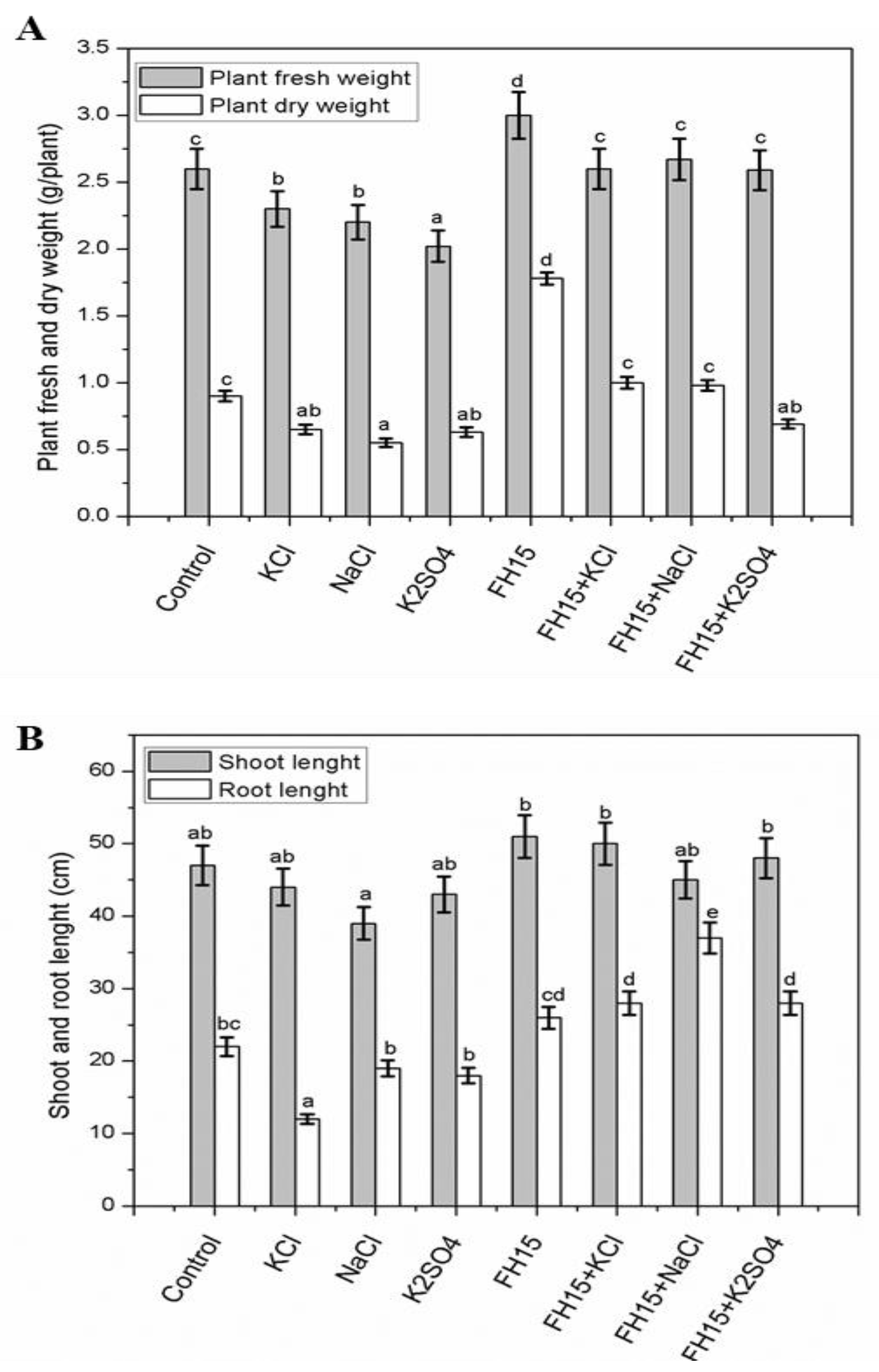
3.4. C. membranifaciens FH15 Augment Maize Plants under Salt Stress

Salinity decreased the morphological and growth attributes of the maize plant, while inoculation of C. membranifaciens FH15 alleviated the salt stress-induced alterations. The plants inoculated with endophytic fungus revealed considerably higher (p > 0.05) plant biomass compare to non-inoculated control plants and the fresh and dry biomass was increased by 20.87% and 16.60%, respectively. Without salinity (KCl, NaCl, and K2SO4) stress, the percent increase in fresh and dry biomass was 15.38% and 13.03%, respectively, upon the inoculation of the endophytic fungus. However, the application of C. membranifaciens FH15 showed its prolific effect and enhanced shoot length (13.55%) under salt stress, and when salt-stressed seedlings were inoculated with the endophytic fungi, restoration of root length was observed in the treated plants. The results also revealed an increase in root length by 18.18% in the treated plants compared to normal control plants, whereas the root length of the endophytic fungus increased by 94.54% under saline conditions (Figure 2A,B).

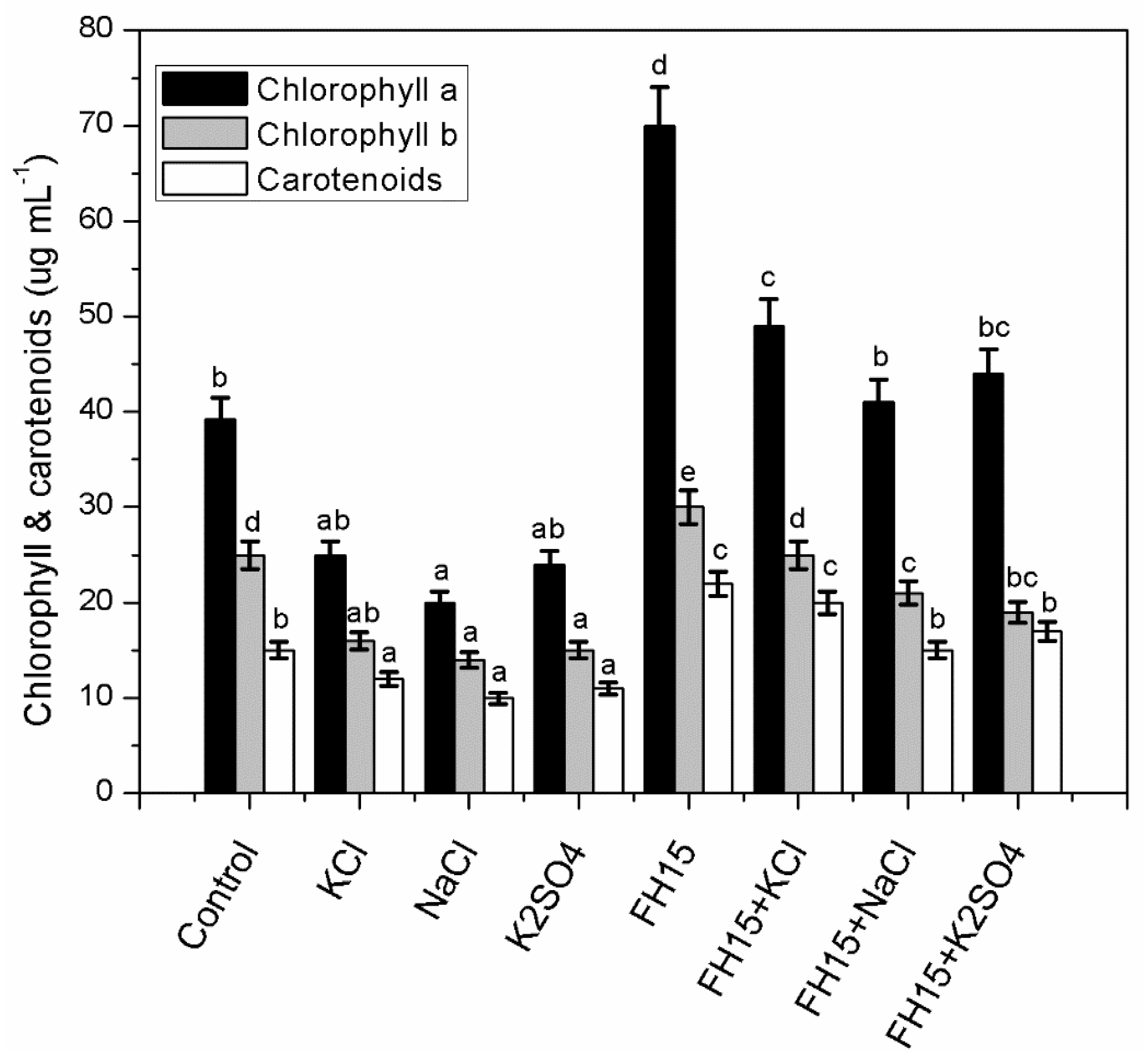
3.5. C. membranifaciens FH15 Improved Chlorophyll and Carotenoids Content in Maize Plants

The percent increase in chlorophyll and carotenoids was 49.29% and 46.67%, respectively, in plants inoculated with C. membranifaciens FH15 as compared to the non-saline control. However, the percent increase in chlorophyll and carotenoids was 69.54% and 57.07%, respectively, when salt-stressed plants were inoculated with endophytic fungus C. membranifaciens FH15. The strain was able to improve pigment concentration under salt stress, suggesting its usefulness as salt-stress alleviator (Figure 3).

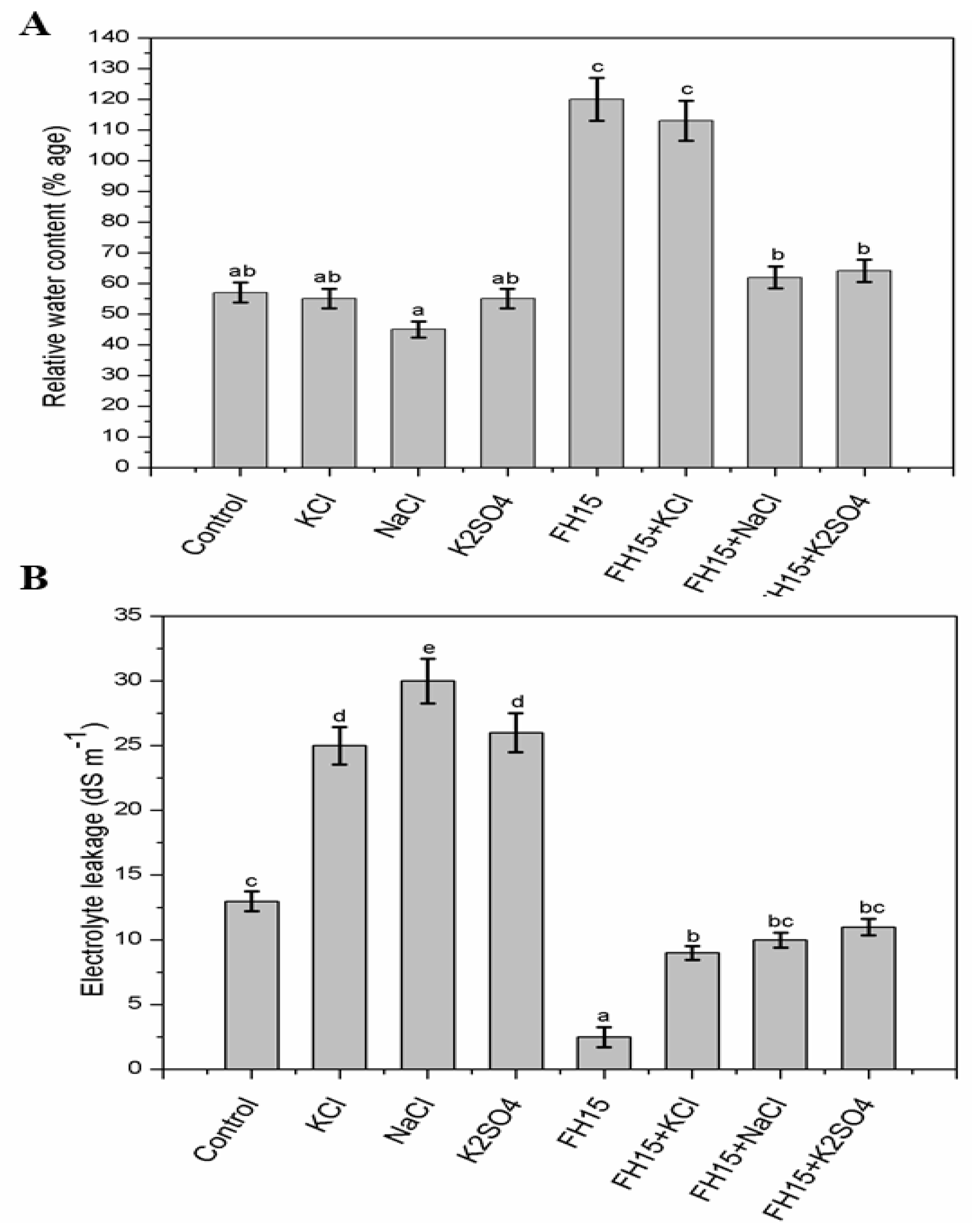
3.6. C. membranifaciens FH15 Improved RWC and Electrolyte Leakage in Maize Plants

Under salinity stress, the RWC of the plants are greatly affected (Figure 4A). However, the application of endophytes to salinity-stressed seedlings enhanced their relative water content values. Increase in RWC was 53.24% under stressed maize plants with C. membranifaciens FH15. The relative water content was increased by 2.5 folds in maize inoculated with C. membranifaciens FH15 as compared to the non-saline control. A significant increase in electrolyte leakage from maize leaf grown under higher (100 mM) salinity (KCl, NaCl, and K2SO4) was observed as compared to the non-saline control (Figure 4B).
A reduction in EL (p < 0.001) was found with application of C. membranifaciens FH15 under all treatments. Percent reduction in EL was 62.79% when salt-treated plants were inoculated with C. membranifaciens FH15. A significant decrease in electrolyte leakage (80.77%) was observed when the non-saline control was inoculated with C. membranifaciens FH15.

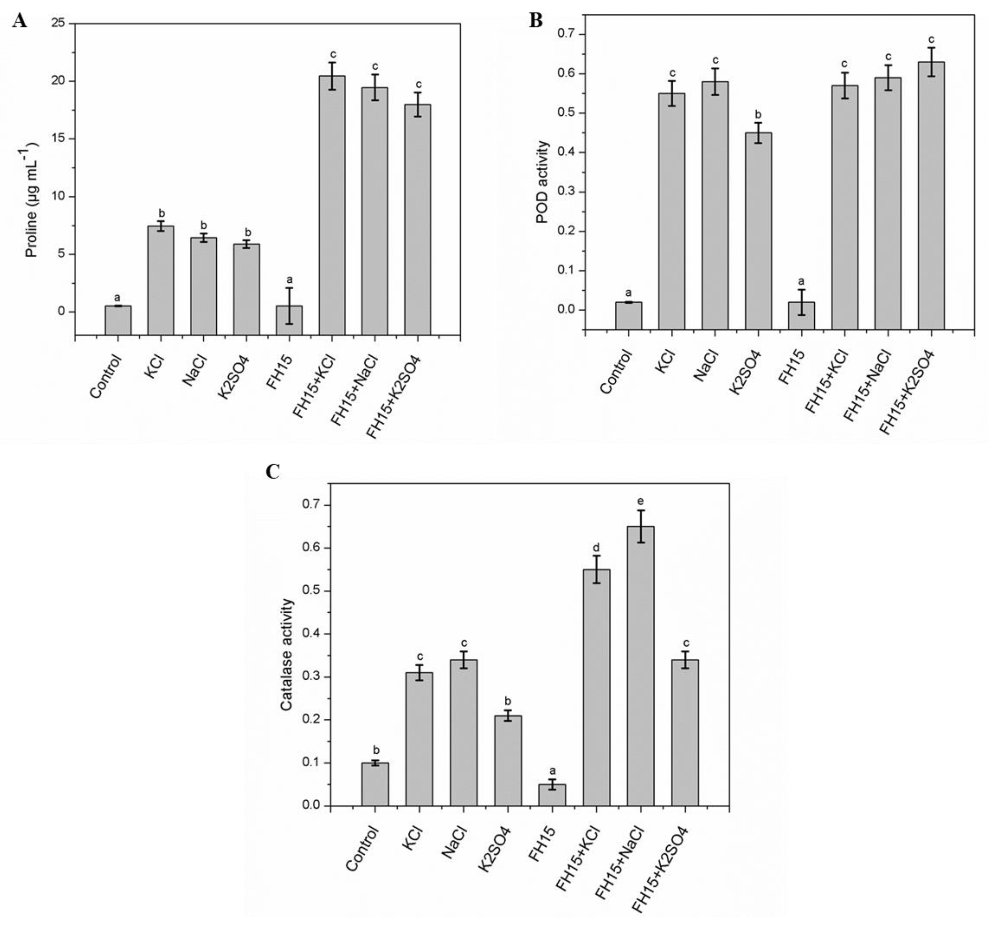
3.7. Effect of C. membranifaciens FH15 on Proline Content and Antioxidant Enzymes System

Proline is an osmoprotectant that accumulates in plants due to salinity stress. In agreement with this, our finding (Figure 5A) revealed that maize with C. membranifaciens FH15 produced a significantly higher level of proline under salinity stress. A 2.9 fold increase in proline content was observed when maize plants were inoculated with C. membranifaciens FH15 under salt stress conditions.
The proline content was decreased several hundred folds when stressed maize was inoculated with C. membranifaciens FH15 as compared to the non-saline control. Similarly, salt stress triggered a significant rise in antioxidant enzymes activities. The percent increase in POD activity was 15.12% when salinity-stressed maize was inoculated with C. membranifaciens FH15 (Figure 5B). Increase in catalase activity was 76.83% when salinity stressed maize was inoculated with C. membranifaciens FH15 (Figure 5C). Catalase and POD activities were decreased by 50% and 90%, respectively, when maize was inoculated with C. membranifaciens FH15 as compared to the non-saline control.

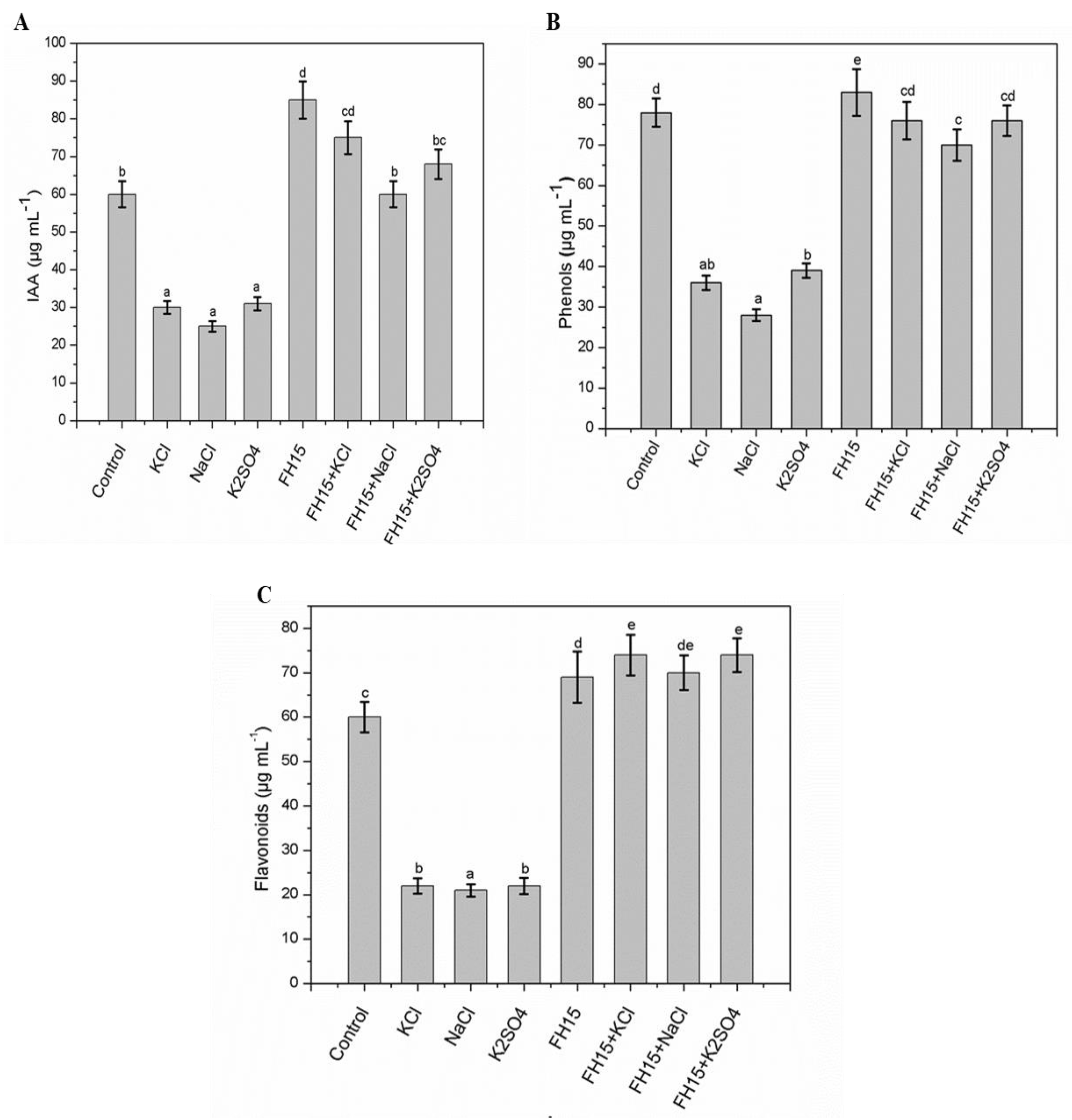
3.8. Effect of C. membranifaciens FH15 on IAA, Total Flavonoids and Phenolics

To understand the effect of the endophytic interaction and its role in salinity stress alleviation, IAA content treated with or without salinity stress was analyzed. The IAA content of maize was increased by 41.67% when inoculated with C. membranifaciens FH15 compared to the non-saline control. A 2.3 fold increase in IAA content in C. membranifaciens FH15-inoculated salinity-stressed maize plants was observed as compared to the salinity-stressed control (Figure 6A). Moreover, a prominent decrease was observed in the phenolic and flavonoid contents of maize plants when subjected to stressful conditions. The average decrease of phenolic content was 34.33 µg/mL, and the average decrease of flavonoids was 21.67 (µg/mL) compared to control without any stress. However, increases of both phenolics and flavonoids were detected upon the inoculation of C. membranifaciens FH15 in the maize plants under stressful conditions. The highest increase, i.e., 76 µg/mL, was observed when maize plants under KCl and K2SO4 stress were inoculated with C. membranifaciens FH15 (Figure 6B,C).

3.9. Identification of Compounds in C. membranifaciens FH15 in the CF by LC-ESI-MS/MS

Utilizing comparative standards data and LC-ESI-MS/MS spectrum data, the active components in the C. membranifaciens extract were identified (Table 2). Presently, compound 1 with t R = 1.6 displayed [M-H]-ive ions at S137.00 and 106.92 m/z fragment ions in its MS 2 spectrum at 137.00 m/z w. After comparing the obtained spectrum data to earlier standard data, molecule 1 was determined to be salicylic acid (Figure S1, Table 2). The results for compound 2 revealed a precursor ion with t R = 3.39 at 269.00 m/z. The substance was categorized as baicalein, since the data were compatible with the structure of baicalein (Table 2, Figure S2). Compound 3’s [M-H]-ive ion displayed t R = 2.58 at 173.00 m/z, and fragments at 115.92 m/z were visible in its MS 2 spectrum. These results were consistent with the structure of aconitic acid (Table 2, Figure S3). Compound 4 (t R= 7.83) displayed a [M-H]-ive ion at 367.00 m/z along with fragment ions at 177.08 m/z. After comparing the spectrum data of compound 4 with the reference standard and literature, compound 4 was identified as feruloylquinic acid (Table 2, Figure S4).
Additionally, the data for compound 5 revealed a precursor ion with t R = 7.79 at 339 m/z. Compound 5’s MS 2 spectra showed fragment ions at 163.00 m/z; the compound was found to be coniferyl aldehyde hexoside (Table 2, Figure S5). Compound 6 revealed a precursor ion at m/z 401.42 when it was eluted at retention time (t R = 5.53). Chemical 6’s MS 2 spectrum also showed a fragment ion at m/z 383. Molecule 6 was identified as Benzyl alcohol hexose pentose, based on the analytical standard, retention time, and MS fragmentation paths (Table 2, Figure S6). When described previously, Compound 7 with t R = 4.55, a distinctive precursor ion at m/z 581.00, and fragment ions at m/z 501.42 was determined to be pentose (Table 2, Figure S7). The molecule was identified as chlorogenic acid by consulting the literature, as the MS/MS of the precursor ion at mass m/z 353 with t R = 7.33 is consistent with the structure of chlorogenic acid (Table 2, Figure S8). Compound 9 was detected at m/z 256.25 and 300.33. Compound 9 was identified as myricetin, based on MS fragmentation routes, the analytical standard, and retention time (Table 2, Figure S9). Compound 10 has a retention time of 4.71 and is shown in Table 2 and Figure S10 to have fragment ions at m/z 322, m/z 296, and m/z 215, as well as a distinctive MS2 fragment with a mass of 340.00. These findings matched the structure of propoxyphene. As a result, when compound 10 was compared to a reference standard and data from the literature, it was determined to be propoxyphene. Compound 11 revealed an [MH] + peak at m/z 284.00 and was eluted at retention time (t R = 3.85). Additionally, fragment ions at m/z 228.17 and 198.08, 184.00, 170.00, 157.00, 143.92, and 129.92 were detected. Compound 11 was identified as aminoflunitrazepam, using MS fragmentation pathways (Table 2, Figure S11).

4. Discussion

New bioactive substances, agrochemicals, and antibiotics which are more effective, maintain low-toxicity, and have less environmental impact are in high demand. The majority of synthetic medications on the market are ineffective and have a number of negative health effects [46]. Thus, there is a universal need to identify and create medicinal compounds, agrochemicals, and antibiotics from endophytic fungi that are highly effective, have low toxicity, and have less or no environmental impact [47]. Endophytic fungi occur asymptomatically and have a symbiotic association with their host plant, which make this connection advantageous for both parties. Mutualism frequently promotes the host’s growth [17]. Currently, a mixture of metabolites in the culture filtrate of our selected fungal isolate were evaluated and screened through a preliminary screening bioassay on waito-c rice.
For maize crops growing on salt-affected land, we endeavored to isolate a halotolerant fungal strain from E. milli L. leaves with the potentiality to readily colonize its host and mitigate salinity stress. The endophytic fungus C. membranifaciens FH15, residing on E. milli L. leaves, was revealed to improve plant growth, produce IAA (evaluated on waito-c rice and maize), and was halotolerant to salinity. Our results revealed that NaCl (100 mM) adversely affects plants’ growth-promoting attributes, while the inoculation of FH15 did not significantly improve the growth under salt stress. Similar results were reported by Hamayun, Hussain, Khan, Kim, Khan, Waqas, Irshad, Iqbal, Rehman, and Jan [26], showing that a higher NaCl stress of 150 mM adversely affected the fungal biomass, reducing it by 26% (fresh biomass) and 29% (dry biomass) relative to fungal strain grown in the control medium, which supports the present data. The strain FH15 also revealed positive results for siderophore production and phosphate solubilization activities. Similar results have been reported by Chadha, Prasad, and Varma [27], showing that fungal endophytes stimulate plant growth through the production of IAA and through their phosphate-solubilizing ability. Salinity stress negatively affects the growing crop plants; symbiotic fungal interactions have shown enhanced plant tolerance to saline environments [48].
The release of plant growth-stimulating substances (IAA and GAs) from endophytic fungi is a source for improved plant growth under abiotic stress environments [49]. Maize plants inoculated with C. membranifaciens FH15 showed an outstanding increase in shoot root length and plant fresh dry weight under salt stress. Inoculation of FH15, in addition to salt stress, alleviated the adverse effect of salt, and a similar result has been obtained by [50] in tomato plants. Previous studies showed that the chlorophyll content of plants is a common indicator of abiotic stress tolerance. Singh and Gautam [51] also reported that plants growing in saline environments had reduced chlorophyll concentration, which led to entire growth retardation. In our present result, we observed that salt stress markedly reduced chlorophyll content in maize plants, however, chlorophyll content was inverted back to the level of the control in maize plants inoculated with FH15.
We also evaluated that chlorophyll content was significantly improved in maize plants due to the inoculation of FH15, with or without the maize plants being exposed to salinity stress. Similar reports were also given by Qi, et al. [52], Rawat, et al. [53], and Zhang, et al. [54], which supports our present study. The strain FH15 enhances the uptake of vital elements, especially (Mg2+), that were adversely affected by salt stress; therefore, the chlorophyll and carotenoid production is enhanced in FH15-treated maize plants. An additional cause for improved pigment content in maize plants might be the synthesis of phytohormones that support the stimulation of pigment contents [55]. The improved pigment contents by FH15 inoculation in maize plants may be due to the inhibition of sodium uptake [56]. Carotenoids exhibit antioxidant abilities and give photo protection to chlorophyll contents by scavenging ROS [57]. Therefore, a reduction in carotenoids content by different salt stresses resulted in an overproduction of ROS that consequently impeded plant growth by stimulating oxidative damage to protein, DNA, and RNA [58].
Phenols and flavonoids are secondary metabolites that act as non-enzymatic antioxidants to scavenge harmful radicals and are essential for plants’ defense [59]. The present results indicated that with salinity stress, the accumulation of phenol and flavonoid contents increases. Therefore, phenol and flavonoid accumulation in salt-resistant crops might be a defensive system meant to scavenge the free radicals of oxygen and inhibit cell membrane damage from salt stress [60]. Our finding of increasing phenol and flavonoid contents with salinity corroborates with the outcomes of [60], which described improved flavonoid and phenol contents in chickpeas under salt stress. The substance that produced a precursor ion at m/z 137.00 during this study’s LC MS/MS observation was identified as salicylic acid.
Salicylic acid is an important phenolic compound that is capable of promoting plant growth and harvesting in certain plants. Moreover, salicylic acid has the ability to affect plant growth, serve as a possible non-enzymatic antioxidant, and significantly influence the synthesis of a number of physiological processes and bioactive molecules in plants [61]. Aconitic acid is a phenolic acid that has two isomers: cis-aconitic acid and trans-aconitic acid. Aconitic acid has a nutty flavor, which makes it valuable as an artificial nut flavor, as shown by [37]. Feruloylquinic acid is phenolic compound that is a potent antioxidant, and has been described to exhibit antiviral, antibacterial, anti-inflammatory, and anti-carcinogenic effects [62]. While a low molecular weight phenolic molecule called coniferylaldehyde hexoside serves as the direct precursor of coniferyl alcohol in the lignin biosynthesis process [63].
Benzyl alcohol hexose pentose is a phenolic compound. Benzyl alcohol is a colorless liquid that exhibits a sharp burning taste and slight odor. It is generally used as a local anesthetic to decrease pain related to Lidocaine injection. Similarly, chlorogenic acids are vital for plant defense mechanisms [64]. Flavonoids are commonly induced by salt stress and have a stimulating role in plant defense [65]. These substances added in plant tissue defend themselves from damaging effects by acting as free radical scavengers due to the presence of hydroxyl groups in their structure. Baicalein is a type of flavonoid that is used against several inflammatory diseases such as nephritis hepatitis, asthma, atopic dermatitis, and bronchitis. In addition, antibacterial, anti-cancer and antiviral activities have been observed. Baicalein’s beneficial effects on human health are also related to its antioxidant properties, as it has good electron and hydrogen donors [66]. A naturally occurring flavonoid is myricetin, commonly referred to as myricetol. The antioxidant abilities of flavonoids are well known, however myricetin stands out as being more effective than other flavonoids in this regard.
Proline is an essential nitrogen source that is accessible for plant retrieval from environmental stress and restoration of plant growth [67]. Proline decreases the uptake of toxic ions [68]. Therefore, proline plays a major role in defending plants from osmotic stress [69]. In our current report, the proline content was improved in maize seedlings grown under salt stress alone, relative to control. Presently, proline was greatly increased in maize plants inoculated with fungal strain C. membranifaciens FH15 under saline environments. The findings of rising proline concentrations in maize plants that had been inoculated with fungal strain FH15 under saline conditions correspond with those of Bagheri, Saadatmand, Niknam, Nejadsatari, and Babaeizad [31]. Under salinity stress, there is an increased generation of ROS in plants, which is thought to be a metabolic change [69]. According to reports, IAA plays a critical function in regulating the primary signaling pathways that contribute to plant development under salt stress [70]. Moreover, IAA plays a key role in the growth and development of the plants, and performs an important function in the root growth [71]. In this work, it was found that plants exposed to salt stress and C. membranifaciens inoculation had significant IAA concentrations.
The increased production of ROS in plants under salt stress is also considered to be a biochemical alteration [72] that is responsible for salinity-induced harm to macro molecules and plants’ cellular structures under stressful conditions [73]. To mitigate the harm related to the increase production of ROS, plants naturally developed a varied series of enzymatic defensive systems in order to detoxify the free radicals, thereby protecting themselves from harmful biochemical (oxidative) damage [74]. The enzymatic antioxidant mechanism is one of the main protective systems in plants; it involves the immediate action of a wide variety of enzymes comprising CAT and POD [75].
The results from the current report determine that salinity stress induced plants to develop increased levels of CAT and POD activity in maize plants relative to un-inoculated plants. In the present result, antioxidant enzymes, higher catalase, and peroxidase activities were detected in FH15-inoculated plants relative to the control plants. Increased catalase activity is related to improved root length and enhanced seedling growth, as publicized by [76]. Similarly, polyphenol oxidase and peroxidase protect cells against the damaging effect of H2O2 by catalyzing its disintegration by the oxidation of phenolic osmolytes [30]. Gusain, et al. [77] revealed that Trichoderma improved the POD and SOD in rice cultivars, granting tolerance to these plants under water stress and different endophytic fungi were reported for the production of enzyme inhibitory metabolites [78].

5. Conclusions

The findings of the present study reveal that plants inoculated with endophytic Candida membranifaciens (FH15) showed low signs of the adverse effects of salinity stress and enhanced the growth parameters of maize plants. Moreover, the fungal endophyte could be used for further field trails on a variety of economically important crop species. However, the pragmatic role of the fungal endophyte still needs to be investigated, particularly the rate of colonization and the give-and-take mechanism of the fungal endophyte and plant root under salt stress conditions.

Supplementary Materials

The following supporting information can be downloaded at: https://www.mdpi.com/article/10.3390/agronomy12102263/s1, Figure S1: Computer reconstructed mass chromatogram for the negative ion electrospray LC ESI-MS/MS analysis of Salicylic acid; Figure S2: Negative product ion mass spectra of [M–H]− of Baicalein, (LC ESI-MS/MS); Figure S3: LC-ES1-MS/MS peak chromatogram of Aconitic acid in aqueous extract of endophytic fungus that were detected in the sample extract in negative ion mode; Figure S4: Computer reconstructed mass chromatogram for the negative ion electrospray LC ESI-MS/MS analysis of Feruloylquinic acid; Figure S5: LC-ES1-MS/MS peak chromatogram of Coniferyl aldehyde hexoside in aqueous extract of endophytic fungus that were detected in the sample extract in negative ionization mode; Figure S6: Computer reconstructed mass chromatogram for the negative ion electrospray LC ESI-MS/MS analysis of Benzyl alcohol hexose pentose; Figure S7: Computer reconstructed mass chromatogram for the negative ion electrospray LC ESI-MS/MS analysis of pentose; Figure S8: Computer-reconstructed mass chromatogram for the negative ion electrospray LC ESI-MS/MS analysis of Chlorogenic acid; Figure S9: Computer reconstructed mass chromatogram for the positive ion electrospray LC ESI-MS/MS analysis of Myricetin; Figure S10: Computer reconstructed mass chromatogram for the positive ion electrospray LC ESI-MS/MS analysis of Propoxyphene; Figure S11: Computer reconstructed mass chromatogram for the positive ion electrospray LC ESI-MS/MS analysis of Aminoflunitrazepam.

Author Contributions

Conceptualization, formal analysis, data curation, F.G.J., M.H. and A.H.; methodology, M.H., S.A.K., and S.A.; software, F.G.J. and A.H.; validation, Investigation, writing—original draft preparation, writing—review & editing, visualization, project administration, resources, G.J., S.A. and G.J.; supervision, M.H., A.H. and G.J., funding acquisition, I.-J.L., and S.A.K. All authors have read and agreed to the published version of the manuscript.

Funding

This work was supported by the National Research Foundation of Korea (NRF) grant funded by the Korea government (MSIT), No. (2022R1A2C1008993).

Institutional Review Board Statement

Not applicable.

Informed Consent Statement

Not applicable.

Data Availability Statement

The data presented in this study are available on request from the corresponding author.

Conflicts of Interest

The authors declare no conflict of interest.

References

  1. Ikram, M.; Ali, N.; Jan, G.; Iqbal, A.; Hamayun, M.; Jan, F.G.; Hussain, A.; Lee, I.-J. Trichoderma reesei improved the nutrition status of wheat crop under salt stress. J. Plant Interact. 2019, 14, 590–602. [Google Scholar] [CrossRef]
  2. Gul, S.L.; Moon, Y.-S.; Hamayun, M.; Khan, S.A.; Iqbal, A.; Khan, M.A.; Hussain, A.; Shafique, M.; Kim, Y.-H.; Ali, S.J.C.M. Porostereum spadiceum-AGH786 Regulates the Growth and Metabolites Production in Triticum aestivum L. Under Salt Stress. Curr. Microbiol. 2022, 79, 159. [Google Scholar] [CrossRef] [PubMed]
  3. Bouzouina, M.; Kouadria, R.; Lotmani, B. Fungal endophytes alleviate salt stress in wheat in terms of growth, ion homeostasis and osmoregulation. J. Appl. Microbiol. 2021, 130, 913–925. [Google Scholar] [CrossRef] [PubMed]
  4. Ali, S.; Khan, S.A.; Hamayun, M.; Iqbal, A.; Khan, A.L.; Hussain, A.; Shah, M. Endophytic fungi from Caralluma acutangula can secrete plant growth promoting enzymes. Fresenius Env. Bull 2019, 28, 2688–2696. [Google Scholar]
  5. Miransari, M. Hyperaccumulators, arbuscular mycorrhizal fungi and stress of heavy metals. Biotechnol. Adv. 2011, 29, 645–653. [Google Scholar] [CrossRef] [PubMed]
  6. Rodriguez, R.J.; Woodward, C.J.; Redman, R.S. Fungal influence on plant tolerance to stress. Biocomplexity Plant-Fungal Interact. 2012, 6, 155–163. [Google Scholar]
  7. Rai, M.; Rathod, D.; Agarkar, G.; Dar, M.; Brestic, M.; Pastore, G.M.; Junior, M.R.M. Fungal growth promotor endophytes: A pragmatic approach towards sustainable food and agriculture. Symbiosis 2014, 62, 63–79. [Google Scholar] [CrossRef]
  8. Zou, W.; Tan, R. Advances in Plant Science; China Higher Education Press: Beijing, China, 1999; Volume 2, pp. 183–190. [Google Scholar]
  9. Souchie, E.L.; Azcón, R.; Barea, J.M.; Saggin-Júnior, O.J.; Silva, E.M.R.D. Phosphate solubilization and synergism between P-solubilizing and arbuscular mycorrhizal fungi. Pesqui. Agropecuária Bras. 2006, 41, 1405–1411. [Google Scholar] [CrossRef]
  10. Haselwandter, K. Structure and function of siderophores produced by mycorrhizal fungi. Mineral. Mag. 2008, 72, 61–64. [Google Scholar] [CrossRef]
  11. Garg, N.; Manchanda, G. ROS generation in plants: Boon or bane? Plant Biosyst. 2009, 143, 81–96. [Google Scholar] [CrossRef]
  12. Ali, S.; Moon, Y.-S.; Hamayun, M.; Khan, M.A.; Bibi, K.; Lee, I.-J. Pragmatic role of microbial plant biostimulants in abiotic stress relief in crop plants. J. Plant Interact. 2022, 17, 705–718. [Google Scholar] [CrossRef]
  13. Waqas, M.; Khan, A.L.; Kamran, M.; Hamayun, M.; Kang, S.-M.; Kim, Y.-H.; Lee, I.-J. Endophytic fungi produce gibberellins and indoleacetic acid and promotes host-plant growth during stress. Molecules 2012, 17, 10754–10773. [Google Scholar] [CrossRef] [PubMed] [Green Version]
  14. Arnold, A.E. Endophytic fungi: Hidden Components of Tropical Community Ecology. In Tropical Forest Community Ecology; Carson, W.P., Schnitzer, S.A., Eds.; Blackwell Publishing Ltd: West Sussex, UK, 2008; pp. 178–188. [Google Scholar]
  15. Harper, J.K.; Arif, A.M.; Ford, E.J.; Strobel, G.A.; Porco, J.A., Jr.; Tomer, D.P.; Oneill, K.L.; Heider, E.M.; Grant, D.M. Pestacin: A 1, 3-dihydro isobenzofuran from Pestalotiopsis microspora possessing antioxidant and antimycotic activities. Tetrahedron 2003, 59, 2471–2476. [Google Scholar] [CrossRef]
  16. Sannigrahi, S.; Mazumder, U.K.; Pal, D.; Mishra, S.L. Hepatoprotective potential of methanol extract of Clerodendrum infortunatum Linn. against CCl4 induced hepatotoxicity in rats. Pharmacogn. Mag. 2009, 5, 394. [Google Scholar]
  17. Khan, S.A.; Hamayun, M.; Yoon, H.; Kim, H.-Y.; Suh, S.-J.; Hwang, S.-K.; Kim, J.-M.; Lee, I.-J.; Choo, Y.-S.; Yoon, U.-H. Plant growth promotion and Penicillium citrinum. BMC Microbiol. 2008, 8, 231. [Google Scholar] [CrossRef] [PubMed]
  18. Waller, F.; Achatz, B.; Baltruschat, H.; Fodor, J.; Becker, K.; Fischer, M.; Heier, T.; Hückelhoven, R.; Neumann, C.; Von Wettstein, D. The endophytic fungus Piriformospora indica reprograms barley to salt-stress tolerance, disease resistance, and higher yield. Proc. Natl. Acad. Sci. USA 2005, 102, 13386–13391. [Google Scholar] [CrossRef] [PubMed]
  19. Shinozaki, K.; Yamaguchi-Shinozaki, K. Gene networks involved in drought stress response and tolerance. J. Exp. Bot. 2007, 58, 221–227. [Google Scholar] [CrossRef]
  20. Yuan, Z.-l.; Zhang, C.-l.; Lin, F.-c. Role of diverse non-systemic fungal endophytes in plant performance and response to stress: Progress and approaches. J. Plant Growth Regul. 2010, 29, 116–126. [Google Scholar] [CrossRef]
  21. Khan, A.L.; Hamayun, M.; Radhakrishnan, R.; Waqas, M.; Kang, S.-M.; Kim, Y.-H.; Shin, J.-H.; Choo, Y.-S.; Kim, J.-G.; Lee, I.-J. Mutualistic association of Paecilomyces formosus LHL10 offers thermotolerance to Cucumis sativus. Antonie Van Leeuwenhoek 2012, 101, 267–279. [Google Scholar] [CrossRef]
  22. Strobel, G.A. Endophytes as sources of bioactive products. Microbes Infect. 2003, 5, 535–544. [Google Scholar] [CrossRef]
  23. Mehdi, S.; Ahsan, M. Genetic coefficient of variation, relative expected genetic advance and inter-relationships in maize (Zea mays L.) for green fodder purposes at seedling stage. Pak. J. Biol. Sci. 2000, 3, 1890–1891. [Google Scholar]
  24. Photita, W.; Lumyong, S.; Lumyong, P.; McKenzie, E.; Hyde, K. Are some endophytes of Musa acuminata latent pathogens? Fungal Divers. 2004, 16, 131–140. [Google Scholar]
  25. Jan, F.G.; Hamayun, M.; Moon, Y.-S.; Jan, G.; Shafique, M.; Ali, S.J.B. Endophytic aspergillus oryzae reprograms Abelmoschus esculentus L. to higher growth under salt stress via regulation of physiochemical attributes and antioxidant system. Biologia 2022, 1–14. [Google Scholar] [CrossRef]
  26. Hamayun, M.; Hussain, A.; Khan, S.A.; Kim, H.-Y.; Khan, A.L.; Waqas, M.; Irshad, M.; Iqbal, A.; Rehman, G.; Jan, S. Gibberellins producing endophytic fungus Porostereum spadiceum AGH786 rescues growth of salt affected soybean. Front. Microbiol. 2017, 8, 686. [Google Scholar] [CrossRef] [PubMed]
  27. Chadha, N.; Prasad, R.; Varma, A. Plant Promoting Activities of Fungal Endophytes Associated with Tomato Roots from Central Himalaya, India and Their Interaction with Piriformospora Indica. IJPBS 2015, 6, 333–343. [Google Scholar]
  28. Bhalodia, N.R.; Acharya, R.; Shukla, V. Evaluation of in vitro Antioxidant Activity of hydroalcoholic seed extratcs of Cassia fistula linn. Free Radic. Antioxid. 2011, 1, 68–76. [Google Scholar] [CrossRef]
  29. Akbay, P.; Basaran, A.A.; Undeger, U.; Basaran, N. In vitro immunomodulatory activity of flavonoid glycosides from Urtica dioica L. Phytother. Res. Int. J. Devoted Pharmacol. Toxicol. Eval. Nat. Prod. Deriv. 2003, 17, 34–37. [Google Scholar] [CrossRef]
  30. Khan, A.L.; Waqas, M.; Hamayun, M.; Al-Harrasi, A.; Al-Rawahi, A.; Lee, I.-J. Co-synergism of endophyte Penicillium resedanum LK6 with salicylic acid helped Capsicum annuum in biomass recovery and osmotic stress mitigation. BMC Microbiol. 2013, 13, 51. [Google Scholar] [CrossRef] [PubMed]
  31. Bagheri, A.A.; Saadatmand, S.; Niknam, V.; Nejadsatari, T.; Babaeizad, V. Effect of endophytic fungus, Piriformospora indica, on growth and activity of antioxidant enzymes of rice (Oryza sativa L.) under salinity stress. Int. J. Adv. Biol. Biomed. Res. 2013, 1, 1337–1350. [Google Scholar]
  32. Sherameti, I.; Shahollari, B.; Venus, Y.; Altschmied, L.; Varma, A.; Oelmüller, R. The endophytic fungus Piriformospora indica stimulates the expression of nitrate reductase and the starch-degrading enzyme glucan-water dikinase in tobacco and Arabidopsis roots through a homeodomain transcription factor that binds to a conserved motif in their promoters. J. Biol. Chem. 2005, 280, 26241–26247. [Google Scholar]
  33. Gurupavithra, S.; Jayachitra, A. Isolation and identification of endophytic fungi from Ocimum sanctum and analyse its antioxidant properties. Int. J. Resear. Pharmaceut. Biomed. Sci 2013, 4, 1120–1125. [Google Scholar]
  34. Zhishen, J.; Mengcheng, T.; Jianming, W. The determination of flavonoid contents in mulberry and their scavenging effects on superoxide radicals. Food Chem. 1999, 64, 555–559. [Google Scholar] [CrossRef]
  35. Khan, I.; Rahman, H.; El-Salam, N.M.A.; Tawab, A.; Hussain, A.; Khan, T.A.; Khan, U.A.; Qasim, M.; Adnan, M.; Azizullah, A. Punica granatum peel extracts: HPLC fractionation and LC MS analysis to quest compounds having activity against multidrug resistant bacteria. BMC Complementary Altern. Med. 2017, 17, 247. [Google Scholar] [CrossRef] [PubMed]
  36. Jebamony, J.R.; Thomas Muthiah, P. 8-Hydroxyquinolinium–Salicylate–Salicylic Acid (1/1/1) Complex, C9H8NO+. C7H5O3−. C7H6O3. Acta Crystallogr. Sect. C Cryst. Struct. Commun. 1998, 54, 539–540. [Google Scholar] [CrossRef]
  37. Jiang, C.; Liang, G.; Ren, Y.; Xu, T.; Song, Y.; Jin, W. An UPLC-MS/MS method for simultaneous quantification of the components of Shenyanyihao oral solution in rat plasma. BioMed Res. Int. 2020, 2020, 4769267. [Google Scholar] [CrossRef]
  38. Said, A.; Aboutabl, E.A.; Melek, F.R.; Abdel Jaleel Raheem Abdel Jaleel, G.; Raslan, M. Phytoconstituents profiling of Cissus rotundifolia (Forssk.) Vahl. by HPLC-MS/MS, and evaluation of its free radical scavenging activity (DPPH) and cytotoxicity. Trends Phytochem. Res. 2018, 2, 65–74. [Google Scholar]
  39. Ghareeb, M.A.; Sobeh, M.; Rezq, S.; El-Shazly, A.M.; Mahmoud, M.F.; Wink, M. HPLC-ESI-MS/MS profiling of polyphenolics of a leaf extract from Alpinia zerumbet (Zingiberaceae) and its anti-inflammatory, anti-nociceptive, and antipyretic activities in vivo. Molecules 2018, 23, 3238. [Google Scholar] [CrossRef] [PubMed]
  40. Sisó-Terraza, P.; Luis-Villarroya, A.; Fourcroy, P.; Briat, J.-F.; Abadía, A.; Gaymard, F.; Abadía, J.; Álvarez-Fernández, A. Accumulation and secretion of coumarinolignans and other coumarins in Arabidopsis thaliana roots in response to iron deficiency at high pH. Front. Plant Sci. 2016, 7, 1711. [Google Scholar] [CrossRef]
  41. Bilia, A.R.; Rubio, M.d.M.E.; Alvarez, M.L.; Morelli, I.; Gonzalez, J.M. New benzyl alcohol glycosides from Pyrus bourgaeana. Planta Med. 1994, 60, 569–571. [Google Scholar] [CrossRef]
  42. Beelders, T.; De Beer, D.; Stander, M.A.; Joubert, E. Comprehensive phenolic profiling of Cyclopia genistoides (L.) Vent. by LC-DAD-MS and-MS/MS reveals novel xanthone and benzophenone constituents. Molecules 2014, 19, 11760–11790. [Google Scholar] [CrossRef] [PubMed]
  43. Koolen, H.H.F.; da Silva, F.M.A.; Gozzo, F.C.; de Souza, A.Q.L.; de Souza, A.D.L. Antioxidant, antimicrobial activities and characterization of phenolic compounds from buriti (Mauritia flexuosa L. f.) by UPLC–ESI-MS/MS. Food Res. Int. 2013, 51, 467–473. [Google Scholar] [CrossRef]
  44. Bonta, R.K. Dietary phenolic acids and flavonoids as potential anti-cancer agents: Current state of the art and future perspectives. Anti Cancer Agents Med. Chem. 2020, 20, 29–48. [Google Scholar] [CrossRef] [PubMed]
  45. Cao, Z.; Kaleta, E.; Wang, P. Simultaneous quantitation of 78 drugs and metabolites in urine with a dilute-and-shoot LC–MS-MS assay. J. Anal. Toxicol. 2015, 39, 335–346. [Google Scholar] [CrossRef] [PubMed]
  46. Tiwari, K. The future products: Endophytic fungal metabolites. J. Biodivers. Bio Prospect. Dev. 2015, 2, 2–7. [Google Scholar]
  47. Tenguria, R.K.; Khan, F.N.; Quereshi, S. Endophytes-mines of pharmacological therapeutics. World J. Sci. Technol. 2011, 1, 127–149. [Google Scholar]
  48. Porcel, R.; Aroca, R.; Ruiz-Lozano, J.M. Salinity stress alleviation using arbuscular mycorrhizal fungi. A review. Agron. Sustain. Dev. 2012, 32, 181–200. [Google Scholar] [CrossRef]
  49. Khan, A.L.; Hamayun, M.; Kim, Y.-H.; Kang, S.-M.; Lee, I.-J. Ameliorative symbiosis of endophyte (Penicillium funiculosum LHL06) under salt stress elevated plant growth of Glycine max L. Plant Physiol. Biochem. 2011, 49, 852–861. [Google Scholar] [CrossRef]
  50. Mastouri, F.; Björkman, T.; Harman, G.E. Trichoderma harzianum enhances antioxidant defense of tomato seedlings and resistance to water deficit. Mol. Plant-Microbe Interact. 2012, 25, 1264–1271. [Google Scholar] [CrossRef]
  51. Singh, P.K.; Gautam, S. Role of salicylic acid on physiological and biochemical mechanism of salinity stress tolerance in plants. Acta Physiol. Plant. 2013, 35, 2345–2353. [Google Scholar] [CrossRef]
  52. Qi, W.-Z.; Liu, H.-H.; Liu, P.; Dong, S.-T.; Zhao, B.-Q.; So, H.B.; Li, G.; Liu, H.-D.; Zhang, J.-W.; Zhao, B. Morphological and physiological characteristics of corn (Zea mays L.) roots from cultivars with different yield potentials. Eur. J. Agron. 2012, 38, 54–63. [Google Scholar] [CrossRef]
  53. Rawat, L.; Singh, Y.; Shukla, N.; Kumar, J. Alleviation of the adverse effects of salinity stress in wheat (Triticum aestivum L.) by seed biopriming with salinity tolerant isolates of Trichoderma harzianum. Plant Soil 2011, 347, 387–400. [Google Scholar] [CrossRef]
  54. Zhang, F.; Yuan, J.; Yang, X.; Cui, Y.; Chen, L.; Ran, W.; Shen, Q. Putative Trichoderma harzianum mutant promotes cucumber growth by enhanced production of indole acetic acid and plant colonization. Plant Soil 2013, 368, 433–444. [Google Scholar] [CrossRef]
  55. Paixão Resende, M.; Jakoby, I.C.M.C.; dos Santos, L.C.R.; Soares, M.A.; Pereira, F.D.; Souchie, E.L.; Silva, F.G. Phosphate solubilization and phytohormone production by endophytic and rhizosphere Trichoderma isolates of guanandi (Calophyllum brasiliense Cambess). Afr. J. Microbiol. Res. 2014, 8, 2616–2623. [Google Scholar]
  56. Iqbal, M.; Ashraf, M. Alleviation of salinity-induced perturbations in ionic and hormonal concentrations in spring wheat through seed preconditioning in synthetic auxins. Acta Physiol. Plant. 2013, 35, 1093–1112. [Google Scholar] [CrossRef]
  57. Behera, R.K.; Mishra, P.C.; Choudhury, N.K. High irradiance and water stress induce alterations in pigment composition and chloroplast activities of primary wheat leaves. J. Plant Physiol. 2002, 159, 967–973. [Google Scholar] [CrossRef]
  58. Ahmad, P.; Jaleel, C.; Sharma, S. Antioxidant defense system, lipid peroxidation, proline-metabolizing enzymes, and biochemical activities in two Morus alba genotypes subjected to NaCl stress. Russ. J. Plant Physiol. 2010, 57, 509–517. [Google Scholar] [CrossRef]
  59. Sarwat, M.; Hashem, A.; Ahanger, M.A.; Abd_Allah, E.F.; Alqarawi, A.; Alyemeni, M.N.; Ahmad, P.; Gucel, S. Mitigation of NaCl stress by arbuscular mycorrhizal fungi through the modulation of osmolytes, antioxidants and secondary metabolites in mustard (Brassica juncea L.) plants. Front. Plant Sci. 2016, 7, 869. [Google Scholar] [CrossRef]
  60. Singh, A. The physiology of salt tolerance in four genotypes of chickpea during germination. J. Agric. Sci. 2018, 6, 87–93. [Google Scholar]
  61. Amanullah, M.M.; Sekar, S.; Vincent, S. Plant growth substances in crop production: A review. Asian J. Plant Sci. 2010, 9, 215–222. [Google Scholar] [CrossRef]
  62. Chen, F.; Long, X.; Liu, Z.; Shao, H.; Liu, L. Analysis of phenolic acids of Jerusalem artichoke (Helianthus tuberosus L.) responding to salt-stress by liquid chromatography/tandem mass spectrometry. Sci. World J. 2014, 2014, 568043. [Google Scholar] [CrossRef]
  63. Tsuji, Y.; Chen, F.; Yasuda, S.; Fukushima, K. Unexpected behavior of coniferin in lignin biosynthesis of Ginkgo biloba L. Planta 2005, 222, 58–69. [Google Scholar] [CrossRef] [PubMed]
  64. Wianowska, D.; Gil, M. Recent advances in extraction and analysis procedures of natural chlorogenic acids. Phytochem. Rev. 2019, 18, 273–302. [Google Scholar] [CrossRef]
  65. Grace, S.C.; Logan, B.A. Energy dissipation and radical scavenging by the plant phenylpropanoid pathway. Philos. Trans. R. Soc. London. Ser. B Biol. Sci. 2000, 355, 1499–1510. [Google Scholar] [CrossRef]
  66. Moghaddam, E.; Teoh, B.-T.; Sam, S.-S.; Lani, R.; Hassandarvish, P.; Chik, Z.; Yueh, A.; Abubakar, S.; Zandi, K. Baicalin, a metabolite of baicalein with antiviral activity against dengue virus. Sci. Rep. 2014, 4, 5452. [Google Scholar] [CrossRef] [PubMed] [Green Version]
  67. Alenazi, M.; Egamberdieva, D.; Ahmad, P. Arbuscular mycorrhizal fungi mitigates NaCl induced adverse effects on Solanum lycopersicum L. Pak. J. Bot 2015, 47, 327–340. [Google Scholar]
  68. Woodward, A.J.; Bennett, I.J. The effect of salt stress and abscisic acid on proline production, chlorophyll content and growth of in vitro propagated shoots of Eucalyptus camaldulensis. Plant Cell Tissue Organ Cult. 2005, 82, 189–200. [Google Scholar] [CrossRef]
  69. Khan, M.N.; Siddiqui, M.H.; Mohammad, F.; Naeem, M.; Khan, M.M.A. Calcium chloride and gibberellic acid protect linseed (Linum usitatissimum L.) from NaCl stress by inducing antioxidative defence system and osmoprotectant accumulation. Acta Physiol. Plant. 2010, 32, 121–132. [Google Scholar] [CrossRef]
  70. Herrera-Medina, M.J.; Steinkellner, S.; Vierheilig, H.; Ocampo Bote, J.A.; García Garrido, J.M. Abscisic acid determines arbuscule development and functionality in the tomato arbuscular mycorrhiza. New Phytol. 2007, 175, 554–564. [Google Scholar] [CrossRef]
  71. Khan, A.L.; Hamayun, M.; Kang, S.-M.; Kim, Y.-H.; Jung, H.-Y.; Lee, J.-H.; Lee, I.-J. Endophytic fungal association via gibberellins and indole acetic acid can improve plant growth under abiotic stress: An example of Paecilomyces formosus LHL10. BMC Microbiol. 2012, 12, 3. [Google Scholar] [CrossRef]
  72. Younis, M.E.; Hasaneen, M.N.; Kazamel, A.M. Exogenously applied ascorbic acid ameliorates detrimental effects of NaCl and mannitol stress in Vicia faba seedlings. Protoplasma 2010, 239, 39–48. [Google Scholar] [CrossRef]
  73. Özdemir, F.; Bor, M.; Demiral, T.; Türkan, İ. Effects of 24-epibrassinolide on seed germination, seedling growth, lipid peroxidation, proline content and antioxidative system of rice (Oryza sativa L.) under salinity stress. Plant Growth Regul. 2004, 42, 203–211. [Google Scholar] [CrossRef]
  74. Li, J.-T.; Qiu, Z.-B.; Zhang, X.-W.; Wang, L.-S. Exogenous hydrogen peroxide can enhance tolerance of wheat seedlings to salt stress. Acta Physiol. Plant. 2011, 33, 835–842. [Google Scholar] [CrossRef]
  75. Ma, L.; Li, Y.; Yu, C.; Wang, Y.; Li, X.; Li, N.; Chen, Q.; Bu, N. Alleviation of exogenous oligochitosan on wheat seedlings growth under salt stress. Protoplasma 2012, 249, 393–399. [Google Scholar] [CrossRef] [PubMed]
  76. Harman, G.E. Multifunctional fungal plant symbionts: New tools to enhance plant growth and productivity. New Phytol. 2011, 189, 647–649. [Google Scholar] [CrossRef]
  77. Gusain, Y.S.; Singh, U.; Sharma, A. Enhance activity of stress related enzymes in rice (Oryza sativa L.) induced by plant growth promoting fungi under drought stress. Afr. J. Agric. Res. 2014, 9, 1430–1434. [Google Scholar]
  78. Ali, S.; Khan, A.L.; Ali, L.; Rizvi, T.S.; Khan, S.A.; Hussain, J.; Al-Harrasi, A. Enzyme inhibitory metabolites from endophytic Penicillium citrinum isolated from Boswellia sacra. Arch. Microbiol. 2017, 199, 691–700. [Google Scholar] [CrossRef]
Figure 1. Molecular identification of the FH15. The analysis included 21 nucleotide sequences using the Neighbor joining tree method according to the model from Tamura. Evolutionary analysis has been carried out in MEGA-7. Our new fungal strain C. membranifaciens strongly recommended as a new strain, the 98% bootstrap support for C. membranifaciens isolate FH15.
Agronomy 12 02263 g001
Figure 2. Impact of various salts on maize (A) fresh and dry biomass and (B) shoot and root length with or without endophytic fungal strain C. membranifaciens FH15 inoculation. The bars represent mean with ± SE and the different letters on the bars represent significance at p = 0.05.
Agronomy 12 02263 g002
Figure 3. Impact of various salts on total chlorophyll and carotenoids contents of maize with or without endophytic fungal strain C. membranifaciens FH15. The bars represent mean with ± SE and the different letters on the bars represent significance at p = 0.05.
Agronomy 12 02263 g003
Figure 4. Effect of different salts on (A) relative water content and (B) electrolyte leakage of maize with or without endophytic fungal strain C. membranifaciens FH15. The bars represent mean with ± SE and the different letters on the bars represent significance at p = 0.05.
Agronomy 12 02263 g004
Figure 5. Effect of different salts on endogenous (A) proline content (B) peroxidase and (C) Catalase activity of maize with or without endophytic fungal strain C. membranifaciens FH15. The bars represent mean with ± SE and the different letters on the bars represent significance at p = 0.05.
Agronomy 12 02263 g005
Figure 6. Impact of various salts on endogenous (A) IAA content (B) phenols and (C) flavonoids contents of maize with or without endophytic fungal strain C. membranifaciens FH15. The bars represent mean with ± SE and the different letters on the bars represent significance at p = 0.05.
Agronomy 12 02263 g006
Table 1. Salt resistance of selected fungal isolate (FH15) and their effect on biomass, IAA, phenols and flavonoids content under salinity stress. The bars represent mean with ± SE and the different letters on the bars represent significance at p = 0.05.
Fungal Fresh BiomassFungal Dry BiomassIAA (µg/mL)Phenols (µg/mL)Flavonoids (µg/mL)
Control3.97 ± 0.2 b1.9 ± 0.2 c21 ± 1.2 c50 ± 2.8 c40 ± 2.3 b
100 mM NaCl3.7 ± 0.2 b1.8 ± 0.1 c35.8 ± 1.3 d70 ± 3.2 d50 ± 2.5 c
150 mM NaCl1.1 ± 0.1 a0.6 ± 0.1 b8 ± 0.5 b20 ± 1.1 b11 ± 0.6 a
200 mM NaCl1 ± 0.1 a0.1 ± 0.1 a2 ± 0.1 a9 ± 0.5 a7 ± 0.4 a
Table 2. Characterization of chemical constituents in the extract of C. membranifaciens FH15 by LC-ES1-MS/MS method.
NOt R
(min)
Proposed
Formula
ModePrecursor Ion, m/zLC-ES1-MS/MS IonsIdentificationReferences
11.66C7H6O3_[M-H] − 137106.92, 92.92Salicylic acid Bduhafsdun et al., 2018 [36]
23.39C15H10O5_[M-H] − 269267, 251, 225, 223, 209, 197, 195, 181, 167, 154BaicaleinSoraia et al., 2009 [37]
32.58C6H6O6_[M-H] − 173115.92, 128.83Aconitic acidSoraia et al., 2009 [38]
47.83C17H20O9_[M-H] − 367177.08Feruloylquinic acidGhareeb et al., 2018 [39]
57.79C16H20O8_[M-H] − 339163.00, 132.92Coniferyl aldehyde hexosideTerraza et al., 2016 [40]
65.53C18H26O10_[M-H] − 401383, 365, 357, 344, 321, 284, 260, 241, 213, 197, 176, 144Benzyl alcohol hexose pentoseBystroma et al., 2008 [41]
78.26C26H29O15_[M-H] − 581501.42PentoseBeelders et al., 2014 [42]
87.33C16H18O9_[M-H] − 353177.00, 163.00Chlorogenic acidKoolen et al., 2013 [43]
94.55C15H10O8+[M+H] + 318300.33, 256.25MyricetinBonta, 2017 [44]
104.71C22H29NO2+[M+H] + 340322, 296, 215, 284, 312PropoxypheneCao et al., 2015 [45]
113.85C16H14FN3O+[M+H] + 284228.17, 198.08, 184.00, 170.00, 157.00, 143.92, 129.92AminoflunitrazepamCao et al., 2015 [45]
Publisher’s Note: MDPI stays neutral with regard to jurisdictional claims in published maps and institutional affiliations.

