Article

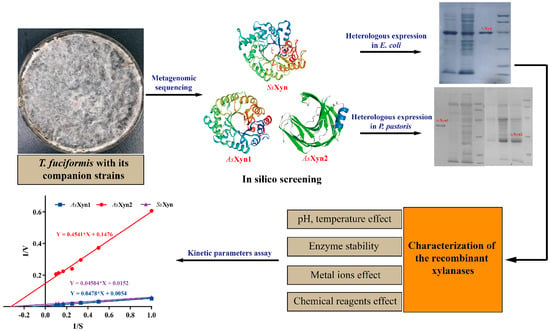
Mining, Identification, and Characterization of Three Xylanases from the Microbiota of T. fuciformis with Its Companion Strains

1
College of Food Engineering, Zhangzhou Institute of Technology, Zhangzhou 363000, China
2
College of Life Sciences, Fujian Agriculture and Forestry University, Fuzhou 350002, China
3
Institute of Edible Fungi, Fujian Academy of Agricultural Sciences, Fuzhou 350012, China
*
Authors to whom correspondence should be addressed.
Catalysts 2024, 14(1), 15; https://doi.org/10.3390/catal14010015
Submission received: 20 November 2023 / Revised: 19 December 2023 / Accepted: 19 December 2023 / Published: 24 December 2023
(This article belongs to the Special Issue Microbial Biocatalysis, 2nd Edition)

Abstract

:
Microbial xylanase has wide application in bioenergy, animal feed, environmental protection, the pulp and paper industry, and agricultural development. In this study, three xylanases from the microbiota of T. fuciformis with its companion strains were identified by metagenomics sequencing. The three enzymes were subjected to cloning and expression in E. coli or P. pastoris, purification, and characterization for their properties. The results showed that AsXyn1, from Annulohypoxylon stygium, among the three enzymes possessed high thermostability at 40 °C and broad pH tolerance in the range of 2.0–10.0, exhibiting its application potential. Furthermore, it was found that post-translational modification (such as glycosylation) of AsXyn1 enzyme modulated its activity, kinetic parameters, and thermostability. These results and findings provided a hint for enzyme modification and design in future.

Graphical Abstract

1. Introduction

Lignocellulosic biomass is the most abundant polysaccharide-containing renewable resource on earth, and mainly consists of a mixture of various complex carbohydrates, namely cellulose, hemicellulose, and lignin [1]. Xylan, as a typical hemicellulose composition, constitutes about one-third of the total dry weight in lignocellulosic biomass [2]. It is a heteroglycan constituted by β-1,4-linked xylose residues with side branches of α-glucuronic acid and α-arabinofuranose. In order to complete the conversion of xylan, an array of carbohydrate-active enzymes (CAZymes) is required, called xylanolytic enzymes [3]. These enzymes are mostly extracellular, are produced mainly by various microorganisms, including bacteria and fungi, which act in a concerted fashion to degrade xylan into its individual monosaccharide components. Of these, xylanase (endo-1,4-β-xylanase, EC 3.2.1.8) is one of the crucial biocatalysts to cleave the β-1,4-glycosidic bonds of the xylan backbone to produce xylooligosaccharides of different lengths [4].
Microbial xylanase has been widely applied in bioenergy, animal feed, environmental protection, the pulp and paper industry, and agricultural development [5]. Therefore, xylanase production with a higher yield and activity at a low cost is desired and required. In this respect, the isolation and identification of new genes encoding xylanases from some novel microbial sources represent alternative methods. Xylanases are widespread in nature, and have been reported in many microorganisms. Among microbial sources, filamentous fungi are relevant in that they secrete extracellular xylanases at higher levels than those observed in yeasts and bacteria. Tremella fuciformis, also known as white jelly mushroom, is an important edible and medicinal fungus containing abundant biologically active polysaccharides [6]. During its cultivation process, the growth and development of T. fuciformis fruiting bodies is extremely dependent on exogenous nutrients supplied by its companion Annulohypoxylon species via degrading biomass substrate. Among the companion strains, the fungus of Annulohypoxylon stygium is the preferred one, which belongs to Xylariaceae, Ascomycota [7,8]. The companion strains, especially A. stygium, have been shown to perform well in lignin and carbohydrate degradation with various glycohydrolases, including pectinases, xylanases, and β-glucanases [9,10]. Moreover, it is indicated that the xylanases secreted by companion Annulohypoxylon species possess higher activity in comparison to those from other edible fungi [11]. However, the genes encoding the xylanases from companion Annulohypoxylon species have yet to be revealed.
T. fuciformis, with its companion strains, constitutes one microbiota in which the majority of these microorganisms have not yet been isolated and identified, thus resulting in a limit on the mining of the xylanase genes. Metagenome sequencing and screening are effective tools, and can be used to search for and identify some novel target enzymes [12]. Several novel xylanases have been identified by metagenomics studies of camel rumen, chicken cecum, termite hindgut, and cow manure [13,14,15,16]. Furthermore, heterologous expressions of these xylanases identified in different hosts by genetic engineering technology could efficiently improve the xylanase yields and exhibit their high thermostability, catalytic efficiency, and organic solvent tolerance [17].
In this study, the microbiota of T. fuciformis with its companion strains were used to perform metagenomics sequencing, and the metagenomics data were used to mine novel xylanase enzymes. Three xylanase candidates were screened and predicted based on genomic annotation in combination with their three-dimensional structure modeling. Furthermore, heterologous expression in two different hosts of E. coli and P. pastoris confirmed that the three candidate enzymes possessed xylanase activities. The three recombinant enzymes were purified and characterized for their enzymatic properties. This is the first report on the xylanase enzymes derived from T. fuciformis with its companion strains.

2. Results and Discussion

2.1. Analysis of Metagenomic Sequencing

In our previous study, T. fuciformis with its companion strains showed a stronger ability to secrete xylanase than other edible fungi. However, the majority of the microbiota have not yet been isolated and identified, which resulting in the difficulty of identifying these xlyanase genes. Herein, the metagenomic sequencing method was used to search the potential xylanase enzymes in the microbiota of T. fuciformis and its companion strains. The metagenomics DNA was extracted from the mycelium of T. fuciformis with its companion strains by the CTAB method, and the extracted DNA quality was assessed by agarose gel electrophoresis. As shown in Figure 1A, the extracted DNA band was clear and bright, with a size of more than 8000 bp, and no tailing could be observed, suggesting that the metagenomic DNA quality satisfied the sequencing demand. The raw data of metagenomics sequencing generated 62,890,030 reads, which were treated via assembly, CDS prediction, and redundancy elimination. The sum of the unigenes with full open reading frames reached 50,904, which occupied 72.17% of the total unigenes and exhibited good fitness. The taxonomic profile of the microbiota, as deduced from the raw data, is visualized in Figure 1C. The results revealed that the microbiota of T. fuciformis with its companion strains contained 55% prokaryotes, 29% eukaryota, and 16% unclassified species, respectively. Among the prokaryotic microorganisms, Sphingobacterium thalpophilum and Achromobacter sp. were the dominant bacteria, with the fractions of 19% and 9%, respectively, while the fungi of Eutypa lata, Pestalotiopsis lata, and Annulohypoxylon stygium as the main species occupied 7%, 5%, and 5% of the eukaryotic microorganisms, respectively. Furthermore, the obtained unigenes underwent BLASTp alignment with protein sequence from the CAZy database, which resulted in 199 unigenes annotated and classified according to enzyme function (Figure 1B). A total of 84 unigenes among the 199 annotated unigenes were predicted as glycoside hydrolase (GH) and exhibited the largest proportion of CAZymes, with 42.2%, demonstrating that a large amount of GH enzymes might be secreted by T. fuciformis with its companion strains.

2.2. In Silico Screening of Xylanase Condidates

Among the annotated unigenes of 88 GHs, 3 unigenes were predicted as xylanase candidate genes originating from A. stygium and Sphingobacterium sp,. respectively (Table 1). BLASTp’s alignment with the CAZy database by DIAMOND software indicated that the unigenes of 11,018 and 14,634, designated as AsXyn1 and AsXyn2, shared the identities of 97% and 100% with those from A. stygium, which belonged to the GH10 and GH11 xylanase families, respectively. On the other hand, the unigene of 52,146, designated as SsXyn, shared a 95% identity with the GH10 family xylanase from Sphingobacterium sp.
To obtain the exact sequence information of xylanase ORFs, the total RNA extraction of the microbiota was performed and used as the template with which to generate the cDNA via the RT-PCR method. Three genes encoding the xylanase candidates of AsXyn1, AsXyn2, and SsXyn were PCR-amplified using the cDNA as the template by the corresponding primers designed according to their unigene sequences. The sequencing results showed that the AsXyn1 gene had a size of 984 bp and encoded 327 amino acids, which was smaller than the size of the AsXyn1 gene (1110 bp) from genomic DNA. Sequence alignment indicated that the ORF of AsXyn1 gene from genomic DNA contained 2 introns (Table S1), while the genes of AsXyn2 and SsXyn had sizes of 651 bp and 1149 bp and encoded 216 and 382 amino acids, respectively, which was consistent with their unigene sequences (Table S1). Furthermore, the structures of three xylanase candidates were predicted by the SWISS-MODEL web server with the best matching templates of xylanase structures (PDB ID: 1K6A for AsXyn1, 3WP3 for AsXyn2, and 4K68 for SsXyn), which were used to confirm their potential functions. As shown in Figure 2A,C, the overall structures of AsXyn1 and SsXyn fit the typical (β/α)8-barrel skeleton, and two conserved residues of Glu155 and Glu262 for AsXyn1 (Glu166 and Glu271 for SsXyn) could be observed in the active pocket as acid/base catalytic residues. The shape of the overall AsXyn2 structure appeared as a “right hand” composed of β-sheets and α-helix, and the β-sheets formed the “palm” and “fingers”, with an angle of nearly 90 degrees. Two active residues of Glu110 and Glu201 as general acid/base catalytic sites were located in this catalytic center region (Figure 2B). The observed information represented the typical characteristics of GH10 family xylanase for AsXyn1 and SsXyn and GH11 family xylanase for AsXyn2, suggesting that the three xylanase candidates had potential xylanase activity [18,19].

2.3. Expression of the Xylanase Candidates in E. coli and P. pastoris

To express the mature enzymes of three xylanase candidates, their signal peptides were predicted and removed by a SignalP 4.1 server (https://services.healthtech.dtu.dk/services/SignalP-4.1/, accessed on 1 February 2020) (Table S2). Subsequently, three mature genes were PCR-amplified and ligated into the pET-28a plasmid to develop expression vectors of pET-28a-AsXyn1, pET-28a-AsXyn2, and pET-28a-SsXyn, which were transformed into E. coli BL21 (DE3) for enzyme expression. The expression level and solubility of the recombinant enzymes were analyzed by SDS-PAGE. As shown in Figure 3A, three clear bands of the recombinant enzymes could be observed in SDS-PAGE gel, showing that the candidate enzymes were expressed successfully in E. coli. Furthermore, the solubility of the recombinant enzymes was analyzed as shown in Figure 3B,C. The results indicated that the recombinant enzyme of SsXyn from Sphingobacterium sp. appeared to have soluble expression with a small amount of inclusion body, and a relatively high activity of 14.36 U/mL was detected by the SsXyn crude enzyme, which was used for purification and further biochemical characterization. On the contrary, the recombinant enzymes of AsXyn1 and AsXyn2 were mainly expressed as inclusion bodies, and less soluble proteins can be observed in Figure 3B. Low activities of crude enzymes were also detected for 0.06 U/mL of AsXyn1 and 0.16 U/mL of AsXyn2, respectively, partially due to low soluble expression (Figure 3D). A possible explanation is that the enzymes of AsXyn1 and AsXyn2 from the fungus A. stygium were not suitable for the expression system of E. coli. To overcome the insoluble expression of AsXyn1 and AsXyn2 in E. coli, the two genes without signal peptides were ligated into the expression pPIC9K vector and transformed into E. coli DH5α. The recombinant vectors were subsequently linearized and transformed into P. pastoris GS115 for genomic integration. The obtained transformants with different copies were screened using fresh YPD medium containing 0–4.0 mg/mL geneticin, as shown in Figure 4. The multi-copy transformants were randomly selected and cultivated in BMGY medium for induced expression by methanol. The culture supernatants containing secreted enzymes were used for enzyme activity assays. The results showed that the activities of two recombinant enzymes in the culture supernatants showed increasing trends with the increase in time, and maximum activities of 1.90 U/mL for AsXyn1 and 2.11 U/mL AsXyn2 could be achieved at 120 h and 96 h, respectively. This indicated that two candidate enzymes were expressed successfully in P. pastoris and exhibited xylanase activity.

2.4. Characterization of the SsXyn Enzyme in E. coli

The recombinant enzyme of SsXyn, with a 6×His tag in E. coli after sonication, was purified by Ni-Sepharose Histrap HP column and then characterized for its enzymatic properties. The purified SsXyn enzyme was analyzed by SDS-PAGE, and a clear band with the size of 43.5 kDa can be observed in Figure 5A, indicating that a pure enzyme was obtained. Furthermore, the effects of temperature and pH on the activity of purified SsXyn enzyme were determined. The results showed that the xylanase activity could be increased with an increase in temperature from 20 °C to 30 °C, and a sharp drop appeared at over 30 °C. A maximum specific activity of 33.1 U/mg could be obtained at 30 °C (Figure 5B). Under the optimum temperature, the SsXyn enzyme exhibited the highest specific activity of 62.5 U/mg at pH 8.0, and more than 50% of the activity was retained between pH 7.0 and pH 10.0 (Figure 5C). The thermostability analysis indicated that the SsXyn enzyme possessed better activity at a low temperature (Figure 5B). Moreover, the excellent stability of SsXyn at pH values from 5.0 to 8.0 could be observed. These results demonstrated that the SsXyn enzyme was suitable for use at room temperature with a relatively broad pH range [20]. Among all the tested ions, Co2+ and Ba2+ were able to improve the enzyme activity of SsXyn by 4% and 10%, respectively. However, obvious inhibition, with an activity decrease of over 40%, could be observed by the ions of Fe2+ and Fe3+ (Figure 5D). Furthermore, some chemical reagents also affected the enzyme activity of SsXyn. As shown in Figure 5E, Tween 80 with 10 mM supplemented in the reaction solution efficiently improved its activity, leading to a 50% increase. Kinetics analysis of the SsXyn enzyme was performed with different concentrations of beechwood xylan at the optimum pH and room temperature, and the results indicated that the estimated values of Km and Vmax were 2.9 mg/mL and 65.8 U/mg according to SsXyn, respectively (Figure 5F).

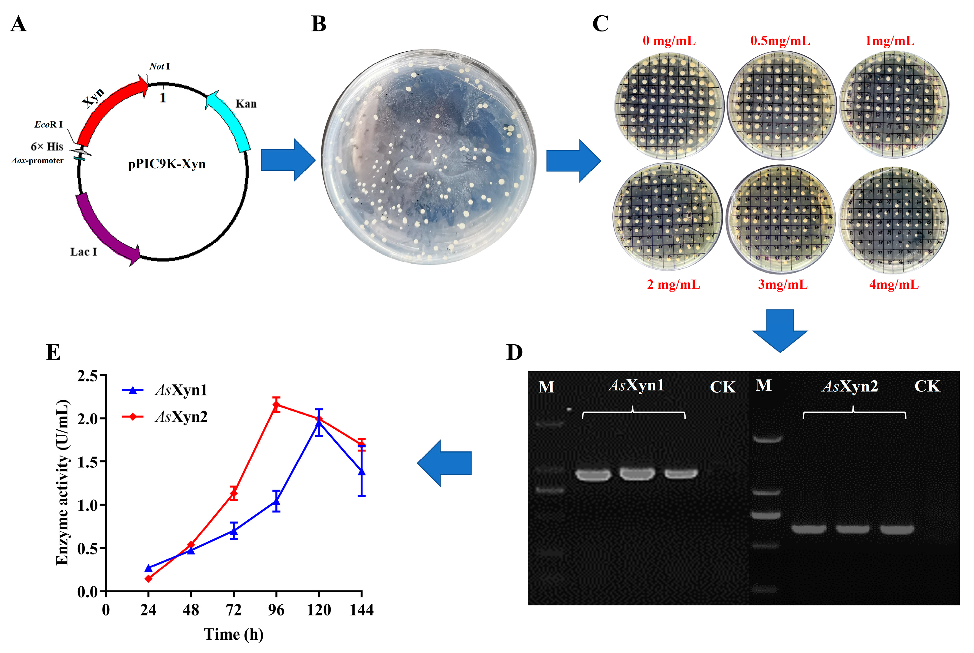
2.5. Characterization of the AsXyn1 and AsXyn2 Enzymes in P. pastoris

The recombinant enzymes of AsXyn1 and AsXyn2 expressed by P. pastoris were purified by the procedure described in “Materials and methods”. After purification, the purity levels of two enzymes were analyzed by SDS-PAGE. As shown in Figure 6A, a high purity was achieved by AsXyn1, while AsXyn2 with partial purification was obtained, as it is not easy to bind Ni resin via a 6-His tag. A possible reason is that the 6-His tag might be buried within the enzyme. The purified enzymes of AsXyn1 and AsXyn2 showed different specific activities at different temperatures. A maximum specific activity of 84.50 U/mg for AsXyn1 could be observed at the optimum temperature of 50 °C, which was close to reported metagenomics GH10 xylanases from chick cecum and the feces of Rhinopithecus bieti [14,21]. The optimum temperatures for AsXyn2 were observed at 40 °C, and the corresponding specific activities reached 4.8 U/mg (Figure 6C). A low enzyme activity for AsXyn2 was partially ascribed to low enzyme purity. The relatively high thermostability could be determined by the purified enzymes of AsXyn1 and AsXyn2, which were able to maintain more than 70% activity of the initial activity after incubation at 40 °C for 1 h (Figure 6D). However, both enzymes showed complete inactivation after pre-incubation at over 50 °C for 1 h. In previous studies, several xylanases belonging to families 10 and 11 were identified from chick cecum and Rhizopus oryzae, exhibiting higher thermostability at 50 °C [14,22]. Under the corresponding optimum temperatures, maximum specific activities of 120.8 U/mg and 5.3 U/mg for AsXyn1 and AsXyn2 could be achieved at a pH of 6.0 (Figure 6E). Furthermore, the two enzymes of AsXyn1 and AsXyn2 exhibited excellent and broad pH stability in the range of 2.0–10.0 (Figure 6F). The AsXyn1 enzyme retained over 60% of its initial enzyme activity when it was incubated at pH values from 2.0 to 10.0 for 1 h, and the residual activity of the AsXyn2 enzyme could be maintained at more than 70% of initial activity after incubation at a pH of 3.0–10.0 for 1 h. Similar results were also found for families 10 and 11 of xylanses from Rhizopus oryzae [22]. A high pH stability could be observed in the range of 4–10 for GH10 xylanase and 4–8 for GH11 xylanase.
The effects of metal ions and chemical reagents on the activities of AsXyn1 and AsXyn2 were determined as shown in Table 2. Among all the tested metal ions and chemical reagents, metal ions of Co2+, Ca2+, Ba2+, Mg2+, and Tween 80 at the concentrations of 1 mM or 10 mM could efficiently improve the enzyme activity of AsXyn1, with maximum increases of 17.3%, 16.0%, 29.7%, 12.0%, and 27.7%, respectively (Table 2). On the other hand, the AsXyn2 enzyme could only be activated by Tween 80 at 10 mM, with an increase of 27.0%. In addition, the AsXyn1 enzyme was shown to be significantly sensitive to Cu2+, Fe3+, and SDS, and resulted in enzyme inactivation. On the contrary, the AsXyn2 enzyme possessed better tolerances to all the tested metal ions than AsXyn1 did. These results demonstrated the property differences of xylanases between the GH10 family and the GH11 family. Furthermore, the kinetics of the AsXyn1 and AsXyn2 enzymes were assayed under the optimum pH value at room temperature by using different birchwood xylan concentrations. The kinetic parameters of Km and Vmax were calculated to be 8.9 mg/mL and 185.2 U/mg for AsXyn1, and 3.1 mg/mL and 6.8 U/mg for AsXyn2 (Figure 6B).
The above enzymatic properties of AsXyn1 and AsXyn2 were similar to those of xylanase purified from spent mushroom compost (SMC) of T. fuciformis in our previous study [11]. Combined with the molecule weight, the AsXyn1 enzyme mined from metagenomic data should have been the same as the xylanase reported in our previous study [11]. However, the activity of the recombinant AsXyn1 enzyme in P. pastoris was obviously lower than that of xylanase purified from the SMC of T. fuciformis. Moreover, the kinetic parameters and thermostability had changed between them [11]. Therefore, we repeated the purification of xylanase from SMC of T. fuciformis and performed a commercial mass spectrum assay. The results of the analysis indicated that the sequence of purified xylanase was consistent with that of AsXyn1 enzyme. Furthermore, we found that the molecular weight of AsXyn1 expressed in P. pastoris was slight higher than that of E. coli (Figure 3 and Figure 6). A possible explanation is that glycosylation of the AsXyn1 enzyme by P. pastoris led to reduced activity when compared with the wild xylanse enzyme from SMC. Thus, a deglycosylation experiment of the recombinant AsXyn1 enzyme expressed by P. pastoris was carried out (Figure 7). The activity of AsXyn1 enzyme after deglycosylation with 62.3 U/mg was obtained under optimal conditions, while the recombinant AsXyn1 enzyme exhibited higher activity of 120.8 U/mg. These results suggest that glycosylated modification of the AsXyn1 enzyme had an important impact on its activity, although the post-translational modification of the AsXyn1 enzyme in a wild strain not yet known. Previous studies have shown that protein glycosylation plays a key role in the regulation of enzyme activity [23,24,25]. Moreover, the covalent addition of glycans to the enzyme surface could modulate the kinetic parameters and thermostability [26]. The xylanase from Bacillus subtilis was engineered by protein glycosylation, which revealed its improved thermostability [24]. In this study, the results show that a significant reduction in enzyme activity and thermostability for AsXyn1 expressed by P. pastoris could be observed in comparison to that of a wild strain. A comprehensive understanding of the potential mechanism between them would favor the design of an enzyme with higher enzymatic activity and thermostability.

3. Materials and Methods

3.1. Strains and Plasmids

The microbiota of T. fuciformis with its companion strains was obtained from the Bioengineering Training Center, Fujian Agriculture and Forestry University (Fujian, China). The E. coli DH5a strain was used as the cloning host for plasmid construction, while the E. coli BL21(DE3) strain was used as an expression host for the heterologous expression of the xylanase candidates using expression vector Novagen® pET28a-(+) (Darmstadt, German). The P. pastoris GS115 strain was used as the expression host for the heterologous expression of the xylanase candidates via expression vector pPIC9K.

3.2. Metagenomic DNA Extraction and Analysis

The microbiota of T. fuciformis with its companion strains was inoculated into a liquid substrate medium (20 g/L sawdust, 5 g/L wheat bran, 2 g/L NaCl, 0.5 g/L MgSO4, and 0.5 g/L KH2PO4) and cultured in the dark at 25 °C. The mycelia were collected and ground with liquid nitrogen using a mortar. The metagenomic DNA extraction was performed using the CTAB method and stored at −80 °C. Subsequently, metagenomic DNA was stored in dry ice and send to a commercial company for metagenomic sequencing. Read-based metagenomic analysis was carried out using the software metaSPAdes 3.11.0. The NR database was employed for species annotation, and extraction sequences belonging to microorganisms from the NR database formed the NR_meta sublibrary. DIAMOND software (https://github.com/bbuchfink/di, accessed on 1 February 2020) was used to compare the above unigenes in the NR_meta sublibrary, combined with the NCBI species classification database, realizing species-specific annotation information for each taxonomic level of the sequence.

3.3. Sequence Analysis

The unigenes obtained from sequencing were aligned with the protein sequence in the CAZy database using DIAMOND software (blastp, evalue ≤ 1 × 10⁻⁵). The highest alignment scores were used for the protein annotations. According to the annotations, the unigene number distribution information of six functional enzymes in the metagenome was calculated according to the CAZy database. Unigenes annotated as xylanase were searched for among the CAZy annotation results. The amino acid sequences encoded by the putative xylanase genes were analyzed by BioEdit software 7.0.9 to identify ORFs, and this was further confirmed using the NCBI BLASTp algorithm (https://blast.ncbi.nlm.nih.gov/Blast.cgi, accessed on 1 February 2020). Physical and chemical properties, including molecular weight, isoelectric point, etc., of the assigned protein were analyzed by the ProtParam (https://web.expasy.org/protparam/, accessed on 1 February 2020). Selected putative xylanases were structurally modeled through the SWISS-MODEL web server (https://swissmodel.expasy.org/interactive, accessed on 1 February 2020) with the best matching template, and protein structures were visualized and analyzed using PyMol 2.4 software.

3.4. RNA Extraction and cDNA Amplification

The microbiota of T. fuciformis with its companion strains was inoculated into a liquid substrate medium and cultured in the dark at 25 °C for 7~9 d. The mycelia were collected, and total RNA extraction was performed via the Trizol method. In order to obtain the cDNA of the xylanase-encoding gene, the reverse transcriptase (RT-PCR) technique was used to synthesize the cDNA via Takara’s Prime Script® RT regent Kit (Takara, Dalian, China) with gDNA Eraser. Candidate xylanase genes were amplified by the prepared cDNA, as the template using the corresponding primers was designed according to metagenomic sequencing results (Table 3). The designed primers are given in Table 1. The PCR amplification reactions consisted of 0.5 ng of cDNA, 1 × TransStart Fast Pfu Buffer, 0.2 mM dNTP, 0.2 μM forward and reverse primers, and 1 unit of Fast Pfu DNA Polymeras (TransGen). The PCR products were analyzed using 1% agarose gel and subsequently purified with the AxyPrep DNA Gel Extraction Kit for commercial sequencing, obtained from Sangon Biotech (Shanghai, China).

3.5. Heterologous Expression of the Putative Xylanase Candidates in E. coli

The PCR amplification products of the xylananse candidates were ligated to the pET-28a expression vector by homologous recombination to generate the recombinant plasmids, which were transformed into E. coli DH5α-competent cells. The obtained recombinant strains were used to perform commercial sequencing to confirm the correct construction. Subsequently, the recombinant plasmids were extracted and transformed into E. coli BL21 (DE3), resulting in the recombinant expression strains. For heterologous expression, the recombinant E. coli BL21 (DE3) strains harboring the expression vectors were cultured in 50 mL LB medium containing kanamycin (50 μg/mL) at 37 °C and 180 rpm for 2.5 h, and IPTG, as an inducer, with a final concentration of 0.5 mM was added into the broth to induce the protein expression at 18 °C and 180 rpm overnight. The induced cells were harvested by centrifugation at 4 °C and 6000× g for 10 min, then resuspended in 100 mM phosphate buffer (pH 8.0). After sonication on ice and centrifugation at 4 °C, the supernatant and precipitation were collected to analyze the expression level and solubility of the recombinant proteins through SDS-PAGE.

3.6. Heterologous Expression of the Putative Xylanase Candidates in P. pastoris

The PCR amplification products of the xylananse candidates were ligated to the pPIC9K plasmid by means of homologous recombination to develop the recombinant plasmids, which were transformed into E. coli DH5α-competent cells for commercial sequencing. The correct recombinant plasmids were linearized by restriction endonuclease of EcoR I or Not I, and then transformed into P. pastoris GS115 by electroporation. The cell suspensions, after transformation, were spread and grown on the MD plates for 2~3 days to select positive transformants at 30 °C. Multicopy transformant selection of P. pastoris was performed on YPD plates containing 1.0–4.0 mg/mL geneticin. The positive transformants with high resistance to geneticin were randomly selected and confirmed by colony PCR, and P. pastoris transformed with the empty pPIC9K vector was used as the control. For heterologous expression, the P. pastoris transformants were inoculated into BMGY medium and grown at 30 °C and 180 rpm until the OD value at 600 nm reached 2.0~6.0. The cells were harvested by centrifugation at 5000× g for 10 min and resuspended to the OD600 of 1.0 using fresh BMMY medium. Subsequently, methanol was supplemented to 0.5% of the final concentration every 24 h to induce the protein expression until 120 h. During the induction process, the samples were taken each 24 h to evaluate the protein expression via by SDS-PAGE and assay the enzyme activity.

3.7. Purification of Recombinant Enzymes

The recombinant E. coli cells were resuspended in lysis buffer (50 mM phosphate, 300 mM NaCl, 10 mM imidazole, pH 8.0) and disrupted by sonication on ice. The supernatant, as the crude enzyme after centrifugation, was loaded into a Ni-Sepharose Histrap HP column. The pre-cooling wash buffer (50 mM phosphate, 300 mM NaCl, 50 mM imidazole, pH 8.0) was used to remove non-specific proteins, and a linear gradient elution with pre-cooling elution buffer (50 mM phosphate, 300 mM NaCl, 50–500 mM imidazole, pH 8.0) was performed to obtain the pure target enzyme. The eluate containing the pure enzyme was desalted and concentrated using a 10 kDa cut-off Centriprep device (GE Health-care, Chicago, IL, USA). For the purification of recombinant enzymes by P. pastoris, the culture supernatant collected by centrifugation was precipitated using 80% ammonium sulfate saturation. The precipitated protein was obtained by centrifugation and resuspended using ultrapure water. A Centriprep device with 10 kDa cut-off was used for the desalination and concentration of the protein sample, which was subsequently subjected to enzymatic purification using a Ni-Sepharose Histrap HP column (GE Health-care, Chicago, IL, USA), as mentioned for the above method. These purified enzymes were stored at −20 °C for further experiments.

3.8. Enzyme Activity Assays

The xylanase activity was assayed by measuring the production of reducing sugar from the hydrolysis of beechwood xylan using the DNS method. The reaction mixture contained 375 µL 1% (w/v) beechwood xylan and the appropriate enzyme solution in 500 µL sodium acetate buffer (50 mM, pH 5.2). The reaction mixture was maintained for 30 min, and the reaction was stopped with 750 μL of DNS solution. The changes in absorbance were detected at the wavelength of 540 nm using a spectrophotometer (UV-1800, Mapada, Shanghai, China), and the xylose concentration was quantified according to the calibration curve of standard xylose. One unit (U) of xylanase activity is defined as the amount of enzyme required to produce 1 μmol of xylose in 1 min. The protein concentrations of crude and purified enzymes were quantified using the Brandford method. All the experiments were performed in triplicate.

3.9. Characterization of Recombinant Enzymes

The effects of temperature and pH on the activities of recombinant xylanases were conducted in the range of 20–80 °C and pH 2.0–10.0. The activities of the purified enzymes were determined at different temperatures using the reaction mixture (50 mM sodium acetate buffer, pH 5.2), and the enzyme activity at an optimum temperature was used as 100%. For the pH effects, the purified enzyme activity was determined by using different buffers. The buffers used in this study included glycine-HCl (50 mM, pH 2.0–3.0), sodium acetate (50 mM, pH 4.0–5.0), sodium phosphate (50 mM, pH 6.0–8.0), and NaOH-glycine (50 mM, pH 9.0–10.0), and the maximum enzyme activity at an optimum pH value was used as 100%. For the enzymatic thermostability, the recombinant xylanase was pre-incubated at different temperatures (20–80 °C) at the optimum pH for 1 h, and the residual enzyme activity was determined at the optimal temperature and pH value. The percentage of the initial activity assayed before pre-incubation under the optimum conditions was calculated as the relative activity at different temperatures. The pH stability assay was determined by pre-incubation of the recombinant xylanase at different pH values of 2.0–10.0 at room temperature for 1 h, and the residual activity was assayed under the optimum temperature and pH value. Similarly, the relative activity was calculated as a percentage of the initial activity assayed before pre-incubation under optimum conditions, which represented 100% enzyme activity. All of the tests were carried out in triplicate.
The substrate concentration of beechwood xylan varied, ranging from 1 to 10 mg/mL, and was used to determine the kinetic constants of recombinant enzymes under the optimum pH value at room temperature. The apparent Km and Vmax values were obtained using the Lineweaver–Burk plot for enzymatic reactions, with 1/V and 1/S as coordinates.

4. Conclusions

In this study, three xylanase genes were identified by the metagenomic sequencing method combined with analysis of the protein tertiary structure from the microbiota of T. fuciformis and its companion strains. Heterologous expression indicated that, among the three enzymes, AsXyn1 possessed high thermostability at 40 °C and broad pH tolerance at the range of 2.0–10.0, exhibiting its application potential. Furthermore, we found that post-translational modification (such as glycosylation) of the AsXyn1 enzyme modulated its activity, kinetic parameters, and thermostability. These results and findings provide a hint for future enzyme modification and design.

Supplementary Materials

The following supporting information can be downloaded at: https://www.mdpi.com/article/10.3390/catal14010015/s1, Table S1: Sequence information of the genes identified in this study; Table S2: Analysis of signal peptides for the xylanases identified in this study.

Author Contributions

L.Z. and H.L. conceived this study and designed the experiments; Y.L. and C.L. performed the analysis of metagenome data and developed all the recombinant strains; Y.L. and C.W. determined the enzyme activity; H.L. and L.Z. wrote and edited the manuscript. All authors have read and agreed to the published version of the manuscript.

Funding

This work was supported by the Fujian Science and technology support program (No.2020S0062).

Data Availability Statement

Data are contained within the article and supplementary materials.

Conflicts of Interest

The authors declare no conflict of interest.

References

  1. Loqué, D.; Scheller, H.V.; Pauly, M. Engineering of plant cell walls for enhanced biofuel production. Curr. Opin. Plant Biol. 2015, 25, 151–161. [Google Scholar] [CrossRef] [PubMed]
  2. Miao, Y.; Li, J.; Xiao, Z.; Shen, Q.; Zhang, R. Characterization and identification of the xylanolytic enzymes from Aspergillus fumigatus Z5. BMC Microbiol. 2015, 15, 126. [Google Scholar] [CrossRef] [PubMed]
  3. Bosetto, A.; Justo, P.I.; Zanardi, B.; Venzon, S.S.; Graciano, L.; dos Santos, E.L.; Simão Rde, C. Research Progress Concerning Fungal and Bacterial β-Xylosidases. Appl. Biochem. Biotechnol. 2016, 178, 766–795. [Google Scholar] [CrossRef] [PubMed]
  4. Moreira, L.R.; Filho, E.X. Insights into the mechanism of enzymatic hydrolysis of xylan. Appl. Microbiol. Biotechnol. 2016, 100, 5205–5214. [Google Scholar] [CrossRef] [PubMed]
  5. Ariaeenejad, S.; Maleki, M.; Hosseini, E.; Kavousi, K.; Moosavi-Movahedi, A.A.; Salekdeh, G.H. Mining of camel rumen metagenome to identify novel alkali-thermostable xylanase capable of enhancing the recalcitrant lignocellulosic biomass conversion. Bioresour. Technol. 2019, 281, 343–350. [Google Scholar] [CrossRef] [PubMed]
  6. Yuan, H.J.; Dong, L.; Zhang, Z.Y.; He, Y.; Ma, X. Production, structure, and bioactivity of polysaccharide isolated from Tremella fuciformis. Food Sci. Hum. Well 2022, 11, 1010–1017. [Google Scholar] [CrossRef]
  7. Liu, D.M.; Zhu, H.Y.; Pujiana, D.; Zheng, L.S.; Chen, L.G.; Ma, A.M. Cloning and functional characterization of gpd and α-tubulin promoters from Annulohypoxylon stygium, a companion fungus of Tremella fuciformis. Mycoscience 2020, 61, 1–8. [Google Scholar] [CrossRef]
  8. Deng, Y.; Hsiang, T.; Li, S.; Lin, L.; Wang, Q.; Chen, Q.; Xie, B.; Ming, R. Comparison of the mitochondrial genome sequences of six Annulohypoxylon stygium isolates suggests short fragment insertions as a potential factor leading to larger genomic size. Front. Microbiol. 2018, 9, 2079. [Google Scholar] [CrossRef]
  9. Robl, D.; Delabona Pda, S.; Mergel, C.M.; Rojas, J.D.; Costa Pdos, S.; Pimentel, I.C.; Vicente, V.A.; da Cruz Pradella, J.G.; Padilla, G. The capability of endophytic fungi for production of hemicellulases and related enzymes. BMC Biotechnol. 2013, 13, 94. [Google Scholar] [CrossRef]
  10. Wingfield, B.D.; Bills, G.F.; Dong, Y.; Huang, W.; Nel, W.J.; Swalarsk-Parry, B.S.; Vaghefi, N.; Wilken, P.M.; An, Z.; de Beer, Z.W.; et al. IMA Genome-F 9: Draft genome sequence of Annulohypoxylon stygium, Aspergillus mulundensis, Berkeleyomyces basicola (syn. Thielaviopsis basicola), Ceratocystis smalleyi, two Cercospora beticola strains, Coleophoma cylindrospora, Fusarium fracticaudum, Phialophora cf. hyalina, and Morchella septimelata. IMA Fungus 2018, 9, 199–223. [Google Scholar]
  11. Lin, H.; Sun, M.; Li, J.; Xu, Q.; Yang, B.; Wang, Q.; Xie, W.; Sun, S.; Hu, K.; Zhang, L. Purification and Characterization of Xylanase from Spent Mushroom Compost and its Application in Saccharification of Biomass Wastes. Bioresources 2018, 13, 220–230. [Google Scholar] [CrossRef]
  12. Hu, Y.; Zhang, G.; Li, A.; Chen, J.; Ma, L. Cloning and enzymatic characterization of a xylanase gene from a soil-derived metagenomics library with an efficient approach. Appl. Microbiol. Biotechnol. 2008, 80, 823. [Google Scholar] [CrossRef] [PubMed]
  13. Ariaeenejad, S.; Hosseini, E.; Maleki, M.; Kavousi, K.; Moosavi-Movahedi, A.A.; Salekdeh, G.H. Identification and characterization of a novel thermostable xylanase from camel rumen metagenome. Int. J. Biol. Macromol. 2019, 1, 1295–1302. [Google Scholar] [CrossRef] [PubMed]
  14. Al-Darkazali, H.; Meevootisom, V.; Isarangkul, D.; Wiyakrutta, S. Gene expression and molecular characterization of a xylanase from chicken cecum metagenome. Int. J. Microbiol. 2017, 2017, 4018398. [Google Scholar] [CrossRef]
  15. Warnecke, F.; Luginbühl, P.; Ivanova, N.; Ghassemian, M.; Richardson, T.H.; Stege, J.T.; Cayouette, M.; McHardy, A.C.; Djordjevic, G.; Aboushadi, N. Metagenomic and functional analysis of hindgut microbiota of a wood-feeding higher termite. Nature 2007, 450, 560. [Google Scholar] [CrossRef]
  16. Li, R.; Kibblewhite, R.; Orts, W.J.; Lee, C.C. Molecular cloning and characterization of multidomain xylanase from manure library. World J. Microbiol. Biotechnol. 2009, 25, 2071–2078. [Google Scholar] [CrossRef]
  17. Cai, J.; Chen, X.L.; Fan, J.X.; Huang, X.M.; Li, R.; Sun, X.D.; Li, Q.Q.; Li, D.Y. Cloning and heterologous expression of a novel xylanase gene TAX1 from Trichoderma atroviride and its application in the deconstruction of corn stover. Appl. Biochem. Biotechnol. 2021, 193, 3029–3044. [Google Scholar] [CrossRef]
  18. Li, H.; Wu, H.; Jiang, F.; Wu, J.; Xue, Y.; Gan, L.; Liu, J.; Long, M. Heterologous Expression and Characterization of an Acidic GH11 Family Xylanase from Hypocrea orientalis. Appl. Biochem. Biotechnol. 2018, 184, 228–238. [Google Scholar] [CrossRef]
  19. Li, G.; Zhou, X.; Li, Z.; Liu, Y.; Liu, D.; Miao, Y.; Wan, Q.; Zhang, R. Significantly improving the thermostability of a hyperthermophilic GH10 family xylanase XynAF1 by semi-rational design. Appl. Microbiol. Biotechnol. 2021, 105, 4561–4576. [Google Scholar] [CrossRef]
  20. Liew, K.J.; Ngooi, C.Y.; Shamsir, M.S.; Sani, R.K.; Chong, C.S.; Goh, K.M. Heterologous expression, purification and biochemical characterization of a new endo-1,4-β-xylanase from Rhodothermaceae bacterium RA. Protein Expres. Purif. 2019, 164, 105464. [Google Scholar] [CrossRef]
  21. Xu, B.; Dai, L.M.; Li, J.J.; Dent, M.; Miao, H.B.; Zhou, J.P.; Mu, Y.L.; Wu, Q.; Tang, X.H.; Yang, Y.J.; et al. Molecular and Biochemical Characterization of a Novel Xylanase from Massilia sp. RBM26 Isolated from the Feces of Rhinopithecus bieti. J. Microbiol. Biotechnol. 2015, 26, 9–19. [Google Scholar] [CrossRef] [PubMed]
  22. Xiao, Z.Z.; Grosse, S.; Bergeron, H.; Lau, P.C.K. Cloning and characterization of the first GH10 and GH11 xylanases from Rhizopus oryzae. Appl. Microbiol. Biotechnol. 2014, 98, 8211–8222. [Google Scholar] [CrossRef] [PubMed]
  23. Shental-Bechor, D.; Levy, Y. Effect of glycosylation on protein folding. A close look at thermodynamic stabilization. Proc. Natl. Acad. Sci. USA 2008, 105, 8256–8261. [Google Scholar] [CrossRef] [PubMed]
  24. Fonseca-Maldonado, R.; Vieira, D.S.; Alponti, J.S.; Bonneil, E.; Thibault, P.; Ward, R.J. Engineering the pattern of protein glycosylation modulates the thermostability of a GH11 xylanase. J. Biol. Chem. 2013, 288, 25522–25534. [Google Scholar] [CrossRef]
  25. Yi, W.; Clark, P.M.; Mason, D.E.; Keenan, M.C.; Hill, C.; Goddard, W.A.; Peters, E.C.; Driggers, E.M.; Hsieh-Wilson, L.C. Phosphofructokinase 1 glycosylation regulates cell growth and metabolism. Science 2012, 337, 975–980. [Google Scholar] [CrossRef]
  26. Helenius, A.; Aebi, M. Roles of N-linked glycans in the endoplasmic reticulum. Annu. Rev. Biochem. 2004, 73, 1019–1049. [Google Scholar] [CrossRef]
Figure 1. Analysis of metagenomics sequencing. (A) Analysis of extracted metagenomic DNA. (B) CAZy classification statistics. (C) Krona species annotation.
Catalysts 14 00015 g001
Figure 2. Analysis of tertiary structure for three xylanase candidates. (A) AsXyn1; (B) AsXyn2; (C) SsXyn.
Catalysts 14 00015 g002
Figure 3. Analysis of expression level and solubility for recombinant enzymes in E. coli. (A) Whole-cell lysate; (B) cell lysate supernatant; (C) cell lysate precipitation; (D) crude enzyme activity (Lane M: Protein Marker; Lane 1: E. coli BL21 (DE3)/pET28a; Lane 2: E. coli BL21 (DE3)/pET28a-AsXyn1; Lane 3, E. coli BL21 (DE3)/pET28a-AsXyn2; Lane 4, E. coli BL21 (DE3)/pET28a-SsXyn).
Catalysts 14 00015 g003
Figure 4. Heterologous expression of AsXyn1 and AsXyn2 in P. pastoris. (A) Construction of recombinant plasmid; (B) preliminary screening; (C) multi-copy transformant selection; (D): colony PCR verification; (E): activity assays of the supernatants by recombinant P. pastoris.
Catalysts 14 00015 g004
Figure 5. Biochemical characterization of recombinant Ssxyn enzyme in E. coli. (A) Recombinant SsXyn purification; (B) temperature effect and thermostability; (C) pH effect and pH stability; (D) metal ion effect; (E): chemical reagents’ effect; (F) Lineweaver–Burk diagram of double reciprocal for SsXyn enzyme (Lane M: protein marker; Lane 1: cell lysate; Lane 2: flowthrough from purification step; Lane 3: purified Ssxyn enzyme).
Catalysts 14 00015 g005
Figure 6. Characterization of the recombinant AsXyn1 and AsXyn2 enzymes by P. pastoris. (A) Enzyme purification; (B) double reciprocal diagram of recombinant AsXyn1 and AsXyn2 xylanases; (C) temperature effects; (D) thermostability; (E) pH effects; (F) pH stability.
Catalysts 14 00015 g006
Figure 7. Deglycosylation of the recombinant AsXyn1 enzyme by End-HF enzyme. Lane M: protein marker; Lane 1: deglycosylation of AsXyn1; Lane 2: AsXyn1 as control.
Catalysts 14 00015 g007
Table 1. Candidate xylanase genes predicted by metagenomic sequencing.
UnigeneNameGene AnnotationStrainFamilyGene LengthIdentity
11,018AsXyn1xylanaseA. stygiumGH10111097%
14,634AsXyn2xylanaseA. stygiumGH11651100%
52,146SsXynxylanaseSphingobacterium sp.GH10114995%
Table 2. Effects of metal ions and chemical reagents on the activities of recombinant xylanases.
Metal Ions and Chemical ReagentsRelative Activity (%)
AsXyn1AsXyn2
1 mM10 mM1 mM10 mM
Control100.0 ± 0.7100.0 ± 0.9100.0 ± 1.1100.0 ± 0.9
Cu2+16 ± 1.62 ± 0.495 ± 2.041 ± 1.6
Fe2+54 ± 2.941 ± 0.476 ± 3.312 ± 1.3
Fe3+21 ± 1.84 ± 2.781 ± 2.246 ± 1.8
Mn2+75 ± 2.075 ± 2.068 ± 2.755 ± 1.8
Co2+110 ± 8.0117 ± 4.489 ± 2.263 ± 1.8
Zn2+71 ± 0.440 ± 1.691 ± 1.160 ± 1.6
Ca2+111 ± 6.2116 ± 3.394 ± 1.695 ± 1.8
K+101 ± 5.1103 ± 7.394 ± 3.198 ± 1.1
Ba2+109 ± 3.3130 ± 3.894 ± 2.096 ± 1.3
Mg2+111 ± 4.9112 ± 4.797 ± 1.397 ± 1.3
EDTA83 ± 3.164 ± 2.985 ± 3.669 ± 6.7
SDS4 ± 1.32 ± 1.684 ± 1.32 ± 1.1
Tween 80115 ± 2.7128 ± 14.495 ± 1.6127 ± 5.3
BME108 ± 1.680 ± 5.388 ± 1.378 ± 2.7
Table 3. Primer sequences used in this study.
NamePrimer Sequence (5′→3′)
28a-AsXyn1-FGGACAGCAAATGGGTCGCGGATCCGCCGACAGCATCGACGCCTT
28a-AsXyn1-RTGGTGCTCGAGTGCGGCCGCAAGCTTTTTCAGGGCGTTCACGACAG
28a-Ssxyn-FGGACAGCAAATGGGTCGCGGATCCAATATACAGGATCTGGAACA
28a-Ssxyn-RGTGGTGCTCGAGTGCGGCCGCAAGCTTCTTTTTTTTTTGAGTCAATG
28a-AsXyn2-FGGACAGCAAATGGGTCGCGGATCCTCGCCGCTCGACCTAATCAC
28a-AsXyn2-RTGCTCGAGTGCGGCCGCAAGCTTAGACTGCTCAACGGTAATC
9k-AsXyn1-FGCTGAAGCTTACGTAGAATTCCATCATCACCATCACCACGCCGACAGCATCGACGCCTT
9k-AsXyn1-RAGGCGAATTAATTCGCGGCCGCTTTCAGGGCGTTCACGACAG
9k-AsXyn2-FCTGAAGCTTACGTAGAATTCTCGCCGCTCGACCTAATCAC
9k-AsXyn2-RGCGAATTAATTCGCGGCCGCATGATGATGATGATGATGAGACTGCTCAACGGTAATC
Disclaimer/Publisher’s Note: The statements, opinions and data contained in all publications are solely those of the individual author(s) and contributor(s) and not of MDPI and/or the editor(s). MDPI and/or the editor(s) disclaim responsibility for any injury to people or property resulting from any ideas, methods, instructions or products referred to in the content.

