Article

Production and Characterization of Durvillaea antarctica Enzyme Extract for Antioxidant and Anti-Metabolic Syndrome Effects

1
Graduate Institute of Food Culture and Innovation, National Kaohsiung University of Hospitality and Tourism, No. 1, Songhe Road, Xiaogang District, Kaohsiung City 812301, Taiwan
2
Department of Seafood Science, National Kaohsiung University of Science and Technology, No. 142, Haijhuan Road, Nanzih District, Kaohsiung City 81157, Taiwan
3
Department of Marine Environmental Engineering, National Kaohsiung University of Science and Technology, No. 142, Haijhuan Road, Nanzih District, Kaohsiung City 81157, Taiwan
4
Sustainable Environment Research Center, Department of Marine Environmental Engineering, National Kaohsiung University of Science and Technology, No. 142, Haijhuan Road, Nanzih District, Kaohsiung City 81157, Taiwan
5
Institute of Aquatic Science and Technology, National Kaohsiung University of Science and Technology, No. 142, Haijhuan Road, Nanzih District, Kaohsiung City 81157, Taiwan
*
Authors to whom correspondence should be addressed.
These authors contributed equally to this work.
Catalysts 2022, 12(10), 1284; https://doi.org/10.3390/catal12101284
Submission received: 22 September 2022 / Revised: 16 October 2022 / Accepted: 19 October 2022 / Published: 21 October 2022
(This article belongs to the Special Issue Enzymes in Biomedical, Cosmetic and Food Application)

Abstract

:
In this study, three enzyme hydrolysate termed Dur-A, Dur-B, and Dur-C, were produced from Durvillaea antarctica biomass using viscozyme, cellulase, and α-amylase, respectively. Dur-A, Dur-B, and Dur-C, exhibited fucose-containing sulfated polysaccharide from chemical composition determination and characterization by FTIR analyses. In addition, Dur-A, Dur-B, and Dur-C, had high extraction yields and low molecular weights. All extracts determined to have antioxidant activities by DPPH (2,2-diphenyl-1-picrylhydrazyl), ABTS (2,20-azino-bis(3-ethylbenzothiazoline-6-sulfonic acid) diammonium salt), and ferrous ion-chelating methods. All extracts were also able to positively suppress the activities of key enzymes involved in metabolic syndrome: angiotensin I-converting enzyme (ACE), α-amylase, α-glucosidase, and pancreatic lipase. In general, Dur-B exhibited higher antioxidant and higher anti-metabolic syndrome effects as compared to the other two extracts. Based on the above health promoting properties, these extracts (especially Dur-B) can be used as potential natural antioxidants and natural anti-metabolic syndrome agents in a variety of food, cosmetic, and nutraceutical products for health applications.

1. Introduction

Oxidative stress occurs when the net amount of reactive oxygen species (ROS) exceeds the antioxidant capacity. This may happen as a result of a general increase in ROS generation, a depression of antioxidant systems, or both [1]. ROS such as superoxide anion radical (O2), hydrogen peroxide (H2O2), hydroxyl radical (OH), singlet oxygen (1O2), and nitric oxide (NO), are metabolic products and may exist in the environment [2]. Excessive production of ROS may damage cellular DNA, proteins, and lipids, resulting in altered biochemical compounds, corroded cell membranes, and possibly even metabolic syndrome [1,3]. Thus, antioxidants are needed to delay or prevent the oxidation of cellular oxidizable substrates by promoting scavenging of ROS and preventing the production of ROS [4].
Diabetes mellitus is a chronic disease characterized by hyperglycemia, a condition in which blood glucose levels are elevated. There are two types of diabetes mellitus, type 1 and type 2, which differ in insulin production and utilization. Type 1 diabetes mellitus is caused by insufficient insulin production by the pancreas, as a result of autoimmune disease. The disease is managed by insulin injections. In type 2 diabetes, there is sufficient insulin but the body’s cells are insulin resistant, a condition which is thought to be caused by multiple factors such as overweight, high cholesterol, and physical inactivity. The vast majority (95%) of diabetic patients have type 2, and strong associations with dietary intake and reduced physical activity have been reported [5]. Metabolic syndrome is a constellation of abnormalities, which include abdominal obesity, dyslipidemia, high blood glucose/impaired glucose tolerance, and high blood pressure. It has been well established that metabolic syndrome increases the risk of overt diabetes mellitus [6]. The prevalence of cardiovascular disease (CVD) is rising rapidly, regardless of age and gender, and the mortality rate of CVD is high. CVD kills more than 18 million people worldwide annually [7]. In fact, CVD is not a single disease; rather it is a group of disorders related to the heart and blood vessels, and includes coronary heart disease, rheumatic heart disease, and cerebrovascular disease, as well as other conditions. Approximately 80% of CVD-related deaths are due to cardiac arrest and strokes, which involve the deposition of fat in blood vessels. A number of other factors are also thought to play a role in CVDs, such as a poor diet, high intake of oils, alcohol consumption, hereditary factors, and low physical activity [8]. Obesity is a metabolic condition in which the body has gained excess weight, primarily as a consequence of the deposition of excess fat. It is considered a gateway for various other metabolic syndromes, such as insulin resistance, dyslipidemia, hypertension, hyperthyroidism, impaired glucose metabolism, and stroke [9], which have been shown to be associated with CVD. Increased oxidative stress has been identified as a key factor in metabolic syndrome and its related pathologies, and it has been postulated that oxidative stress is mechanistically involved in the progression of this disease. ROS play an important role in the development and progression of CVDs [10]. Furthermore, oxidative stress has been shown to play an important role in the mechanism of micro- and macrovascular complications in metabolic syndrome [11]. Hence, it is crucially important to identify and develop antioxidants (particularly those from naturally occurring sources) that are capable of eliminating ROS. Antioxidant-containing nutraceuticals could be useful in treating or preventing metabolic syndrome-related disorders.
In recent decades, there has been a growing awareness that traditional foods and naturally occurring compounds could be hugely beneficial to digestion and other aspects of human health. Indeed, the importance of consuming complete and nutritious food is increasingly being recognized [12,13]. Marine algae and seaweeds have attracted considerable interest due to their nutritional components and highly bioactive properties. Seaweeds are a part of the traditional diet of coastal-dwelling cultures, particularly in Asia, and are also used in folk medicines. It has been reported that Japan, North Korea, Taiwan, and China, are the largest consumers of seaweeds in Asia. Japanese people consume approximately 5.3 g seaweed per day−1. Interestingly, Japan also has one of the longest lifespans worldwide, as well as low rates of CVDs [5,8,9]. At the cellular level, seaweed-derived polysaccharides exhibit high biological activity, such as antioxidant, anti-cancer, anti-inflammatory, and immunomodulatory properties, although these activities are limited by various characteristics, including molecular weight, glycosidic bonds, solubility, spatial configuration, and main-chain configuration [14]. Thus, these biological activities can be enhanced by applying various molecular modification techniques that can be classified into physical, chemical, and biological methods. The physical method involves reducing the molecular weight of polysaccharides by breaking the glucoside chain using ultrasonic disruption [15], radiation [16], or microwave denaturation [17]. These high energies result in denaturation of glucoside bonds, reducing them to oligomers and monomers. In the chemical method, functional groups such as sulfate, phosphate, selenium, iron, and alkyl groups, are introduced to the polysaccharides, which improves solubility and bioactivity [14]. The biological method also makes use of molecular modification, which is achieved by enzymatic hydrolysis of glycosidic bonds. In comparison with the two aforementioned methods, the enzymatic method is more accurate, more efficient, highly specific, has a higher yield-coefficient, releases novel bio-compounds, and has few side effects. Moreover, a recent study suggested that the enzyme-assisted method promotes the bioactivity of cell wall polymers during extraction [18].
In the present study, various hydrolysates of D. antarctica were produced using the enzyme-assisted extraction (EAE) method, and their antioxidant and inhibitory activities against key metabolic syndrome-related enzymes (ACE, α-amylase, α-glucosidase, and pancreatic lipase) were investigated. The EAE method has attracted a lot of attention in the past decade as it exhibits high selectivity and it allows a large amount of biological active compounds to be recovered [19]. Kulshreshtha et al. (2015) employed commercial protease and carbohydrases to extract bioactive materials from Codium fragile and Chondrus crispus and achieved an extraction yield of 40–70% (dry basis) [19]. Nguyen et al. (2020) applied combined commercial enzymes, cellulase (Cellic® CTec2) and alginate lyase, which were derived from Sphingomonas sp. in a single-step process, to extract fucoidan from brown seaweeds, Fucus distichus subsp. evanescens and Saccharina latissimi [20], and reported an extraction yield of 29–40%. Moreover, Olivares-Molina and Fernández (2016) used an enzymatic method (cellulase and α-amylase) to extract compounds from three brown seaweeds, Lessonia nigrescens, D. antarctica, and Macrocystis pyrifera, and found the extraction yields ranged from 9–38% [18]. Herein, we used viscozyme, cellulose, and α-amylase to extract bioactive substances from D. antarctica. Viscozyme® L (Novozyme Corp.) is a blend of β-glucanases, hemicellulases, pectinases, and xylanases, which is a cell wall-degrading enzyme complex produced by Aspergillus sp. Cellulase is a member of the glycoside hydrolase family, and is secreted by a number of cellulolytic microorganisms. Cellulase produced by Aspergillus niger catalyzes the hydrolysis of endo-1,4-β-D-glycosidic linkages in cellulose, cereal beta-D-glucans, lichenin, and the cellooligosaccharides cellotriose to cellohexaose. As cellulose molecules are strongly bound to one another, it is more difficult to break it down by cellulolysis compared with polysaccharides such as starch. Microbial amylases are exoenzymes that are utilized in various industrial applications, such as bread making, maltose syrups, and the fermentation of soya sauce, miso, and so on. The α-amylase derived from Aspergillus oryzae acts as a catalyst in the hydrolysis of α-1,4 glycosidic bonds in soluble starches and other related substrates. These substrates are then further broken down, releasing short oligosaccharides and α-limit dextrins.
Different enzymes exert different degrees of action on target polysaccharides, and the activities of these enzymes have been analyzed to determine the optimum conditions [14,21]. In the present study, Durvillaea antarctica, a type of brown seaweed, was investigated in an experimental model. Seaweed samples were hydrolyzed using three different enzymes, viscozyme, cellulase, and α-amylase, while maintaining a uniform condition in order to compare the physicochemical properties of the hydrolysates and their biological activities. As this study was specifically concerned with investigating antioxidants and their potential role in metabolic syndrome-related enzyme inhibition, various antioxidant assays such as DPPH (2,2-diphenyl-1-picrylhydrazyl), ABTS (2,20-azino-bis(3-ethylbenzothiazoline-6-sulfonic acid) diammonium salt), and ferrous ion-chelating ability, were employed. The physiochemical characterization of hydrolyzed products was performed using FTIR and NMR techniques to analyze functional groups. Chemical composition, monosaccharide composition, and molecular weight analyses, were performed by various assays and HPLC techniques. Finally, the metabolic syndrome-related enzyme inhibition studies were conducted by evaluating the inhibition of angiotensin I-converting enzyme (ACE), α-glucosidase, α-amylase, and pancreatic lipase enzymes. The overall aim of these experiments was to explore the potential of enzymatic hydrolysates from D. antarctica to serve as a natural antioxidant for use in the food, cosmetic, and nutraceutical industries and which may be beneficial in the prevention or treatment of metabolic syndrome.

2. Results

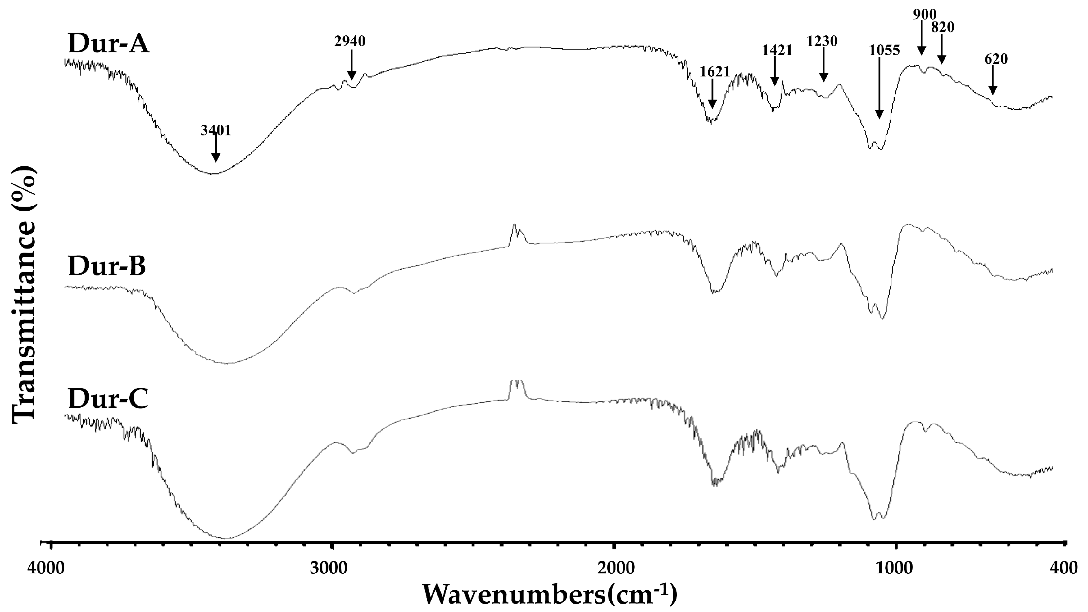
2.1. Preparation of Enzymatic Extracts (Dur-A, Dur-B, and Dur-C) from D. antarctica, and Physicochemical Characteristics of Dur-A, Dur-B, and Dur-C

In the current study, D. antarctica was utilized for hydrolysis using viscozyme, cellulase, or α-amylase in order to obtain hydrolysis products, namely Dur-A, Dur-B, and Dur-C. Table 1 shows the hydrolysis variables of Dur-A, Dur-B, and Dur-C, and the extraction yields of Dur-A, Dur-B, and Dur-C were 38.4% ± 1.1%, 43.0% ± 0.2%, and 41.4% ± 0.6%, respectively, suggesting that Dur-B and Dur-C have higher extraction yields than that of Dur-A. However, the difference was not remarkable. Table 2 shows the results of the physicochemical properties of Dur-A, Dur-B, and Dur-C. The size exclusion chromatographic method revealed the MW distribution in Dur-A, Dur-B, and Dur-C (Table 2). Among these three hydrolysis products, two peaks were observed. For Dur-A, the peak for higher MW was 171.4 kDa, with a peak area of approximately 99.5% and the peak for lower MW was 8.61 kDa, with a peak area of approximately 0.5%. For Dur-B, the peak for higher MW was 215.9 kDa with a peak area approximately 99.5%, and the peak for lower MW was 10.0 kDa, with a peak area of approximately 0.5%. For Dur-C, the peak for higher MW was 183.3 kDa, with a peak area of approximately 51.6%, and the peak for lower MW was 2.19 kDa, with a peak area approximately 48.4%. These data indicate that Dur-C had more oligosaccharides (the molecular weight was about 2.19 kDa) as compared to Dur-A and Dur-B. Total sugar, fucose, sulfate, uronic acid, alginic acid, polyphenols, and proteins contents, as well as the monosaccharide composition of Dur-A, Dur-B, and Dur-C, are presented in Table 2. The total sugar contents of Dur-A, Dur-B, and Dur-C were 44.4% ± 0.2%, 40.4% ± 0.3%, and 72.3% ± 0.6%, respectively. The fucose contents of Dur-A, Dur-B, and Dur-C were 13.5% ± 0.5%, 18.2% ± 0.3%, and 19.9% ± 0.3%, respectively. In general, Dur-C had the highest total sugar content and fucose content as compared to Dur-A and Dur-B. The sulfate contents of Dur-A, Dur-B, and Dur-C were 38.5% ± 0.0%, 41.2% ± 0.0%, and 24.7% ± 0.1%, respectively. The percentage contents of uronic acid in Dur-A, Dur-B, and Dur-C were 16.9% ± 0.3% 16.7% ± 0.6%, and 16.8% ± 0.2%, respectively. The alginic acid content percentages of Dur-A, Dur-B, and Dur-C were 6.99% ± 0.20%, 6.50% ± 0.20%, and 7.27% ± 0.54%, respectively. The percentage contents of polyphenols in Dur-A, Dur-B, and Dur-C were 0.46% ± 0.02%, 0.39% ± 0.02%, and 0.42% ± 0.04%, respectively. Moreover, the percentage contents of protein in Dur-A, Dur-B, and Dur-C were 1.08% ± 0.02%, 0.93% ± 0.03%, and 0.97% ± 0.03%, respectively. Generally, Dur-B had the highest sulfate content, and the uronic acid content, alginic acid content, polyphenols content, and proteins content in Dur-A, Dur-B, and Dur-C were similar. Table 2 also shows the monosaccharide compositions of Dur-A, Dur-B, and Dur-C. In brief, fucose, rhamnose, and glucuronic acid were the most prevalent sugar units in Dur-A. Fucose, rhamnose, and galacturonic acid were the most prevalent sugar units in Dur-B; fucose, rhamnose, galacturonic acid, glucose, and xylose were the most prevalent sugar units in Dur-C. Taken together, the monosaccharide compositions of Dur-A, Dur-B, and Dur-C varied. In summary, Dur-A, Dur-B, and Dur-C, had low molecular weights and showed the characteristic compositions of fucose-containing sulfated polysaccharides.
FTIR and NMR techniques were used to perform structural analyses of Dur-A, Dur-B, and Dur-C. Figure 1 shows the IR bands at 3401 cm−1 and 2940 cm−1 which correspond to OH and H2O stretching vibration and C–H stretching of the pyranoid ring or the C-6 group of the fucose and galactose units [22,23]. Absorption bands were detected at 1621 cm−1 and 1421 cm−1 indicating the scissoring vibration of H2O and vibrations of the in-plane ring CCH, COH, and OCH, which is a known absorption pattern of polysaccharides [22,23,24]. The peaks at 1230 cm−1 and 1055 cm−1 can be attributed to the presence of asymmetric stretching of S=O and C–O–C stretching vibrations in the ring or C–O–H of the glucosidal bond [22,23]. Bands at 900 cm−1 corresponded to C1–H bending vibration in β-anomeric units (probably galactose) [25]. The peak at 820 cm−1 corresponded to the bending vibrations of C-O-S of sulfate [26]. The bands at 620 cm−1 may correspond to symmetric O=S=O deformations [25]. Figure 1 provides evidence that Dur-A, Dur-B, and Dur-C exhibited the characteristic IR absorptions of fucose-containing sulfated polysaccharides, but the differences in their FTIR results are not obvious. Figure S1A depicts the 1H-NMR spectra of Dur-A, Dur-B, and Dur-C. The signals at 4.57 ppm and 4.46 ppm indicate the presence of H-2 in a 2-sulfated fucopyranose residue [27], and the signal at 4.13 ppm (4[H]) is suggestive of the presence of 3-linked α-l-fucose [24]. Signals with a ppm of 4.07/3.95 (6[H]/6′[H]) denote the presence of a (1-6)-β-D-linked galacton [28]. The signals detected at 3.78 ppm and 3.72 ppm may be indicative of the existence of (3[H]) 4-linked β-D-galactose and (4[H]) 2,3-linked α-β-mannose, respectively [24]. In addition, the signal at 1.32 ppm indicates the presence of C6 methyl protons of L-fucopyranose [29]. Due to the dissimilarity of the 1H-NMR spectra, it can thus be deduced that Dur-A, Dur-B, and Dur-C may have different structures. In addition, Figure S1B shows the 13C-NMR spectra of Dur-A, Dur-B, and Dur-C. The signals at 75.86 ppm attributes to the presence of C-5 of M-block in a mannuronate homo-oligomer [30], indicating the mannuronic acid of alginic acid is probably detectable in Dur-A, Dur-B, and Dur-C.

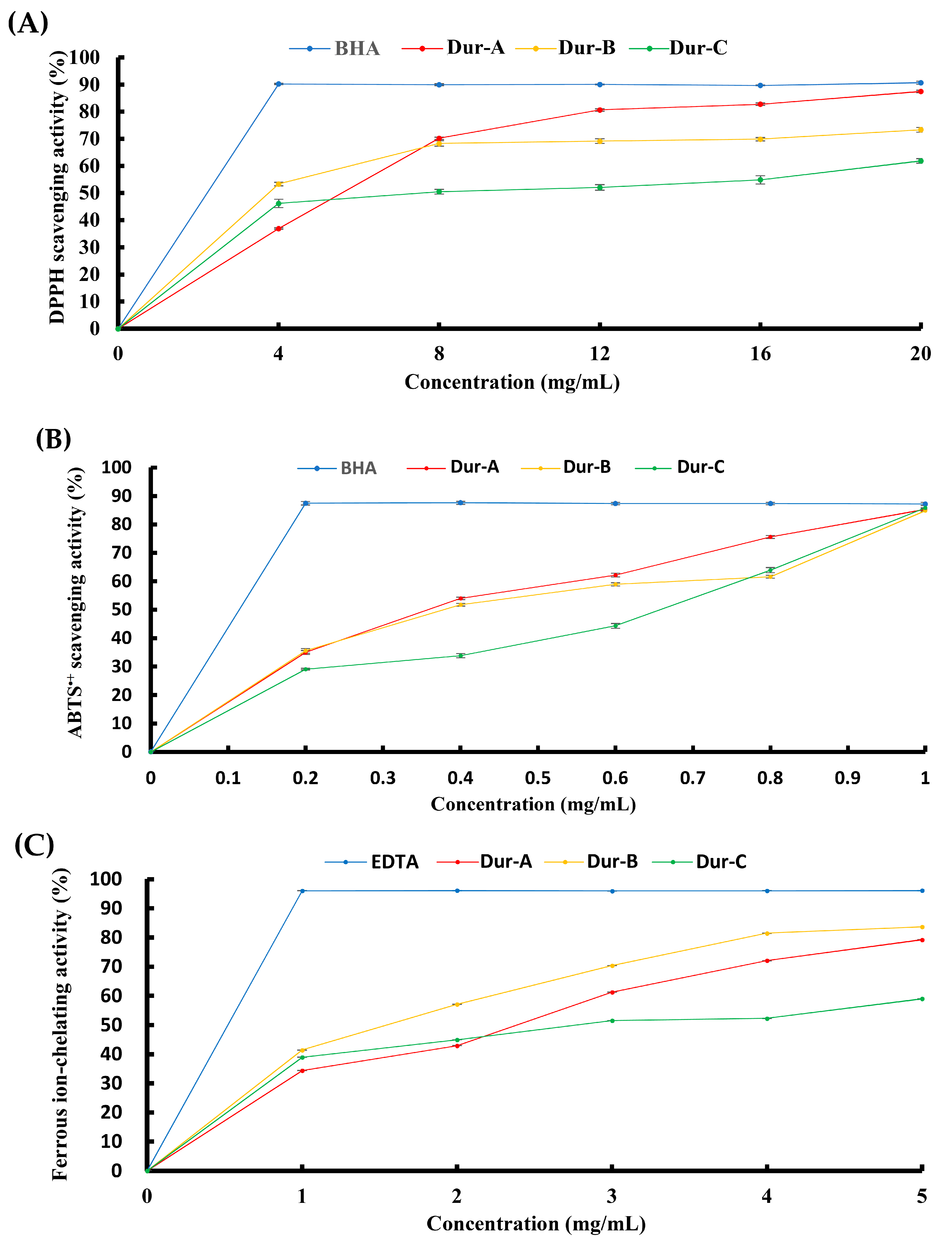
2.2. Effect of Enzymatic Extracts (Dur-A, Dur-B, and Dur-C) on Antioxidant Activities

The antioxidant activities of Dur-A, Dur-B, and Dur-C, were examined by DPPH, ABTS, and ferrous ion-chelating analyses, and the data are presented in Figure 2. The scavenging effects of Dur-A, Dur-B, and Dur-C on DPPH free radicals are shown in Figure 2A. These extracts exhibited DPPH scavenging properties in a dose-dependent manner. The observed results also suggest that Dur-B has the most potent effect on DPPH scavenging ability, followed by Dur-A, and then Dur-C. The scavenging effects of Dur-A, Dur-B, and Dur-C on ABTS•+ free radicals are shown in Figure 2B. These extracts exhibited ABTS•+ scavenging properties in a dose-dependent manner. The observed results suggest that Dur-A has the most potent effect on ABTS•+ radical scavenging ability, followed by Dur-B, and then Dur-C. The scavenging effects of Dur-A, Dur-B, and Dur-C on ferrous ion-chelating are shown in Figure 2C. These extracts exhibited ferrous ion-chelating activities in a dose-dependent manner. The observed results suggest that Dur-B has the most potent effect on ferrous ion-chelating activity, followed by Dur-A, and then Dur-C. All of these extracts showed antioxidant activities and thus further investigations are warranted to explore their anti-metabolic syndrome capacities.

2.3. Effect of Enzymatic Extracts (Dur-A, Dur-B, and Dur-C) on the Inhibition against Key Metabolic Syndrome-Related Enzymes

The inhibitory activities against key metabolic syndrome-related enzymes of Dur-A, Dur-B, and Dur-C were investigated, and the results are shown in Table 3. For the inhibition of ACE, the concentration of Dur-A, Dur-B, and Dur-C used was 1 mg/mL, and the inhibitory rates for Dur-A, Dur-B, and Dur-C were 72.5% ± 1.4%, 80.7% ± 1.6%, and 62.9% ± 0.6%, respectively. The observed results suggest that Dur-B had the most ACE inhibitory activity, followed by Dur-A, and then Dur-C. For the inhibition of α-amylase, the concentration of Dur-A, Dur-B, and Dur-C used was 3 mg/mL, and the inhibitory rates for Dur-A, Dur-B, and Dur-C were 53.3% ± 0.4%, 69.8% ± 0.5%, and 61.6% ± 1.0%, respectively. The observed results suggest that Dur-B had the most α-amylase inhibitory activity, followed by Dur-C, and then Dur-A. For the inhibition of α-glucosidase, the concentration of Dur-A, Dur-B, and Dur-C used was 3 mg/mL, and the inhibitory rates for Dur-A, Dur-B, and Dur-C were 26.9% ± 0.6%, 30.1% ± 4.8%, and 60.6% ± 0.9%, respectively. The observed results suggest that Dur-C has the most α-glucosidase inhibitory activity, followed by Dur-A and Dur-B. For the inhibition of pancreatic lipase, the concentration of Dur-A, Dur-B, and Dur-C used was 0.3 mg/mL, and the inhibitory rates for Dur-A, Dur-B, and Dur-C were 38.1% ± 5.0%, 33.6% ± 6.0%, and 17.1% ± 3.0%, respectively. The observed results suggest that Dur-A and Dur-B have better pancreatic lipase inhibitory activity than Dur-C. In summary, all extracts showed inhibitory effects on metabolic syndrome-related enzymes.

3. Discussion

In this study, we investigated the antioxidant and inhibitory activities of enzymatic extract of D. antarctica biomass against key metabolic syndrome-related enzymes (ACE, α-amylase, α-glucosidase, and pancreatic lipase), which were extracted using the EAE method. The extraction yields of D. antarctica obtained with viscozyme (Dur-A), cellulase (Dur-B), and α-amylase (Dur-C), were 38.4%, 43.0%, and 41.4%, respectively (Table 1), indicating that Dur-B and Dur-C had higher extraction yields than that of Dur-A. However, the difference in yield was not significant. Hammed et al. (2017) investigated viscozyme, cellulase, and amyloglucosidase, as potential enzymes for the extraction of sulfated polysaccharides from the brown seaweed Turbinaria turbinate under different extraction conditions. This study obtained a maximum extraction yield of about 24–25% [21]. In short, the present study used an extraction model that produced remarkable extraction yields that were similar or superior to previously reported findings.
With respect to bioactive polysaccharides, the molecular structures, sulfate content, type of sugar, and molecular weight, play important roles in their biological functions. Among these variables, the molecular weight of polysaccharides appears to be the most important factor that determines the biological characteristics [31]. High molecular weight polysaccharides do not exhibit good solubility and cannot be processed easily, thereby limiting their capacity to penetrate cells. However, polysaccharides that have a low molecular weight possess more biological functions, such as immunostimulation, anticancer, antioxidant activities, and anticoagulation [2,32,33]. The EAE method has been successfully used to obtain LMW polysaccharides with superior biological functions as compared to compounds obtained using conventional hot-water extraction [34]. In the current study, among the three extracts, higher molecular weight distribution was between 171.4 kDa and 215.9 kDa, and the lower molecular weight distribution was found between 2.19 kDa and 10.0 kDa (Table 2). It has been reported that the molecular weight of a fucose-containing sulfated polysaccharide extracted by hot water was approximately 690.8 kDa (higher molecular weight) and 327.1 kDa (lower molecular weight) [35], and therefore the EAE method applied herein facilitated the production of polysaccharides with lower molecular weights. Furthermore, Dur-C contained more short oligosaccharides (approx. 2.19 kDa) in comparison with those of Dur-A and Dur-B (Table 2), suggesting that α-amylase was more effective for releasing short oligosaccharides compared to the other two enzymes. Whether the oligosaccharides content of extracts is related to their biological functions remains to be elucidated.
Table 2 depicts the chemical compositions of Dur-A, Dur-B, and Dur-C. It can be seen that Dur-C has the highest total sugar content and fucose content, but has the lowest sulfate content, in comparison with Dur-A and Dur-B. Other compounds, such as uronic acid content, polyphenols content, and alginic acid content, showed similar amounts among Dur-A, Dur-B, and Dur-C. Regarding monosaccharide compounds (Table 2), the sugars in Dur-A were largely fucose, rhamnose, and glucuronic acid; the sugars in Dur-B were predominantly fucose, rhamnose, and galacturonic acid; in Dur-C, the predominant sugars were fucose, rhamnose, galacturonic acid, xylose, and glucose. The results imply that these extracts are largely fucose-containing sulfated polysaccharides. FTIR is a powerful tool that is used for the detection of a range of functional groups. It can also provide information on the chemical composition of compounds [36]. The data shown in Figure 1 indicate that Dur-A, Dur-B, and Dur-C, exhibit the characteristic IR peaks of fucose-containing sulfated polysaccharide. We then assessed the 1H-NMR spectra of Dur-A, Dur-B, and Dur-C (Figure S1A), and the results showed notable differences among Dur-A, Dur-B, and Dur-C. As there were differences in total sugar content, fucose content, sulfate content, monosaccharide composition, and structure among Dur-A, Dur-B, and Dur-C, we were interested in further characterizing their biological activities, particularly with respect to their antioxidant properties and their potential to exert mitigating effects against metabolic syndrome.
We evaluated the antioxidant activities of Dur-A, Dur-B, and Dur-C, using DPPH, ABTS, and ferrous ion-chelating analyses. DPPH is a widely used technique for evaluating antioxidant activity within a relatively short duration [37]. Figure 2A shows the DPPH radical-scavenging activities of Dur-A, Dur-B, and Dur-C, and BHA (as a reference). The results reveal that all extracts exhibited DPPH radical-scavenging activity dose-dependently. The IC50 values (the concentration of a sample capable of scavenging 50% of DPPH radicals) of the extracted samples (Dur-A, Dur-B, and Dur-C) on DPPH radical-scavenging activity were 5.60, 3.78, and 7.52 mg/mL, respectively (Figure 2A). Hence, Dur-B showed the highest DPPH radical-scavenging activity, followed by Dur-A, and then Dur-C. The key mechanism of the ABTS radical decolorization assay involves the decolorization of ABTS+ when it reacts with a hydrogen-donating antioxidant [38]. Figure 2B shows the ABTS+ scavenging properties of Dur-A, Dur-B, and Dur-C, and BHA (as a reference). In all extracts, the ABTS+ scavenging activity was found to have occurred dose-dependently. The IC50 values (the concentration of a sample capable of scavenging 50% of ABTS+) of the extracted samples (Dur-A, Dur-B, and Dur-C) on ABTS+ scavenging activity were found to be 0.36, 0.38, and 0.66 mg/mL, respectively (Figure 2B). Therefore, Dur-A showed the highest ABTS+ scavenging activity, followed by Dur-B, and then Dur-C. However, there was no notable difference between Dur-A and Dur-B. Ferrozine is capable of quantitatively forming complexes with Fe2+, and absorbance is seen at 562 nm. In the presence of a chelating agent, the complexes are disrupted resulting in a decrease in the red color of the complexes. Hence, measuring the color reduction is an indicator of the ferrous ion-chelating effect [39]. Ferrous ions have been shown to stimulate lipid peroxidation and are known to be an effective pro-oxidant in food systems [40]. Figure 2C depicts the ferrous ion-chelating characteristics of Dur-A, Dur-B, and Dur-C, and EDTA (as a reference). The results reveal that all of the extracts demonstrated ferrous ion-chelating activity in a dose-dependent manner. The IC50 values (the concentration of a sample capable of chelating 50% of ferrous ion) of the extracted samples (Dur-A, Dur-B, and Dur-C) on ferrous ion-chelating activity were found to be 2.40, 1.55, and 2.78 mg/mL, respectively (Figure 2C). Thus, Dur-B showed the highest ferrous ion-chelating activity, followed by Dur-A, and then Dur-C. These results, taken together, demonstrate that all three of the tested extracts (Dur-A, Dur-B, and Dur-C) possessed antioxidant capabilities. Overall, the antioxidant activities of Dur-B were superior in comparison with the other extracts. The high antioxidant activities of Dur-B may be related to its high sulfate content (Table 2). Further studies on oversulfation or desulfation of polysaccharides are required to better understand the antioxidant properties of Dur-B.
In previous studies, it has been suggested that the predominant external sources of oxidative stress, i.e., cigarette smoke and air pollution, are associated with the development of metabolic syndrome and CAD. There is also evidence that the antioxidant characteristics of pharmacological agents, such as statins, metformin, angiotensin II type I receptor blockers (ARBs), and ACE inhibitors, are helpful in preventing and treating cardiovascular complications of metabolic syndrome [10]. As Dur-A, Dur-B, and Dur-C, demonstrated antioxidant capabilities, we conducted additional experiments to explore their potential to combat metabolic syndrome.
Metabolic syndrome is a pathological condition that is characterized by dysfunctional metabolism of fats, proteins, carbohydrates, and other substances in the body. It is thought that metabolic syndrome is a precursor to cardiovascular and cerebrovascular diseases and diabetes. The preventive and therapeutic strategies that are applied in the management of these life-threatening ailments typically involve the use of synthetic drugs, which often cause adverse side effects. Therefore, nutritional modalities have been explored as alternative approaches aimed at preventing or managing metabolic syndrome [41]. Fucose-containing sulfated polysaccharide has been reported to ameliorate metabolic syndrome-related disorders, such as hypertension, obesity, hyperglycemia, and hyperlipidemia, via various regulatory mechanisms [41]. Hence, we investigated the inhibitory effects of Dur-A, Dur-B, and Dur-C, on metabolic syndrome-related enzymes, including ACE, α-glucosidase, α-amylase, and pancreatic lipase. ACE plays a vital role in blood pressure regulation and normal cardiovascular function. It catalyzes the conversion of angiotensin I to angiotensin II, which raises blood pressure. Therefore, inhibiting ACE could be useful in the control of hypertension [42]. The findings displayed in Table 3 indicate that for captopril, a positive control, the ACE inhibitory activity was 93.6% at a concentration of 1.10 μg/mL. Previous studies suggested that the IC50 values of crude extracts obtained from Sargassum siliquosum and Sargassum polycystum biomass ranged between 0.03–1.53 mg/mL for ACE inhibitory activity [43]. Thus, a concentration of 1 mg/mL for Dur-A, Dur-B, and Dur-C was used for ACE inhibitory experiments. The ACE inhibitory activity of our extracted samples (Dur-A, Dur-B, and Dur-C) were 72.5%, 80.7%, and 62.9%, at a concentration of 1 mg/mL (Table 3). Among the studied samples, Dur-B displayed the most potent ACE inhibitory activity, which was consistent with its antioxidant activities (Figure 2) and sulfate content (Table 2), both of which were highest in Dur-B. The most widely applied therapeutic approach in the treatment of type 2 diabetes mellitus is to reduce postprandial hyperglycemia. The role of α-amylase is to break up large polysaccharides into sugars. Naturally occurring α-amylase inhibitors, therefore, could be a useful therapeutic tool for treating postprandial hyperglycemia, as they could decrease the release of glucose from starch [44]. α-glucosidase is an enzyme that hydrolyses starch and disaccharides to glucose. The inhibition of α-glucosidase has been applied in the management of type 2 diabetes [45]. α -glucosidase inhibitors could blunt the rapid hydrolysis of dietary carbohydrates, thereby suppressing postprandial hyperglycemia. The inhibitory effects of Dur-A, Dur-B, Dur-C, and acarbose (a commercial inhibitor of α-amylase and α-glucosidase) on digestive enzymes (α-amylase and α-glucosidase), are displayed in Table 3. Regarding the inhibition of α-amylase by acarbose, at a concentration of 0.25 mg/mL, the inhibitory activity was 81.7%. Previous studies suggested that the IC50 values of fucoidan obtained from Ascophyllum nodosum ranged between 0.12–4.64 mg/mL [46]. Thus, a concentration of 3 mg/mL for Dur-A, Dur-B, and Dur-C, was used for α-amylase inhibitory experiments. The α-amylase inhibitory activity of the extracted samples in this study (Dur-A, Dur-B, and Dur-C) were 53.3%, 69.8%, and 61.6%, at a concentration of 3 mg/mL (Table 3). Among the tested samples, Dur-B showed the strongest α-amylase inhibitory activity, which was in the line with its antioxidant activities (Figure 2) and sulfate content (Table 2), both of which were highest for Dur-B. For the inhibition of α-glucosidase by acarbose, at a concentration of 0.60 mg/mL, the inhibitory activity was 97.7%. For the ease of comparison with α-amylase, 3 mg/mL concentration of Dur-A, Dur-B, and Dur-C, was applied for α-glucosidase inhibitory experiments. The α-glucosidase inhibitory activity of the extracted samples (Dur-A, Dur-B, and Dur-C) were 26.9%, 30.1%, and 60.6%, at a concentration of 3 mg/mL (Table 3). Among the tested samples, Dur-C displayed the most potent α- glucosidase inhibitory activity, followed by Dur-B and Dur-A. Lipase, which is predominantly produced in the pancreas, hydrolyses lipids to form fatty acids, which are then absorbed in the digestive tract [45]. In order for fat to be absorbed it must first be digested, and therefore, the inhibition of pancreatic lipase, the most important enzyme in the digestion of dietary triglycerides, is one of the various methods that can be employed to reduce the uptake of fat. This inhibitory approach can be utilized to manage weight and potentially reduce the severity of obesity [47]. Fucose-containing sulfated polysaccharide has been shown to have an excellent attenuating effect on lipid accumulation in 3T3-L1 adipocytes [48]. As shown in Table 3, orlistat, which was used as a positive control, at a concentration of 20 μg/mL, had a pancreatic lipase inhibitory activity of 95.8%. Previous studies suggested that the IC50 values of extracts obtained from Ascophyllum nodosum, Fucus vesiculosus, and Pelvetia canaliculata ranged between 0.119–0.969 mg/mL for lipase inhibitory activity [49]. Thus, a concentration of 0.3 mg/mL for Dur-A, Dur-B, and Dur-C, was used for lipase inhibitory experiments. The pancreatic lipase inhibitory activity of the extracted samples (Dur-A, Dur-B, and Dur-C) were 38.1%, 33.6%, and 17.1%, at a concentration of 0.3 mg/mL (Table 3). Among the tested samples, Dur-A and Dur-B had the strongest pancreatic lipase inhibitory activity, followed by Dur-C. Thus, these data suggest that Dur-A, Dur-B, and Dur-C, can suppress the activities of some of the key enzymes involved in metabolic syndrome, i.e., ACE, α-glucosidase, α-amylase, and pancreatic lipase.

4. Materials and Methods

4.1. Materials

A sample of D. antarctica was purchased from a local grocery market in Kaohsiung City, Taiwan. It was oven-dried, and then kept in plastic bags at 4 °C until use. L-fucose, L-rhamnose, D-glucose, D-glucuronic acid, D-galactose, D-xylose, D-galacturonic acid, gallic acid, sodium carbonate, potassium sulfate, 2,2-diphenyl-1-picrylhydrazyl (DPPH), 2-azino-bis(3-ethylbenzothiazoline-6-sulphonic acid) diammonium salt (ABTS), 3-(2-pyridyl)-5,6-bis (4-phenylsulfonic acid)-1,2,4-triazine (ferrozine), 2,4,6-Tris(2-pyridyl)-s-triazine (TPTZ), sodium acetate trihydrate, thioglycolic acid solution, bovine serum albumin (BSA), potassium bromide (KBr), potassium persulfate, sodium sulphite, ferrous chloride, hippuryl-histidyl-leucine (HHL), hyppuric acid (HA), and purified angiotensin I-converting enzyme (ACE) (EC 3.4.15.1) from rabbit lung, 2,2,2-trifluoroacetic acid (TFA), captopril, α-amylase, acarbose, α-glucosidase, lipase, orlistat, and Bradford reagent, were obtained from Sigma-Aldrich (St. Louis, MO, USA). All other reagents were of analytical grade or the best grade available.

4.2. Seaweed Extraction by Enzymes

The enzymatic extraction was conducted using a hydrolytic reaction of viscozyme (≥100 FBGU/g), cellulase (~0.8 units/mg solid), or α-amylase (≥5 units/mg solid) with the seaweeds according to previously published methods [18,50], with minor modifications. One hundred mL of the ddH2O (adjust to pH 6.0) was added to 1 g of the dried algal sample, and 100 μL of viscozyme or 100 mg of cellulase or α-amylase were mixed in. The conditions for the enzymatic reactions were 40 °C for 17 h with continuous shaking (250 rpm). The samples were centrifuged at 8000 rpm at 4 °C for 30 min and then vacuum-filtered through 0.45-μm PVDF filters to remove the unhydrolyzed residues. The extracts were frozen and freeze dried, and the resulting powder was stored at −20 °C for subsequent analysis. The extraction yield was calculated using the following equation:
Extraction yield (%) = (gA/gB) × 100
where gA represents the weight of the extracted solid on a dry basis, and gB is the weight of the sample on a dry basis.

4.3. Chemical Methods

The phenol-sulfuric acid colorimetric method was used to determine the total sugar content, and galactose was used as the standard. The fucose content was determined by the previous method [51], and L-fucose was used as the standard. Protein in the extract was quantified by the Bradford method using BSA as the standard. Uronic acids were estimated by the colorimetric method using d-galacturonic acid as the standard [37]. Alginate content was measured according to the previous method [38]. Polyphenols were analyzed by the Folin-Ciocalteu method and gallic acid was used as the standard. Sulfate content was determined by first hydrolyzing the sample with 1 N HCl solution for 5 h at 105 °C. The hydrolysate was then quantified based on the percentage of sulfate composition using Dionex ICS-1500 Ion Chromatography (Sunnyvale, CA, USA) with an IonPac AS9-HC column (4 × 250 mm) at a flow rate of 1 mL/min at 30 °C with conductometric detection. The eluent was 9 mM Na2CO3, and K2SO4 was utilized as the standard.

4.4. Analysis of Monosaccharide Composition

The monosaccharide composition was analyzed according to our previously described method [52], using l-fucose, d-xylose, d-galactose, d-glucose, d-glucuronic acid, l-rhamnose, and D-galacturonic acid, as the standards.

4.5. NMR Spectroscopy

A 20 mg sample was dissolved in 550 μL of 99.9% deuterium oxide (D2O) in a NMR tube and 1H NMR spectrum was recorded at 60 °C on a Varian 400-MR (400 MHz) spectrometer (Agilent Technologies, Santa Clara, CA, USA) for proton detection. The proton chemical shift was expressed in ppm.

4.6. Molecular Weight Analysis

The molecular weight analysis of the polysaccharides was conducted according to the method of Yang [37]. The standards used to calibrate the column were various dextrans with different molecular weights (1, 12, 50, 150, and 670 kDa), which were obtained from Sigma-Aldrich (Sigma-Aldrich, St. Louis, MO, USA).

4.7. FTIR Spectroscopy

The FTIR analysis was performed according to the method of Huang [53]. In brief, sample and KBr (w/w, 1:50) were mixed and ground evenly until particles measured less than 2.5 μm in size. The transparent KBr pellets were made at 500 kg/cm2 under vacuum. The FTIR spectra were obtained using a FT-730 spectrometer (Horiba, Kyoto, Japan), and the absorbance was read between 400 and 4000 cm1. Pellet of KBr alone was used as a background.

4.8. DPPH Radical Scavenging Activity

The DPPH radical scavenging activity was determined according to a method described elsewhere [54]. Briefly, 50 µL of sample was added to 150 µL 0.1 mM freshly prepared DPPH solution (in methanol). The mixture was shaken vigorously for 1 min, left to stand for 30 min in the dark at room temperature, and the absorbance of all sample solutions was measured at 517 nm using a microplate reader (SPECTROstar Nano; BMG Labtech, Ortenberg, Germany). The radical-scavenging activity was calculated using the following equation:
DPPH radical - scavenging   ( % ) = [ 1 A sample A control   ]   ×   100
where Asample is the absorbance of the methanol solution of DPPH with tested samples, and Acontrol represents the absorbance of the methanol solution of DPPH without the sample.

4.9. ABTS Radical Cation Scavenging Activity

The scavenging activity of the samples against ABTS radical cation was measured according to a method described elsewhere [35]. In brief, the ABTS+ solution was prepared by reacting 5 mL of ABTS solution (7 mM) with 88 µL of potassium persulfate (140 mM), and the mixture was kept in the dark at room temperature for 16 h. The solution was diluted with 95% ethanol to obtain an absorbance of 0.70 ± 0.05 at 734 nm. To start the assay, 100 µL diluted ABTS+ solution was mixed with 100 µL of various sample solutions. The mixture was allowed to react at room temperature for 6 min, and the absorbance of all sample solutions at 734 nm was measured using a microplate reader (SPECTROstar Nano; BMG Labtech, Germany). The blank was prepared in the same manner, except that distilled water was used instead of the sample. The activity of scavenging ABTS+ was calculated according to the following equation:
ABTS cation   radical - scavenging   ( % ) = [ 1 A sample A control   ]   ×   100
where Asample is the absorbance of ABTS with tested samples, and Acontrol represents the absorbance of ABTS without the sample.

4.10. Ferrous Ion-Chelating Activity

The ferrous ion-chelating activity of polysaccharides was measured using the previous method [35]. Briefly, 200 μL of sample, 740 μL of methanol, and 20 μL of FeCl2 solution (2 mM) were mixed. The mixture was incubated for 30 s followed by the addition of 5 mM ferrozine (40 μL). After allowing the reaction to continue for 10 min at room temperature, the absorbance of the mixture was measured at 562 nm using a microplate reader (SPECTROstar Nano; BMG Labtech, Germany). The chelating activity of ferrous ion was calculated as follows:
Chelating activity (%) = (1 − Asample/Acontrol) × 100
where Acontrol represents absorbance without the sample, and Asample is absorbance with tested samples.

4.11. ACE-Inhibiting Activity

The ACE-inhibiting activities of samples were assayed by measuring the concentration of hippuric acid liberated from HHL according to a previous method [42], with some modifications. For each assay, 50 μL of sample solution with 50 μL of ACE solution (a purified enzyme from rabbit lung) (60 mU in sodium borate buffer, pH 8.3) were pre-incubated at 37 °C for 20 min and then incubated with 200 μL of substrate (0.67 mM HHL in 0.05 M sodium borate buffer at pH 8.3) at 37 °C for 60 min. The reaction was then stopped by adding 250 μL of 1 N HCl. Hippuric acid concentration was determined using a Waters HPLC system (Waters Corp., Milford, MA, USA) with an Inspire C18 column (4.6 mm × 250 mm, 5 μm) (Dikma Technologies Inc., Lake Forest, CA, USA). The column was operated at 25 °C. The mobile phase consisted of 0.1% (v/v) TFA in 50% methanol. The spectra were monitored at 228 nm and performed at a flow rate of 0.8 mL/min.

4.12. α-Amylase Inhibitory Activity

A volume of 40 μL of sample, positive control (acarbose), or negative control (distilled water) were added to 20 μL α-amylase solution (2 U/mL in 0.02 M sodium phosphate buffer pH 6.9). Test tubes were incubated at room temperature for 1 h. Later, 20 μL of 1% potato soluble starch solution (previously dissolved in 0.02 M sodium phosphate buffer pH 6.9 and boiled for 15 min) was added to each tube and incubated at 37 °C for 10 min. Finally, 40 μL of dinitrosalicylic acid solution was added, and the tubes were placed in a 100 °C water bath for 8 min. A volume of 780 μL of distilled water was added to the mixture, after centrifugation at 12,000× g for 5 min, and the absorbance was read at 540 nm in a microplate reader (SPECTROstar Nano; BMG Labtech, Germany). The percentage inhibition was calculated relative to the negative control with 100% enzyme activity.

4.13. α-Glucosidase Inhibition

The α-glucosidase inhibition assay was performed following a previous method [55]. A quantity of 72 μL of samples, positive control (acarbose), or negative control (distilled water) were added to 72 μL of rat intestine α-glucosidase (1 U/mL in 0.1 M maleate buffer pH 6.9). Test tubes were incubated at 37 °C for 30 min. After pre-incubation, 144 μL of substrate (2 mM maltose or 20 mM sucrose) was added to each tube. The reaction mixtures were incubated at 37 °C for 20 min. Finally, reactions were stopped by adding 576 μL of 1 M Na2CO3, after centrifugation at 12,000× g for 5 min, and the absorbance was read at 405 nm in a microplate reader (SPECTROstar Nano; BMG Labtech, Germany). The percentage inhibition was calculated relative to the negative control with 100% enzyme activity.

4.14. Lipase Inhibition Assay

The inhibition of pancreatic lipase activity was performed using the modified method with p-nitrophenyl palmitate (p-NPP) as the substrate and porcine pancreatic lipase [56]. A quantity of 330 μL of samples, positive control (orlistat), or negative control (distilled water) were added to 70 μL of pancreatic lipase (0.4 U). Test tubes were incubated at 37 °C for 30 min. After pre-incubation, 300 μL of substrate (0.8 mmol L–1 p-NPP) was added to each tube. The reaction mixtures were incubated at 37 °C for 30 min. After centrifugation at 12,000× g for 5 min, the absorbance was read at 405 nm in a microplate reader (SPECTROstar Nano; BMG Labtech, Germany). The percentage inhibition was calculated relative to the negative control with 100% enzyme activity.

4.15. Statistical Analysis

All experiments were performed in triplicate, and the results were the average of three independent experiments. Measurements were presented as means ± standard deviation. Statistical analysis was performed using the Statistical Package for the Social Sciences (SPSS). The results obtained were analyzed using one-way analysis of variance (ANOVA), followed by Duncan’s Multiple Range tests. A probability value of p < 0.05 was considered statistically significant.

5. Conclusions

All the extracted samples in this study had high extraction yields, low molecular weights, and the characteristics of fucose-containing sulfated polysaccharides. These extracts appeared to have different structural qualities as demonstrated by 1H-NMR. Dur-A, Dur-B, and Dur-C, showed antioxidant activities as determined by DPPH, ABTS, and ferrous ion-chelating analyses, and were capable of suppressing the activities of some of the key enzymes involved in metabolic syndrome, i.e., ACE, α-glucosidase, α-amylase, and pancreatic lipase. Therefore, the three extracts evaluated in this study (particularly Dur-B) show promise as naturally occurring antioxidants and anti-metabolic syndrome agents that could be used in various food, cosmetic, and nutraceutical products. Further studies are thus warranted using an in vivo model to confirm the abilities of Dur-A, Dur-B, and Dur-C, to prevent metabolic syndrome-related health ailments.

Supplementary Materials

The following supporting information can be downloaded at: https://www.mdpi.com/article/10.3390/catal12101284/s1, Figure S1: NMR analyses of Dur-A, Dur-B, and Dur-C. (A) 1H-NMR spectra of Dur-A, Dur-B, and Dur-C. (B) 13C-NMR spectra of Dur-A, Dur-B, and Dur-C. The characteristic peaks are indicated in each graph.

Author Contributions

Conceptualization, M.-K.S. and C.-H.K.; methodology, Y.-H.T.; software, C.-Y.H. (Chih-Yao Hou); validation, M.-C.L., C.-D.D., and C.-Y.H. (Chun-Yung Huang); formal analysis, P.K.P. and M.-C.L.; investigation, Y.-H.T., C.-H.K., and C.-D.D.; resources, C.-H.K. and C.-Y.H. (Chih-Yao Hou); data curation, Z.-Y.X., M.-C.L., and M.-K.S.; writing—original draft preparation, M.-K.S. and C.-H.K.; writing—review and editing, A.K.P., C.-H.K., and C.-Y.H. (Chun-Yung Huang); supervision, M.-K.S. and C.-Y.H. (Chih-Yao Hou); project administration, C.-Y.H. (Chih-Yao Hou) and C.-Y.H. (Chun-Yung Huang); funding acquisition, C.-Y.H. (Chih-Yao Hou), M.-K.S., and C.-Y.H. (Chun-Yung Huang). All authors have read and agreed to the published version of the manuscript.

Funding

This research was funded by grants provided by the National Science and Technology Council, Taiwan, grant numbers MOST 111-2221-E-992-024, awarded to Chun-Yung Huang; 110-2320-B-992-001-MY3 and 111-2622-E-992-002, awarded to Chih-Yao Hou; and 111-2221-E-328-001-MY3, awarded to Ming-Kuei Shih, respectively.

Data Availability Statement

Data is contained within the article.

Conflicts of Interest

The authors declare no conflict of interest.

References

  1. Roberts, C.K.; Sindhu, K.K. Oxidative stress and metabolic syndrome. Life Sci. 2009, 84, 705–712. [Google Scholar] [CrossRef]
  2. Wang, J.; Zhang, Q.; Zhang, Z.; Song, H.; Li, P. Potential antioxidant and anticoagulant capacity of low molecular weight fucoidan fractions extracted from Laminaria japonica. Int. J. Biol. Macromol. 2010, 46, 6–12. [Google Scholar] [CrossRef]
  3. Sallmyr, A.; Fan, J.; Rassool, F.V. Genomic instability in myeloid malignancies: Increased reactive oxygen species (ROS), DNA double strand breaks (DSBs) and error-prone repair. Cancer Lett. 2008, 270, 1–9. [Google Scholar] [CrossRef]
  4. Halliwell, B. Antioxidants in human health and disease. Annu. Rev. Nutr. 1996, 16, 33–50. [Google Scholar] [CrossRef] [PubMed]
  5. Sharifuddin, Y.; Chin, Y.X.; Lim, P.E.; Phang, S.M. Potential bioactive compounds from seaweed for diabetes management. Mar. Drugs 2015, 13, 5447–5491. [Google Scholar] [CrossRef] [PubMed] [Green Version]
  6. Haffner, S.M. The metabolic syndrome: Inflammation, diabetes mellitus, and cardiovascular disease. Am. J. Cardiol. 2006, 97, 3–11. [Google Scholar] [CrossRef]
  7. Frieden, T.R.; Jaffe, M.G. Saving 100 million lives by improving global treatment of hypertension and reducing cardiovascular disease risk factors. J. Clin. Hypertens. 2018, 20, 208. [Google Scholar] [CrossRef] [PubMed] [Green Version]
  8. Cardoso, S.M.; Pereira, O.R.; Seca, A.M.; Pinto, D.C.; Silva, A.M. Seaweeds as preventive agents for cardiovascular diseases: From nutrients to functional foods. Mar. Drugs 2015, 13, 6838–6865. [Google Scholar] [CrossRef] [Green Version]
  9. Seca, A.M.; Pinto, D.C. Overview on the antihypertensive and anti-obesity effects of secondary metabolites from seaweeds. Mar. Drugs 2018, 16, 237. [Google Scholar] [CrossRef] [PubMed] [Green Version]
  10. Hutcheson, R.; Rocic, P. The metabolic syndrome, oxidative stress, environment, and cardiovascular disease: The great exploration. Exp. Diabetes Res. 2012, 2012, 271028. [Google Scholar] [CrossRef]
  11. Folli, F.; Corradi, D.; Fanti, P.; Davalli, A.; Paez, A.; Giaccari, A.; Perego, C.; Muscogiuri, G. The role of oxidative stress in the pathogenesis of type 2 diabetes mellitus micro-and macrovascular complications: Avenues for a mechanistic-based therapeutic approach. Curr. Diabetes Rev. 2011, 7, 313–324. [Google Scholar] [CrossRef] [PubMed]
  12. Zaky, A.A.; Simal-Gandara, J.; Eun, J.B.; Shim, J.H.; Abd El-Aty, A. Bioactivities, applications, safety, and health benefits of bioactive peptides from food and by-products: A review. Front. Nutr. 2021, 8, 815640. [Google Scholar] [CrossRef] [PubMed]
  13. Lafarga, T.; Acién-Fernández, F.G.; Garcia-Vaquero, M. Bioactive peptides and carbohydrates from seaweed for food applications: Natural occurrence, isolation, purification, and identification. Algal Res. 2020, 48, 101909. [Google Scholar] [CrossRef]
  14. Li, S.; Xiong, Q.; Lai, X.; Li, X.; Wan, M.; Zhang, J.; Yan, Y.; Cao, M.; Lu, L.; Guan, J. Molecular modification of polysaccharides and resulting bioactivities. Compr. Rev. Food Sci. Food Saf. 2016, 15, 237–250. [Google Scholar] [CrossRef] [PubMed] [Green Version]
  15. Zhong, K.; Zhang, Q.; Tong, L.; Liu, L.; Zhou, X.; Zhou, S. Molecular weight degradation and rheological properties of schizophyllan under ultrasonic treatment. Ultrason. Sonochem. 2015, 23, 75–80. [Google Scholar] [CrossRef] [PubMed]
  16. Choi, W.S.; Ahn, K.J.; Lee, D.W.; Byun, M.W.; Park, H.J. Preparation of chitosan oligomers by irradiation. Polym. Degrad. Stab. 2002, 78, 533–538. [Google Scholar] [CrossRef]
  17. Tao, Y.; Xu, W. Microwave-assisted solubilization and solution properties of hyperbranched polysaccharide. Carbohydr. Res. 2008, 343, 3071–3078. [Google Scholar] [CrossRef] [PubMed]
  18. Olivares-Molina, A.; Fernández, K. Comparison of different extraction techniques for obtaining extracts from brown seaweeds and their potential effects as angiotensin I-converting enzyme (ACE) inhibitors. J. Appl. Phycol. 2016, 28, 1295–1302. [Google Scholar] [CrossRef]
  19. Kulshreshtha, G.; Burlot, A.S.; Marty, C.; Critchley, A.; Hafting, J.; Bedoux, G.; Bourgougnon, N.; Prithiviraj, B. Enzyme-assisted extraction of bioactive material from Chondrus crispus and Codium fragile and its effect on herpes simplex virus (HSV-1). Mar. Drugs 2015, 13, 558–580. [Google Scholar] [CrossRef] [Green Version]
  20. Nguyen, T.T.; Mikkelsen, M.D.; Tran, V.H.N.; Trang, V.T.D.; Rhein-Knudsen, N.; Holck, J.; Rasin, A.B.; Cao, H.T.T.; Van, T.T.T.; Meyer, A.S. Enzyme-assisted fucoidan extraction from brown macroalgae Fucus distichus subsp. evanescens and Saccharina latissima. Mar. Drugs 2020, 18, 296. [Google Scholar] [CrossRef]
  21. Hammed, A.; Jaswir, I.; Simsek, S.; Alam, Z.; Amid, A. Enzyme aided extraction of sulfated polysaccharides from Turbinaria turbinata brown seaweed. Int. Food Res. J. 2017, 24, 1660–1666. [Google Scholar]
  22. Movasaghi, Z.; Rehman, S.; ur Rehman, D.I. Fourier transform infrared (FTIR) spectroscopy of biological tissues. Appl. Spectrosc. Rev. 2008, 43, 134–179. [Google Scholar] [CrossRef]
  23. Shao, P.; Pei, Y.P.; Fang, Z.X.; Sun, P.L. Effects of partial desulfation on antioxidant and inhibition of DLD cancer cell of Ulva fasciata polysaccharide. Int. J. Biol. Macromol. 2014, 65, 307–313. [Google Scholar] [CrossRef]
  24. Palanisamy, S.; Vinosha, M.; Marudhupandi, T.; Rajasekar, P.; Prabhu, N.M. Isolation of fucoidan from Sargassum polycystum brown algae: Structural characterization, in vitro antioxidant and anticancer activity. Int. J. Biol. Macromol. 2017, 102, 405–412. [Google Scholar] [CrossRef] [PubMed]
  25. Synytsya, A.; Bleha, R.; Synytsya, A.; Pohl, R.; Hayashi, K.; Yoshinaga, K.; Nakano, T.; Hayashi, T. Mekabu fucoidan: Structural complexity and defensive effects against avian influenza A viruses. Carbohydr. Polym. 2014, 111, 633–644. [Google Scholar] [CrossRef] [PubMed]
  26. Vijayabaskar, P.; Vaseela, N.; Thirumaran, G. Potential antibacterial and antioxidant properties of a sulfated polysaccharide from the brown marine algae Sargassum swartzii. Chin. J. Nat. Med. 2012, 10, 421–428. [Google Scholar] [CrossRef]
  27. Tako, M.; Nakada, T.; Hongou, F. Chemical characterization of fucoidan from commercially cultured Nemacystus decipiens (Itomozuku). Biosci. Biotechnol. Biochem. 1999, 63, 1813–1815. [Google Scholar] [CrossRef] [Green Version]
  28. Immanuel, G.; Sivagnanavelmurugan, M.; Marudhupandi, T.; Radhakrishnan, S.; Palavesam, A. The effect of fucoidan from brown seaweed Sargassum wightii on WSSV resistance and immune activity in shrimp Penaeus monodon (Fab). Fish Shellfish Immunol. 2012, 32, 551–564. [Google Scholar] [CrossRef]
  29. Synytsya, A.; Kim, W.J.; Kim, S.M.; Pohl, R.; Synytsya, A.; Kvasnička, F.; Čopíková, J.; Park, Y.I. Structure and antitumour activity of fucoidan isolated from sporophyll of Korean brown seaweed Undaria pinnatifida. Carbohydr. Polym. 2010, 81, 41–48. [Google Scholar] [CrossRef]
  30. Zhang, Z.; Yu, G.; Zhao, X.; Liu, H.; Guan, H.; Lawson, A.M.; Chai, W. Sequence analysis of alginate-derived oligosaccharides by negative-ion electrospray tandem mass spectrometry. J. Am. Soc. Mass Spectrom. 2006, 17, 621–630. [Google Scholar] [CrossRef] [Green Version]
  31. García-Vaquero, M.; Rajauria, G.; O’Doherty, J.V.; Sweeney, T. Polysaccharides from macroalgae: Recent advances, innovative technologies and challenges in extraction and purification. Food Res. Int. 2017, 99, 1011–1020. [Google Scholar]
  32. Hsiao, W.C.; Hong, Y.H.; Tsai, Y.H.; Lee, Y.C.; Patel, A.K.; Guo, H.R.; Kuo, C.H.; Huang, C.Y. Extraction, biochemical characterization, and health effects of native and degraded fucoidans from Sargassum crispifolium. Polymers 2022, 14, 1812. [Google Scholar] [CrossRef] [PubMed]
  33. Choi, J.i.; Kim, H.J. Preparation of low molecular weight fucoidan by gamma-irradiation and its anticancer activity. Carbohydr. Polym. 2013, 97, 358–362. [Google Scholar] [CrossRef] [PubMed]
  34. Song, Y.R.; Sung, S.K.; Jang, M.; Lim, T.G.; Cho, C.W.; Han, C.J.; Hong, H.D. Enzyme-assisted extraction, chemical characteristics, and immunostimulatory activity of polysaccharides from Korean ginseng (Panax ginseng Meyer). Int. J. Biol. Macromol. 2018, 116, 1089–1097. [Google Scholar] [CrossRef]
  35. Huang, C.Y.; Wu, S.J.; Yang, W.N.; Kuan, A.W.; Chen, C.Y. Antioxidant activities of crude extracts of fucoidan extracted from Sargassum glaucescens by a compressional-puffing-hydrothermal extraction process. Food Chem. 2016, 197, 1121–1129. [Google Scholar] [CrossRef]
  36. Amir, R.M.; Anjum, F.M.; Khan, M.I.; Khan, M.R.; Pasha, I.; Nadeem, M. Application of Fourier transform infrared (FTIR) spectroscopy for the identification of wheat varieties. J. Food Sci. Technol. 2013, 50, 1018–1023. [Google Scholar] [CrossRef] [Green Version]
  37. Yang, W.N.; Chen, P.W.; Huang, C.Y. Compositional characteristics and in vitro evaluations of antioxidant and neuroprotective properties of crude extracts of fucoidan prepared from compressional puffing-pretreated Sargassum crassifolium. Mar. Drugs 2017, 15, 183. [Google Scholar] [CrossRef] [Green Version]
  38. Huang, C.Y.; Kuo, C.H.; Chen, P.W. Compressional-puffing pretreatment enhances neuroprotective effects of fucoidans from the brown seaweed Sargassum hemiphyllum on 6-hydroxydopamine-induced apoptosis in SH-SY5Y cells. Molecules 2018, 23, 78. [Google Scholar] [CrossRef] [PubMed] [Green Version]
  39. Wang, C.Y.; Wu, T.C.; Hsieh, S.L.; Tsai, Y.H.; Yeh, C.W.; Huang, C.Y. Antioxidant activity and growth inhibition of human colon cancer cells by crude and purified fucoidan preparations extracted from Sargassum cristaefolium. J. Food Drug Anal. 2015, 23, 766–777. [Google Scholar] [CrossRef] [Green Version]
  40. Wang, C.Y.; Wu, T.C.; Wu, C.H.; Tsai, Y.H.; Chung, S.M.; Hong, Y.H.; Huang, C.Y. Antioxidant, anti-inflammatory, and HEP G2 cell growth-inhibitory effects of aqueous-ethanol extracts obtained from non-puffed and compressional-puffed Sargassum crassifolium. J. Mar. Sci. Technol. 2020, 28, 5. [Google Scholar]
  41. Devalaraja, S.; Jain, S.; Yadav, H. Exotic fruits as therapeutic complements for diabetes, obesity and metabolic syndrome. Food Res. Int. 2011, 44, 1856–1865. [Google Scholar] [CrossRef] [PubMed] [Green Version]
  42. Huang, C.Y.; Tsai, Y.H.; Hong, Y.H.; Hsieh, S.L.; Huang, R.H. Characterization and antioxidant and angiotensin I-converting enzyme (ACE)-inhibitory activities of gelatin hydrolysates prepared from extrusion-pretreated milkfish (Chanos chanos) scale. Mar. Drugs 2018, 16, 346. [Google Scholar] [CrossRef] [Green Version]
  43. Nagappan, H.; Pee, P.P.; Kee, S.H.Y.; Ow, J.T.; Yan, S.W.; Chew, L.Y.; Kong, K.W. Malaysian brown seaweeds Sargassum siliquosum and Sargassum polycystum: Low density lipoprotein (LDL) oxidation, angiotensin converting enzyme (ACE), α-amylase, and α-glucosidase inhibition activities. Food Res. Int. 2017, 99, 950–958. [Google Scholar] [CrossRef]
  44. Apostolidis, E.; Kwon, Y.I.; Shetty, K.; Apostolidis, E.; Kwon, Y.I. Potential of cranberry-based herbal synergies for diabetes and hypertension management. Asia Pac. J. Clin. Nutr. 2006, 15, 433–441. [Google Scholar]
  45. Sakulnarmrat, K.; Konczak, I. Composition of native Australian herbs polyphenolic-rich fractions and in vitro inhibitory activities against key enzymes relevant to metabolic syndrome. Food Chem. 2012, 134, 1011–1019. [Google Scholar] [CrossRef] [PubMed]
  46. Kim, K.T.; Rioux, L.E.; Turgeon, S.L. Alpha-amylase and alpha-glucosidase inhibition is differentially modulated by fucoidan obtained from Fucus vesiculosus and Ascophyllum nodosum. Phytochemistry 2014, 98, 27–33. [Google Scholar] [CrossRef]
  47. Kaisoon, O.; Konczak, I.; Siriamornpun, S. Potential health enhancing properties of edible flowers from Thailand. Food Res. Int. 2012, 46, 563–571. [Google Scholar] [CrossRef]
  48. Huang, C.Y.; Kuo, C.H.; Lee, C.H. Antibacterial and antioxidant capacities and attenuation of lipid accumulation in 3T3-L1 adipocytes by low-molecular-weight fucoidans prepared from compressional-puffing-pretreated Sargassum crassifolium. Mar. Drugs 2018, 16, 24. [Google Scholar] [CrossRef] [Green Version]
  49. Chater, P.I.; Wilcox, M.; Cherry, P.; Herford, A.; Mustar, S.; Wheater, H.; Brownlee, I.; Seal, C.; Pearson, J. Inhibitory activity of extracts of Hebridean brown seaweeds on lipase activity. J. Appl. Phycol. 2016, 28, 1303–1313. [Google Scholar] [CrossRef] [Green Version]
  50. Heo, S.J.; Park, E.J.; Lee, K.W.; Jeon, Y.J. Antioxidant activities of enzymatic extracts from brown seaweeds. Bioresour. Technol. 2005, 96, 1613–1623. [Google Scholar] [CrossRef]
  51. Wu, T.C.; Hong, Y.H.; Tsai, Y.H.; Hsieh, S.L.; Huang, R.H.; Kuo, C.H.; Huang, C.Y. Degradation of Sargassum crassifolium fucoidan by ascorbic acid and hydrogen peroxide, and compositional, structural, and in vitro anti-Lung cancer analyses of the degradation products. Mar. Drugs 2020, 18, 334. [Google Scholar] [CrossRef] [PubMed]
  52. Hsiao, H.H.; Wu, T.C.; Tsai, Y.H.; Kuo, C.H.; Huang, R.H.; Hong, Y.H.; Huang, C.Y. Effect of oversulfation on the composition, structure, and in vitro anti-lung cancer activity of fucoidans extracted from Sargassum aquifolium. Mar. Drugs 2021, 19, 215. [Google Scholar] [CrossRef] [PubMed]
  53. Huang, C.Y.; Kuo, J.M.; Wu, S.J.; Tsai, H.T. Isolation and characterization of fish scale collagen from tilapia (Oreochromis sp.) by a novel extrusion-hydro-extraction process. Food Chem. 2016, 190, 997–1006. [Google Scholar] [CrossRef] [PubMed]
  54. Shiao, W.C.; Wu, T.C.; Kuo, C.H.; Tsai, Y.H.; Tsai, M.L.; Hong, Y.H.; Huang, C.Y. Physicochemical and antioxidant properties of gelatin and gelatin hydrolysates obtained from extrusion-pretreated fish (Oreochromis sp.) scales. Mar. Drugs 2021, 19, 275. [Google Scholar] [CrossRef]
  55. Eruygur, N.; Dincel, B.; Dincel, N.G.K.; Ucar, E. Comparative study of in vitro antioxidant, acetylcholinesterase and butyrylcholinesterase activity of alfalfa (Medicago sativa L.) collected during different growth stages. Open Chem. 2018, 16, 963–967. [Google Scholar] [CrossRef]
  56. Tiss, M.; Souiy, Z.; ben Abdeljelil, N.; Njima, M.; Achour, L.; Hamden, K. Fermented soy milk prepared using kefir grains prevents and ameliorates obesity, type 2 diabetes, hyperlipidemia and Liver-Kidney toxicities in HFFD-rats. J. Funct. Foods 2020, 67, 103869. [Google Scholar] [CrossRef]
Figure 1. FTIR spectra for Dur-A, Dur-B, and Dur-C. Absorption bands at 3401, 2940, 1621, 1421, 1230, 1055, 900, 820, and 620 cm−1 are indicated.
Catalysts 12 01284 g001
Figure 2. Antioxidant activities of Dur-A, Dur-B, and Dur-C. (A) DPPH radical-scavenging activity for Dur-A, Dur-B, Dur-C, and BHA. (B) ABTS radical-scavenging activity for Dur-A, Dur-B, Dur-C, and BHA. (C) Ferrous ion-chelating activity for Dur-A, Dur-B, Dur-C, and EDTA.
Catalysts 12 01284 g002
Table 1. Hydrolysis variables and extraction yields of enzymatic hydrolysis products (Dur-A, Dur-B, and Dur-C) from D. antarctica.
Variables of HydrolysisDur-ADur-BDur-C
Enzyme usedViscozymeCellulaseα-Amylase
Hydrolysis conditionspH 6.0, 40 °C, 17 hpH 6.0, 40 °C, 17 hpH 6.0, 40 °C, 17 h
Extraction YieldDur-ADur-BDur-C
Extraction yield (%) 38.4 ± 1.1 1,a43.0 ± 0.2 b41.4 ± 0.6 b
1 Values are mean ± SD (n = 3); values in the same row with different letters (in a and b) are significantly different (p < 0.05).
Table 2. Physicochemical analyses for Dur-A, Dur-B, and Dur-C.
Molecular WeightDur-ADur-BDur-C
Peak 1 (MW (kDa)/Peak area (%))171.4/99.5215.9/99.5183.3/51.6
Peak 2 (MW (kDa)/Peak area (%))8.61/0.510.0/0.52.19/48.4
Chemical CompositionDur-ADur-BDur-C
Total sugar (%) 144.4 ± 0.2 b40.4 ± 0.3 a72.3 ± 0.6 c
Fucose (%) 113.5 ± 0.5 a18.2 ± 0.3 b19.9 ± 0.3 c
Sulfate (%) 138.5 ± 0.0 b41.2 ± 0.0 c24.7 ± 0.1 a
Uronic acid (%) 116.9 ± 0.3 a16.7 ± 0.6 a16.8 ± 0.2 a
Alginic acid (%) 16.99 ± 0.20 a6.50 ± 0.20 a7.27 ± 0.54 a
Polyphenols (%) 10.46 ± 0.02 a0.39 ± 0.02 a0.42 ± 0.04 a
Proteins (%) 11.08 ± 0.02 b0.93 ± 0.03 a0.97 ± 0.03 a
Monosaccharide Composition (Molar Ratio)Dur-ADur-BDur-C
Fucose111
Rhamnose0.161.711.69
Glucuronic acid0.120.020.05
Galacturonic acid0.080.190.23
Glucose0.050.050.79
Galactose0.030.010.01
Xylose0.020.050.16
1 Total sugars (%), fucose (%), sulfate (%), uronic acid (%), alginic acid (%), polyphenols (%), and protein (%), = (g/gsample, dry basis) × 100; Values are mean ± SD (n = 3); values in the same row with different letters (in a, b, and c) are significantly different (p < 0.05).
Table 3. Inhibitory activities against key metabolic syndrome-related enzymes of Dur-A, Dur-B, and Dur-C.
Inhibitory ActivitiesRelative Inhibitors 1Dur-ADur-BDur-C
Angiotensin I-converting enzyme (ACE) 293.6 ± 0.9 2,3,d72.5 ± 1.4 b80.7 ± 1.6 c62.9 ± 0.6 a
α-Amylase 281.7 ± 0.4 2,d53.3 ± 0.4 a69.8 ± 0.5 c61.6 ± 1.0 b
α-Glucosidase 297.7 ± 0.2 2,c26.9 ± 0.6 a30.1 ± 4.8 a60.6 ± 0.9 b
Pancreatic lipase 295.8 ± 2.5 2,c38.1 ± 5.0 b33.6 ± 6.0 b17.1 ± 3.0 a
1 Inhibitor for Angiotensin I-Converting Enzyme: captopril (1.10 μg/mL); inhibitor for α-amylase: acarbose (0.25 mg/mL); inhibitor for α-glucosidase: acarbose (0.60 mg/mL); inhibitor for lipase: orlistat (20 μg/mL). 2 For the inhibition of angiotensin I-converting enzyme, the concentration of Dur-A, Dur-B, and Dur-C used was 1 mg/mL; for the inhibition of α-amylase, the concentration of Dur-A, Dur-B, and Dur-C used was 3 mg/mL; for the inhibition of α-glucosidase, the concentration of Dur-A, Dur-B, and Dur-C used was 3 mg/mL; for the inhibition of pancreatic lipase, the concentration of Dur-A, Dur-B, and Dur-C used was 0.3 mg/mL. 3 Values are mean ± SD (n = 3); values in the same row with different letters (in a, b, c, and d) are significantly different (p < 0.05).
Publisher’s Note: MDPI stays neutral with regard to jurisdictional claims in published maps and institutional affiliations.

