Next Issue
Volume 10, December
Previous Issue
Volume 10, October


Micromachines, Volume 10, Issue 11 (November 2019) – 80 articles

Cover Story (view full-size image): 
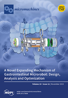
The morbidity and mortality of gastrointestinal malignancy and related diseases are increasing year by year, which has seriously threatened people’s health and even life. Research focused on the gastrointestinal microrobot, which can move autonomically and expand the intestinal tract, is important. The expanding mechanism is the key device for the gastrointestinal microrobot to achieve active locomotion and anchoring in the intestinal tract. The performance of the gastrointestinal microrobot is directly related to the gastrointestinal tract safety, applicability, and work efficiency. View this paper
  • Issues are regarded as officially published after their release is announced to the table of contents alert mailing list.
  • You may sign up for e-mail alerts to receive table of contents of newly released issues.
  • PDF is the official format for papers published in both, html and pdf forms. To view the papers in pdf format, click on the "PDF Full-text" link, and use the free Adobe Reader to open them.
Order results
Result details
Section
Select all
Export citation of selected articles as:
13 pages, 6942 KiB  
Article
Influence of Waterproof Films on the Atomization Behavior of Surface Acoustic Waves
by Qing-Yun Huang, Hong Hu, Jun-Long Han, Yu-Lin Lei and Xiao-Qing Yang
Micromachines 2019, 10(11), 794; https://doi.org/10.3390/mi10110794 - 19 Nov 2019
Cited by 4 | Viewed by 2614
Abstract
One of the reasons why commercial application of surface acoustic wave (SAW) atomization is not possible is due to the condensation of aerosol droplets generated during atomization, which drip on the interdigitated transducer (IDT), thereby causing electrodes to short-circuit. In order to solve [...] Read more.
One of the reasons why commercial application of surface acoustic wave (SAW) atomization is not possible is due to the condensation of aerosol droplets generated during atomization, which drip on the interdigitated transducer (IDT), thereby causing electrodes to short-circuit. In order to solve this problem, a SU-8-2002 film coating on an IDT is proposed in this paper. The waterproof performance of the film coating was tested on a surface acoustic wave (SAW) device several times. The experimental results reveal that the film coating was robust. The experiment also investigated the effects of the SU-8-2002 film on atomization behavior and heating. Full article
Show Figures

Figure 1

13 pages, 3303 KiB  
Article
Tetrafluoroethylene-Propylene Elastomer for Fabrication of Microfluidic Organs-on-Chips Resistant to Drug Absorption
by Emi Sano, Chihiro Mori, Naoki Matsuoka, Yuka Ozaki, Keisuke Yagi, Aya Wada, Koichi Tashima, Shinsuke Yamasaki, Kana Tanabe, Kayo Yano and Yu-suke Torisawa
Micromachines 2019, 10(11), 793; https://doi.org/10.3390/mi10110793 - 19 Nov 2019
Cited by 38 | Viewed by 5314
Abstract
Organs-on-chips are microfluidic devices typically fabricated from polydimethylsiloxane (PDMS). Since PDMS has many attractive properties including high optical clarity and compliance, PDMS is very useful for cell culture applications; however, PDMS possesses a significant drawback in that small hydrophobic molecules are strongly absorbed. [...] Read more.
Organs-on-chips are microfluidic devices typically fabricated from polydimethylsiloxane (PDMS). Since PDMS has many attractive properties including high optical clarity and compliance, PDMS is very useful for cell culture applications; however, PDMS possesses a significant drawback in that small hydrophobic molecules are strongly absorbed. This drawback hinders widespread use of PDMS-based devices for drug discovery and development. Here, we describe a microfluidic cell culture system made of a tetrafluoroethylene-propylene (FEPM) elastomer. We demonstrated that FEPM does not absorb small hydrophobic compounds including rhodamine B and three types of drugs, nifedipine, coumarin, and Bay K8644, whereas PDMS absorbs them strongly. The device consists of two FEPM layers of microchannels separated by a thin collagen vitrigel membrane. Since FEPM is flexible and biocompatible, this microfluidic device can be used to culture cells while applying mechanical strain. When human umbilical vein endothelial cells (HUVECs) were subjected to cyclic strain (~10%) for 4 h in this device, HUVECs reoriented and aligned perpendicularly in response to the cyclic stretch. Moreover, we demonstrated that this device can be used to replicate the epithelial–endothelial interface as well as to provide physiological mechanical strain and fluid flow. This method offers a robust platform to produce organs-on-chips for drug discovery and development. Full article
(This article belongs to the Special Issue Organs-on-chips)
Show Figures

Figure 1

12 pages, 7309 KiB  
Article
A Robust Fully-Integrated Digital-Output Inductive CMOS-MEMS Accelerometer with Improved Inductor Quality Factor
by Yi Chiu, Hsuan-Wu Liu and Hao-Chiao Hong
Micromachines 2019, 10(11), 792; https://doi.org/10.3390/mi10110792 - 18 Nov 2019
Cited by 1 | Viewed by 3309
Abstract
This paper presents the design, fabrication, and characterization of an inductive complementary metal oxide semiconductor micro-electromechanical systems (CMOS-MEMS) accelerometer with on-chip digital output based on LC oscillators. While most MEMS accelerometers employ capacitive detection schemes, the proposed inductive detection scheme is less susceptible [...] Read more.
This paper presents the design, fabrication, and characterization of an inductive complementary metal oxide semiconductor micro-electromechanical systems (CMOS-MEMS) accelerometer with on-chip digital output based on LC oscillators. While most MEMS accelerometers employ capacitive detection schemes, the proposed inductive detection scheme is less susceptible to the stress-induced structural curling and deformation that are commonly seen in CMOS-MEMS devices. Oscillator-based frequency readout does not need analog to digital conversion and thus can simplify the overall system design. In this paper, a high-Q CMOS inductor was connected in series with the low-Q MEMS sensing inductor to improve its quality factor. Measurement results showed the proposed device had an offset frequency of 85.5 MHz, sensitivity of 41.6 kHz/g, noise floor of 8.2 mg/√Hz, bias instability of 0.94 kHz (11 ppm) at an average time of 2.16 s, and nonlinearity of 1.5% full-scale. Full article
(This article belongs to the Special Issue Advanced MEMS/NEMS Technology, Volume II)
Show Figures

Figure 1

13 pages, 8192 KiB  
Article
The Analysis of the Influence of Threshold on the Dynamic Contact Process of a Fabricated Vertically Driven MEMS Inertial Switch
by Wenguo Chen, Rui Wang, Huiying Wang, Dejian Kong and Shulei Sun
Micromachines 2019, 10(11), 791; https://doi.org/10.3390/mi10110791 - 18 Nov 2019
Cited by 2 | Viewed by 2292
Abstract
In this work, to evaluate the influence of the threshold on the dynamic contact process, five models (number 1, 2, 3, 4, 5) with different thresholds were proposed and fabricated with surface micromachining technology. The contact time and response time were used to [...] Read more.
In this work, to evaluate the influence of the threshold on the dynamic contact process, five models (number 1, 2, 3, 4, 5) with different thresholds were proposed and fabricated with surface micromachining technology. The contact time and response time were used to characterize the dynamic contact performance. The dynamic contact processes of the inertial switches with gradually increasing thresholds were researched using analytical, simulation, and experimental methods. The basic working principle analysis of the inertial switch shows that the contact time of the inertial switch with a low-g value can be extended by using a simply supported beam as the fixed electrode, but the high-G inertial needs more elasticity for fixed electrode. The simulation results indicate that the response time and contact time decrease with the increment in the designed threshold. Prototypes were tested using a dropping hammer system, and the test result indicates that the contact time of the inertial switch with a fixed electrode of the simply supported beam is about 15 and 5 μs when the threshold is about 280 and 580 g, respectively. Meanwhile, the contact time can be extended to 100 μs for the inertial switch using a spring as the fixed electrode when the threshold is about 280 and 580 g. These test results not only prove that the spring fixed electrode can effectively extend the contact time, but also prove that the style of the fixed electrode is the deciding factor affecting the contact time of the high-G inertial switch. Full article
(This article belongs to the Special Issue MEMS/NEMS Sensors: Fabrication and Application, Volume II)
Show Figures

Figure 1

13 pages, 2879 KiB  
Article
Engineering a Bi-Conical Microchip as Vascular Stenosis Model
by Yan Li, Jianchun Wang, Wei Wan, Chengmin Chen, Xueying Wang, Pei Zhao, Yanjin Hou, Hanmei Tian, Jianmei Wang, Krishnaswamy Nandakumar and Liqiu Wang
Micromachines 2019, 10(11), 790; https://doi.org/10.3390/mi10110790 - 18 Nov 2019
Viewed by 3110
Abstract
Vascular stenosis is always associated with hemodynamic changes, especially shear stress alterations. Herein, bi-conical shaped microvessels were developed through flexibly and precisely controlled templated methods for hydrogel blood-vessel-like microchip. The blood-vessel-like microvessels demonstrated tunable dimensions, perfusable ability, and good cytocompatibility. The microchips showed [...] Read more.
Vascular stenosis is always associated with hemodynamic changes, especially shear stress alterations. Herein, bi-conical shaped microvessels were developed through flexibly and precisely controlled templated methods for hydrogel blood-vessel-like microchip. The blood-vessel-like microvessels demonstrated tunable dimensions, perfusable ability, and good cytocompatibility. The microchips showed blood-vessel-like lumens through fine embeddedness of human umbilical vein endothelial cells (HUVECs) on the interior surface of hydrogel microchannels, which closely reproduced the morphology and functions of human blood vessels. In the gradual narrowing region of bi-conical shape, fluid flow generated wall shear stress, which caused cell morphology variations. Wall shear rates at the gradual narrowing region were simulated by FLUENT software. The results showed that our microchannels qualified for performance as a vascular stenosis-like model in evaluating blood hydrodynamics. In general, our blood-vessel-on-a-chip could offer potential applications in the prevention, diagnosis, and therapy of arterial thrombosis. Full article
(This article belongs to the Special Issue Microengineering Techniques for Disease Modeling and Drug Discovery)
Show Figures

Graphical abstract

9 pages, 1195 KiB  
Communication
An Implantable Cranial Window Using a Collagen Membrane for Chronic Voltage-Sensitive Dye Imaging
by Nobuo Kunori and Ichiro Takashima
Micromachines 2019, 10(11), 789; https://doi.org/10.3390/mi10110789 - 18 Nov 2019
Cited by 11 | Viewed by 4458
Abstract
Incorporating optical methods into implantable neural sensing devices is a challenging approach for brain–machine interfacing. Specifically, voltage-sensitive dye (VSD) imaging is a powerful tool enabling visualization of the network activity of thousands of neurons at high spatiotemporal resolution. However, VSD imaging usually requires [...] Read more.
Incorporating optical methods into implantable neural sensing devices is a challenging approach for brain–machine interfacing. Specifically, voltage-sensitive dye (VSD) imaging is a powerful tool enabling visualization of the network activity of thousands of neurons at high spatiotemporal resolution. However, VSD imaging usually requires removal of the dura mater for dye staining, and thereafter the exposed cortex needs to be protected using an optically transparent artificial dura. This is a major disadvantage that limits repeated VSD imaging over the long term. To address this issue, we propose to use an atelocollagen membrane as the dura substitute. We fabricated a small cranial chamber device, which is a tubular structure equipped with a collagen membrane at one end of the tube. We implanted the device into rats and monitored neural activity in the frontal cortex 1 week following surgery. The results indicate that the collagen membrane was chemically transparent, allowing VSD staining across the membrane material. The membrane was also optically transparent enough to pass light; forelimb-evoked neural activity was successfully visualized through the artificial dura. Because of its ideal chemical and optical manipulation capability, this collagen membrane may be widely applicable in various implantable neural sensors. Full article
(This article belongs to the Special Issue Implantable Neural Sensors for the Brain Machine Interface)
Show Figures

Figure 1

12 pages, 2890 KiB  
Article
Preparing Polypyrrole-Coated Stretchable Textile via Low-Temperature Interfacial Polymerization for Highly Sensitive Strain Sensor
by Xiaodie Chen, Bintian Li, Yan Qiao and Zhisong Lu
Micromachines 2019, 10(11), 788; https://doi.org/10.3390/mi10110788 - 17 Nov 2019
Cited by 29 | Viewed by 5260
Abstract
The stretchable sensor has been considered as the most important component in a wearable device. However, it is still a great challenge to develop a highly sensitive textile-based strain sensor with good flexibility, excellent skin affinity, and large dynamic range. Herein, polypyrrole (PPy) [...] Read more.
The stretchable sensor has been considered as the most important component in a wearable device. However, it is still a great challenge to develop a highly sensitive textile-based strain sensor with good flexibility, excellent skin affinity, and large dynamic range. Herein, polypyrrole (PPy) was immobilized on a stretchable textile knitted by polyester and spandex via low-temperature interfacial polymerization to prepare a conductive strain sensor for human motion and respiration measurements. Scanning electron microscopy, Fourier transform infrared spectrometry, and thermal gravimetric data verify that a thin layer of PPy has been successfully coated on the textile with a high density and very uniform distribution. The resistance of the as-prepared textile is 21.25 Ω/cm2 and the PPy-coated textile could be used as an electric conductor to light up a LED lamp. Moreover, the textile could tolerate folding at an angle of 180° and 500 times of bending-twisting cycles without significant changes on its resistance. A negative correlation between the resistance change and the applied strain is observed for the textile-based sensor in the strain ranging from 0 to 71% with the gauge factor of −0.46. After more than 200 cycles of stretching-releasing under the strain of 26%, there is no obvious alteration on the sensing responses. The sensors were attached on volunteers’ body or clothes for the real-time measurement of human motions and respiration, demonstrating that the textile-based sensor could sensitively detect finger, elbow, and knee bending and differentiate deep, normal, and fast breath. This work may provide an approach to uniform and dense coating conductive polymers on textiles for highly sensitive and stretchable sensors, which possess great potentials in practical applications for real-time monitoring human motions and physiological signs. Full article
Show Figures

Figure 1

17 pages, 1339 KiB  
Review
Whole Blood Based Multiparameter Assessment of Thrombus Formation in Standard Microfluidic Devices to Proxy In Vivo Haemostasis and Thrombosis
by Isabella Provenzale, Sanne L. N. Brouns, Paola E. J. van der Meijden, Frauke Swieringa and Johan W. M. Heemskerk
Micromachines 2019, 10(11), 787; https://doi.org/10.3390/mi10110787 - 16 Nov 2019
Cited by 16 | Viewed by 6352
Abstract
Microfluidic assays are versatile tests which, using only small amounts of blood, enable high throughput analyses of platelet function in several minutes. In combination with fluorescence microscopy, these flow tests allow real-time visualisation of platelet activation with the possibility of examining combinatorial effects [...] Read more.
Microfluidic assays are versatile tests which, using only small amounts of blood, enable high throughput analyses of platelet function in several minutes. In combination with fluorescence microscopy, these flow tests allow real-time visualisation of platelet activation with the possibility of examining combinatorial effects of wall shear rate, coagulation and modulation by endothelial cells. In particular, the ability to use blood and blood cells from healthy subjects or patients makes this technology promising, both for research and (pre)clinical diagnostic purposes. In the present review, we describe how microfluidic devices are used to assess the roles of platelets in thrombosis and haemostasis. We place emphasis on technical aspects and on experimental designs that make the concept of “blood-vessel-component-on-a-chip” an attractive, rapidly developing technology for the study of the complex biological processes of blood coagulability in the presence of flow. Full article
(This article belongs to the Special Issue Blood-on-a-Chip)
Show Figures

Figure 1

16 pages, 2556 KiB  
Article
Mixing Performance of a Cost-effective Split-and-Recombine 3D Micromixer Fabricated by Xurographic Method
by Ramezan Ali Taheri, Vahabodin Goodarzi and Abdollah Allahverdi
Micromachines 2019, 10(11), 786; https://doi.org/10.3390/mi10110786 - 16 Nov 2019
Cited by 19 | Viewed by 3765
Abstract
This paper presents experimental and numerical investigations of a novel passive micromixer based on the lamination of fluid layers. Lamination-based mixers benefit from increasing the contact surface between two fluid phases by enhancing molecular diffusion to achieve a faster mixing. Novel three-dimensional split [...] Read more.
This paper presents experimental and numerical investigations of a novel passive micromixer based on the lamination of fluid layers. Lamination-based mixers benefit from increasing the contact surface between two fluid phases by enhancing molecular diffusion to achieve a faster mixing. Novel three-dimensional split and recombine (SAR) structures are proposed to generate fluid laminations. Numerical simulations were conducted to model the mixer performance. Furthermore, experiments were conducted using dyes to observe fluid laminations and evaluate the proposed mixer’s characteristics. Mixing quality was experimentally obtained by means of image-based mixing index (MI) measurement. The multi-layer device was fabricated utilizing the Xurography method, which is a simple and low-cost method to fabricate 3D microfluidic devices. Mixing indexes of 96% and 90% were obtained at Reynolds numbers of 0.1 and 1, respectively. Moreover, the device had an MI value of 67% at a Reynolds number of 10 (flow rate of 116 µL/min for each inlet). The proposed micromixer, with its novel design and fabrication method, is expected to benefit a wide range of lab-on-a-chip applications, due to its high efficiency, low cost, high throughput and ease of fabrication. Full article
Show Figures

Figure 1

13 pages, 8550 KiB  
Article
Robust Tracking of a Cost-Effective Micro-Stereolithography System Based on a Compliant Nanomanipulator
by Yue Cao and Zhen Zhang
Micromachines 2019, 10(11), 785; https://doi.org/10.3390/mi10110785 - 16 Nov 2019
Cited by 3 | Viewed by 2708
Abstract
Micro-stereolithography (MSL) has emerged as a promising and challenging technique in micro-/nano-scale additive manufacturing. Besides the requirement of the light source, the motion system requires ultra-high-precision tracking capability to reach the right location for every solidification event. To achieve single-digit micron feature size [...] Read more.
Micro-stereolithography (MSL) has emerged as a promising and challenging technique in micro-/nano-scale additive manufacturing. Besides the requirement of the light source, the motion system requires ultra-high-precision tracking capability to reach the right location for every solidification event. To achieve single-digit micron feature size of the fabrication, we propose a robust control strategy to support a self-developed cost-effective MSL prototype based on a compliant nanomanipulator and a blue light-emitting diode (LED) module. In particular, the nonlinearity and parameter-variation of the compliant manipulator are dealt with by a robust radial basis function (RBF)-based neural network, and the repetitive control (RC) is innovatively integrated with RBF to improve the tracking performance of a closed pattern. Various simulations and real-time experiments are conducted to validate the proposed control strategy. The fabrication of a closed pattern will not begin by turning on the laser source until the tracking error reaches submicrons, and the fabrication results demonstrate that the cost-effective MSL system is capable of fabricating 2.5 µm feature size in a 0.5 mm working range. Full article
(This article belongs to the Special Issue Manipulation, Manufacturing and Measurement on the Nanoscale 2019)
Show Figures

Figure 1

15 pages, 5266 KiB  
Article
Configuration and Design of Electromagnets for Rapid and Precise Manipulation of Magnetic Beads in Biosensing Applications
by Moshe Stern, Meir Cohen and Amos Danielli
Micromachines 2019, 10(11), 784; https://doi.org/10.3390/mi10110784 - 15 Nov 2019
Cited by 9 | Viewed by 3546
Abstract
Rapid and precise manipulation of magnetic beads on the nano and micro scales is essential in many biosensing applications, such as separating target molecules from background molecules and detecting specific proteins and DNA sequences in plasma. Accurately moving magnetic beads back and forth [...] Read more.
Rapid and precise manipulation of magnetic beads on the nano and micro scales is essential in many biosensing applications, such as separating target molecules from background molecules and detecting specific proteins and DNA sequences in plasma. Accurately moving magnetic beads back and forth requires at least two adjustable magnetic field gradients. Unlike permanent magnets, electromagnets are easy to design and can produce strong and adjustable magnetic field gradients without mechanical motion, making them desirable for use in robust and safe medical devices. However, using multiple magnetic field sources to manipulate magnetic beads presents several challenges, including overlapping magnetic fields, added bulk, increased cost, and reduced durability. Here, we provide a thorough analysis, including analytical calculations, numerical simulations, and experimental measurements, of using two electromagnets to manipulate magnetic beads inside a miniature glass cell. We analyze and experimentally demonstrate different aspects of the electromagnets’ design, such as their mutual influence, the advantages and disadvantages of different pole tip geometries, and the correlation between the electromagnets’ positions and the beads’ aggregation during movement. Finally, we have devised a protocol to maximize the magnetic forces acting on magnetic beads in a two-electromagnet setup while minimizing the electromagnets’ size. We used two such electromagnets in a small footprint magnetic modulation biosensing system and detected as little as 13 ng/L of recombinant Zika virus antibodies, which enables detection of Zika IgM antibodies as early as 5 days and as late as 180 days post symptoms onset, significantly extending the number of days that the antibodies are detectable. Full article
(This article belongs to the Special Issue Magnetic Biosensors)
Show Figures

Figure 1

9 pages, 5211 KiB  
Article
Imine Reductase Based All-Enzyme Hydrogel with Intrinsic Cofactor Regeneration for Flow Biocatalysis
by Patrick Bitterwolf, Felix Ott, Kersten S. Rabe and Christof M. Niemeyer
Micromachines 2019, 10(11), 783; https://doi.org/10.3390/mi10110783 - 15 Nov 2019
Cited by 19 | Viewed by 4148
Abstract
All-enzyme hydrogels are biocatalytic materials, with which various enzymes can be immobilized in microreactors in a simple, mild, and efficient manner to be used for continuous flow processes. Here we present the construction and application of a cofactor regenerating hydrogel based on the [...] Read more.
All-enzyme hydrogels are biocatalytic materials, with which various enzymes can be immobilized in microreactors in a simple, mild, and efficient manner to be used for continuous flow processes. Here we present the construction and application of a cofactor regenerating hydrogel based on the imine reductase GF3546 from Streptomyces sp. combined with the cofactor regenerating glucose-1-dehydrogenase from Bacillus subtilis. The resulting hydrogel materials were characterized in terms of binding kinetics and viscoelastic properties. The materials were formed by rapid covalent crosslinking in less than 5 min, and they showed a typical mesh size of 67 ± 2 nm. The gels were applied for continuous flow biocatalysis. In a microfluidic reactor setup, the hydrogels showed excellent conversions of imines to amines for up to 40 h in continuous flow mode. Variation of flow rates led to a process where the gels showed a maximum space-time-yield of 150 g·(L·day)−1 at 100 μL/min. Full article
Show Figures

Graphical abstract

16 pages, 10400 KiB  
Article
The Influence of Piezoelectric Transducer Stimulating Sites on the Performance of Implantable Middle Ear Hearing Devices: A Numerical Analysis
by Houguang Liu, Yu Zhao, Jianhua Yang and Zhushi Rao
Micromachines 2019, 10(11), 782; https://doi.org/10.3390/mi10110782 - 14 Nov 2019
Cited by 4 | Viewed by 4760
Abstract
To overcome the inherent deficiencies of hearing aids, implantable middle ear hearing devices (IMEHDs) have emerged as a new treatment for hearing loss. However, clinical results show that the IMEHD performance varies with its transducer’s stimulating site. To numerically analyze the influence of [...] Read more.
To overcome the inherent deficiencies of hearing aids, implantable middle ear hearing devices (IMEHDs) have emerged as a new treatment for hearing loss. However, clinical results show that the IMEHD performance varies with its transducer’s stimulating site. To numerically analyze the influence of the piezoelectric transducer’s stimulating sites on its hearing compensation performance, we constructed a human ear finite element model and confirmed its validity. Based on this finite element model, the displacement stimulation, which simulates the piezoelectric transducer’s stimulation, was applied to the umbo, the incus long process, the incus body, the stapes, and the round window membrane, respectively. Then, the stimulating site’s effect of the piezoelectric transducer was analyzed by comparing the corresponding displacements of the basilar membrane. Besides, the stimulating site’s sensitivity to the direction of excitation was also studied. The result of the finite element analysis shows that stimulating the incus body is least efficient for the piezoelectric transducer. Meanwhile, stimulating the round window membrane or the stapes generates a higher basilar membrane displacement than stimulating the eardrum or the incus long process. However, the performance of these two ideal sites’ stimulation is sensitive to the changes in the excitation’s direction. Thus, the round window membrane and the stapes is the ideal stimulating sites for the piezoelectric transducer regarding the driving efficiency. The direction of the excitation should be guaranteed for these ideal sites. Full article
(This article belongs to the Special Issue Piezoelectric Transducers: Materials, Devices and Applications)
Show Figures

Figure 1

13 pages, 3837 KiB  
Communication
Automated Analysis of Platelet Aggregation on Cultured Endothelium in a Microfluidic Chip Perfused with Human Whole Blood
by Hugo J. Albers, Robert Passier, Albert van den Berg and Andries D. van der Meer
Micromachines 2019, 10(11), 781; https://doi.org/10.3390/mi10110781 - 14 Nov 2019
Cited by 10 | Viewed by 5279
Abstract
Organ-on-a-chip models with incorporated vasculature are becoming more popular to study platelet biology. A large variety of image analysis techniques are currently used to determine platelet coverage, ranging from manually setting thresholds to scoring platelet aggregates. In this communication, an automated methodology is [...] Read more.
Organ-on-a-chip models with incorporated vasculature are becoming more popular to study platelet biology. A large variety of image analysis techniques are currently used to determine platelet coverage, ranging from manually setting thresholds to scoring platelet aggregates. In this communication, an automated methodology is introduced, which corrects misalignment of a microfluidic channel, automatically defines regions of interest and utilizes a triangle threshold to determine platelet coverages and platelet aggregate size distributions. A comparison between the automated methodology and manual identification of platelet aggregates shows a high accuracy of the triangle methodology. Furthermore, the image analysis methodology can determine platelet coverages and platelet size distributions in microfluidic channels lined with either untreated or activated endothelium used for whole blood perfusion, proving the robustness of the method. Full article
(This article belongs to the Special Issue Blood-on-a-Chip)
Show Figures

Figure 1

12 pages, 4825 KiB  
Article
Rapid Design and Analysis of Microtube Pneumatic Actuators Using Line-Segment and Multi-Segment Euler–Bernoulli Beam Models
by Myunggi Ji, Qiang Li, In Ho Cho and Jaeyoun Kim
Micromachines 2019, 10(11), 780; https://doi.org/10.3390/mi10110780 - 14 Nov 2019
Cited by 3 | Viewed by 2787
Abstract
Soft material-based pneumatic microtube actuators are attracting intense interest, since their bending motion is potentially useful for the safe manipulation of delicate biological objects. To increase their utility in biomedicine, researchers have begun to apply shape-engineering to the microtubes to diversify their bending [...] Read more.
Soft material-based pneumatic microtube actuators are attracting intense interest, since their bending motion is potentially useful for the safe manipulation of delicate biological objects. To increase their utility in biomedicine, researchers have begun to apply shape-engineering to the microtubes to diversify their bending patterns. However, design and analysis of such microtube actuators are challenging in general, due to their continuum natures and small dimensions. In this paper, we establish two methods for rapid design, analysis, and optimization of such complex, shape-engineered microtube actuators that are based on the line-segment model and the multi-segment Euler–Bernoulli’s beam model, respectively, and are less computation-intensive than the more conventional method based on finite element analysis. To validate the models, we first realized multi-segment microtube actuators physically, then compared their experimentally observed motions against those obtained from the models. We obtained good agreements between the three sets of results with their maximum bending-angle errors falling within ±11%. In terms of computational efficiency, our models decreased the simulation time significantly, down to a few seconds, in contrast with the finite element analysis that sometimes can take hours. The models reported in this paper exhibit great potential for rapid and facile design and optimization of shape-engineered soft actuators. Full article
(This article belongs to the Special Issue 10th Anniversary of Micromachines)
Show Figures

Graphical abstract

8 pages, 2443 KiB  
Article
Electrical Performance and Bias-Stress Stability of Amorphous InGaZnO Thin-Film Transistors with Buried-Channel Layers
by Ying Zhang, Haiting Xie and Chengyuan Dong
Micromachines 2019, 10(11), 779; https://doi.org/10.3390/mi10110779 - 14 Nov 2019
Cited by 7 | Viewed by 4075
Abstract
To improve the electrical performance and bias-stress stability of amorphous InGaZnO thin-film transistors (a-IGZO TFTs), we fabricated and characterized buried-channel devices with multiple-stacked channel layers, i.e., a nitrogen-doped a-IGZO film (front-channel layer), a conventional a-IGZO film (buried-channel layer), and a nitrogen-doped a-IGZO film [...] Read more.
To improve the electrical performance and bias-stress stability of amorphous InGaZnO thin-film transistors (a-IGZO TFTs), we fabricated and characterized buried-channel devices with multiple-stacked channel layers, i.e., a nitrogen-doped a-IGZO film (front-channel layer), a conventional a-IGZO film (buried-channel layer), and a nitrogen-doped a-IGZO film (back-channel layer). The larger field-effect mobility (5.8 cm2V−1s−1), the smaller subthreshold swing value (0.8 V/dec, and the better stability (smaller threshold voltage shifts during bias-stress and light illumination tests) were obtained for the buried-channel device relative to the conventional a-IGZO TFT. The specially designed channel-layer structure resulted in multiple conduction channels and hence large field-effect mobility. The in situ nitrogen-doping caused reductions in both the front-channel interface trap density and the density of deep states in the bulk channel layers, leading to a small subthreshold swing value. The better stability properties may be related to both the reduced trap states by nitrogen-doping and the passivation effect of the nitrogen-doped a-IGZO films at the device back channels. Full article
(This article belongs to the Special Issue Wide Bandgap Based Devices: Design, Fabrication and Applications)
Show Figures

Figure 1

15 pages, 11235 KiB  
Article
Dynamics Behaviors of Droplet on Hydrophobic Surfaces Driven by Electric Field
by Jie Liu and Sheng Liu
Micromachines 2019, 10(11), 778; https://doi.org/10.3390/mi10110778 - 14 Nov 2019
Cited by 8 | Viewed by 3322
Abstract
Droplet microfluidic technology achieves precise manipulation of droplet behaviors by designing and controlling the flow and interaction of various incompatible fluids. The electric field provides a non-contact, pollution-free, designable and promising method for droplet microfluidics. Since the droplet behaviors in many industrial and [...] Read more.
Droplet microfluidic technology achieves precise manipulation of droplet behaviors by designing and controlling the flow and interaction of various incompatible fluids. The electric field provides a non-contact, pollution-free, designable and promising method for droplet microfluidics. Since the droplet behaviors in many industrial and biological applications occur on the contact surface and the properties of droplets and the surrounding environment are not consistent, it is essential to understand fundamentally the sessile droplet motion and deformation under various conditions. This paper reports a technique using the pin-plate electrode to generate non-uniform dielectrophoresis (DEP) force to control sessile droplets on hydrophobic surfaces. The electrohydrodynamics phenomena of the droplet motion and deformation are simulated using the phase-field method. It is found that the droplet moves along the substrate surface to the direction of higher electric field strength, and is accompanied with a certain offset displacement. In addition, the effect of pin electric potentials, surface contact angles and droplet volumes on the droplet motion and deformation are also studied and compared. The results show that higher potentials, more hydrophobic surfaces and larger droplet volumes exhibit greater droplet horizontal displacement and offset displacement. But for the droplet vertical displacement, it is found that during the first revert process, the release of the surface tension can make the droplet with low potentials, small contact angles or small droplet volumes span from negative to positive. These results will be helpful for future operations encountered in sessile droplets under non-uniform electric fields towards the droplet microfluidics applications. Full article
(This article belongs to the Special Issue Micro/Nano-Chip Electrokinetics, Volume III)
Show Figures

Figure 1

11 pages, 1950 KiB  
Article
Fast and Parallel Detection of Four Ebola Virus Species on a Microfluidic-Chip-Based Portable Reverse Transcription Loop-Mediated Isothermal Amplification System
by Xue Lin, Xiangyu Jin, Bin Xu, Ruliang Wang, Rongxin Fu, Ya Su, Kai Jiang, Han Yang, Ying Lu, Yong Guo and Guoliang Huang
Micromachines 2019, 10(11), 777; https://doi.org/10.3390/mi10110777 - 14 Nov 2019
Cited by 20 | Viewed by 3599
Abstract
Considering the lack of official vaccines and medicines for Ebola virus infection, reliable diagnostic methods are necessary for the control of the outbreak and the spread of the disease. We developed a microfluidic-chip-based portable system for fast and parallel detection of four Ebola [...] Read more.
Considering the lack of official vaccines and medicines for Ebola virus infection, reliable diagnostic methods are necessary for the control of the outbreak and the spread of the disease. We developed a microfluidic-chip-based portable system for fast and parallel detection of four Ebola virus species. The system is based on reverse transcription loop-mediated isothermal amplification (RT-LAMP) and consists of four specific LAMP primers, a disc microfluidic chip, and a portable real-time fluorescence detector. It could specifically and parallelly distinguish four species of the Ebola virus after only one sampling, including the Zaire Ebola virus, the Sudan Ebola virus, the Bundibugyo Ebola virus, and the Tai Forest Ebola virus, without cross-contamination. The limit of detection was as small as 10 copies per reaction, while the total consumption of sample and reagent was 0.94 μL per reaction. The final results could be obtained in 50 min after one addition of sample and reagent mixture. This approach provides simplicity, high sensitivity, and multi-target parallel detection at a low cost, which could enable convenient and effective on-site detections of the Ebola virus in the outdoors, remote areas, and modern hospitals. Full article
(This article belongs to the Special Issue Microsystems for Point-of-Care Testing)
Show Figures

Figure 1

10 pages, 1807 KiB  
Article
Calibration Technique of a Curved Zoom Compound Eye Imaging System
by Fengli Liu, Xiaolei Diao, Lun Li and Yongping Hao
Micromachines 2019, 10(11), 776; https://doi.org/10.3390/mi10110776 - 13 Nov 2019
Cited by 3 | Viewed by 1830
Abstract
A calibration method for the designed curved zoom compound eye is studied in order to achieve detection and positioning of spatial objects. The structure of the curved zoom compound eye is introduced. A calibration test platform is designed and built based on the [...] Read more.
A calibration method for the designed curved zoom compound eye is studied in order to achieve detection and positioning of spatial objects. The structure of the curved zoom compound eye is introduced. A calibration test platform is designed and built based on the image characteristics of the compound eye, which can be constructed in the large field view for the calibration target. The spot images are obtained through image processing. The center of the spot is calculated by Gauss fitting method. This method is highly simple and intuitive, and it can be used in a zoom surface compound eye without any complex procedures. Finally, the corresponding relationship between the spot center coordinates and the incident light vector of the corresponding sub-eye is established, and the calibration of the multi vision positioning system is completed. Full article
(This article belongs to the Special Issue MOEMS: Micro-Optical MEMS)
Show Figures

Figure 1

11 pages, 3349 KiB  
Article
Generation of Pure State Photon Triplets in the C-Band
by Xi-Rong Su, Yi-Wen Huang, Tong Xiang, Yuan-Hua Li and Xian-Feng Chen
Micromachines 2019, 10(11), 775; https://doi.org/10.3390/mi10110775 - 13 Nov 2019
Cited by 1 | Viewed by 2660
Abstract
In this work, the cascaded second-order spontaneous parametric down-conversion (SPDC) is considered to produce pure state photon triplets in periodically poled lithium niobite (PPLN) doped with 5% MgO. A set of parameters are optimized through calculating the Schmidt number of two-photon states generated [...] Read more.
In this work, the cascaded second-order spontaneous parametric down-conversion (SPDC) is considered to produce pure state photon triplets in periodically poled lithium niobite (PPLN) doped with 5% MgO. A set of parameters are optimized through calculating the Schmidt number of two-photon states generated by each down-conversion process with different pump durations and crystal lengths. We use a Gaussian filter in part and obtain three photons with 100% purity in spectrum. We provide a feasible and unprecedented scheme to manipulate the spectrum purity of photon triplets in the communication band (C-band). Full article
(This article belongs to the Special Issue Nonlinear Photonics Devices)
Show Figures

Figure 1

17 pages, 4794 KiB  
Article
Controlled Focused Ion Beam Milling of Composite Solid State Nanopore Arrays for Molecule Sensing
by Péter Fürjes
Micromachines 2019, 10(11), 774; https://doi.org/10.3390/mi10110774 - 13 Nov 2019
Cited by 14 | Viewed by 4150
Abstract
Various nanoscale fabrication techniques are elaborated to form artificial nanoporous/nanochannel membranes to be applied for biosensing: one of the most prevalent is the micro-electromechanical systems (MEMS) compatible focused ion beam (FIB) milling. This technique can be easily adopted in micro- and nanomachining process [...] Read more.
Various nanoscale fabrication techniques are elaborated to form artificial nanoporous/nanochannel membranes to be applied for biosensing: one of the most prevalent is the micro-electromechanical systems (MEMS) compatible focused ion beam (FIB) milling. This technique can be easily adopted in micro- and nanomachining process sequences to develop composite multi-pore structures, although its precision and reproducibility are key points in the case of these thick multi-layered membranes. This work is to demonstrate a comprehensive characterisation of FIB milling to improve the reliability of the fabrication of solid state nanopore arrays with precisely predetermined pore geometries for a targeted molecule type to be recognised. The statistical geometric features of the fabricated nanopores were recorded as the function of the process parameters, and the resulting geometries were analysed in detail by high resolution scanning electron microscope (SEM), transmission electron microscope (TEM) and ion scanning microscopy. Continuous function of the pore diameter evolution rate was derived from the experimental results in the case of different material structures, and compared to former dissentient estimations. The additional metal layer was deposited onto the backside of the membrane and grounded during the ion milling to prevent the electrical charging of dielectric layers. The study proved that the conformity of the pore geometry and the reliability of their fabrication could be improved significantly. The applicability of the developed nanopore arrays for molecule detection was also considered by characterising the pore diameter dependent sensitivity of the membrane impedance modulation based measurement method. Full article
(This article belongs to the Special Issue Nanomaterials-Based Biosensors)
Show Figures

Figure 1

10 pages, 2406 KiB  
Article
Highly Sensitive Temperature Sensing Performance of a Microfiber Fabry-Perot Interferometer with Sealed Micro-Spherical Reflector
by Jin Li, Juntong Yang and Jinna Ma
Micromachines 2019, 10(11), 773; https://doi.org/10.3390/mi10110773 - 12 Nov 2019
Cited by 9 | Viewed by 2522
Abstract
A temperature probe has been proposed by inserting a microfiber taper into a silica hollow core fiber with a microsphere end. The sealed air cavity in the microsphere and the inserted microfiber acted as the two reflectors of a Fabry-Perot interferometer, respectively. The [...] Read more.
A temperature probe has been proposed by inserting a microfiber taper into a silica hollow core fiber with a microsphere end. The sealed air cavity in the microsphere and the inserted microfiber acted as the two reflectors of a Fabry-Perot interferometer, respectively. The contribution of both microfiber diameter and cavity length on the interference spectra was analyzed and discussed in detail. The temperature change was experimentally determined by monitoring the wavelength location of the special resonance dip. By filling the air cavity with poly-dimethylsiloxane (PDMS), a high temperature sensitivity of 3.90 nm/°C was experimentally demonstrated. This temperature probe with the diameter of 150 μm and length of 10 mm will be a promising candidate for exploring the miniature or implantable sensors. Full article
Show Figures

Figure 1

19 pages, 6326 KiB  
Article
Investigation of Leukocyte Viability and Damage in Spiral Microchannel and Contraction-Expansion Array
by Thammawit Suwannaphan, Werayut Srituravanich, Achariya Sailasuta, Prapruddee Piyaviriyakul, Suchaya Bhanpattanakul, Wutthinan Jeamsaksiri, Witsaroot Sripumkhai and Alongkorn Pimpin
Micromachines 2019, 10(11), 772; https://doi.org/10.3390/mi10110772 - 12 Nov 2019
Cited by 10 | Viewed by 3692
Abstract
Inertial separation techniques in a microfluidic system have been widely employed in the field of medical diagnosis for a long time. Despite no requirement of external forces, it requires strong hydrodynamic forces that could potentially cause cell damage or loss during the separation [...] Read more.
Inertial separation techniques in a microfluidic system have been widely employed in the field of medical diagnosis for a long time. Despite no requirement of external forces, it requires strong hydrodynamic forces that could potentially cause cell damage or loss during the separation process. This might lead to the wrong interpretation of laboratory results since the change of structures and functional characteristics of cells due to the hydrodynamic forces that occur are not taken into account. Therefore, it is important to investigate the cell viability and damage along with the separation efficacy of the device in the design process. In this study, two inertial separation techniques—spiral microchannel and contraction-expansion array (CEA)—were examined to evaluate cell viability, morphology and intracellular structures using a trypan blue assay (TB), Scanning Electron Microscopy (SEM) and Wright-Giemsa stain. We discovered that cell loss was not significantly found in a feeding system, i.e., syringe, needle and tube, but mostly occurred in the inertial separation devices while the change of cell morphology and intracellular structures were found in the feeding system and inertial separation devices. Furthermore, percentage of cell loss was not significant in both devices (7–10%). However, the change of cell morphology was considerably increased (30%) in spiral microchannel (shear stress dominated) rather than in CEA (12%). In contrast, the disruption of intracellular structures was increased (14%) in CEA (extensional and shear stress dominated equally) rather than spiral microchannel (2%). In these experiments, leukocytes of canine were used as samples because their sizes are varied in a range between 7–12 µm, and they are commonly used as a biomarker in many clinical and medical applications. Full article
(This article belongs to the Special Issue Biomedical Microfluidic Devices 2019)
Show Figures

Figure 1

13 pages, 4051 KiB  
Article
Design and Waveform Assessment of a Flexible-Structure-Based Inertia-Drive Motor
by Junyang Wei, Sergej Fatikow, Hai Li and Xianmin Zhang
Micromachines 2019, 10(11), 771; https://doi.org/10.3390/mi10110771 - 12 Nov 2019
Cited by 3 | Viewed by 2189
Abstract
This paper reports the mechanical design, waveform investigation and experimental validation of an flexible-structure-based inertia-drive linear motor. The flexible structure is designed and verified with finite element analysis to meet the bandwidth requirement for high-frequency actuation. In order to improve the output velocity, [...] Read more.
This paper reports the mechanical design, waveform investigation and experimental validation of an flexible-structure-based inertia-drive linear motor. The flexible structure is designed and verified with finite element analysis to meet the bandwidth requirement for high-frequency actuation. In order to improve the output velocity, non-resonance low-harmonic driving waveform is implemented and evaluated. Experimental results show that the motor is capable of an output velocity of 2.41 mm/s with the waveform, compared to 0.73 mm/s with the classic saw-tooth waveform actuation. The improvement of the non-resonance low-harmonic waveform for the flexible-structure-based motor is confirmed. Full article
(This article belongs to the Special Issue Robotic Micromanipulation)
Show Figures

Figure 1

12 pages, 11729 KiB  
Article
Aptamer Affinity-Bead Mediated Capture and Displacement of Gram-Negative Bacteria Using Acoustophoresis
by SangWook Lee, Byung Woo Kim, Hye-Su Shin, Anna Go, Min-Ho Lee, Dong-Ki Lee, Soyoun Kim and Ok Chan Jeong
Micromachines 2019, 10(11), 770; https://doi.org/10.3390/mi10110770 - 11 Nov 2019
Cited by 7 | Viewed by 3500
Abstract
Here, we report a simple and effective method for capturing and displacement of gram-negative bacteria using aptamer-modified microbeads and acoustophoresis. As acoustophoresis allows for simultaneous washing and size-dependent separation in continuous flow mode, we efficiently obtained gram-negative bacteria that showed high affinity without [...] Read more.
Here, we report a simple and effective method for capturing and displacement of gram-negative bacteria using aptamer-modified microbeads and acoustophoresis. As acoustophoresis allows for simultaneous washing and size-dependent separation in continuous flow mode, we efficiently obtained gram-negative bacteria that showed high affinity without any additional washing steps. The proposed device has a simple and efficient channel design, utilizing a long, square-shaped microchannel that shows excellent separation performance in terms of the purity, recovery, and concentration factor. Microbeads (10 µm) coated with the GN6 aptamer can specifically bind gram-negative bacteria. After incubation of bacteria culture sample with aptamer affinity bead, gram-negative bacteria-bound microbeads, and other unbound/contaminants can be separated by size with high purity and recovery. The device demonstrated excellent separation performance, with high recovery (up to 98%), high purity (up to 99%), and a high-volume rate (500 µL/min). The acoustophoretic separation performances were conducted using 5 Gram-negative bacteria and 5 Gram-positive bacteria. Thanks to GN6 aptamer’s binding affinity, aptamer affinity bead also showed binding affinity to multiple strains of gram-negative bacteria, but not to gram-positive bacteria. GN6 coated bead can capture Gram-negative bacteria but not Gram-positive bacteria. This study may present a different perspective in the field of early diagnosis in bacterial infectious diseases. In addition to detecting living bacteria or bacteria-derived biomarkers, this protocol can be extended to monitoring the contamination of water resources and may aid quick responses to bioterrorism and pathogenic bacterial infections. Full article
Show Figures

Figure 1

12 pages, 3675 KiB  
Article
A Liquid-Metal-Based Dielectrophoretic Microdroplet Generator
by Ronghang Wang, Lunjia Zhang, Meng Gao, Qifu Wang, Zhongshan Deng and Lin Gui
Micromachines 2019, 10(11), 769; https://doi.org/10.3390/mi10110769 - 11 Nov 2019
Cited by 7 | Viewed by 2720
Abstract
This paper proposes a novel microdroplet generator based on the dielectrophoretic (DEP) force. Unlike the conventional continuous microfluidic droplet generator, this droplet generator is more like “invisible electric scissors”. It can cut the droplet off from the fluid matrix and modify droplets’ length [...] Read more.
This paper proposes a novel microdroplet generator based on the dielectrophoretic (DEP) force. Unlike the conventional continuous microfluidic droplet generator, this droplet generator is more like “invisible electric scissors”. It can cut the droplet off from the fluid matrix and modify droplets’ length precisely by controlling the electrodes’ length and position. These electrodes are made of liquid metal by injection. By applying a certain voltage on the liquid-metal electrodes, the electrodes generate an uneven electric field inside the main microfluidic channel. Then, the uneven electric field generates DEP force inside the fluid. The DEP force shears off part from the main matrix, in order to generate droplets. To reveal the mechanism, numerical simulations were performed to analyze the DEP force. A detailed experimental parametric study was also performed. Unlike the traditional droplet generators, the main separating force of this work is DEP force only, which can produce one droplet at a time in a more precise way. Full article
(This article belongs to the Special Issue Electrokinetics in Micro-/nanofluidic Devices)
Show Figures

Graphical abstract

18 pages, 19038 KiB  
Article
Accelerated Particle Separation in a DLD Device at Re > 1 Investigated by Means of µPIV
by Jonathan Kottmeier, Maike Wullenweber, Sebastian Blahout, Jeanette Hussong, Ingo Kampen, Arno Kwade and Andreas Dietzel
Micromachines 2019, 10(11), 768; https://doi.org/10.3390/mi10110768 - 11 Nov 2019
Cited by 12 | Viewed by 3071
Abstract
A pressure resistant and optically accessible deterministic lateral displacement (DLD) device was designed and microfabricated from silicon and glass for high-throughput fractionation of particles between 3.0 and 7.0 µm comprising array segments of varying tilt angles with a post size of 5 µm. [...] Read more.
A pressure resistant and optically accessible deterministic lateral displacement (DLD) device was designed and microfabricated from silicon and glass for high-throughput fractionation of particles between 3.0 and 7.0 µm comprising array segments of varying tilt angles with a post size of 5 µm. The design was supported by computational fluid dynamic (CFD) simulations using OpenFOAM software. Simulations indicated a change in the critical particle diameter for fractionation at higher Reynolds numbers. This was experimentally confirmed by microparticle image velocimetry (µPIV) in the DLD device with tracer particles of 0.86 µm. At Reynolds numbers above 8 an asymmetric flow field pattern between posts could be observed. Furthermore, the new DLD device allowed successful fractionation of 2 µm and 5 µm fluorescent polystyrene particles at Re = 0.5–25. Full article
(This article belongs to the Special Issue Particles Separation in Microfluidic Devices)
Show Figures

Figure 1

8 pages, 2744 KiB  
Article
Study on Modulation Bandwidth and Light Extraction Efficiency of Flip-Chip Light-Emitting Diode with Photonic Crystals
by Hong Wang, Ming Zhong, Lijun Tan, Wei Shi and Quanbin Zhou
Micromachines 2019, 10(11), 767; https://doi.org/10.3390/mi10110767 - 11 Nov 2019
Cited by 7 | Viewed by 2480
Abstract
In this study, the photonic crystal structure is employed to increase both the light extraction efficiency and the modulation bandwidth of flip-chip GaN-based light-emitting diodes (LEDs). The finite difference time domain method is utilized to investigate the influence of structure of photonic crystals [...] Read more.
In this study, the photonic crystal structure is employed to increase both the light extraction efficiency and the modulation bandwidth of flip-chip GaN-based light-emitting diodes (LEDs). The finite difference time domain method is utilized to investigate the influence of structure of photonic crystals on the Purcell factor and light extraction efficiency of flip-chip GaN-based LEDs. Simulation results show that the modulation bandwidth is estimated to be 202 MHz at current densities of 1000 A/cm2. The experimental result of modulation bandwidth is in accord with the simulation. The optical f-3dB of the device achieves 212 MHz at current densities of 1000 A/cm2 and up to 285 MHz at current densities of 2000 A/cm2. This design of photonic crystal flip-chip LED has the potential for applications in high-frequency visible light communication. Full article
(This article belongs to the Special Issue Nanostructured Light-Emitters)
Show Figures

Figure 1

14 pages, 5130 KiB  
Review
Molecularly Imprinted Polymer-Based Microfluidic Systems for Point-of-Care Applications
by Yeşeren Saylan and Adil Denizli
Micromachines 2019, 10(11), 766; https://doi.org/10.3390/mi10110766 - 11 Nov 2019
Cited by 30 | Viewed by 4001
Abstract
Fast progress has been witnessed in the field of microfluidic systems and allowed outstanding approaches to portable, disposable, low-cost, and easy-to-operate platforms especially for monitoring health status and point-of-care applications. For this purpose, molecularly imprinted polymer (MIP)-based microfluidics systems can be synthesized using [...] Read more.
Fast progress has been witnessed in the field of microfluidic systems and allowed outstanding approaches to portable, disposable, low-cost, and easy-to-operate platforms especially for monitoring health status and point-of-care applications. For this purpose, molecularly imprinted polymer (MIP)-based microfluidics systems can be synthesized using desired templates to create specific and selective cavities for interaction. This technique guarantees a wide range of versatility to imprint diverse sets of biomolecules with different structures, sizes, and physical and chemical features. Owing to their physical and chemical robustness, cost-friendliness, high stability, and reusability, MIP-based microfluidics systems have become very attractive modalities. This review is structured according to the principles of MIPs and microfluidic systems, the integration of MIPs with microfluidic systems, the latest strategies and uses for point-of-care applications and, finally, conclusions and future perspectives. Full article
Show Figures

Graphical abstract

30 pages, 21601 KiB  
Review
Chitosans for Tissue Repair and Organ Three-Dimensional (3D) Bioprinting
by Shenglong Li, Xiaohong Tian, Jun Fan, Hao Tong, Qiang Ao and Xiaohong Wang
Micromachines 2019, 10(11), 765; https://doi.org/10.3390/mi10110765 - 11 Nov 2019
Cited by 59 | Viewed by 5830
Abstract
Chitosan is a unique natural resourced polysaccharide derived from chitin with special biocompatibility, biodegradability, and antimicrobial activity. During the past three decades, chitosan has gradually become an excellent candidate for various biomedical applications with prominent characteristics. Chitosan molecules can be chemically modified, adapting [...] Read more.
Chitosan is a unique natural resourced polysaccharide derived from chitin with special biocompatibility, biodegradability, and antimicrobial activity. During the past three decades, chitosan has gradually become an excellent candidate for various biomedical applications with prominent characteristics. Chitosan molecules can be chemically modified, adapting to all kinds of cells in the body, and endowed with specific biochemical and physiological functions. In this review, the intrinsic/extrinsic properties of chitosan molecules in skin, bone, cartilage, liver tissue repair, and organ three-dimensional (3D) bioprinting have been outlined. Several successful models for large scale-up vascularized and innervated organ 3D bioprinting have been demonstrated. Challenges and perspectives in future complex organ 3D bioprinting areas have been analyzed. Full article
(This article belongs to the Special Issue 3D Printing for Tissue Engineering and Regenerative Medicine)
Show Figures

Figure 1

Previous Issue
Next Issue
Back to TopTop