Article

Transcriptome Analysis Reveals the Hormone Signalling Coexpression Pathways Involved in Adventitious Root Formation in Populus

1
National Key Laboratory for Germplasm Innovation and Utilization Crops, College of Horticulture and Forestry, Huazhong Agriculture University, Wuhan 430070, China
2
State Key Laboratory of Tree Genetics and Breeding, Research Institute of Forestry, Chinese Academy of Forestry, Beijing 100091, China
3
Hubei Key Laboratory of Economic Forest Germplasm Improvement and Resources Comprehensive Utilization, Hubei Collaborative Innovation Center for the Characteristic Resources Exploitation of Dabie Mountains, College of Biology and Agricultural Resources, Huanggang Normal University, Huanggang 438000, China
*
Authors to whom correspondence should be addressed.
These authors contributed equally to this work.
Forests 2023, 14(7), 1436; https://doi.org/10.3390/f14071436
Submission received: 26 May 2023 / Revised: 5 July 2023 / Accepted: 10 July 2023 / Published: 13 July 2023
(This article belongs to the Special Issue Abiotic Stress in Tree Species)

Abstract

:
Adventitious roots (ARs) occur naturally in many species and are important for plants to absorb nutrients and water. AR formation can also be induced from explants of trees, whose clonal propagation is needed. AR formation is gridlock for many woody plant mass propagations. Plant hormones have been regarded as playing a key role in AR formation, and the molecular regulatory mechanisms need to be elucidated. In this study, RNA-Seq was performed to reveal the molecular mechanisms in the different periods of AR formation from hybrid poplar clone 84K (Populus alba × P. glandulosa) and AUXIN SIGNALING F-BOX (PagFBL1-OE). To understand the importance of differentially expressed genes (DEGs), we found that many genes involved in signal transduction mechanisms were induced at 12, 24 and 48 h in 84K and PagFBL1-OE cells by NOG classification. We also found that many DEGs were enriched in hormone signal transduction only for the first 12 h in 84K and PagFBL1-OE by KEGG pathway enrichment. Notably, more DEGs appeared in indole-3-acetic acid (IAA), abscisic acid (ABA), ethylene (ETH), jasmonic acid (JA), brassinolide (BR), cytokinin (CTK) and gibberellin (GA) signal transduction for the first 12 h in PagFBL1-OE than in 84K. Moreover, ARF (Pop_G01G075686), IAA14 (Pop_A10G047257), SAURs (Pop_A03G019756, Pop_A12G067965, Pop_G03G055849 and Pop_G12G008821), JAR1s (Pop_A14G000375 and Pop_G14G044264), CTR1 (Pop_A17G052594 and Pop_G09G030293), CRE1s (Pop_G07G086605 and Pop_G07G086618), GID1 (Pop_A04G026477), BKI1 (Pop_A02G066155), PYR/PYLs (Pop_A03G050217 and Pop_G01G089222), and TGAs (Pop_A04G059310, Pop_G04G060065 and Pop_G05G008153) were only specifically expressed in PagFBL1-OE and could play an important role in AR formation, especially in the first 12 h under plant hormone signal transduction. These results show that the complex biological process of AR formation is primarily influenced by the hormone signalling pathway in Populus. This study reveals the initial regulation of AR formation in woody plant cuttings and thus contributes to further elucidating the molecular mechanism by which hormones interact.

1. Introduction

Roots play a key role in verb form, e.g., absorbing or transporting [1]. Unlike most animals, plants have evolved to have tremendous capacities for propagation and regeneration. Inter alia, they can reproduce both sexually and asexually, especially their abilities to develop adventitious roots (ARs) from aboveground organs, which leads to clonal breeding development [2]. In contrast to lateral roots (LRs), which are formed from primary roots and originate from pericycle cells, ARs are formed from aboveground organs such as hypocotyls, leaves and stems and initiated from adjacent vascular cells or cambial cells [1,3]. Therefore, AR and LR emergence could be subject to different regulatory mechanisms, although they share many common regulators [4].
Clonal propagation in forest nurseries can transiently amplify millions of cuttings from elite clones for commercial production [1,5]. Rapid clonal propagation is AR formation by artificial induction through hormone application or wounding of many tree species [1]. However, the AR formation processes are complex, and the most widely recognized phases can be seen in four phases starting with the induction phase, initiation phase, activation of the root primordium phase and out-growth phase [3,6]. These processes are influenced by a large set of endogenous and exogenous factors, such as the genetic background of the mother plants and the application of hormones [1,2,7]. Among the endogenous factors, plant hormones are the most important modulators of AR development [3]. AR initiation (ARI) is mainly integrated with one another in complex networks by phytohormone pathways [1,8], which involve negative and positive regulators, including auxin crosstalk and are supported by other phytohormones [5,6]. However, auxin (IAA) is the central hormone, and ethylene (ET) and brassinosteroid (BR) promote ARI, while cytokinin (CTK), gibberellic acid (GA), jasmonic acid (JA) and abscisic acid (ABA) are negative regulators [4,9].
Each hormone has a unique signal transduction mechanism. For example, the perception of auxin appears through the association of the auxin receptor (SCFTIR1/AFB) with the AUXIN/INDOLE ACETIC ACID (Aux/IAA) family. However, Aux/IAA inhibit AUXIN RESPONSE FACTORS (ARFs), which repress or activate downstream genes upon release from repression Aux/IAAs in the absence of auxin [1,10,11]; GAs play their roles and mostly involve the degradation of DELLA proteins by transcriptional regulation, but the DELLA domain is responsible for interacting with GIBBERELLIN INSENSITIVE DWARF1 (GID1), the GA nuclear receptor. After binding to GAs, the GID1-DELLA interaction allows recruitment of the F-box protein GIBBERELLIN INSENSITIVE DWARF2/SLEEPY1 (GID2/SLY1) [11]; BRs, which bind BL to BRI1/BAK1, trigger dissociation and phosphorylation of the receptor complex of BKI1, which causes dissociation of BKI1 and activation of BRI1 from the cytoplasmatic domain of BRI1. However, free BKI1 can release BES1/BZR1-type transcription factors in the nucleus [12]; the CTK receptor CRE1/AHK4 was identified, and the CTK signal transducer ARR1 (a transcription-factor-type (type-B) response regulator) directly transactivates a primary CTK responsive gene. However, histidine-containing phosphotransfer (HPt) factors (AHPs) interact physically with ARR1 and functionally with CRE1/AHK4 [13]; ET receptors (ETR1) maintain constitutive triple response 1 (CTR1) in an active state, which serves to repress ET responses in air. In ET, repression is relieved, ethylene-insensitive 2 (EIN2) is activated, and ET response factors (ERFs) are initiated [14]. Coronatine insensitive 1 (COI1) is the receptor for JA signalling, and jasmonate zinc finger inflorescence meristem (ZIM)-domain protein (JAZ) acts as a repressor. However, JAZ proteins have two conserved domains, ZIM and Jas, that interact with COI1 by the Jas domain and MYC2 by the ZIM domain and are degraded after being transferred to the 26S proteasome, and some transcription factors (TFs) are simultaneously released to activate downstream gene expression [15]. At low SA levels, nonexpresser of pathogenesis-related genes (NPR1) forms oligomers and stays in the cytosol, but in the nucleus, NPR4 and NPR3 prevent NPR1 function. With high SA levels, monomeric NPR1 enters the nucleus, and SA binds to NPR4 and NPR3 to hinder their transcriptional repression activity. Moreover, NPR1 interacts with TGACG-binding factors (TGAs) using SA-responsive promoters and leads to the activation of defence responses [16]; ABA signalling involves short- and long-distance perception and transport by PYR/PYL/RCAR (PYRABACTIN RESISTANCE 1/PYR1-like/REGULATORY COMPONENTS OF ABA RECEPTOR) ABA receptors. Upon ABA binding, ABA receptors interact with and restrain clade A type 2C protein phosphatases (PP2Cs). However, ABA binding relieves the PP2C-mediated inhibition of SNF1-related protein kinases (SnRK2), and SnRK2 phosphorylation directly regulates ABI5/ABF but is further activated by B2/B3-type RAF kinases [17].
To date, some tree taxa are difficult to root. For example, some Pinus and Eucalyptus species poorly form ARs without exogenous phytohormone applications [2]. Some recent discoveries in Populus indicate that it is possible to identify genes and their pathways in cells prior to AR initiation [1]. Therefore, studying the molecular mechanisms of the phytohormone signalling pathway underlying adventitious rooting is helpful to provide insight into the control of AR formation, as well as to improve adventitious rooting in trees. Although there have been some studies on hormone signalling pathways in AR formation in Arabidopsis thaliana and there is some structural similarity between Arabidopsis thaliana and wood plants, the underlying mechanisms of some developmental aspects of the induction and formation of ARs may be uncommon between them [1].
Despite the importance of clonal propagation in tree systems, knowledge about the mechanisms of adventitious rooting in trees is limited [3]. The plant hormone signalling pathway in AR formation in woody species still needs to be clarified [1]. We previously analysed poplar TIR homologues and found that overexpressing PagFBL1 (PagFBL1-OE) (AUXIN SIGNALING F-BOX) stimulated AR formation, increased root biomass, and was a critical regulator in the auxin signalling pathway to induce adventitious rooting [1]. To gain molecular insights into the phytohormone signalling pathway in AR formation, a transcriptome analysis was performed to identify differentially expressed genes (DEGs) in PagFBL1-OE and 84K (Populus alba × P. glandulosa). Our aim is to focus on hormonal signalling-related genes during AR formation in Populus and help reveal the complex hormonal signalling regulatory networks involved in PagFBL1-OE. These results further provide an understanding of the regulatory mechanisms of ARs and genetic resources associated with woody plant propagation.

2. Materials and Methods

2.1. Sequence Annotation and DEG Analysis

To gain a deeper understanding of the regulatory mechanisms, we performed RNA-Seq on stem bases to examine the gene expression patterns. Leafy stems of 1-month-old seedlings of 84K and PagFBL1-OE were sampled from 30 explants at 0, 12, 24 and 48 h during AR formation, and an Illumina HiSeq 2500 platform was used for sequencing [1]. The validation and mapping of the RNA-Seq data quality were compared with the reference sequence of the hybrid poplar clone 84K [18]. The raw reads were further processed with a bioinformatics pipeline tool under the BMKCloud (www.biocloud.net, accessed on 29 December 2016) online platform. To further determine whether plant hormone signal transduction was modulated significantly (p ≤ 0.05) during AR formation, the function of genes was annotated according to the following databases: EggNOG-mapper (http://eggnog-mapper.embl.de/, accessed on 29 December 2016) and KEGG (https://www.genome.jp/kegg/pathway.html, accessed on 29 December 2016) to reveal pathway enrichment in the transcriptomes at three developmental stages [7]. To understand the molecular roles of the phytohormone signalling pathways in adventitious rooting, transcriptome analysis was performed to identify differentially expressed genes (DEGs) in AR formation for 84K and PagFBL1-OE, and DEGs at three developmental stages were identified using IDEG6. DEGs were determined to have |log2(fold change)| > 1 and p ≤ 0.01 in at least one rooting stage with three biological replicates [1]. The RNA-Seq data were stored in the NCBI SRA database with accession number PRJNA379047 [1].

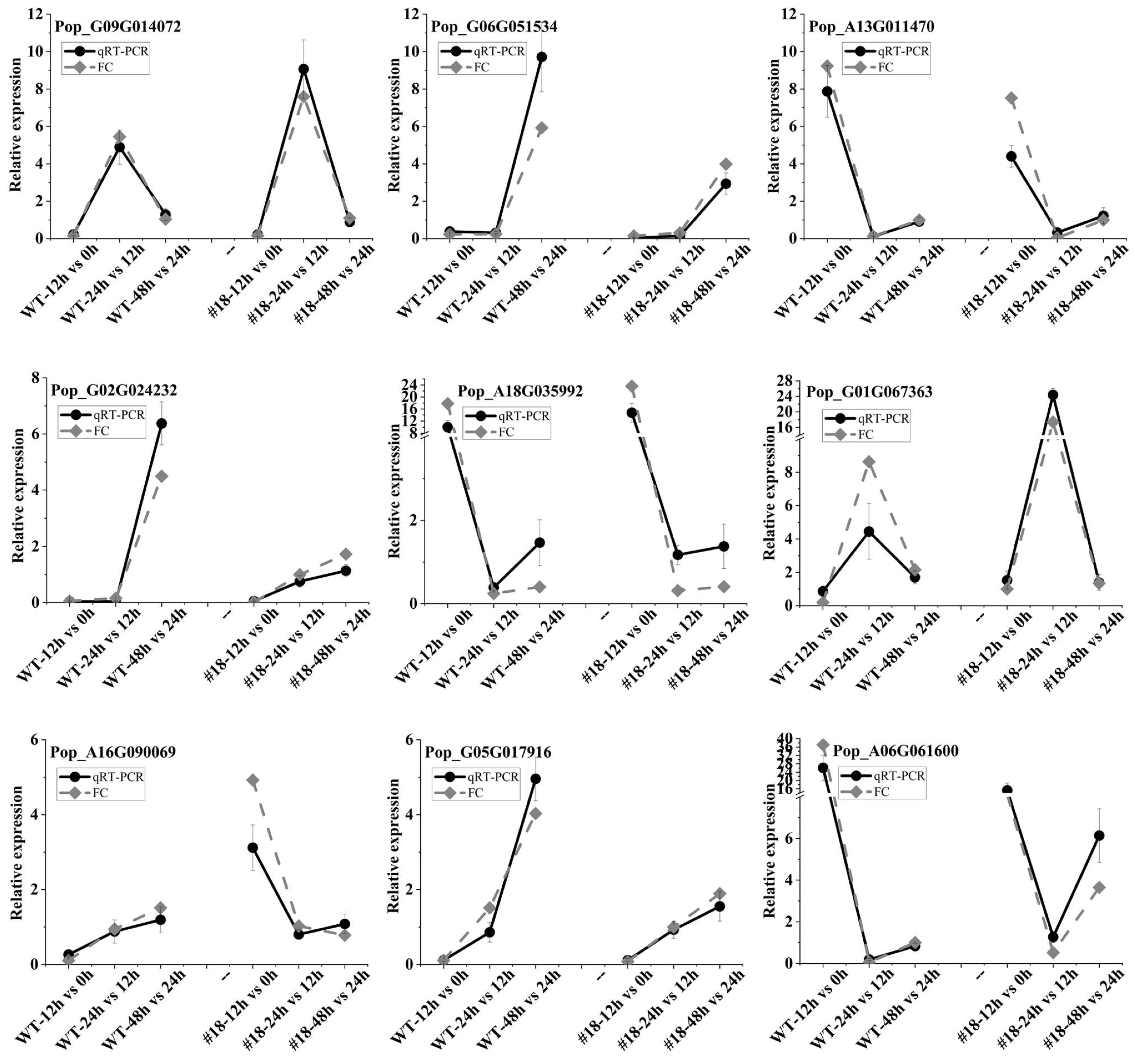
2.2. RNA Isolation and qRT-PCR

Total RNA was extracted from the same 30 samples collected at 0, 12, 24 and 48 h during AR formation, and their quality and quantity were checked according to a previously described method [1]. First-strand cDNA of all samples was synthesized with approximately 3 μg RNA using the Superscript III reverse transcription kit (Life Technologies, Carlsbad, CA, USA) according to the manufacturer’s instructions [1,10]. The amplified fragments were confirmed using agarose gel electrophoresis. Real-time quantitative PCR (qRT-PCR) was used to verify the FPKM values of the DEGs in the transcriptome. We selected the housekeeping gene PagUBQ [1] as an internal reference gene for qRT-PCR measurement. qRT-PCR was performed as described by Shu [1,10]. Nine plant hormone signalling-related genes were selected for qRT-PCR, and all primer sequences used in the qRT-PCR are described in Table S1.

3. Results

3.1. PagFBL1-OE Stimulates the Remodelling of Gene Expression in Transgenic Poplar

PagFBL1, the auxin receptor, showed the same dynamic changes with auxin distribution during AR formation [1,19] and participated in the auxin signalling pathway to regulate AR induction and initiation [1]. For 84K (N samples = 4), a total of 8081 genes were significantly differentially expressed between the 0–12 h AR induction time, with 4140 upregulated and 3941 downregulated (Figure 1a,b). However, only 4774 DEGs, including 1995 upregulated and 2779 downregulated genes, were detected from 12–24 h with initiation (Figure 1a,b), and 3870 DEGs with 2841 upregulated and 1029 downregulated from 24–48 h with expression were obtained (Figure 1a,b). However, for PagFBL1-OE (N samples = 4), a total of 9493 DEGs with 4920 upregulated genes and 4573 downregulated genes were detected from samples with an AR induction time of 0–12 h (Figure 2a,b). Only 5141 DEGs, including 2277 upregulated genes and 2864 downregulated genes (Figure 2a,b), were found in the sample with a 12–24 h AR initiation time, and 3044 DEGs, with 2059 upregulated genes and 985 downregulated genes, were found in the sample with a 24–48 h AR primordium activation time (Figure 2a,b). These results suggest that there were more upregulated genes, i.e., 780 genes and 282 genes for PagFBL1-OE versus 84K in the induction phase and initiation phase, respectively. Moreover, the numbers of DEGs between 0–12 h in PagFBL1-OE were much larger than those between 12–24 h. Therefore, PagFBL1-OE stimulates the expression of a larger number of genes within 12 h of AR induction prior to AR initiation.

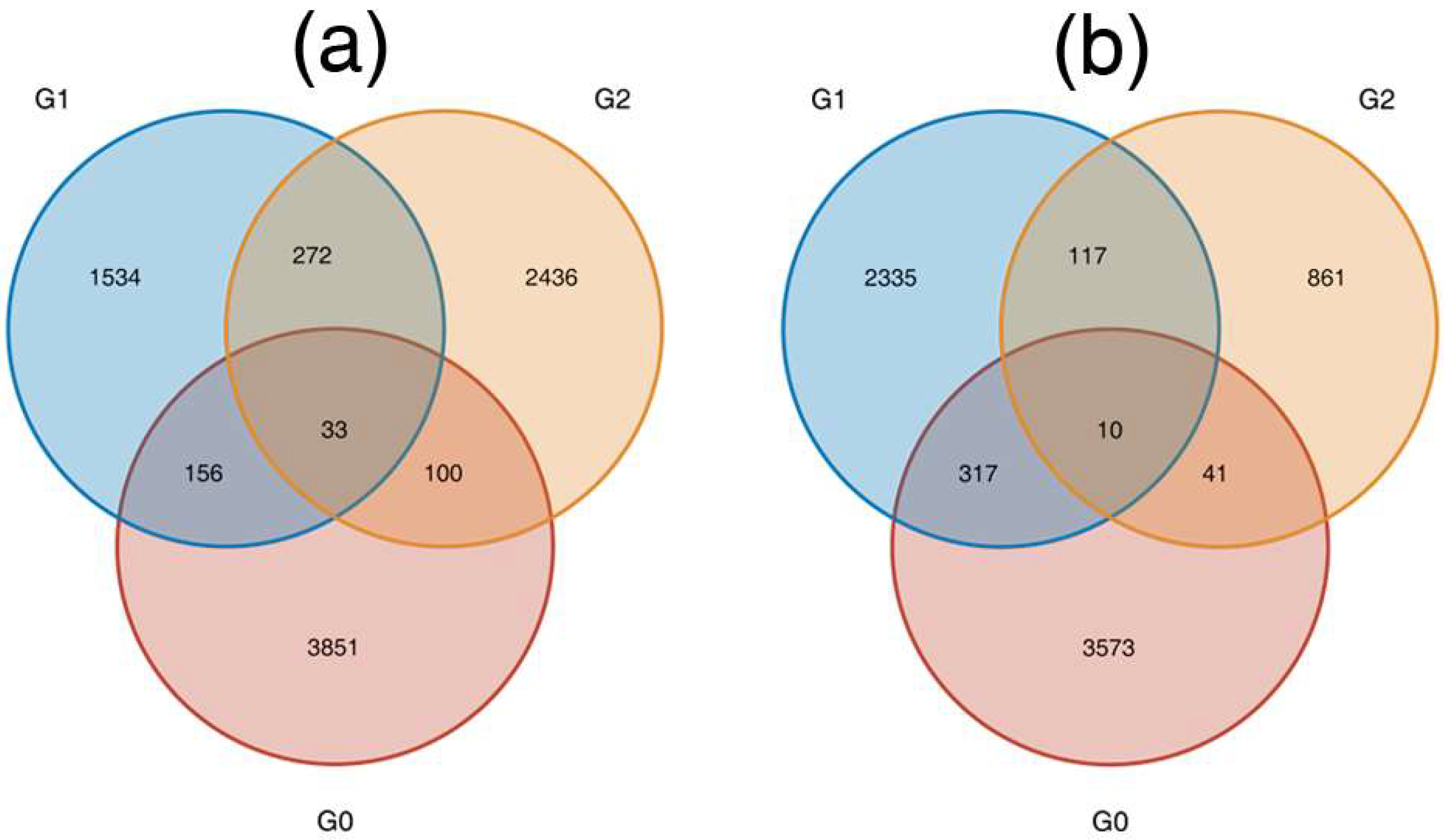
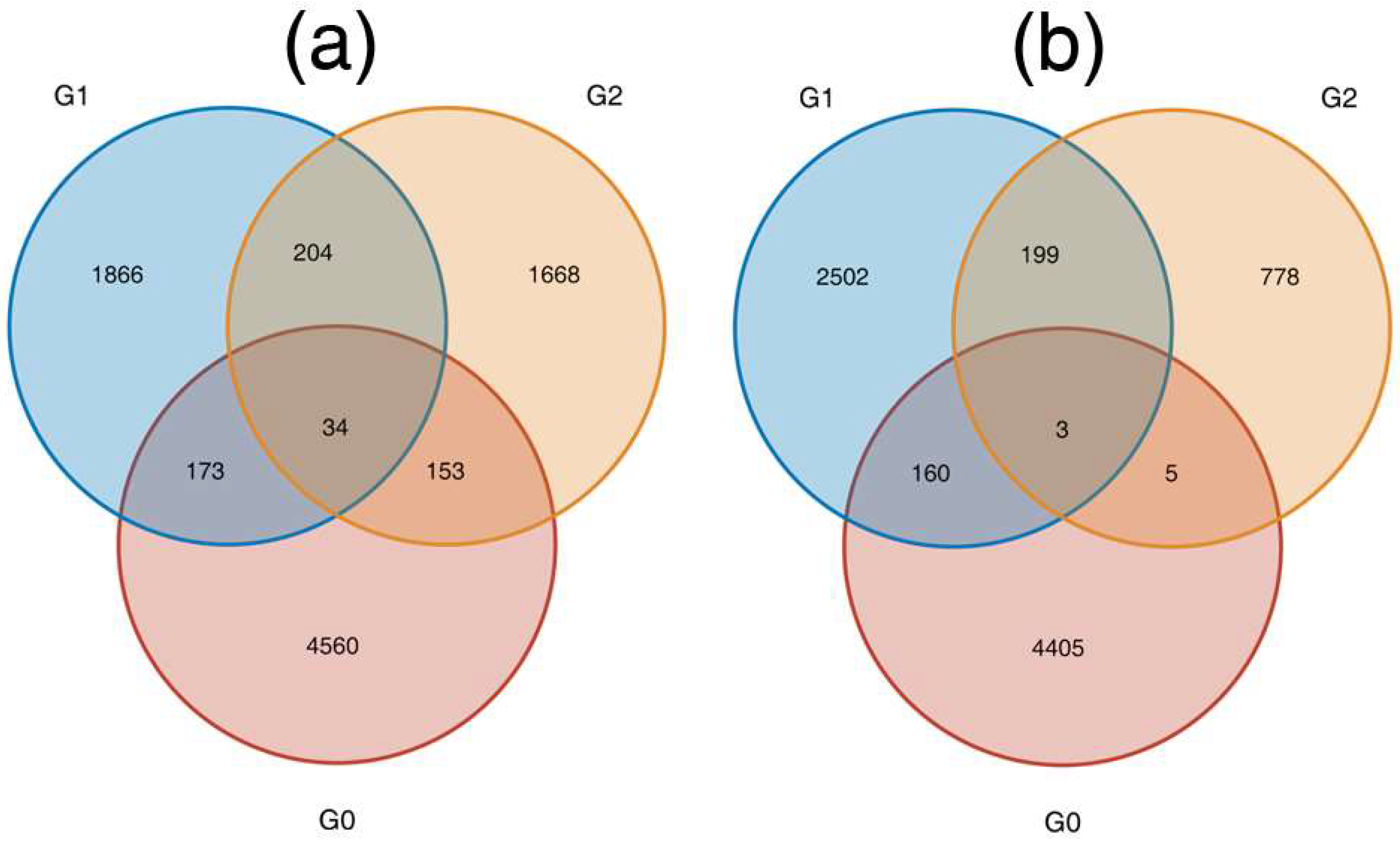
3.2. NOG and KEGG Pathway Enrichment Analysis

To understand the meaning of DEGs, Evolutionary Genealogy of Genes: Nonsupervised Orthologous Groups (NOG) classification was determined (Figures S1 and S2). Some genes involved in signal transduction mechanisms were induced at 12, 24 and 48 h of AR formation in 84K and PagFBL1-OE cells. Comparing the three AR formation stages in 84K and PagFBL1-OE, at every point, PagFBL1-OE expressed more DEGs than 84K (Figures S1 and S2). We also found that a large number of DEGs were only enriched in plant hormone signal transduction in the first 12 h in 84K and PagFBL1-OE (Figures S3 and S4), and eight hormone signalling-related genes encoding receptors were identified. Notably, 316 DEGs (10.98%) and 352 DEGs (10.47%) were expressed in plant hormone signal transduction for the first 12 h in 84K (Figure S3) and PagFBL1-OE (Figure S4), respectively, and there were more than 36 upregulated genes, which is considered significant for PagFBL1-OE versus 84K, in which PagFBL1-OE promotes AR expression [1]. These results suggest that PagFBL1-OE could promote the expression of more DEGs for the first 12 h in the plant hormone signal transduction pathway in favour of AR formation to promote adventitious rooting.

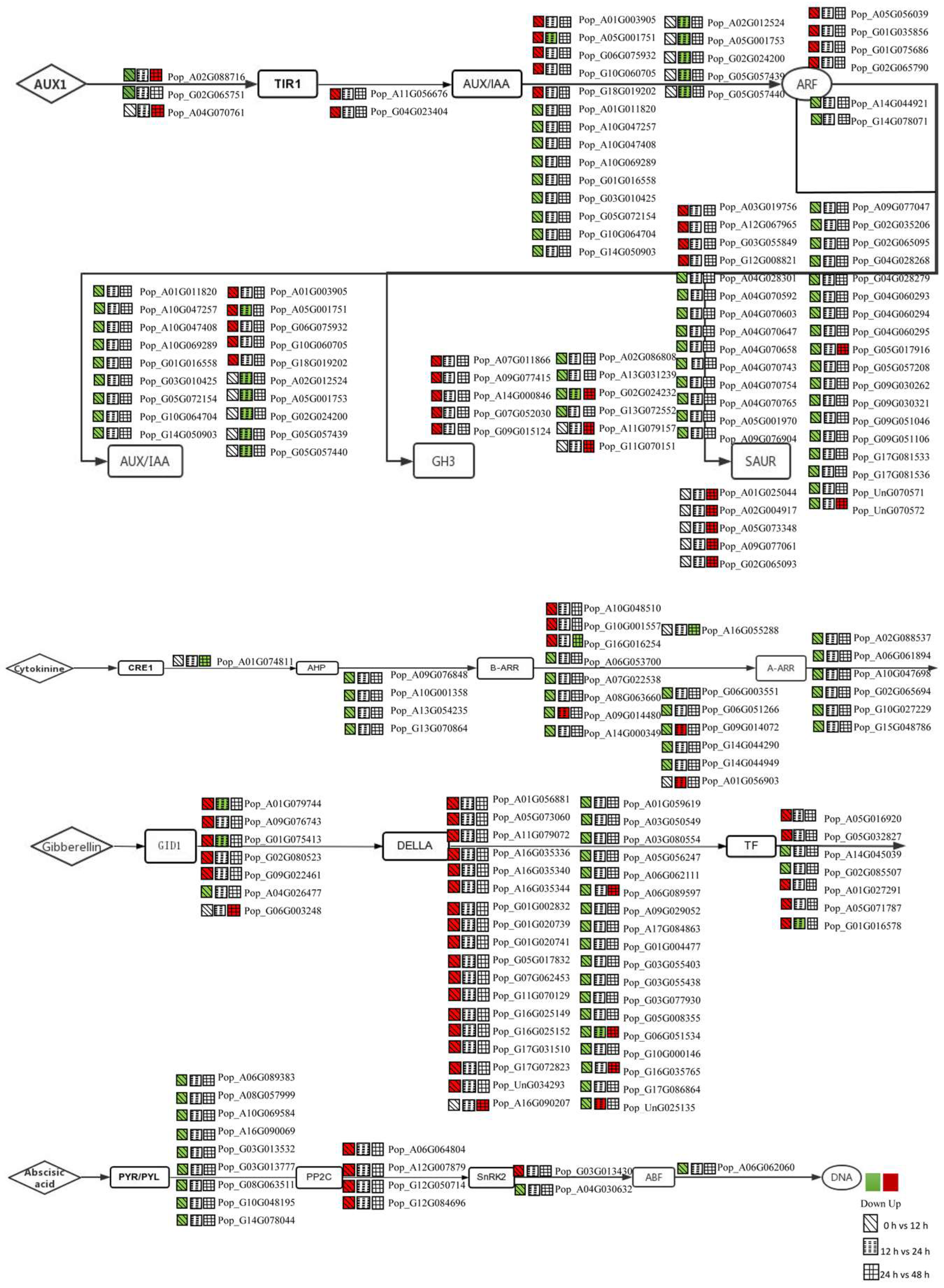
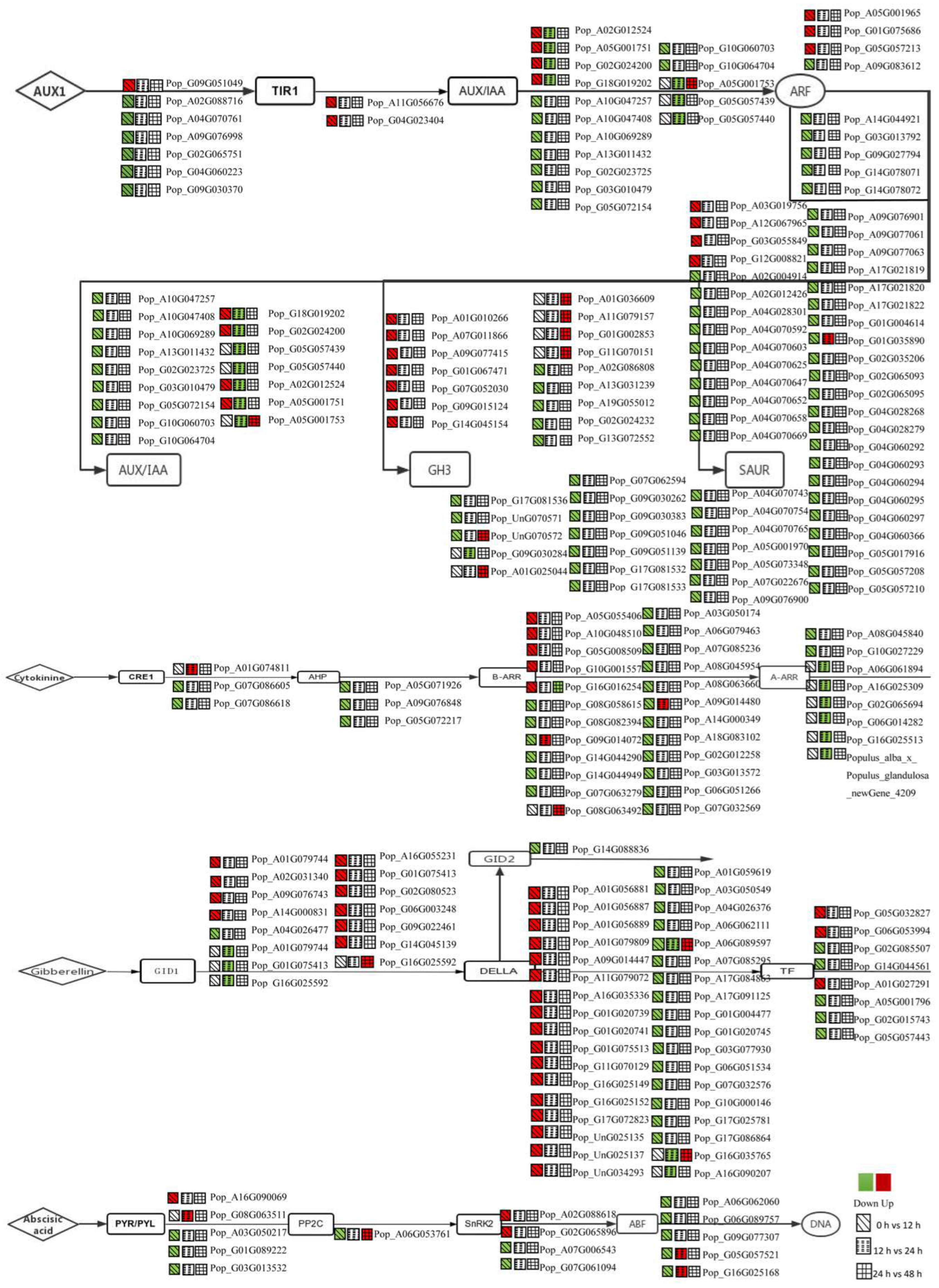
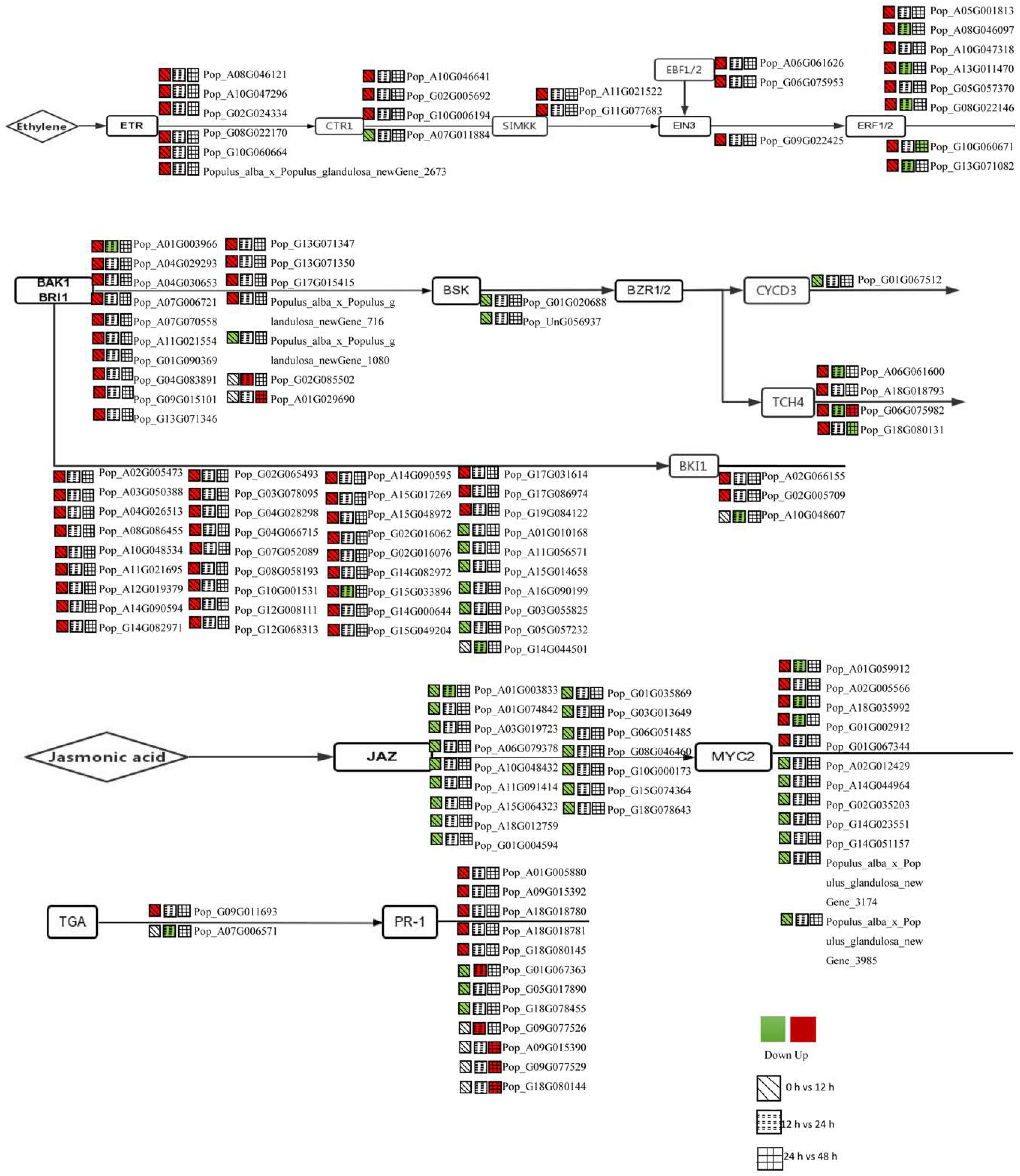
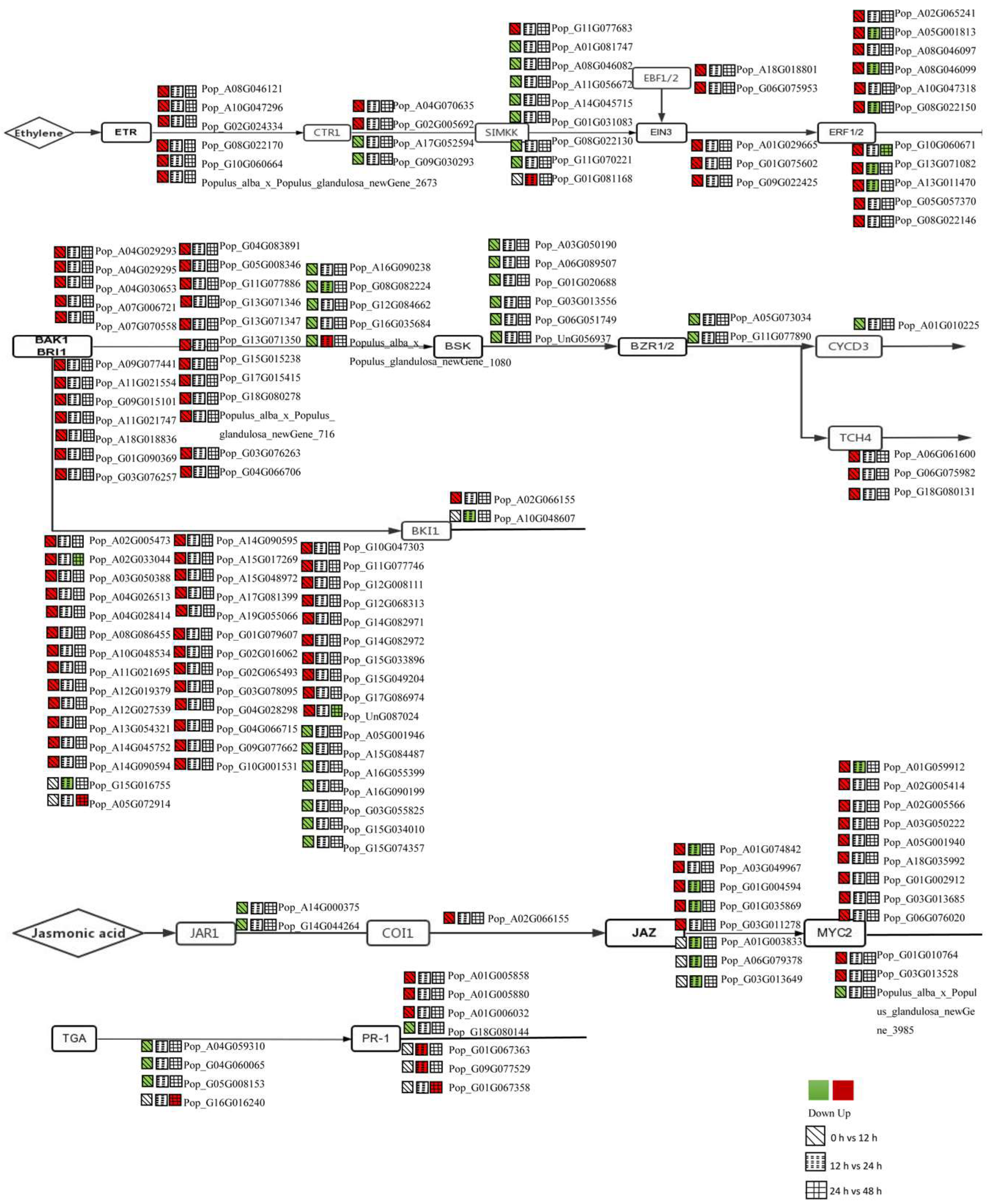
3.3. DEGs in the Plant Hormone Signalling Pathway Were Regulated during AR Formation

To understand the roles of pant hormones in AR formation, we summarized the dynamic gene expression in three stages of AR formation and development and studied phytohormone-related genes in poplar. After analysing the DEGs obtained in the present study, many transcripts were found to be involved in phytohormone-mediated signalling. Among the plant hormone receptor genes, there were data for eight hormone-related genes that can be used to study the expression patterns of 84K and PagFBL1-OE (Figure 3, Figure 4, Figure 5 and Figure 6 and Tables S1 and S2). Thus, these genes were searched among all DEGs. A total of 299 and 374 genes with both |log2(fold change)| > 1 and FPKM ≥ 5 were identified for 84K and PagFBL1-OE, respectively, including 78 and 105 auxin signalling pathway genes, 22 and 35 ethylene signalling pathway genes, 26 and 38 CTK signalling pathway genes, 50 and 59 gibberellin-related genes, 16 and 15 abscisic acid-related genes, 28 and 23 JA-related genes, 64 and 88 BR-related genes and 14 and 11 SA-related genes (Tables S1 and S2).

3.4. Expression Analysis of Auxin-Related Genes for the First 12 h

The presented section summarizes the expression levels of auxin-related genes in 84K poplar (Figure 3 and Table S1) and PagFBL1-OE, and many genes were activated or repressed for the first 12 h (Figure 4 and Table S2). Three auxin transporter-like protein 3 genes (AUX1) have two genes in 84K (Figure 3 and Table S1) and seven genes in PagFBL1-OE (Figure 4 and Table S2); AUX/IAAs have 14 genes in 84K (Figure 3 and Table S1) and 13 genes in PagFBL1-OE (Figure 4 and Table S2); ARFs have six genes in 84K (Figure 3 and Table S1) and nine genes in PagFBL1-OE (Figure 4 and Table S2); Gretchen hagen 3 (GH3) genes have nine genes in 84K (Figure 3 and Table S1) and 12 genes in PagFBL1-OE (Figure 4 and Table S2); small auxin up RNAs (SAURs) have 32 genes in 84K (Figure 3 and Table S1) and 53 genes in PagFBL1-OE (Figure 4 and Table S2). However, the expression levels of Pop_A03G019756, Pop_A12G067965, Pop_G03G055849 and Pop_G12G008821 were significantly increased in 84K and PagFBL1-OE.

3.5. Expression Analysis of CTK-, GA- and ABA-Related Genes for the First 12 h

CTK is a significant regulator of root development [20]. The genes encoding histidine-containing phosphotransferase (AHP), a positive control for CTK signalling, were downregulated in 84K (Figure 3 and Table S1), whereas three genes were repressed in PagFBL1-OE (Figure 4 and Table S2). Two CTK receptors (CRE1) were only repressed in PagFBL1-OE (Figure 4 and Table S2). Six type-A response regulators (A-ARR) were repressed in 84K (Figure 3 and Table S1), whereas two genes were repressed in PagFBL1-OE (Figure 4 and Table S2). Twelve type-B response regulators (B-ARR) were activated or repressed in 84K (Figure 3 and Table S1), whereas 23 genes were activated or repressed in PagFBL1-OE (Figure 4 and Table S2).
In addition, ABA-related genes are mainly triggered by abiotic stress conditions [20]. Nine abscisic acid receptors (PYR/PYL) were repressed in 84K cells (Figure 3 and Table S1), whereas four genes were activated or repressed in PagFBL1-OE cells (Figure 4 and Table S2). The expression levels of Pop_A16G090069 and Pop_G03G013532 were significantly decreased in 84K and PagFBL1-OE for the first 12 h. Four probable protein phosphatase 2C (PP2C) genes were also activated in 84K (Figure 3 and Table S1), whereas one gene was activated or repressed in PagFBL1-OE (Figure 4 and Table S2). Two SnRK2 genes were activated or repressed in 84K (Figure 3 and Table S1), whereas four genes were activated or repressed in PagFBL1-OE (Figure 4 and Table S2). One ABF gene was repressed in 84K (Figure 3 and Table S1), whereas five genes were activated or repressed in PagFBL1-OE (Figure 4 and Table S2). The expression level of Pop_A06G062060 was significantly decreased in 84K and PagFBL1-OE cells for the first 12 h.
Interestingly, six gibberellin-insensitive dwarf1 (GID1) genes were activated or repressed in 84K (Figure 3 and Table S1), whereas 11 genes were activated or repressed in PagFBL1-OE (Figure 4 and Table S2). The expression levels of Pop_A01G079744, Pop_A09G076743, Pop_G01G075413, Pop_G02G080523 and Pop_G09G022461 were significantly increased, and Pop_A04G026477 was significantly decreased in 84K and PagFBL1-OE; one gibberellin-insensitive dwarf2 (GID2) was only repressed in PagFBL1-OE (Figure 4 and Table S2); seven transcription factors (TF) were activated or repressed in 84K (Figure 3 and Table S1), whereas eight genes were activated or repressed in PagFBL1-OE (Figure 4 and Table S2). The expression levels of Pop_G05G032827 and Pop_A01G027291 were significantly increased in 84K and PagFBL1-OE, but Pop_G02G085507 was significantly decreased in 84K and PagFBL1-O; thirty-five other related genes (DELLA) were activated or repressed in 84K (Figure 3 and Table S1), whereas 33 genes were activated or repressed in PagFBL1-OE (Figure 4 and Table S2).

3.6. Expression Analysis of BR-, JA-, SA- and ETH-Related Genes for the First 12 h

BR availability triggers dual effects on tree growth at both doses. Fifteen brassinosteroid insensitive1-associated receptor kinase1 (BAK1) genes were activated or repressed to BSK in 84K (Figure 5 and Table S1), whereas 27 genes were activated or repressed in PagFBL1-OE (Figure 6 and Table S2). The expression level of Pop_A04G029293, Pop_A04G030653, Pop_A07G006721, Pop_A07G070558, Pop_A11G021554, Pop_G01G090369, Pop_G04G083891, Pop_G13G071346, Pop_G13G071347, Pop_G13G071350, Pop_G17G015415 and Populusalba_x_Populus_glandulosa_newGene_716 were significantly increased in 84K and PagFBL1-OE for first 12 h; Two BSK were repressed in 84K (Figure 5 and Table S1), whereas six genes were repressed in PagFBL1-OE (Figure 6 and Table S2); one CYCD3 was repressed in 84K (Figure 5 and Table S1), whereas one gene was repressed in PagFBL1-OE (Figure 6 and Table S2); four TCH4 were activated or repressed in 84K (Figure 5 and Table S1), whereas three genes were activated in PagFBL1-OE (Figure 6 and Table S2). But thirty-seven Brassinosteroid insensitive1 (BRI1) were activated or repressed to BKI1 in 84K (Figure 5 and Table S1), whereas 45 genes were activated or repressed in PagFBL1-OE (Figure 6 and Table S2). The expression levels of Pop_A02G005473, Pop_A03G050388, Pop_A04G026513, Pop_A08G086455, Pop_A10G048534, Pop_A11G021695, Pop_A12G019379, Pop_A14G090594, Pop_G02G065493, Pop_G03G078095, Pop_G04G028298, Pop_G04G066715, Pop_G10G001531, Pop_G12G00811, Pop_G14G082972, Pop_G15G033896, Pop_G15G049204 and Pop_G17G086974 were significantly increased in 84K and PagFBL1-OE for the first 12 h, but Pop_A16G090199 and Pop_G03G055825 were significantly decreased in 84K and PagFBL1-OE for the first 12 h. Two BKI1 genes were activated or repressed in 84K (Figure 5 and Table S1), whereas one gene was activated in PagFBL1-OE (Figure 6 and Table S2). The expression level of Pop_A02G066155 was significantly increased in 84K and PagFBL1-OE cells for the first 12 h.
Moreover, AR formation repressed sixteen jasmonate ZIM domains (JAZ) in 84K (Figure 5 and Table S1), whereas five genes were activated in PagFBL1-OE (Figure 6 and Table S2). Twelve MYC2 genes were activated or repressed in 84K (Figure 5 and Table S1), whereas 12 genes were activated or repressed in PagFBL1-OE (Figure 6 and Table S2). Likewise, one coronatine-insensitive protein (COI) was only activated, and two JAR1 genes were repressed in PagFBL1-OE (Figure 6 and Table S2). Additionally, six ethylene receptors (ETRs) were activated in 84K (Figure 5 and Table S1) and PagFBL1-OE cells (Figure 6 and Table S2). The expression levels of Pop_A08G046121, Pop_A10G047296, Pop_G02G024334, Pop_G08G022170, Pop_G10G060664 and Populus_alba_x_Populus_glandulosa_newGene_2673 were significantly increased in 84K and PagFBL1-OE for the first 12 h; one EIN3-binding gene and two EBF1/2 genes were activated in 84K (Figure 5 and Table S1), whereas three genes and two genes were activated in PagFBL1-OE (Figure 6 and Table S2), respectively; two SIMKK genes were activated in 84K (Figure 5 and Table S1), whereas eight genes were activated or repressed in PagFBL1-OE (Figure 6 and Table S2); and four CTR1 and eight ERF1/2 genes were activated or repressed in 84K (Figure 5 and Table S1), whereas four genes and 11 genes were activated or repressed in PagFBL1-OE (Figure 6 and Table S2). One phenylalanine metabolism (TGA) was upregulated in 84K (Figure 5 and Table S1), whereas three genes were repressed in PagFBL1-OE (Figure 6 and Table S2); eight pathogenesis-related (PR-1) proteins were upregulated and downregulated in 84K (Figure 5 and Table S1), whereas four genes were activated or repressed in PagFBL1-OE (Figure 6 and Table S2). These results suggest that phytohormones might indirectly or directly regulate the expression of these genes during AR formation in Populus.

3.7. Validation of Gene Expression by qRT-PCR

To validate the gene expression data of RNA-Seq analysis, nine genes of plant hormone signal transduction with differential expression patterns at three time points were selected at random for qRT-PCR assay. Detailed information regarding these genes is presented in Tables S1 and S2. Gene expression levels, as measured by qRT-PCR, showed a strong correlation with the RNA-Seq data (Figure 7) [1]. This result is mainly consistent with the DEG analysis of the RNA-Seq data and further shows the role of 84K and PagFBL1-OE in regulating gene expression. The qPCR analysis confirmed that the RNA-Seq method provided reliable data for differential gene expression in the AR developmental stages of Populus cuttings.

4. Discussion

Plants form a large number of ARs in vitro when cultured under certain conditions, but the abundance of ARs varies with explant type. The mechanism of AR growth and development by hypocotyl pericycle cells in the root–hypocotyl junction of planta is rarely studied [4]. The formation of ARs is a complex biological pathway and requires modifying the expression of a set of genes to induce stem cuttings to form new roots [20]. Plant growth and development are mainly affected by hormonal contents [20]. Plant hormones are thought to be key regulators of AR formation. During AR formation, different hormones interact to form a large and complex network that regulates dynamic biological processes. Plant hormones are known to play vital roles in adventitious rooting. AR induction is dependent on the interaction of different hormone networks [21]. However, we do not know how to promote adventitious rooting by regulating the expression of genes related to plant hormone signalling. Crosstalk with other hormones plays a significant role at different developmental stages of AR formation either in combination or individually [7,22]. Although time scales vary between species, auxin has been confirmed as a mediator of AR formation by interacting with other plant hormones, such as CTK, ET, JA, SA and BRs. However, ET, auxin, JA, GA, CTK and ABA affect AR formation at the induction stage [22,23,24]. Populus is an important commercial tree, and AR formation is a major setback to its asexual reproduction. AR formation is a complex biological pathway that requires modifying the expression of a set of genes to induce stem cuttings to produce new roots. Transcriptome analyses of AR formation have been recently performed to identify candidate genes in some different Populus species in response to auxin and other hormones [23,25,26]. However, until now, there has been no study in which AR formation for the phytohormone signalling pathway in Populus has been systematically reported. Therefore, we attempted to understand the phytohormone signalling process of in vitro ARs in Populus.

4.1. Regulation of AR Formation by Hormone Signalling

Plant growth and development are mainly affected by hormonal contents and metabolic factors [20]. AR induction is an important stage of molecular reprogramming, and the initiation stage is critical for AR primordium formation [20]. Generally, auxin promotes AR initiation, whereas strigolactones and cytokinin inhibit AR initiation [7,21]. However, the interactions between different hormone networks play a key role in plant growth [20]. The AR formation process can be divided into three stages, namely, 0–12 h is the induction stage, 12–24 h is the initiation stage and 24–48 h is the activation stage in Populus [1,27,28], which is slightly different from Camellia sinensis [29] and Cryptomeria japonica [24]. However, the AR induction stage signifies a vital step for molecular reprogramming, and the initiation stage is essential for AR primordium formation [20]. In the present study, KEGG biological function analysis showed that many genes related to hormone signal transduction were upregulated (Figure 3, Figure 4, Figure 5 and Figure 6) in 84K and PagFBL1-OE, indicating that AR growth and development were mainly promoted at the transcriptional level at 0–12 h. Among the DEGs, plant hormone-related genes were identified. These included transcriptional controls of auxin, GA, CTK, BRs, SA and JA signalling (Figure 3, Figure 4, Figure 5 and Figure 6). This result is similar to the mapping of the Populus trichocarpa reference genome [1]. According to the expression profiles of the plant hormone-related genes, we were able to deduce the roles of eight investigated plant hormones, including Auxin, ABA, BR, CTK, ET, GA, SA and JA, which may play dual roles (inhibitory or regulatory) in AR formation (Tables S1 and S2). The interactions between different hormone networks play a key role in plant growth [20]. Since continuous comparisons may reliably reveal the dynamic changes in gene expression during AR development, this result might indicate that the period of time from 0–12 h is the most important stage in the plant hormone signal transduction pathway for AR formation (Figures S1 and S2). The induction of auxin-related processes in AR formation is a complex regulatory, balanced and signalling interaction between auxin and other phytohormones [30]. Plant hormones play a key role in different developmental stages of AR formation either in combination or individually [30]. Therefore, it is necessary to maintain the endogenous hormone balance in AR formation during clonal reproduction.

4.2. Regulation of AR Formation by Auxin Signalling for the First 12 h

Most tree species share a common trait that auxin is closely related to the start of AR formation [8,22,24,25]. Auxin signalling acts through three transcription factors, AUX/IAA, GH3 and SAUR, from the ARF gene family in Arabidopsis thaliana, in which AtARF17 positively regulates the downstream GH3 family, while AtARF6/8 negatively regulates the downstream GH3 family [31]. This study shows the upregulation and downregulation of AUX, AUX/IAA, ARF, GH3 and SAUR (Figure 3 and Figure 4 and Tables S1 and S2). Depending on auxin distribution, its signal transduction stimulates the transcription of downstream genes or crosstalk with other hormonal signals [20]. During AR formation, auxin behaves as a stimulator [19] and shows an upregulated expression pattern in Arabidopsis thaliana [31]. All these genes affected by auxin are regulated by ARF activation [20]. Hence, ARF upregulation might be the reason for the high incidence of ARs [20]. In Populus, PeARF17.1 and PeARF17.2 negatively regulate AR formation [32,33]; clone T89: P. tremula × P. tremuloides PeARF8.1/2 and P. davidiana × P. bolleana PaARF17.1/2 promote AR formation [26]. However, in Eucalyptus globulus, AR formation in cuttings is correlated with ARF6 or ARF8 expression levels [7]. A study on AR formation in cuttings of mulberry hardwood showed that 13 ARF genes involved in auxin signal transduction were significantly differentially expressed [33]. In this study, Pop_G01G075686 of ARFs showed a positive role due to increased expression in 84K and PagFBL1-OE during induction (0–12 h) (Figure 3 and Figure 4). In addition, when auxin signalling is activated, AUX/IAAs and ARFs form dimers to regulate the expression of auxin response element-related genes and promote AR formation [33]. Recently, in cotyledon segments of Mangifera indica, the local upregulation of several Aux/IAA-like genes was associated with the formation of exclusive AR formation in proximal cut surfaces compared to distal cut surfaces [7]. However, the NL895 hybrid P. deltoides × P. euramericana and the Shanxinyang hybrid P. davidiana × P. bolleana IAA4 inhibit AR growth [26]. Herein, five auxin responsive genes (IAA13 (Pop_A10G047408 and Pop_G10G064704), IAA14 (Pop_A10G047257), IAA22 (Pop_G05G072154) and IAA32 (Pop_A10G069289)) were significantly decreased in 84K and PagFBL1-OE, and these genes are the same pertains to IAA29 in apple stem cuttings [22] and but difference pertains to IAA14, IAA18 and IAA28 in Arabidopsis during AR formation [22].
Additionally, auxin-responsive members of the GH3 family regulate auxin homeostasis in higher plants [32]. Hence, it is essential that members of the GH3 family are a group of early auxin-responsive genes [20]. In Arabidopsis thaliana, auxin also stimulated the expression of GH3 (GH3.3, GH3.5 and GH3.6) during AR formation [22]. This result is inconsistent with our study. It has been reported that the GH3 family auxin response gene BpGH3.5 may regulate root elongation in Betula platyphylla × Betula pendula [33]. In addition, GH3 protein was also found to be upregulated during root formation and emergence in stem tissues in Taxodium ‘Zhongshanshan’ [33]. In apple plants, MsGH3.5-OE significantly increased the content of some IAA–amino acid conjugates and reduced the content of free IAA, and MsGH3.5-OE produced fewer ARs than the control [34]. These results demonstrated that GH3 proteins were intricately involved in the development of ARs but did not only play a positive role [34]. In this study, GH3.1 (Pop_A07G011866, Pop_A09G077415, Pop_G07G052030 and Pop_G09G015124) was significantly upregulated and showed higher expression during the early induction phase (0–12 h), which is consistent with apple rootstock [22]. SAUR is the largest family of auxin-responsive factors in plants, responding to early auxin signals [32]. SAUR proteins are also related to JA and ETH and are involved in the downstream implementation of hormone-mediated processes [7]. SAUR proteins may exhibit a species- or type-dependent positive function in AR formation. In E. excelsum, three SAUR genes indicated a close association with AR formation [34]. Recently, the local upregulation of one SAUR-like gene in proximal cut surfaces was associated with exclusive AR formation in Mangifera indica [7]. In this study, the expression levels of four SAURs were significantly increased in 84K and PagFBL1-OE cells for the first 12 h (Figure 4 and Table S2). These DEGs for AUX, AUX/IAA, ARF, GH3 and SAUR have some commonalities with previous studies [16,22,35,36]. Most likely, ARF (Pop_G01G075686), IAA14 (Pop_A10G047257) and SAURs (Pop_A03G019756, Pop_A12G067965, Pop_G03G055849 and Pop_G12G008821) may play an important role in the first 12 h in Populus.

4.3. Regulation of AR Formation by JA, ETH, CTK and GA Signalling for the First 12 h

In addition, in this study, the interaction of auxin with CTK and JA during AR formation was also apparent [22]. Comparably, the role of JA is species-specific during AR formation, and it negatively regulates ARs in Arabidopsis thaliana [20,22]. In apple, it plays a key role in regulating the induction stages of ARs [20]. After wounding, JA is induced and activates ETHYLENE RESPONSE FACTOR109 (ERF109), which also induces auxin biosynthesis and promotes ARs in Arabidopsis [26]. Studies have also shown that in the presence of exogenous JA, the regeneration ability of ARs involves crosstalk with ethylene through the ETHYLENE INSENSITIVE3/ETHYLENE INSENSITIVE3-LIKE1 (EIN3/EIL1) signalling pathway [26]. In Arabidopsis, JA inhibits AR initiation in hypocotyls by the COI1 signalling pathway, where GH3 and ARF regulate the level of the JA active form and inhibit adventitious rooting by the COI1-MYC2-dependent pathway, but JA may be a positive regulator of AR formation in Pisum sativum [26]. In apple, it plays a key role in regulating the induction stages of ARs [1]. Similar results were also found in this study, where JA plays positive roles in the formation of ARs, and PagFBL1-OE increased the expression of the JA genes JAZ, MYC2, COI and JAR1 (Figure 5 and Figure 6). During AR formation, JA signalling genes were differentially expressed in 84K and PagFBL1-OE, suggesting that JA might also be involved in AR formation. Considering that JA is a positive regulator of complete hypocotyl-derived ARs, we conclude that PagFBL1-OE may have increased JAR1 (Pop_A14G000375 and Pop_G14G044264) expression, which has an important biological correlation in the IAA–JA interaction. In addition, researchers also found that PttMYC2.1 negatively regulates AR formation in poplar [32,33], but PuMYC2 positively regulates AR formation in Populus ussuriensis [29]. In this study, with regard to the JA signalling pathway, almost all DEGs of MYC2 were significantly increased in PagFBL1-OE for the first 12 h.
Moreover, MYC2 also regulates the expression of ETHYLENE RESPONSE FACTORs (ERFs) by mechanical damage [22]. Ethylene plays a key role in root development in many species, such as apple, sunflower and petunia [33]. Ethylene is also associated with auxin metabolism during AR formation and has been reported to be induced by wounding [24]. Ethylene and its crosstalk with auxin have been shown to be necessary for AR formation [37]. ETH could be involved in the early phase of AR formation and is a negative regulator of AR development in Populus [26]. Largely, in Eucalyptus cuttings, ETH inhibits AR formation at high auxin levels [26], whereas in Populus [28], the overexpression of a cytokinin response regulator (PtRR13) repressed AR development. The AP2/ERF family members are involved in the rooting of P. trichocarpa cuttings [26]. Auxin and ethylene act either synergistically or antagonistically, and the effects are opposite for AR formation [24,31]. Ethylene-insensitive protein 3 (EIN3), a core factor in ethylene-regulated signalling pathways, is involved in root development [37]. The ethylene receptor (ETR) recognizes and transduces ethylene signals, and ethylene insensitive 3 (EIN3) acts as a forwards downstream regulator [33]. Expression of the AP2/ERF gene PtaERF003 in Populus tremula × Populus alba is induced by auxin and has been shown to control the intensity of AR formation in cuttings, which may act as a broad growth regulator [7]. In this study (Figure 5 and Figure 6 and Tables S1 and S2), CTR1 (Pop_A17G052594 and Pop_G09G030293) and SIMKK (Pop_A01G081747, Pop_A08G046082, Pop_A11G056672, Pop_A14G045715, Pop_G01G031083, Pop_G08G022130 and Pop_G11G070221) were only significantly decreased in PagFBL1-OE. These studies demonstrate the important role of ethylene in poplar AR formation and its interaction with other plant hormones in this process. Although some genes have been shown to be involved in AR formation, further studies are needed to reveal their roles and how they interact.
Notably, Auxin and CTK play opposite roles in AR formation [22,38], and AR formation is related to the ratios of auxin and cytokinin [24]. However, CTKs are also auxin antagonistic and repress AR formation in apples, Taxodium and poplar [20,33]. Nevertheless, low CTK concentrations are beneficial in the early stages of AR formation in Monterey pine (Pinus radiata) and apple (Malus pumila Mill) cuttings [31]. Similar to Auxin, CTK also seems to be actively involved in the regulation of ARs [20]. Previously, CTK was shown to be involved in regulating auxin-related gene expression [11]. In this study, most CRE1, AHP, B-ARRs and A-ARRs were also specifically expressed during the first 12 h, and it is clear that the negative role of CTK is phase-dependent in Populus. In the current study, Populus CRE1s (Pop_G07G086605 and Pop_G07G086618), encoding a membrane-localized receptor of CTK signalling, were only significantly decreased in PagFBL1-OE. Combined with expression data for genes associated with auxin and cytokinin responses, our transcriptome analyses show that the interplay between auxin and CTK is important for AR formation in Populus, as shown anteriorly in herbaceous plants [24]. These results suggest that auxin inhibited cytokinin activity by regulating the expression of cytokinin-related genes and that CTK could be involved in the final steps of AR development. In conclusion, CTK- and JA-related genes may interact with auxin signalling during Populus AR formation.
Auxin reduced the expression of GA synthesis genes, resulting in inhibition of GA synthesis [37]. In a previous study, the application of IBA to induce GA-related gene expression may lead to increased GA degradation levels and resultantly promote AR formation [26]. Exogenous application of GA inhibits AR formation in rice [31]. During AR formation, GA blocks the transport of polar auxin through the DELLA protein, and upon GA binding, the GID1 receptor induces DELLA degradation [22]. Thus, this suggests that auxin crosstalks with GA during AR formation. Regarding the GA signalling pathway, it has been reported that the DELLA family recognizes GA transduction signals and interacts with the gibberellin receptor GID1 in poplar [22]. In the present work, GID1 (Pop_A04G026477) only showed downregulation in PagFBL1-OE (Figure 3 and Figure 4). These results are consistent with previous studies showing that the combination of gibberellin with GID1 induces the formation of the GID1–DELLA complex and plays a key role in AR formation [22]. The P. tremula × P. alba GIBERELLINS INSENSITIVE (GAI) gene is an inhibitor of GA signalling, and its overexpression can increase the number of ARs [26]. Therefore, the widespread existence of GA-auxin-related genes might play a crucial role, and GAs may act as negative regulators of AR formation in Populus.

4.4. Regulation of AR Formation by BR, ABA and SA Signalling for the First 12 h

Similarly, other hormones, including BR, ABA and SA, also influence AR formation [22]. Similar to auxin, BR also has a positive effect on root growth [20]. In addition, auxin–BR interactions are also a necessary condition for AR formation [22]. BR application promotes the expression of auxin-responsive genes and root development [20,26]. In a previous study, BRI1 interacted with the BAK1 receptor to form the BRI1/BAK1 complex [22]. This complex activates BR-responsive genes for BZR1/2 (a positive BR signalling modulator) or BKI1. Most BZR1/2 and TCH4, a downstream regulator of BR signalling, showed the highest expression at 12 h (early induction phase). In the present investigation (Figure 5 and Figure 6 and Tables S1 and S2), twelve BAK1s were upregulated and seemed to interact with eighteen BRI1 receptors to form the BRI1/BAK1 complex for 0–12 h in 84K and PagFBL1-OE cells. This complex is known to restrain GLYCOGEN SYNTHASE KINASE3 (GSK3)/SHAGGY, which in turn activates BR-responsive genes, i.e., BSK (Pop_A06G061600, Pop_G06G075982 and Pop_G18G080131), TCH4 (Pop_A06G061600, Pop_G18G080131 and Pop_G06G075982) and BKI1 (Pop_A02G066155) were also upregulated for 0–12 h in 84K and PagFBL1-OE. These results show that these genes could play a critical role in AR formation in Populus.
Exogenous application of ABA had adverse effects on adventitious rooting in grape and Populus [26]. ABA is a negative regulator of AR development [31]. ABFs are core components of the ABA signalling pathway [33]. Previous studies have shown that SnRK2 can activate the expression of ABF, promote the binding of ABF to ABA-responsive promoter elements (ABREs) and induce the expression of ABA-responsive genes, which is crucial to the control of AR development [33]. Moreover, downregulation of ABA-responsive elements is responsible for more ARs. The expression levels of PYR/PYLs (Pop_A03G050217 and Pop_G01G089222) were only significantly decreased in PagFBL1-OE. These results are consistent with previous reports that ABA negatively regulates the formation of ARs during induction phases and also is involved in developmental processes. SA has a variety of functions, mainly in plant immunity, but is also involved in developmental processes. Notably, changes in the level of expression of gene families associated with SA signalling pathways affect root development [33]. In this study (Figure 5 and Figure 6 and Tables S1 and S2), the expression levels of TGAs (Pop_A04G059310, Pop_G04G060065 and Pop_G05G008153) were only significantly decreased in PagFBL1-OE compared with 84K. The results show that SA negatively regulates AR formation in Populus.
Overall, different plant hormones form a complex network that interacts at different levels to influence the formation of ARs in vitro. From the above results and discussion, it can be inferred that DEGs related to auxin, JA, CTK, GA and BR signalling play a key role in AR formation in Populus. Taken together, PagFBL1-OE triggered multiple phytohormone signalling pathways and promoted more AR formation than 84K. At present, although we cannot definitively identify which hormone-related genes are specifically responsible for promoting AR formation, we can further explore the transcriptome as well as other genome-wide analyses in the future to identify specific candidate genes that control AR formation.

5. Conclusions

Considering the potential of in vitro induction of ARs, it is necessary to understand the induction mechanism and identify the key factors for the effective development of processes. In this study, PagFBL1-OE promoted the formation of ARs compared with control cuttings. Analysis of specific biological processes showed that many genes related to hormone signalling were upregulated. AR formation in Populus was mainly influenced by the auxin signalling pathway. The results showed that there was a large and complex regulatory network of diverse plant hormones for the first 12 h. However, individual genes need to be validated before they can be used in commercial in vitro systems. These results can improve AR formation abilities and promote the process of asexual reproduction of difficult-to-root woody plants.

Supplementary Materials

The following supporting information can be downloaded at https://www.mdpi.com/article/10.3390/f14071436/s1, Figure S1: Venn diagram of the number of DEGs classified into groups at 0, 12, 24 and 48 h after 84K AR induction for the EggNOG pathway for major hormones in plant hormone signal transduction. (a) DEGs from 84K-12 h vs. 84K-0 h. (b) DEGs from 84K-24 h vs. 84K-12 h. (c) DEGs from 84K-48 h vs. 84K-24 h; Figure S2: Venn diagram of the number of DEGs classified into groups at 0, 12, 24 and 48 h after FBL1 AR induction for the EggNOG pathway for major hormones in plant hormone signal transduction. (a) DEGs from PagFBL1-12 h vs. PagFBL1-0 h. (b) DEGs from PagFBL1-24 h vs. PagFBL1-12 h. (c) DEGs from PagFBL1-48 h vs. PagFBL1-24 h; Figure S3: Venn diagram of the number of DEGs classified into groups at 0, 12, 24 and 48 h after 84K AR induction for KEGG pathways for major hormones in plant hormone signal transduction. (a) DEGs from 84K-12 h vs. 84K-0 h. (b) DEGs from 84K-24 h vs. 84K-12 h. (c) DEGs from 84K-48 h vs. 84K-24 h; Figure S4: Venn diagram of the number of DEGs classified into groups at 0, 12, 24 and 48 h after PagFBL1 AR induction for KEGG pathways for major hormones in plant hormone signal transduction. (a) DEGs from PagFBL1-12 h vs. PagFBL1-0 h. (b) DEGs from PagFBL1-24 h vs. PagFBL1-12 h. (c) DEGs from PagFBL1-48 h vs. PagFBL1-24 h; Table S1: Upregulated and downregulated genes of hormone signalling pathways related to AR induction at different time points in WT; Table S2: Upregulated and downregulated genes of hormone signalling pathways related to AR induction at different time points in PagFBL1-OE cells; Table S3: The primer sequences used in real-time quantitative PCR.

Author Contributions

W.S. and J.L. carried out all the analyses and drafted the manuscript. F.J. and F.T. helped in collecting Populus materials. N.S. and Y.P. helped with seedling total RNA extraction and RT-PCR analyses. Q.Z. and M.S. helped with bioinformatics data analyses. M.L. and L.C. contributed intellectually to all aspects of this research and helped in finalizing the manuscript. All authors have read and agreed to the published version of the manuscript.

Funding

This work was financially supported by the Zhejiang Science and Technology Major Program on Agricultural New Variety Breeding (2021C02070-1), Hubei Key Laboratory of Economic Forest Germplasm Improvement and Resources Comprehensive Utilization (202303102) and Science and Technology Innovation Fund of College of Horticulture and Forestry, Huazhong Agricultural University (2023YLSRF14).

Data Availability Statement

The raw data of all RNA-Seq samples obtained in this study were deposited in the NCBI Sequence Read Archive under the project with identification number PRJNA379047.

Acknowledgments

We thank Houjun Zhou, Jin Zhang and Shutang Zhao of the State Key Laboratory of Tree Genetics and Breeding, Research Institute of Forestry, Chinese Academy of Forestry for their valuable technical contributions.

Conflicts of Interest

The authors declare no conflict of interest.

References

  1. Shu, W.; Zhou, H.; Jiang, C.; Zhao, S.; Wang, L.; Li, Q.; Yang, Z.; Groover, A.; Lu, M.-Z. The auxin receptor TIR1 homolog (PagFBL1) regulates adventitious rooting through interactions with Aux/IAA28 in Populus. Plant Biotechnol. J. 2018, 17, 338–349. [Google Scholar] [CrossRef] [PubMed] [Green Version]
  2. Lakehal, A.; Bellini, C. Control of adventitious root formation: Insights into synergistic and antagonistic hormonal interactions. Physiol. Plant. 2018, 165, 90–100. [Google Scholar] [CrossRef] [PubMed] [Green Version]
  3. Legué, V.; Rigal, A.; Bhalerao, R.P. Adventitious root formation in tree species: Involvement of transcription factors. Physiol. Plant. 2014, 151, 192–198. [Google Scholar] [CrossRef] [Green Version]
  4. Bai, Z.; Zhang, J.; Ning, X.; Gou, H.; Xu, X.; Huang, X.; Wang, Y.; Hu, Z.; Lu, C.; Zhang, L.; et al. A Ki-nase-Phosphatase-Transcription Factor Module Regulates Adventitious Root Emergence in Arabidopsis Root-Hypocotyl Junc-tions. Mol. Plant 2020, 13, 1162–1177. [Google Scholar] [CrossRef]
  5. Sengupta, S.; Chaudhuri, R.N. ABI3 plays a role in de-novo root regeneration from Arabidopsis thaliana callus cells. Plant Signal. Behav. 2020, 15, 1794147. [Google Scholar] [CrossRef]
  6. Joshi, M.; Ginberg, I. Adventitious root formation in crops-Potato as an example. Physiol. Plant. 2021, 172, 124–133. [Google Scholar] [CrossRef]
  7. Druege, U.; Hilo, A.; Pérez-Pérez, J.M.; Klopotek, Y.; Acosta, M.; Shahinnia, F.; Zerche, S.; Franken, P.; Hajirezaei, M.R. Mo-lecular and physiological control of adventitious rooting in cuttings: Phytohormone action meets resource allocation. Ann. Bot. 2019, 123, 929–949. [Google Scholar] [CrossRef] [Green Version]
  8. Koike, I.; Watanabe, S.; Okazaki, K.; Hayashi, K.-I.; Kasahara, H.; Shimomura, K.; Umehara, M. Endogenous auxin determines the pattern of adventitious shoot formation on internodal segments of ipecac. Planta 2020, 251, 73. [Google Scholar] [CrossRef]
  9. Lakehal, A.; Dob, A.; Novák, O.; Bellini, C. A DAO1-Mediated Circuit Controls Auxin and Jasmonate Crosstalk Robustness during Adventitious Root Initiation in Arabidopsis. Int. J. Mol. Sci. 2019, 20, 4428. [Google Scholar] [CrossRef] [Green Version]
  10. Shu, W.; Liu, Y.; Guo, Y.; Zhou, H.; Zhang, J.; Zhao, S.; Lu, M. A Populus TIR1 gene family survey reveals differential expression patterns and responses to 1-naphthaleneacetic acid and stress treatments. Front. Plant Sci. 2015, 6, 719. [Google Scholar] [CrossRef] [Green Version]
  11. Blázquez, M.A.; Nelson, D.C.; Weijers, D. Evolution of Plant Hormone Response Pathways. Annu. Rev. Plant Biol. 2020, 71, 327–353. [Google Scholar] [CrossRef] [Green Version]
  12. Rozhon, W.; Akter, S.; Fernandez, A.; Poppenberger, B. Inhibitors of Brassinosteroid Biosynthesis and Signal Transduction. Molecules 2019, 24, 4372. [Google Scholar] [CrossRef] [Green Version]
  13. Aoyama, T.; Oka, A. Cytokinin signal transduction in plant cells. J. Plant Res. 2003, 116, 221–231. [Google Scholar] [CrossRef] [PubMed]
  14. Chen, Y.-F.; Etheridge, N.; Schaller, G.E. Ethylene Signal Transduction. Ann. Bot. 2005, 95, 901–915. [Google Scholar] [CrossRef] [PubMed]
  15. Ruan, J.; Zhou, Y.; Zhou, M.; Yan, J.; Khurshid, M.; Weng, W.; Cheng, J.; Zhang, K. Jasmonic Acid Signaling Pathway in Plants. Int. J. Mol. Sci. 2019, 20, 2479. [Google Scholar] [CrossRef] [PubMed] [Green Version]
  16. Li, N.; Han, X.; Feng, D.; Yuan, D.; Huang, L.J. Signaling Crosstalk between Salicylic Acid and Ethylene/Jasmonate in Plant Defense: Do We Understand What They Are Whispering? Int. J. Mol. Sci. 2019, 20, 671. [Google Scholar] [CrossRef] [PubMed] [Green Version]
  17. Ali, A.; Pardo, J.M.; Yun, D.-J. Desensitization of ABA-Signaling: The Swing From Activation to Degradation. Front. Plant Sci. 2020, 11, 379. [Google Scholar] [CrossRef] [PubMed] [Green Version]
  18. Qiu, D.; Bai, S.; Ma, J.; Zhang, L.; Shao, F.; Zhang, K.; Yang, Y.; Sun, T.; Huang, J.; Zhou, Y.; et al. The genome of Populus alba x Populus tremula var. glandulosa clone 84K. DNA Res. 2019, 26, 423–431. [Google Scholar] [CrossRef]
  19. Liu, B.; Wang, L.; Zhang, J.; Li, J.; Zheng, H.; Chen, J.; Lu, M. WUSCHEL-related Homeobox genes in Populus tomentosa: Di-versified expression patterns and a functional similarity in adventitious root formation. BMC Genom. 2014, 15, 296. [Google Scholar] [CrossRef] [Green Version]
  20. Tahir, M.M.; Chen, S.; Ma, X.; Li, S.; Zhang, X.; Shao, Y.; Shalmani, A.; Zhao, C.; Bao, L.; Zhang, D. Transcriptome analysis reveals the promotive effect of potassium by hormones and sugar signaling pathways during adventitious roots formation in the apple rootstock. Plant Physiol. Biochem. 2021, 165, 123–136. [Google Scholar] [CrossRef]
  21. Steffens, B.; Rasmussen, A. The Physiology of Adventitious Roots. Plant Physiol. 2015, 170, 603–617. [Google Scholar] [CrossRef] [Green Version]
  22. Devi, J.; Kaur, E.; Swarnkar, M.K.; Acharya, V.; Bhushan, S. De novo transcriptome analysis provides insights into formation of in vitro adventitious root from leaf explants of Arnebia euchroma. BMC Plant Biol. 2021, 21, 414. [Google Scholar] [CrossRef]
  23. Zhang, Y.; Xiao, Z.; Zhan, C.; Liu, M.; Xia, W.; Wang, N. Comprehensive analysis of dynamic gene expression and investigation of the roles of hydrogen peroxide during adventitious rooting in poplar. BMC Plant Biol. 2019, 19, 99. [Google Scholar] [CrossRef] [Green Version]
  24. Fukuda, Y.; Hirao, T.; Mishima, K.; Ohira, M.; Hiraoka, Y.; Takahashi, M.; Watanabe, A. Transcriptome dynamics of rooting zone and aboveground parts of cuttings during adventitious root formation in Cryptomeria japonica D. Don. BMC Plant Biol. 2018, 18, 201. [Google Scholar] [CrossRef]
  25. Díaz-Sala, C. A Perspective on Adventitious Root Formation in Tree Species. Plants 2020, 9, 1789. [Google Scholar] [CrossRef] [PubMed]
  26. Bannoud, F.; Bellini, C. Adventitious Rooting in Populus Species: Update and Perspectives. Front. Plant Sci. 2021, 12, 668837. [Google Scholar] [CrossRef]
  27. Rigal, A.; Yordanov, Y.S.; Perrone, I.; Karlberg, A.; Tisserant, E.; Bellini, C.; Busov, V.B.; Martin, F.; Kohler, A.; Bhalerao, R.; et al. The AINTEGUMENTA LIKE1 Homeotic Transcription Factor PtAIL1 Controls the Formation of Adventitious Root Primordia in Poplar. Plant Physiol. 2012, 160, 1996–2006. [Google Scholar] [CrossRef] [PubMed] [Green Version]
  28. Ramírez-Carvajal, G.A.; Morse, A.M.; Dervinis, C.; Davis, J.M. The Cytokinin Type-B Response Regulator PtRR13 Is a Negative Regulator of Adventitious Root Development in Populus. Plant Physiol. 2009, 150, 759–771. [Google Scholar] [CrossRef] [Green Version]
  29. Wei, M.; Liu, Q.; Wang, Z.; Yang, J.; Li, W.; Chen, Y.; Lu, H.; Nie, J.; Liu, B.; Lv, K.; et al. PuHox52-mediated hierarchical mul-tilayered gene regulatory network promotes adventitious root formation in Populus ussuriensis. New Phytol. 2020, 228, 1369–1385. [Google Scholar] [CrossRef] [PubMed]
  30. Zhao, Y.; Chen, Y.; Jiang, C.; Lu, M.-Z.; Zhang, J. Exogenous hormones supplementation improve adventitious root formation in woody plants. Front. Bioeng. Biotechnol. 2022, 10, 1009531. [Google Scholar] [CrossRef]
  31. Bellini, C.; Pacurar, D.I.; Perrone, I. Adventitious Roots and Lateral Roots: Similarities and Differences. Annu. Rev. Plant Biol. 2014, 65, 639–666. [Google Scholar] [CrossRef] [PubMed]
  32. Yu, Y.; Meng, N.; Chen, S.; Zhang, H.; Liu, Z.; Wang, Y.; Jing, Y.; Wang, Y.; Chen, S. Transcriptomic profiles of poplar (Populus simonii × P. nigra) cuttings during adventitious root formation. Front. Genet. 2022, 13, 968544. [Google Scholar] [CrossRef] [PubMed]
  33. Cai, K.; Zhang, D.; Li, X.; Zhang, Q.; Jiang, L.; Li, Y.; Song, R.; Sun, S.; Guo, R.; Han, R.; et al. Exogenous phytohormone ap-plication and transcriptome analysis provides insights for adventitious root formation in Taxus cuspidata S. et Z. Plant Growth Regul. 2022, 100, 33–53. [Google Scholar] [CrossRef]
  34. Xiong, Y.; Chen, S.; Wei, Z.; Chen, X.; Guo, B.; Zhang, T.; Yin, Y.; Yu, X.; Pang, J.; Niu, M.; et al. Transcriptomic analyses provide insight into adventitious root formation of Euryodendron excelsum H. T. Chang during ex vitro rooting. Plant Cell Tissue Organ. Cult. 2022, 148, 649–666. [Google Scholar] [CrossRef]
  35. Ranjan, A.; Perrone, I.; Alallaq, S.; Singh, R.; Rigal, A.; Brunoni, F.; Chitarra, W.; Guinet, F.; Kohler, A.; Martin, F.; et al. Mo-lecular basis of differential adventitious rooting competence in poplar genotypes. J. Exp. Bot. 2022, 73, 4046–4064. [Google Scholar] [CrossRef]
  36. Ji, X.-L.; Li, H.-L.; Qiao, Z.-W.; Zhang, J.-C.; Sun, W.-J.; You, C.-X.; Hao, Y.-J.; Wang, X.-F. The BTB protein MdBT2 recruits auxin signaling components to regulate adventitious root formation in apple. Plant Physiol. 2022, 189, 1005–1020. [Google Scholar] [CrossRef] [PubMed]
  37. Li, S.-W.; Leng, Y.; Shi, R.-F. Transcriptomic profiling provides molecular insights into hydrogen peroxide-induced adventitious rooting in mung bean seedlings. BMC Genom. 2017, 18, 188. [Google Scholar] [CrossRef] [Green Version]
  38. de Klerk, G.-J.; van der Krieken, W.; de Jong, J.C. Review the formation of adventitious roots: New concepts, new possibilities. Vitr. Cell. Dev. Biol. Plant 1999, 35, 189–199. [Google Scholar] [CrossRef]
Figure 1. Venn diagrams showing the number of DEGs classified into groups at 0, 12, 24 and 48 h of AR induction in 84K poplars. G0: 84K-12 h vs. 84K-0 h, G1: 84K-24 h vs. 84K-12 h, G2: 84K-48 h vs. 84K-24 h. (a) Upregulated differentially expressed genes; (b) Downregulated differentially expressed genes.
Forests 14 01436 g001
Figure 2. Venn diagrams showing the number of DEGs classified into groups at 0, 12, 24 and 48 h of AR induction in PagFBL1-OE poplars. G0: PagFBL1-OE-12 h vs. PagFBL1-OE-0 h, G1: PagFBL1-OE-24 h vs. PagFBL1-OE-12 h, G2: PagFBL1-OE-48 h vs. PagFBL1-OE-24 h. (a) Upregulated differentially expressed genes; (b) Downregulated differentially expressed genes.
Forests 14 01436 g002
Figure 3. Wild-type 84K poplar hormone signal transduction diagram for IAA, CK, GA and ABA.
Forests 14 01436 g003
Figure 4. PagFBL1-OE poplar hormone signal transduction diagram for IAA, CK, GA and ABA.
Forests 14 01436 g004
Figure 5. Wild-type 84K poplar hormone signal transduction diagram for ET, BR, JA and SL.
Forests 14 01436 g005
Figure 6. PagFBL1-OE poplar hormone signal transduction diagram for ET, BR, JA and SL.
Forests 14 01436 g006
Figure 7. The expression of genes (Tables S1 and S2) related to auxin signalling pathways at different time points during AR formation using qRT-PCR and fold change (FC) for FPKM.
Forests 14 01436 g007
Disclaimer/Publisher’s Note: The statements, opinions and data contained in all publications are solely those of the individual author(s) and contributor(s) and not of MDPI and/or the editor(s). MDPI and/or the editor(s) disclaim responsibility for any injury to people or property resulting from any ideas, methods, instructions or products referred to in the content.

