Article

Heterochlamydomonas uralensis sp. nov. (Chlorophyta, Chlamydomonadaceae), New Species Described from the Mountain Tundra Community in the Subpolar Urals (Russia)

by
Irina Vladimirovna Novakovskaya
1,*,
Olga Nikolaevna Boldina
2,
Dmitry Mikhailovich Shadrin
1 and
Elena Nikolaevna Patova
1
1
Institute of Biology of Komi Science Centre of the Ural Branch of the Russian Academy of Sciences, Kommunisticheskaya, 28, Syktyvkar 167000, Russia
2
Komarov Botanical Institute, Russian Academy of Sciences, Prof. Popov Str., 2, Saint Petersburg 197376, Russia
*
Author to whom correspondence should be addressed.
Diversity 2023, 15(5), 673; https://doi.org/10.3390/d15050673
Submission received: 17 February 2023 / Revised: 10 May 2023 / Accepted: 11 May 2023 / Published: 16 May 2023
(This article belongs to the Special Issue Biodiversity and Biogeography of Terrestrial Algae and Cyanobacteria)

Abstract

:
This paper describes a new species from the class Chlorophyceae, Heterochlamydomonas uralensis sp. nov., isolated from the soil of the carex-moss-lichen tundra in the Subpolar Urals Mountains, Russia. The taxon is studied using morphological methods and DNA identification. Heterochlamydomonas uralensis differs from other Heterochlamydomonas spp. by cellular morphology, detected on light and ultrastructural levels, and 18S and ITS2 rDNA sequences. These results support the separation of the species Heterochlamydomonas uralensis sp. nov. This study is important for connecting data from DNA metabarcoding with species described on the basis of morphological characteristics.

1. Introduction

The monophyletic genus Heterochlamydomonas was described in 1969 using light microscopy by Cox and Deason [1]. The genus comprises a small group of single-celled green algae with motile cells having two flagella of different lengths and parallel basal bodies [2]. Now, Heterochlamydomonas includes five species: H. callunae (H. Ettl) Mikhailyuk & Demchenko, H. inaequalis Ed.R. Cox & Deason, H. lobata M.F. Langford & Ed.R. Cox, H. monticola (Shin Watan.) Shin Watan. and H. rugosa M.F. Langford & E.R. Cox [3,4]. The type species (holotype) of the genus H. inaequalis was isolated by Cox and Deason from the soil of a cedar glade in Cedars of Lebanon State Forest, Wilson County, TN, USA, and was described in 1969 [1]. Two species, H. lobata and H. rugosa, were described by Langford and Cox in 1971 [5]. These species were collected from surface soil along the rim of the deep gorges in Fall Creek Falls State Park (Van Buren, TN, USA). H. lobata was also found in Canada, Devon Island, in freshwater, and H. rugosa in Scotland, Isle of Ulva, in freshwater [4]. H. callunae and H. monticola were assigned to this genus recently [3,6]. Previously, both taxa were members of the genus Chlamydomonas [7,8]. Their redescription was made based on DNA marker sequence analysis (SSU rRNA and ITS regions) and morphological data [3,6]. H. callunae probably lives in the soil. The authentic strain of H. callunae/C. callunae was isolated from the soil of a degraded pine forest with a predominance of Calluna vulgaris, Czech Republic [7,9]. It has also been found in the soil of various natural zones in Germany, Ukraine and Russia [6,10,11,12]. H. monticola was isolated from the soil sample collected from Mount Hakuba (Mount Shirouma), Japan [8].
The genus Heterochlamydomonas includes a small number of species, which is associated with morphological features and insufficient study of the genus. Motile vegetative cells of Heterochlamydomonas species are morphologically similar to those of the group Chlamydomonas sensu lato. At the ultrastructural level, they have a similar structure of distal and proximal fibers connecting basal bodies [13]. Molecular genetic analysis also shows high similarities between Heterochlamydomonas and Chlamydomonas sensu stricto. Both form a separate Reinhardtinia clade, diverged from many other unicellular chlamydomonades [2,6,14].
Despite the superficial similarity, the genus Heterochlamydomonas differs from other members of Chlamydomonadales by the presence of parallel basal bodies and thin walls in zoospores and vegetative cells, flagella of different lengths and non-striated distal fibers [7,15]. In view of the motility of the cells and their small size, it is difficult to diagnose such morphological features without a polyphasic approach. In addition, it is difficult to identify Heterochlamydomonas species because the identification guides provide only information on their zoospores and young vegetative cells. Our observations, as well as those of other authors [5,6], show that these algae are motile for a short time and then rapidly evolve into a new state, forming mucilage colonies with immotile cells. The use of a polyphasic approach increased knowledge about the genus Chlamydomonas and led to the identity and description of new taxa, proposing new combinations for some members of the genus [15].
Here, we use morphological, ultrastructural and eco-geographical data and DNA identification to describe a new species of the genus Heterochlamydomonas from a soil mountain tundra community in the Subpolar Urals.

2. Materials and Methods

2.1. Study Sites, Sample Collection and Culturing Conditions

The alga was isolated from the mixed soil–algae sample (an area of 3–5 cm2 and a thickness of 2 cm) collected in July 2010 from the carex-moss-lichen tundra on the Subpolar Ural Mountains (Yugyd Va National Park), Russia (65.317 N; 60.516 E, 989 m above sea level). The sample was taken with a sterile knife into sterile paper bags. Isolation of algal cells was made by standard techniques using a Pasteur pipette, streaking and unialgal culture [16]. The strain (SYKOA Ch-009-10) is kept in the Culture Collection of Algae of the Institute of Biology, Syktyvkar, Russia. The strain (SYKOA Ch-009-10) was cultivated in a growth chamber (Biryusa 310ER, Biryusa, Krasnoyarsk, Russia) equipped with lamps (Uniel ULI-P11-35W/SPFR IP40 WHITE, Uniel, Hangzhou, China; 10 µmol photons m−2s−1) at 10–13 °C with a 12:12 h light–dark regime, as well as at room temperature (20–25 °C) and at the above-mentioned light conditions for two months.
The sample from the upper soil horizons at a depth of 0–10 cm was analyzed using traditional methods (GOST 17.4.3.01-83) in the Ecoanalitycal Laboratory of the Institute of Biology, Komi Science Center, Ural Branch, Russian Academy of Sciences. The following methods were exploited for the soil chemical analysis: pHsalt and pHwater using potentiometry (GOST 26423-85 and GOST 25483-85, respectively); total content of organic carbon and nitrogen with the help of the CHNS-element analyzer EA 1110 (Carlo Erba, Milan, Italy); mobile P and K, via the Kirsanov method modified by TsINaO (Central Research Institute of Agrochemical Service of Agriculture); exchangeable cations, after Gedroits with the displacement by 1M NH4Cl and determination by means of ICP Spectro Ciros CCD. Mobile forms of elements were extracted from soil samples in an acetate-ammonium buffer (pH 4.8) and isolation in acidic extraction (1N HC1). The contents of chemical elements were carried out using SPECTRO CIROS ICP (Germany). When collecting soil–alga samples, soil moisture (in vol%) was measured (10 replicates) using a Field Scout TDR-100 (Spectrum Technologies, Haltom, TX, USA) hydrometer. Soil temperature in the upper layer (0–1 cm) of frost boil was measured using DS 1922L (Dallas Semiconductor, Dallas, TX, USA) temperature loggers.

2.2. Light and Transmission Electron Microscopy

For morphological observations, monoculture was grown in liquid and agar medium 3N-BBM, Bg11 [16,17]. LM was carried out using a Nikon Eclipse80i microscope (Nikon, Tokyo, Japan) under phase contrast and Nomarski differential interference contrast optics at 100×. Microphotographs were made by a Nikon Digital Sight Ds—2Mv camera (Nikon, Tokyo, Japan). An image analysis system (AxioVision, White Plains, NY, USA) was used to determine cell widths and lengths: 100 independent measurements of motile and non-motile vegetative cells were made and the means and standard deviations were calculated. Mucilage was visualized with one drop of aqueous methylene blue (1.5%).
For transmission electron microscopy, the cells of 7–10-day-old agar cultures of the SYKOA Ch-009-10 strain were preliminary concluded in warm agar and cut into small blocks [18]. Cells in blocks were fixed in 2.5% glutaraldehyde on ice for 4 h. After washing in culture medium, they were post-fixed in 1% OsO4 for 1 h at the same conditions. Both fixators were dissolved in the liquid culture medium. The procedure was followed by dehydration series in acetone and embedding series in Epon–Araldite resin mixture with DMP–30. For polymerization, the resin samples were kept for two days at room temperature and two more days at 60 °C. Ultra-thin sections were prepared with the LKB III ultratome and glass knives. The sections were placed on copper grids and contrasted with lead citrate for 10 min. Cells were viewed and photographed by means of the transmission electron microscope Libra 120+ (Carl Zeiss, Jena, Germany).

2.3. DNA Extraction, Amplification and Sequencing

DNA was extracted from living algal strains using a kit (FastDNASpinKit, QBioGne, Toronto, Canada) following the manufacturer’s instructions and stored at –20 °C until use. Amplification of the 18S gene was performed in a 25 µL reaction mixture containing 5 µL of ScreenMix (Evrogen, Moscow, Russia), 5 µL of each primer (final concentration 0.3 µM) (Evrogen, Moscow, Russia), 9 µL of nuclease-free water and 1 µL of DNA (10–100 ng). The 18S gene was amplified by PCR with primers 18S-FA—5′-AACCTGGTTGATCCTGCCAGT-3′, 18S-RB—5′-TGATCCTTCTGCAGGTTCACCTAC-3′, 18S-FC—5′-GGGAGGTAGTGACAAIAAATA-3′, 18S-RD—5′-GCTGGCACCAGACTTGC CCTC-3′, 18S-FE—5′-GGGAGTATGGTCGCAAGGCTG-3′, 18S-RF—5′-CCCGTGTTGAG TCAAATTAAG-3′, 18S-FA2—5′-ACCTGGTTGATCCTGCCAGTA-3′, 18S-RB2—5′-GATCC TTCTGCAGGTTCACCTACG-3′ [19,20,21]. For the amplification of ITS1-5.8S-ITS2, we selected primers ITS-5 (5′-GGAAGTAAAAGTCGTAACAAGG-3′) and ITS-4 (5′-TCCTCCGCTTATTGATATGC-3′) [22]. The PCR conditions included an initial denaturation step at 95 °C for 5 min, 33 cycles of 94 °C for 30 s, annealing at 55 °C (for 18S rDNA) or at 58 °C (for ITS) for 30 s and 72 °C for 1 min, and final extension at 72 °C for 5 min, using a DNA Engine Dyad thermal cycler (MJ Research, Reno, NV, USA) or T100 Thermal Cycler (Bio-Rad, Hercules, CA, USA). PCR products were visualized under UV light after electrophoresis in 1% agarose gel stained with ethidium bromide. DNA Ladder SM0313 (100–10,000 bp) (Thermo Scientific, Waltham, MA, USA) was exploited to analyze the size of amplicons. PCR products were purified from the gel using a QIAquick Gel Extraction Kit (Qiagen, Hilden, Germany). Sequencing was performed at the Molecular Biology Shared Resource Center (Institute of Biology, Komi Science Center, Ural Branch of the Russian Academy of Sciences), using a Nanophore 05 genetic analyzer (Syntol, Moscow, Russia) and a BigDye Terminator v. 3.1 kit (Invitrogen, Carlsbad, CA, USA).
The nucleotide sequences obtained in this study were submitted to the GenBank database under accession numbers OQ134141 for the partial SSU rRNA gene and OQ134140 for ITS2.
A total of 25 sequences of Heterochlamydomonas and members of the family Chlamydomonadaceae were retrieved from GenBank and used for phylogenetic analysis of the 18S sequences of strains and 8 sequences for ITS2. For tree-building sequences of strains from the Göttingen University Cultural Collection (SAG), Algae and Protozoa Cultural Collection (CCAP), Microbial Culture Collection at the National Institute for Environmental Studies (NIES) and Algae Culture Collection at the University of Texas at Austin (UTEX) were exploited. In the analysis of the 18S rDNA sequences, Bracteacoccus was selected as an out group. A BLAST (https://blast.ncbi.nlm.nih.gov, 25 February 2023) search was performed to find related sequences in GenBank (https://www.ncbi.nlm.nih.gov/nucleotide/, 25 February 2023) to Heterochlamydomonas and other strains of Chlamydomonadaceae. The alignments and phylogenetic trees were constructed using MEGA X software (http://www.megasoftware.net, 25 February 2023).
To align sequences, we used the ClustalW algorithm. We applied the neighbor-joining (NJ) and maximum-likelihood (ML) estimation methods for the reconstruction of the molecular phylogenetic tree. Since both methods resulted in a similar outcome, here we present the trees obtained by the NJ method for 18S rDNA and ML—ITS2 rDNA. The default parameters for tree building were selected (the Tamura-Nei model). The robustness of the resulting topology was supported with bootstrap tests 1000 replicates for both methods (NJ and ML).

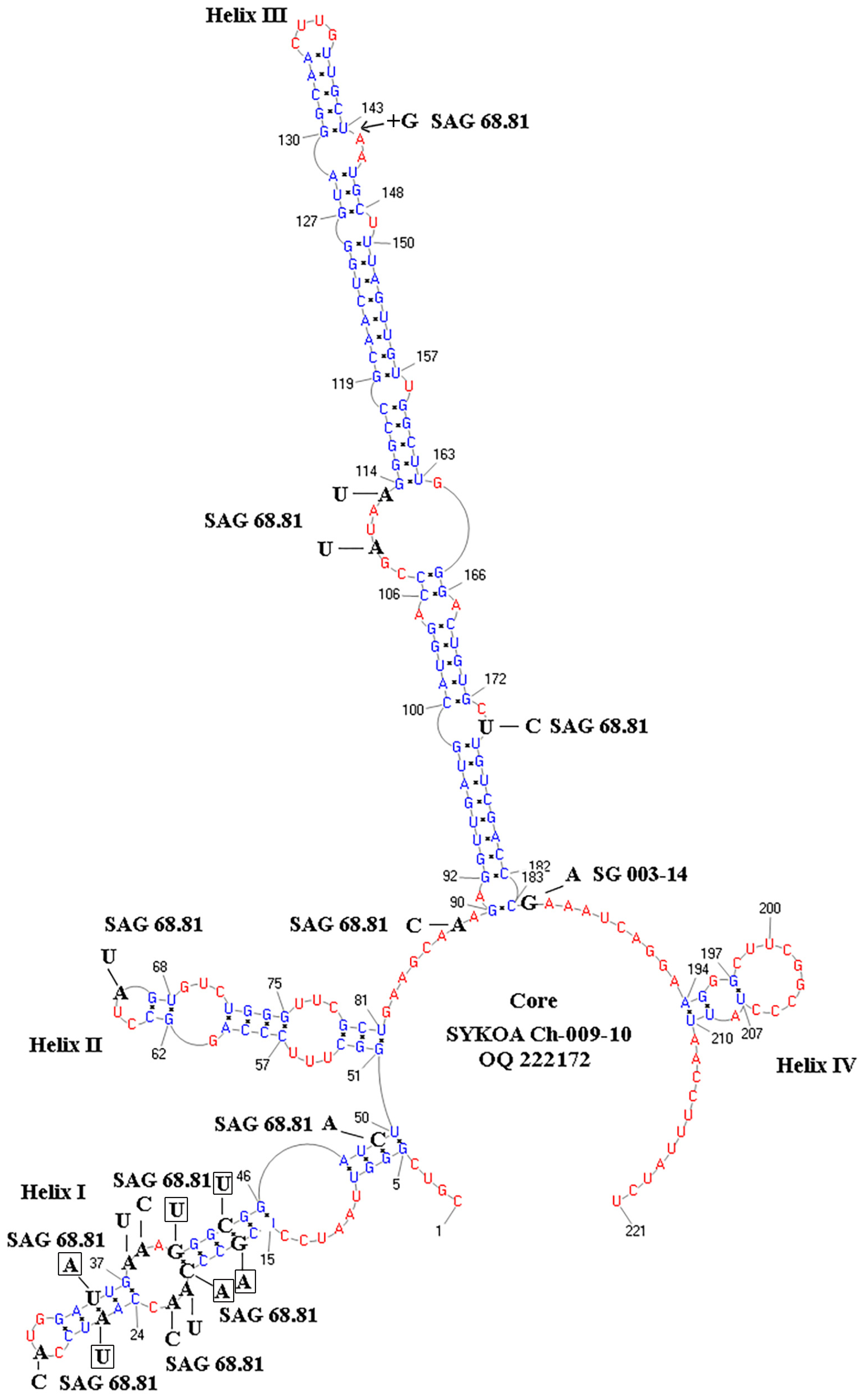
2.4. Modeling the Secondary Structure of the ITS2 Region

The Ribosomal RNA Database [23,24] used for the modeling of the secondary structure of ITS2. The model was built considering the presence of a pyrimidine-pyrimidine unpaired site in Helix II, as well as the length and nucleotide composition of the spacers in the core of the model, which determine the boundaries of the Helix [25,26]. The PseudoViewer3 program [27] was used to visualize the resulting secondary structure. The sequences and their secondary structures were aligned using 4SALE 1.7.1 [28,29] and manually checked. All pairs of bases of the four helices were analyzed for the presence of compensatory base changes [30].

3. Results

3.1. Characteristics of Habitat Conditions

The investigated strain SYKOA Ch-009-10 is a soil habitant, as all earlier described species of this genus. The alga was isolated from biological soil crust collected at frost boils in the high mountain screes of the Subpolar Urals. The ecological conditions of the studied habitat are harsh. The strain was isolated from acidic tundra soil with low exchangeable bases, low content of nitrogen and other important nutrients. Soil moisture was relatively high for frost boils [31] during the research period (Table 1).

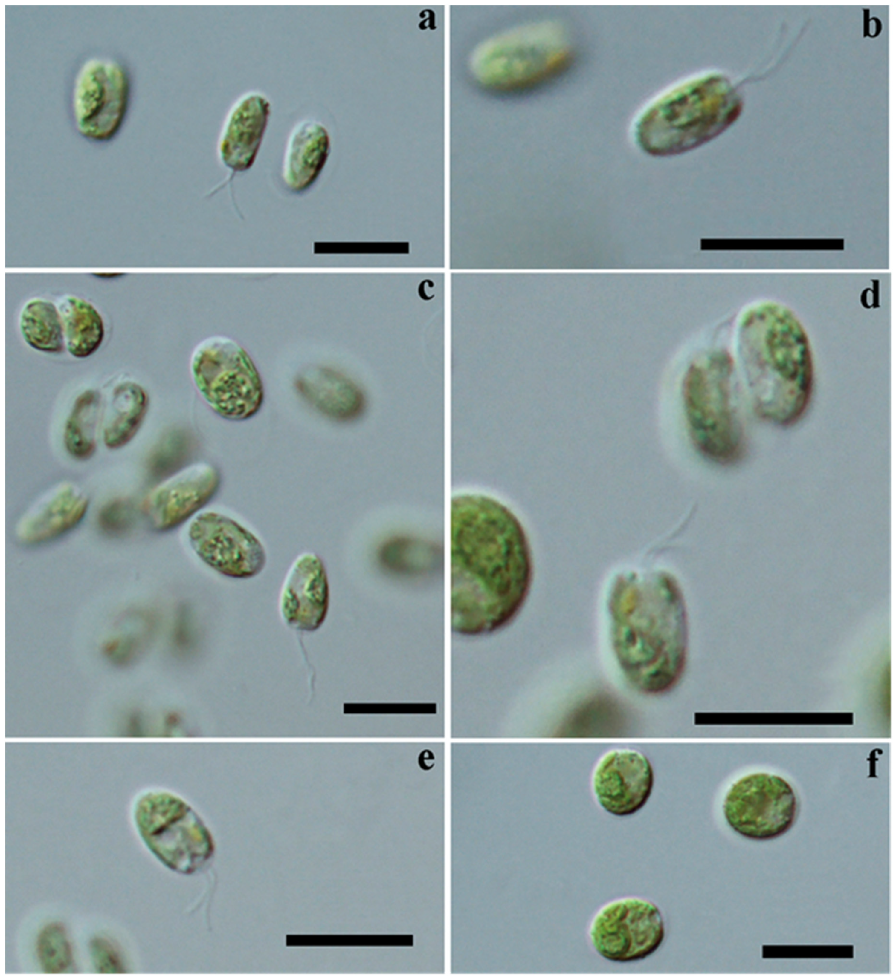
3.2. Cell Morphology: Light Microscopy

Studied motile cells are obovate, slightly asymmetrical, truncated and blunt in upper part, narrowed (some cells are strongly narrowed at end) and rounded in basal part (Figure 1a–e and Figure 2a). Cells are (5.0) 6.4–8.7 µm long and (2.0) 4–4.7 (4.9) µm wide. Cell wall is thin, without papilla. Length of flagella equals length of cell. Flagella are slightly unequal in length. Two contractile vacuoles are apical. Chloroplast is parietal, with incisions on surface, forming 2–4 (5–6) lobes by 1–2 narrow horizontal incisures. Stigma is pale, orange, large, ellipsoid and anterior. Pyrenoid is single, spherical in lateral, middle or upper part of cell, surrounded by several different-sized, mostly small, starch grains. Young cells and zoospores have mainly basal nucleus; in mature cells, nucleus often migrates to central or apical part of cells (Figure 1a–e and Figure 2). Motile stage is brief; cells quickly lose flagella, increase their size and form non-motile vegetative cells in mucous colonies. Adult cells are spherical, broadly oval, without stigma, with two contractile vacuoles, with an anterior or central nucleus. Cells are surrounded by delicate mucilage (individual mucilage for each cell) and collected in amorphous colonies with slightly stratified mucilage (Figure 2b and Figure 3a–d,). Adult cells are (min. 7.9) 14–16 (max. 22.5) µm in diameter. Asexual reproduction by zoospores formed in sporangia with 2–4 cells (mainly 4, 8 or 16). Cells easily leave palmelloid stage after a transfer to fresh liquid culture medium and rapidly form zoospores. Sexual reproduction is not observed. Old culture remains green or becomes yellow-green. Surface of chloroplast in old cells is granular; lobes and incisions are not expressed. Stigma is invisible. Flagella are absent.

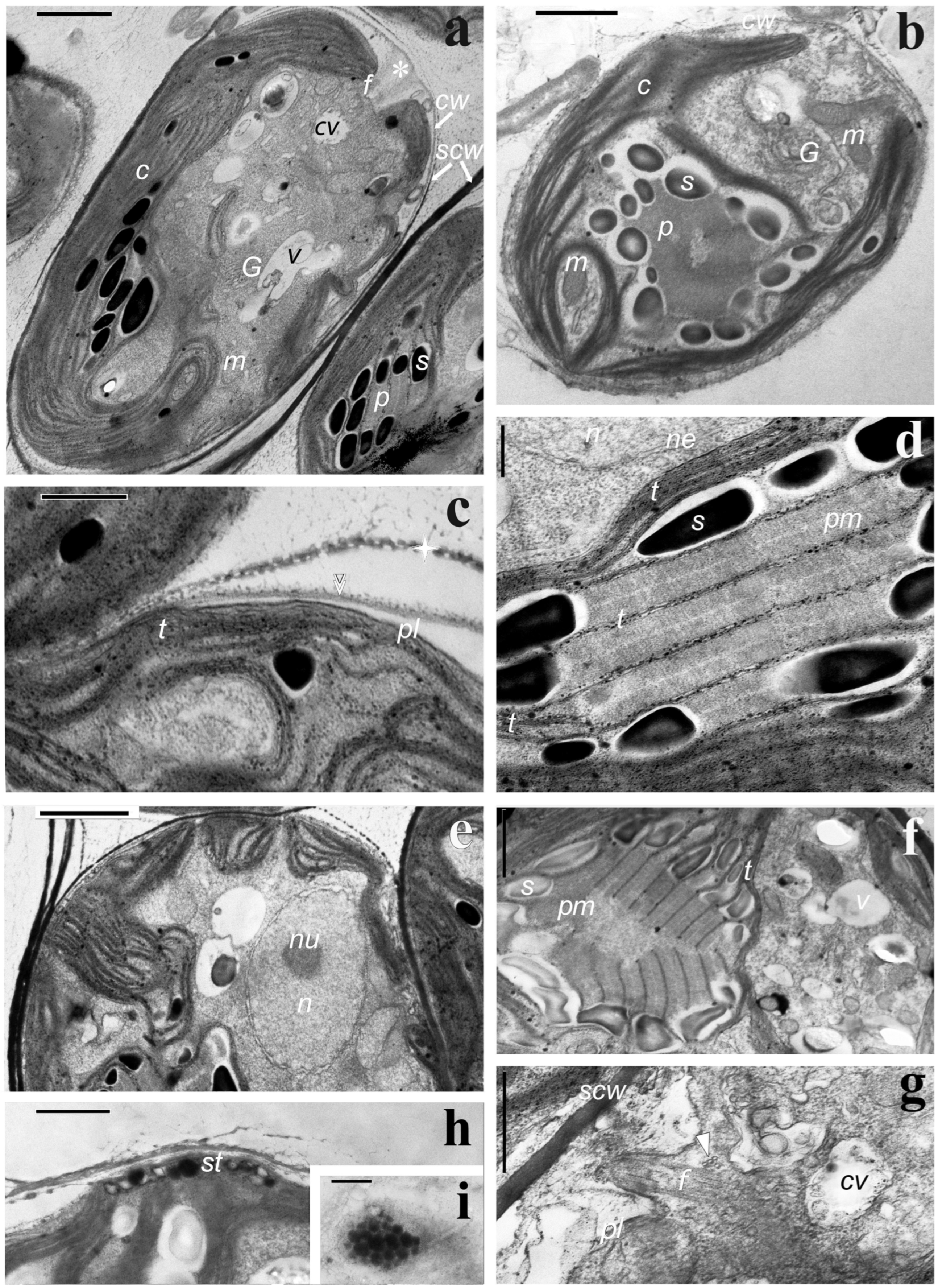
3.3. Cell Ultrastructure. Transmission Electron Microscopy

Cells are covered by a thin (~100 nm) cell wall (Figure 4a–c), consisting of a small portion of fibrils and thin, tightly enclosed electron-dense lamella (Figure 4c). Papilla is absent, but the cell wall is considerably thicker at the apical end, in the region of the flagella outlet (Figure 4a). The dark mucilage-like component is visible outside the individual cell wall, and it contains perforations (Figure 4c). The latter can partially peel off from individual sporangial cells; if not, it forms a very dark sporangial cell wall, which, as the sporangia grows, also stratifies into thicker fragments (Figure 4a,e,g). Plasmalemma is smooth and contacts with a chloroplast envelope only in a few areas (Figure 4c). Chloroplast thylakoids are predominantly long, straight, sometimes curved, and organized in packs of two and three units each, rarely reaching 5–6 units (Figure 4a–f). The chloroplast thylakoid packs are common with the same of a pyrenoid body and likely do not change their form entering but reduce their number up to two (Figure 4d). Pyrenoid sheath consists of 10–20 small starch grains with various shapes and sizes and surrounds the pyrenoid body. The pyrenoid body is highly ordered. It consists of equal portions of the matrix and paired thylakoids regularly penetrating it. Thylakoids are generally straight (Figure 4d,e). Occasionally, thylakoids in the pyrenoid lie in different directions (Figure 4f). In a few sections, the pyrenoid body looks almost homogeneous (Figure 4b). This is due to the architecture and the dissection site of the pyrenoid itself (Figure 4f). Stigma is rarely observed. It is located under the chloroplast envelope and formed by one layer of densely adjacent dark globules (Figure 4h,i). Starch grains in chloroplast are few; their form and sizes are similar to that of the pyrenoid envelope (Figure 4a–c,e). The single nucleus is bounded by a double membrane with almost regular pores and contains weakly condensed chromatin and a central nucleolus with an ordered arrangement of granular and fibrillar components (Figure 4e). Near the nucleus, there are usually one or two Golgi bodies of about five cisternae and a small number of vesicles. Mitochondria are predominantly centrally located (near the nucleus and flagellar apparatus) and are represented mainly by small profiles; their cristae are small and flattened. There are the following types of vacuoles: homogeneous light, with dark contents, with membrane components and two contractile vacuoles (Figure 4a,b,e–g). The latter are observed at the apical cell end, near the flagella. The flagellar bases are located almost parallel to each other (Figure 4a). On some sections, a 3 + 1 fragment of the microtubule root and the transition zone of flagella (Figure 4g) is distinguished.

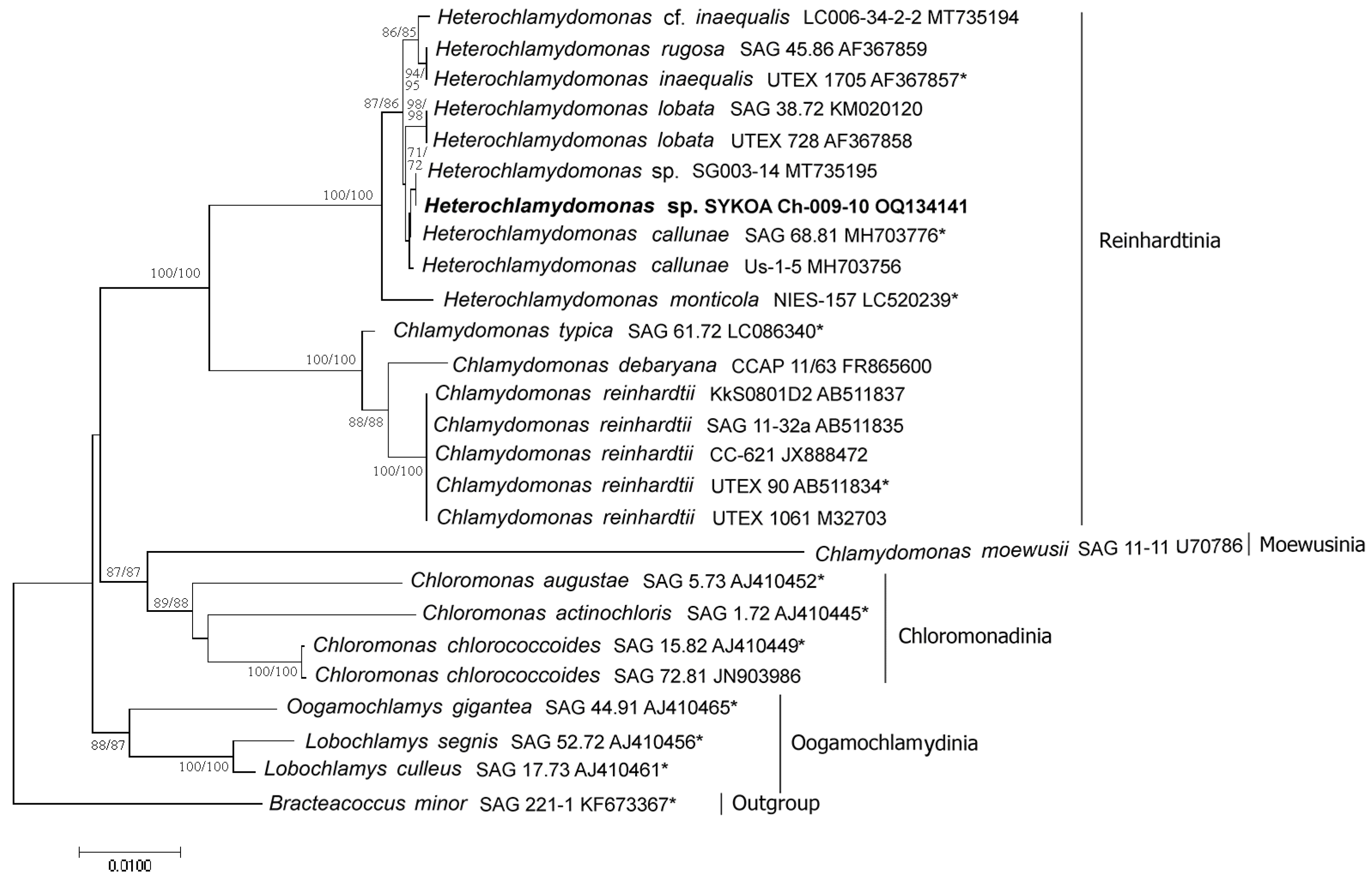
3.4. Molecular Phylogenetic Analysis

The analysis of 18S rDNA sequences combined all studied strains into one well-supported (100%, NJ) clade of Heterochlamydomonas sensu lato (Figure 5). In general, the topology of the 18S rDNA tree is consistent with previously published data [3,6,32]. H. monticola was an early offshoot and sister to other species of the genus. H. rugosa and H. inaequalis constituted a moderately supported clade. H. callunae and H. lobata formed separate branches. The sequence of the studied strain had a high nucleotide similarity (100%) with the strain Heterochlamydomonas sp. SG003-14 [32].
The analysis of ITS2 rDNA sequences is difficult since the scarcity of data on the genus Heterochlamydomonas in GenBank (only one belongs to the authentic strain SAG 68.81 out of the presented sequences). The Heterochlamydomonas clade with high bootstrap support (100%) splits into two subclades on the tree. The first subclade combined strains of Heterochlamydomonas sp. SYKOA Ch-009-10 and SG003-14, as well as their sister Heterochlamydomonas cf. inaequalis LC006-34-2-2. The second subclade included samples of H. callunae SAG 68.81 (authentic strain) and H. callunae Us-1-5. The ITS2 rDNA sequences of the SYKOA Ch-009-10 strain showed maximum similarity (99%) to the SG003-14 strain (Figure 6).
Analysis of the nucleotide sequence ITS2 of SYKOA Ch-009-10 was clearly different from that of the authentic strain of H. callunae SAG 68.81 (Figure 7 and Figure 8). In total, 17 substitutions and 3 indels (positions 33, 143/144 and 175) were identified between the strains SYKOA Ch-009-10 and SAG 68.81 (Figure 7 and Figure 8). Twelve of them are in the Helix I variable region of the secondary structure of ITS2. Three compensatory (CBC) changes are found in Helix I at positions 17 and 44 (G-C on A-U); at 20 and 41 (C-G on A-U); at 26 and 35 (A-U on U-A) (Figure 7). A comparison of the ITS2 secondary structure of conserved regions (Helix II and Helix III) of SYKOA Ch-009-10 with SAG 68.81 found four substitutions and one indel. One replacement in Helix II is the terminal (at position 66) and three substitutions and one indel in Helix III. One substitution was found in the core at position 88 (Figure 7).
Analysis of the secondary structure of ITS2 of SYKOA Ch-009-10 and Heterochlamydomonas sp. SG003-14 strains showed polymorphism in only one position 184 (G/A).
Analysis of another variable region of the ITS2 sequence secondary structure (Helix IV) did not reveal any differences between the strains studied.

4. Discussion

One more strain from the collection of microalgae strains in the Institute of Biology of Komi Science Centre (SYKOA)—SYKOA Ch-009-10 was studied by morphological and ultrastructural methods and by DNA identification. The algae were isolated from biological soil crust in the high mountain screes of the Subpolar Urals. The habitats are characterized by low temperatures, relatively high humidity and low nutrient content. The strain SYKOA Ch-009-10 was originally identified as H. callunae because of its similarities to the authentic strain SAG 68.81. Morphologically the SYKOA Ch-009-10 strain differs from most other species of Heterochlamydomonas by the presence of young cells with an acutely truncated basal end and bluntly cut apex, chloroplast topology (usually 3–4, rarely 5–6 lobes), granularity and absence of pronounced incisions in old cells. However, they are very similar to H. callunae cells. Moreover, cell dimensions of SYKOA Ch-009-10 also correspond to the dimensional range of H. callunae. Nevertheless, the mature cells of SYKOA Ch-009-10 are larger than all previously discovered Heterochlamydomonas.
Cell ultrastructure for different Heterochlamydomonas is studied poorly and concerns only two species—H. inaequalis and H. monticola [3,15]. Like the described Heterochlamydomonas spp., the SYKOA Ch-009-10 has a one-layered stigma, nearly parallel flagella and common traits of flagellar apparatus. The cell wall is thin, but in the region of flagella, output is rather thick. At the same time, the absence of papilla is confirmed on the ultrastructural level for all the above-mentioned species. A low visible or indistinct outer layer characterizes the cell wall of these three species. It is represented by only one dark lamella, and the other two—dark and intermediate white of “central triplet” sensu K. Roberts [33]—are possibly reduced. This contrasts with the well-organized layers of the Chlamydomonas cell walls of core Reinhardtinia, e.g., C. reinhardtii [33]. Additionally, the cells of the SYKOA Ch-009-10 strain have a dark mucilage-like component with perforations located outside the individual cell walls. It remains unclear whether this is a feature of the species or it is common to the genus.
The thylakoid system of the chloroplast of the studied strain has peculiar characteristics. The thylakoids are predominantly long, not connected in large packs, and rarely reach 5–6 units. According to Figure 5 [15], the chloroplast of H. inaequalis equipped by the thylakoids is organized just in the same manner, but with shorter packs. These contrast with the thylakoid system of H. monticola, consisting of short thick packs, even in gametes [3].
The pyrenoid structure of SYKOA Ch-009-10 also has bright distinctive characteristics. Its starch sheath consists of numerous small grains in contrast to H. inaequalis and is similar to that of H. monticola. However, the pyrenoid may look different depending on the sectional plane. On most sections, there is a highly ordered system of paired thylakoids and dark heterogeneous matrix parts between them. At the same time, in some sections, one can see only a homogenous matrix and a few profiles of thylakoids. Meanwhile, the pyrenoid structure of SYKOA Ch-009-10 is similar to that of H. monticola (consisting of many “straight or curved rod-like structures of relatively regular thickness and various lengths, with which thin thylakoid membranes were rarely associated”), as described by S. Watanabe [3]. These pyrenoid peculiarities are different from those of H. inaequalis [15], as well as of C. reinhardtii [18], which have pyrenoids with distended thylakoid bundles and an irregularly penetrating matrix. Perhaps, well-organized pyrenoid structures of SYKOA Ch-009-10 and H. monticola are rare features for green monad algae species. Previously, we observed a similar but always ordered pyrenoid structure (named III* type) in only 1 of 26 chlamydomonad species—C. nasuta, SAG 11.72 [18].
Analysis of the 18S and ITS2 rDNA sequences of the SYKOA Ch-009-10 strain showed high similarity of 100% and 99%, respectively, with Heterochlamydomonas sp. SG003-14 [32]. Analysis of the secondary structure of ITS2 of Ch-009-10 and SG003-14 strains showed polymorphism in only one position, 184. Secondary structure analysis of the Ch-009-10 strain and the authentic SAG 68.81 strain revealed 17 substitutions (including 3 CBCs) and 3 indels. All CBCs substitutions are located in the Helix I region. Currently, the concept of Coleman [34] to distinguish taxa based on compensatory substitutions in conserved ITS2 sites is not relevant for all species of Chlorophyta [35]. However, for the asexual-propagating algae, compensatory substitutions are important molecular indicators for distinguishing the species. We identified three CBCs between the strains SYKOA Ch-009-10 and SAG 68.81. Despite the morphological similarity of the studied strain SYKOA Ch-009-10 compared with the SAG 68.81 strain, the quantity of changes found in ITS2 may be an additional sign of the species specificity of SYKOA Ch-009-10. The strain SYKOA Ch-009-10 has the most similarity with Heterochlamydomonas sp. SG003-14 is based on both 18S gene data and the secondary structure of ITS2. In the strains studied, from a total alignment length of 223 nucleotide positions, we identified 33 polymorphic sites in ITS2. Seventeen of them were parsimony informative (Figure 8). The genetic distance calculated from the p-distance between the strains of Heterochlamydomonas sp. SYKOA Ch-009-10 and SG003-14 was 0.5%, while with other strains, it was about 8%.
Thus, the studied strain has the maximum similarity to the strain Heterochlamydomonas sp. SG003-14. Unfortunately, the article by Samolov et al. [32], which mentions Heterochlamydomonas sp. SG003-14, contains no morphological or ultrastructural description of the species. Thus, it is not possible to verify how close the two strains are in terms of morphology. The strain SG003-14 isolated from the habitat in the semiarid zone of South America (Nature Reserve Santa Gracia) strongly differed from the habitat of SYKOA Ch-009-10 (cold and moist soil, Subpolar Urals, Yugyd Va National Park). So, we suggest the SYKOA strain Ch-009-10 as a new species.

Taxonomic Assessment

Heterochlamydomonas uralensis Novakovskaya, Boldina, Shadrin, Patova sp. nov. (Figure 1, Figure 2 and Figure 3).
Diagnosis: Heterochlamydomonas uralensis is phenotypically distinct from other Heterochlamydomonas species by form of mobile cells, chloroplast and pyrenoid structure (except that of H. monticola), large cell dimensions, and nucleus disposition. Differs from other species of genus Heterochlamydomonas by phylogenetic position based on partial 18S and ITS2 rDNA gene phylogenies.
Description: Motile cells are obovate, in upper part truncated and blunt, in basal part narrowed. Cells are (5.0) 6.4–8.7 µm long and (2.0) 4–4.7 (4.9) µm wide. Cell wall thin, papilla is absent. Two flagella are slightly unequal in length. There are two frontal contractile vacuoles. Chloroplast is parietal, with incisions on surface. Stigma is large, ellipsoid and anterior. Pyrenoid is spherical in lateral, middle or upper part of cell, surrounded by several different-sized, mostly small, starch grains. Young cells and zoospores have basal nucleus; mature cells—apical. Adult cells are spherical, without stigma, with two contractile vacuoles, with an anterior or central nucleus. Cells are surrounded by delicate mucilage and collected in amorphous colonies with slightly stratified mucilage. Adult cells are (7.9) 14–16 (22.5) µm in diameter. Asexual reproduction by 2–4–8 (16) zoospores.
Holotype: Resin-embedded specimen for TEM, deposited into herbarium of Institute of Biology of Komi Science Centre of the Ural Branch of the Russian Academy of Sciences (IB FRC Komi SC UB RAS) (SYKOA), Syktyvkar, Russia, under following accession number: SYKO No. 1015 R. Paratype—dried specimen also deposited into herbarium of Institute of Biology under following accession number SYKO No. 1015.
Reference strain: SYKOA Ch-009-10 (isolated into culture by Novakovskaya), deposited in the Culture Collection of Algae of the Institute of Biology under the number SYKOA Ch-009-10.
NCBI GenBank Accession number: OQ134141 (18S); OQ134140 (ITS2).
Type locality: Russia. Subpolar Urals Mountains (National Park “Yugyd Va”), 65.317 N; 60.516 E, elevation: 989 m. Carex-moss-lichen tundra collected by Novakovskaya on 23 July 2010.
Etymology: H. uralensis = The species was named for its habitat of Ural.
The indicative key for identifying species from the genus Heterochlamydomonas.
1. Motile cells are truncated and blunt in upper part, narrowed and rounded in basal part………………………………………………………………………………………………2
- Motile cells only rounded in basal part …………………………………………………4
2. Pyrenoid basal………………………………………………………………H. monticola
- Pyrenoid lateral, central or apical……………………………………………………3
3. Old cells up to 22 μm long ………………………………………………H. uralensis
- Old cells up to 16 μm long……………………………………………………H. callunae
4. Cell walls of motile cells thicken up to 3.0 μm ………………………H. inaequalis
- Cell walls of motile cells do not thicken ………………………………………………5
5. Chloroplast with a smooth surface and conspicuously lobed margin; old non-motile vegetative cells up to 10 μm long ………………………………………………H. lobate
- Chloroplast with a rugose surface and several to many perforations; old non-motile vegetative cells up to 13.5 μm …………………………………………………………H. rugosa

Author Contributions

Conceptualization, I.V.N.; methodology, I.V.N., O.N.B. and D.M.S.; formal analysis, I.V.N., O.N.B. and E.N.P.; investigation, I.V.N., O.N.B. and D.M.S.; resources, E.N.P.; data curation, I.V.N. and O.N.B.; writing—original draft preparation, I.V.N. and O.N.B.; writing—review and editing, I.V.N., O.N.B. and E.N.P.; visualization, I.V.N., O.N.B. and D.M.S.; supervision, I.V.N.; project administration, I.V.N. and O.N.B.; funding acquisition, E.N.P. and I.V.N. All authors have read and agreed to the published version of the manuscript.

Funding

This research was supported by the state tasks IB FRC Komi SC UB RAS No. 122040600026-9 and the Komarov Botanical Institute RAS No. 121021600184-6.

Institutional Review Board Statement

Not applicable.

Informed Consent Statement

Not applicable.

Data Availability Statement

The data presented in this study are available on request from the corresponding author.

Conflicts of Interest

The authors declare no conflict of interest. The funders had no role in the design of the study; in the collection, analyses, or interpretation of data; in the writing of the manuscript; or in the decision to publish the results.

References

  1. Cox, E.R.; Deason, T.R. Heterochlamydomonas, a new alga from Tennessee. J. Tenn. Acad. Sci. 1969, 44, 105–107. [Google Scholar]
  2. Shoup, S.; Lewis, L.A. Polyphyletic origin of parallel basal bodies in swimming cells of chlorophycean green algae (Chlorophyta). J. Phycol. 2003, 39, 789–796. [Google Scholar] [CrossRef]
  3. Watanabe, S. Reclassification of Chlamydomonas monticola as Heterochlamydomonas (Volvocales, Chlorophyceae). Phycol. Res. 2020, 68, 332–335. [Google Scholar] [CrossRef]
  4. Guiry, M.D.; Guiry, G.M. AlgaeBase. In World-Wide Electronic Publication; National University of Ireland: Galway, Ireland, 2023. [Google Scholar]
  5. Langford, M.F.; Cox, E.R. Additions to the genus Heterochlamydomonas (Chlorophycophyta). J. Tenn. Acad. Sci. 1971, 46, 66–71. [Google Scholar]
  6. Mikhailyuk, T.; Glaser, K.; Tsarenko, P.; Demchenko, E.; Karsten, U. Composition of biological soil crusts from sand dunes of the Baltic Sea coast in the context of an integrative approach to the taxonomy of microalgae and cyanobacteria. Eur. J. Phycol. 2019, 54, 263–290. [Google Scholar] [CrossRef]
  7. Ettl, H. Die Gattung Chlamydomonas Ehrenberg. Beih. Zur Nova Hedwig. 1976, 49, 1–1122. [Google Scholar]
  8. Watanabe, S. New and interesting green algae from soils of some Asian and Oceanian regions. Arch. Für Protistenkd. 1983, 127, 223–270. [Google Scholar] [CrossRef]
  9. Ettl, H.; Gärtner, G. Syllabus der Boden-, Luft-und Flechtenalgen; Auflage 2; Springer: Heidelberg/Berlin, Germany, 2014; p. 773. [Google Scholar]
  10. Kostikov, I.Y.; Romanenko, P.O.; Demchenko, E.M.; Darienko, T.M.; Mikhailyuk, T.I.; Rybchinskii, O.V.; Solonenko, A.M. Soil Algae of Ukraine: History and Methods of Analysis, System, and List of Flora; Fitosotsiotsentr: Kiev, Ukraine, 2001; p. 300. [Google Scholar]
  11. Zimonina, N.M. Soil Algae of the Oil Contaminated Land; VSPU: Kirov, Russia, 1998; p. 170. (In Russian) [Google Scholar]
  12. Patova, E.N.; Novakovskaya, I.V. Soil algae of the Northeastern European Russia. Nov. Sist. Nizshikh Rastenii 2018, 52, 311–353. [Google Scholar] [CrossRef]
  13. Watanabe, S.; Floyd, G.L. Comparative ultrastructure of zoospores with parallel basal bodies from the green algae Dictyochloris fragrans and Bracteacoccus sp. Am. J. Bot. 1992, 79, 551–555. [Google Scholar] [CrossRef]
  14. Nakada, T.; Misawa, K.; Nozaki, H. Molecular systematics of Volvocales (Chlorophyceae, Chlorophyta) based on exhaustive 18S rRNA phylogenetic analyses. Mol. Phylogenetics Evol. 2008, 48, 281–291. [Google Scholar] [CrossRef]
  15. Floyd, G.L.; Wilcox, L.W.; Watanabe, S. Structure and flagellar beat in Heterochlamydomonas inaequalis, a motile, unicellular, green alga with unequal flagella and parallel basal bodies. Crypt. Bot. 1990, 1, 332–339. [Google Scholar]
  16. Andersen, R.A. Algal Culturing Techniques; Elsevier: New York, NY, USA, 2005; p. 589. [Google Scholar]
  17. Andreeva, V.M. Soil and Aerophilic Green Algae (Chlorophyta: Tetrasporales Chlorococcales, Chlorosarcinales); Nauka: St. Petersburg, Russia, 1998; p. 351. (In Russian) [Google Scholar]
  18. Konstantinova, I.A.; Boldina, O.N. Comparative analysis of the pyrenoid ultrastructure in green monad and coccoid algae. Russ. J. Plant Physiol. 2000, 47, 655–659. [Google Scholar]
  19. Nakazawa, A.; Nozaki, H. Phylogenetic analysis of the Tetrasporalean genus Asterococcus (Chlorophyceae) based on 18S ribosomal RNA gene sequences. J. Jpn. Bot. 2004, 79, 255–261. [Google Scholar]
  20. Nakada, T.; Nozaki, H. Re-evaluation of three Chlorogonium (Volvocales, Chlorophyceae) species based on 18S ribosomal RNA gene phylogeny. Eur. J. Phycol. 2007, 42, 177–182. [Google Scholar] [CrossRef]
  21. Nakada, T.; Shinkawa, H.; Ito, T.; Tomita, M. Recharacterization of Chlamydomonas reinhardtii and its relatives with new isolates from Japan. J. Plant Res. 2010, 123, 67–78. [Google Scholar] [CrossRef]
  22. White, T.J.; Bruns, T.D.; Lee, S.B.; Taylor, J.W. Amplification and direct sequencing of fungal ribosomal RNA genes for phylogenetics. In PCR Protocols: A Guide to Methods and Applications; Innis, M.A., Ed.; Academic Press: San Diego, CA, USA, 1990; pp. 315–322. [Google Scholar]
  23. Schultz, J.; Müller, T.; Achtziger, M.; Seibel, P.N.; Dandekar, T.; Wolf, M. The internal transcribed spacer 2 database-a web server for (not only) low level phylogenetic analyses. Nucleic Acids Res. 2006, 34, 704–707. [Google Scholar] [CrossRef]
  24. Ankenbrand, M.J.; Keller, A.; Wolf, M.; Schultz, J.; Förster, F. ITS2 database V: Twice as much. Mol. Biol. Evol. 2015, 32, 3030–3032. [Google Scholar] [CrossRef]
  25. Caisová, L.; Marin, B.; Melkonian, M.A. Consensus Secondary Structure of ITS2 in the Chlorophyta Identied by Phylogenetic Reconstruction. Protist 2013, 164, 482–486. [Google Scholar] [CrossRef]
  26. Temraleeva, A.D.; Mincheva, E.V.; Bukin, Y.S.; Andreeva, A.M. Modern Methods of Isolation, Cultivation and Identification of Green Algae (Chlorophyta); Kostroma Printing House: Kostroma, Russia, 2014; 215p. (In Russian) [Google Scholar]
  27. Byun, Y.; Han, K. PseudoViewer: Web application and web service for visualizing RNA pseudoknots and secondary structures. Nucleic Acids Res. 2006, 34, 416–422. [Google Scholar] [CrossRef]
  28. Seibel, P.N.; Müller, T.; Dandekar, T.; Schultz, J.; Wolf, M. 4SALE—A tool for synchronous RNA sequence and secondary structure alignment and editing. BMC Bioinform. 2006, 7, 498. [Google Scholar] [CrossRef]
  29. Seibel, P.N.; Müller, T.; Dandekar, T.; Wolf, M. Synchronous visual analysis and editing of RNA sequence and secondary structure alignments using 4SALE. BMC Res. Notes 2008, 1, 91. [Google Scholar] [CrossRef]
  30. Coleman, A.W. Is there a molecular key to the level of ‘biological species’ in eukaryotes? A DNA guide. Mol. Phylogenetics. Evol. 2009, 50, 197–203. [Google Scholar] [CrossRef]
  31. Patova, E.N.; Novakovskaya, I.V.; Sivkova, M.D. Cyanobacteria and Algae in Biological Soil Crusts of Frost Boils in the Mountain Tundra of the Urals. Eurasian Soil Sci. 2023, 56, 184–197. [Google Scholar] [CrossRef]
  32. Samolov, E.; Baumann, K.; Büdel, B.; Jung, P.; Leinweber, P.; Mikhailyuk, T.; Karsten, U.; Glaser, K. Biodiversity of Algae and Cyanobacteria in Biological Soil Crusts Collected Along a Climatic Gradient in Chile Using an Integrative Approach. Microorganisms 2020, 8, 1047. [Google Scholar] [CrossRef]
  33. Roberts, K. Crystalline glycoprotein cell walls of algae: Their structure, composition and assembly. Philos. Trans. R. Soc. B 1974, 268, 129–146. [Google Scholar] [CrossRef]
  34. Coleman, A.W.; van Oppen, M.J.H. Secondary structure of the rRNA ITS2 region reveals key evolutionary patterns in acroporid corals. J. Mol. Evol. 2008, 67, 389–396. [Google Scholar] [CrossRef]
  35. Caisová, L.; Marin, B.; Melkonian, M. A close-up view on ITS2 evolution and speciation—A case study in the Ulvophyceae (Chlorophyta, Viridiplantae). BMC Evol. Biol. 2011, 11, 262. [Google Scholar] [CrossRef] [PubMed]
Figure 1. Heterochlamydomonas uralensis sp. nov.: (ae) motile cells, with slightly uneven flagella; (f) non-motile vegetative cells. Scale bars: 10 μm.
Diversity 15 00673 g001
Figure 2. Drawings of Heterochlamydomonas uralensis sp. nov. (a)—motile cells, (b)—old non-motile vegetative cells with mucilage. Scale bar: 10 μm.
Diversity 15 00673 g002
Figure 3. Heterochlamydomonas uralensis sp. nov. (a)—zoospore release from zoosporangium, (bd)—old non-motile vegetative cells, (cd)—cells surrounded by delicate mucilage. Mucilage was visualized with methylene blue. Scale bars: 10 μm.
Diversity 15 00673 g003
Figure 4. Heterochlamydomonas uralensis sp. nov. (a,b)—zoospores and young vegetative cells: (a)—a motile zoospore, note parallel arrangement of flagella channels in the cell wall (asterisk), (c)—a fragment of two contacting zoospores in sporangia with an emphasis on their cell walls, see median layer (double arrowheads) and mucilage-like layer (star), (d)—pyrenoid body with ordered system of two-thylakoid packs, dissecting heterogeneous stroma parts and surrounded by starch grains, here it is close to the nucleus, (e)—a cell fragment with nucleus, (f)—pyrenoid body with thylakoids, lying in two different directions and an area of low dense between them, g—a cell fragment with dense sporangial wall and flagella, note 3+1 microtubular root (arrowhead) and H-form transitional zone, (h,i)—stigma. c—chloroplast, cv—contractile vacuole, cw—cell wall, G—Golgi body, f—flagellum, m—mitochondria, n—nucleus, nu—nucleolus, ne—nuclear envelope, p—pyrenoid, pl—plasmalemma, pm—pyrenoids matrix, s—starch in the pyrenoid envelope, scw—sporangium cell wall, st—stigma, t—thylakoids, v—vacuole. Scale bars: a, b, e, f—1 μm; c, d, g, h, i—0.5 μm.
Diversity 15 00673 g004
Figure 5. Phylogenetic tree built using NJ method based on 18S rDNA sequences. *—authentic strains. Bootstrap values (≥70%) shown on the figure for both analyses (NJ/ML).
Diversity 15 00673 g005
Figure 6. Phylogenetic tree built using ML method based on ITS2 rDNA sequences. *—authentic strains. Bootstrap values (≥70%) shown on the figure for both analyses (NJ/ML).
Diversity 15 00673 g006
Figure 7. The ITS2 secondary structure of Heterochlamydomonas sp. SYKOA Ch-009-10. The ITS2 model of Heterochlamydomonas sp. SYKOA Ch-009-10 was built based on the strain model Chlamydomonas pygmaea SAG 5.93 (NCBI: AJ749619). Variable positions of analyzed strains are marked in bold. Compensatory base changes in conservative regions are square. “+”— insertion.
Diversity 15 00673 g007
Figure 8. The variable sites from an alignment of ITS2 rDNA sequences of Heterochlamydomonas species. *—authentic strains; Diversity 15 00673 i001—gap between positions 143 and 144.
Diversity 15 00673 g008
Table 1. Chemical properties of the mountain tundra soil of Subpolar Urals.
Soil Moisture, %Temperature (Min.; Max.), °CC, %N, %Fe, mg/kgMn, mg/kgpH (H2O)pH (KCl)Exchangeable Cations, cmolc/kgP2O5 mg/kgK2O mg/kg
Ca2+Mg2+
ω ± Δω ± Δω ± Δω ± ΔpH ± ΔpH ± Δn ± Δn ± Δω ± Δω ± Δ
29.7−19.0
+13.1
0.43
0.13
0.038
0.010
57
17
610
170
6.265.215.28
0.40
2.12
0.16
447.8
67.17
96.76
14.51
Disclaimer/Publisher’s Note: The statements, opinions and data contained in all publications are solely those of the individual author(s) and contributor(s) and not of MDPI and/or the editor(s). MDPI and/or the editor(s) disclaim responsibility for any injury to people or property resulting from any ideas, methods, instructions or products referred to in the content.

