Article

Diversity of Silica-Scaled Chrysophytes (Stramenopiles: Chrysophyceae) from Indonesian Papua

1
A.N. Severtsov Institute of Ecology and Evolution, Russian Academy of Sciences, Leninsky Prospect 33, 119071 Moscow, Russia
2
K.A. Timiryazev Institute of Plant Physiology, Russian Academy of Sciences, Botanical Street 35, 127276 Moscow, Russia
*
Author to whom correspondence should be addressed.
Diversity 2022, 14(9), 726; https://doi.org/10.3390/d14090726
Submission received: 8 August 2022 / Revised: 26 August 2022 / Accepted: 30 August 2022 / Published: 2 September 2022
(This article belongs to the Special Issue Algae Biodiversity: Natural and Anthropogenic Impacts)

Abstract

:
The silica-scaled chrysophyte flora from Indonesian Papua was investigated by means of electron microscopy. A total of twenty-four taxa were recorded, including five belonging to Paraphysomonas, one to Chrysosphaerella, one to Spiniferomonas, fifteen to Mallomonas and two to Synura. Thirteen taxa were recorded for the first time in Indonesia. Comparison of the species composition of silica-scaled chrysophytes from different parts of the Island of New Guinea shows significant differences. A “living fossil” was discovered, Mallomonas preisigii, which was described previously from Eocene deposits in Northern Canada. Although the scales from Papua slightly differ from those of the fossil one, we believe they belong to the same morphospecies. Mallomonas preisigii can be considered as a paleoendemic species.

1. Introduction

Silica-scaled chrysophytes comprise heterokont protists from different evolutionary lineages belonging to three orders (Paraphysomonadales, Chromulinales and Synurales) within a single class Chrysophyceae. The scale ultrastructure is species-specific. Therefore, there is a well-developed morphological species concept for several chrysophyte genera (e.g., Synura, Mallomonas and Paraphysomonas) that is congruent with molecular phylogenetic data [1,2]. Electron micrographs, published in the last 70 years, can be used for floristic and biogeographic analysis. Thus, chrysophytes can be considered as a model group for biogeographical studies [3,4,5].
Despite numerous floristic and taxonomic studies of silica-scaled chrysophytes performed all over the world, there is a predominance of such studies in the temperate zone, mainly in Europe and Northern America [6]. In particular, there are numerous unstudied areas exist in tropical regions. During the last two decades, diverse tropical flora has been documented [4,7,8,9,10,11,12,13,14,15,16], and our knowledge on the biodiversity of silica-scaled chrysophytes in tropical regions has been significantly expanded. Every additional investigation from tropical zones results in discoveries of new species to science, e.g., from Africa [17,18,19] and Asia [11,12,20,21,22,23,24,25,26,27,28].
Numerous Indonesian islands belong to the Indo-Malaysian-North-Australian phycogeographical region—one of ten regions defined by Krieger [29] based on the composition of desmid floras. The study on Indonesian algal flora has been performed for a long time and made it possible to identify diverse floras of desmids [29,30,31], euglenoids [30,32] and diatoms [33,34,35]. Many generic (e.g., Ichthyodontum, Tetralunata, Celebesia and Alveocymba) and species endemics among desmids [36] and diatoms [37,38,39,40,41,42] have been described in these studies. Therefore, algal floristic studies of this region are essential for the understanding of the biodiversity and biogeography of different algal groups in the Asian tropics.

2. Materials and Methods

Samples for this study were collected in November 2015 by E. Gusev and M. Kulikovskiy in Indonesian Papua (Table 1, Figure 1). Two areas were studied in Papua province (Jayapura and Jayawijaya Regency), Indonesia. Lake Sentani and the surrounding water bodies are located in Jayapura Regency. This territory is adjacent to the sea coast and has an elevation of 81–119 m a.s.l. In Jayawijaya Regency two highland areas were studied: Lake Habbema and surrounding swamps and streams at elevation 3328–3337 m a.s.l. and water bodies in Baliem Valley in the suburbs of Wamena City at elevation 1654–1730 m a.s.l.
Water specific conductance, pH and temperature measurements were performed using the Hanna Combo (Hanna HI 9828) device (Hanna Instruments, Inc., Ann Arbor, MI, USA). For electron microscopy studies, an aliquot of each sample was rinsed in deionized water in a centrifuge. Drops of the rinsed sample were dried or digested in sulfuric acid with potassium dichromate. For scanning electron microscope (SEM) studies, the samples were placed on the SEM stub and coated with gold for 10 min. Observations were performed with JEOL 6510 LV. For transmission electron microscope (TEM) studies formvar-coated grids (EMS FF200-Cu-50, Electron Microscopy Sciences, Hatfield, PA, USA) were used, and observations were made on a JEM-1011 (Papanin Institute for Biology of Inland Waters RAS, Borok, Russia).

3. Results

A total of twenty-four taxa of silica-scaled chrysophytes were recorded in the water bodies of Indonesian Papua (Table 2), including five belonging to Paraphysomonas De Saedeleer emend. Scoble and Cavalier-Smith (Figure 2A–D,F,G), one to Chrysosphaerella Lauterborn (Figure 2H and Figure 3A), one to Spiniferomonas E. Takahashi (Figure 2E), fifteen to Mallomonas Perty (Figure 3B–L and Figure 4A–J) and two to Synura Ehrenberg (Figure 4K,L). Thirteen taxa were recorded for the first time in Indonesia. Several noteworthy taxa are characterized below.
Several new records of Paraphysomonas in the Indonesian flora were observed in our investigations.
Paraphysomonas cf. segmenta (Figure 2A) was identified on the basis of a group of five scales; however, only a single scale had a well-developed spine. The base-plate is round (diameter 0.41–0.47 µm) with a thickened rim; the spine is two-segmented, 0.47 µm in length; S/P ratio of 1. According to Scoble and Cavalier-Smith [2], this species has a base-plate that is 0.40–0.53 µm in diameter, a spine length of 0.52–0.73 µm and a spine length to base-plate diameter ratio (S/P ratio) of 1.3–1.7. Therefore, our specimen has a slightly shorter spine. Additional material is needed for accurate identification. This species has previously been recorded from the United Kingdom [2].
One unidentified taxon from this genus was observed. Paraphysomonas sp. (Figure 2B,C) is similar to Paraphysomonas uniformis subsp. hemiradia, described by Scoble and Cavalier-Smith [2], in which they have four to six radial ribs on the basal plate of the scales. However, Paraphysomonas sp. significantly differs from P. uniformis subsp. hemiradia by its scale dimensions. Scales have a spine length of 8.93 µm, a spine base width of 0.25 µm, a base-plate diameter of 2.50–2.54 µm and an S/P ratio of 3.57. Furthermore, the spine length and base-plate diameter in our species are significantly larger than in P. longispina Scoble and Cavalier-Smith. It is likely that these scales belong to a yet undescribed species; however, more material is needed for a formal description.
Paraphysomonas uniformis subsp. uniformis (Figure 2D) is characterized by scales with a round to oval base-plate, 1.71–2.02 µm in diameter. The spine is straight, gently tapered with small oblique blunt point, 4.62–4.8 µm in length and with an S/P ratio of 2.3–2.8. The species has previously been recorded from Austria [2].
Paraphysomonas vulgaris subsp. vulgaris (Figure 2F) has a round base-plate (2.0–2.5 µm in diameter) with a dense rim. The spine is 3.3–4.3 µm in length with a bulbous base and oblique dull-pointed tip; S/P ratio of 1.6–1.8. This taxon has previously been recorded from the United Kingdom, Switzerland [2] and Vietnam [43].
Paraphysomonas vulgaris subsp. brevispina (Figure 2G) differs from P. vulgaris subsp. vulgaris in having shorter spines (1.8–3.1 µm). The scales in our finding have the following dimensions: a base-plate with a diameter of 1.63–1.76 µm, a spine length of 2.06–2.39 µm and an S/P ratio of 1.17–1.47. This taxon has previously been recorded from the United Kingdom, India [2], Austria [44] and Vietnam [43].
Mallomonas furtiva (Figure 3G) is a recently described taxon from Vietnam [45], which is morphologically similar to M. kalinae Řezáčová. Both species differ in nucleotide sequences of the SSU rDNA, LSU rDNA and rbcL genes, as well as by minute, but statistically significant, morphological differences in the structure of the silica scales. These differences were prominent in the number of papillae on the shield, the presence of papillae on the dome and the scale length.
Mallomonas cf. mangofera var. reticulata (Figure 3I,J) belongs to the complex and ambiguous group of taxa from the section Torquatae series Mangoferae, which include many morphospecies. Some of them were described from the tropics as M. crocodilorum, M. lemuriocellata, M. madagascariensis [46], M. minuscula [26] and Mallomonas mangofera var. reticulata [47]. Several characteristics that were used for distinguishing taxa have unclear taxonomic significance. In particular, this applies to the presence or absence of reticulation and its form (triangular, polygonal and circular). Originally, scales of Mallomonas mangofera f. reticulata, according to the original description and holotype given by Cronberg [47], were considered to have a network of triangular meshes. Later, many authors identified this variety based on scales with polygonal or circular reticulation similar to our scales from Indonesia [11,48,49]. Further investigations including molecular studies are needed to clarify the taxonomic status of this organism and taxonomic significance of such character as reticulation on the shield.
Mallomonas preisigii (Figure 5 and Figure 6) is the most intriguing species in our study. This species was described from Eocene deposits in the Northwest Territories of Canada [50], and it was considered to be extinct. Our finding suggests that M. preisigii is a paleo-endemic and relict species. Two populations were found in highlands at different elevations (3300 and 1650 m a.s.l). Both sites are located in the Baliem River basin. The scales of our specimens slightly differ from those described by Siver [50]. Scales from Papua are larger than fossil scales (4.7–5.2 × 2.8–3.2 µm vs. 3.5–4 × 2.2 µm). The shield of fossil scales is smooth, whereas some Papuan scales have scattered papillae on the shield in one population from site 14. Fossil and modern scales have shallow domes, expanded laterally and recessed from the distal margin (domeless scales also existed in fossil material). In the M. preisigii formal description, the information on bristle structure is lacking.
In our material, the bristle shafts were gutter-shaped with a flattened and usually bifurcated apical tip and a hooked-shaped foot. P. Siver (pers. com.) observed similar shaped bristles in the fossil samples containing M. preisigii scales; however, because the samples contained numerous scale types, he was not able to definitively link them to M. preisigii.
Mallomonas solea-ferrea var. irregularis (Figure 4A–C) is a recently described taxon [51], which differs from the type variety in having a less distinctive shield pattern and a more prominent (higher) peak of the dome in collar scales. The body scales differ significantly from the type variety in shape and in the more irregular pattern of the shield where the meshes of the reticulum enclose various numbers of pores. Mallomonas solea-ferrea var. irregularis was described from the Czech Republic, where it was found only at two sites. Our finding is the first observation of this species outside Europe.
Scales of Mallomonas sp. 2 (Figure 4J) resemble those of Mallomonas pseudomatvienkoae Jo, Shin, Kim, Siver and Andersen, but differ in the form of reticulation in the distal part and by the presence of papillae on the shield. They are similar to scales found in Vietnam [12,13]. Scales from Indonesia and Vietnam are larger than in Mallomonas pseudomatvienkoae (5.3–5.9 × 3.7–4.2 μm instead of 3–5 × 2–3 μm in type specimens). Our unpublished data, based on collection of images and molecular studies of cultures, indicate that it is a common organism in tropical Asia.
Synura cf. longitubularis (Figure 4K). Synura longitubularis was described recently from South Korea based on data obtained from molecular analysis [52]. It resembles S. curtispina (Petersen and Hansen) Asmund by the morphological structure of scales. Although S. longitubularis has a slightly larger mesh diameter of the secondary silica layer and longer tubular caudal scales, their values overlap with other species, which prevents them from being distinguished morphologically. A reliable identification of the species is possible using molecular data. Studies of strains from this complex, isolated from water bodies of Vietnam, demonstrate that only S. longitubularis is present in this tropical country (our unpublished data). Therefore, we can suggest that Indonesian scales belong to Synura longitubularis but this assumption must be confirmed by molecular methods.
Among other Mallomonas and Synura taxa M. cratis, M. cyathellata, M. cf. elongata, M. mangofera var. foveata, M. multisetigera, M. papillosa, M. portae-ferreae, M. striata var. serrata and S. mammillosa are widely distributed [53], and M. papuensis is endemic for Indonesian Papua [54].

4. Discussion

4.1. Comparison of Silica-Scaled Chrysophyte Floras of Indonesian Papua and Papua New Guinea

Previously, Vyverman [55] reported twelve taxa (including two unidentified Mallomonas species) of silica-scaled chrysophytes from lakes and swamps in Wasur National Park and Yos Sudarso Island (formerly Frederik Hendrik Island). Surprisingly, there is no common taxa between our findings and those of Vyverman’s. Nevertheless, the species composition of silica-scaled chrysophytes from Wasur National Park and Yos Sudarso island is extremely similar to the species composition found in Australia [56]. Indeed, among the twelve taxa listed by Vyverman [55], nine taxa were recorded from Australia (although taxonomic inconsistencies are likely) and only Mallomonas bangladeshica is not known there.
It is common to compare the composition of silica-scaled chrysophyte floras of Indonesian Papua and Papua New Guinea. The study performed by Vyverman and Cronberg [57] is the only work published on silica-scaled chrysophytes from Papua New Guinea. The authors identified 20 taxa of scale-bearing chrysophytes and therefore the two floras are comparable. Although their list requires taxonomic revision, a comparison is possible. In freshwaters of Papua New Guinea as well as of Indonesian Papua most taxa, predictably, belong to Mallomonas because it is the largest genus of chrysophytes.
The only common taxon that existed is M. striata var. serrata, which is considered to be a cosmopolitan [51]. In Papua New Guinea, five Synura species were recorded while only two species were found in Indonesian Papua. One taxon, each of Chrysosphaerella and Spiniferomonas, was recorded from both countries. However, we could not identify our Spiniferomonas species based on isolated plate scales only.
It is likely that those isolated plate scales might belong to a cosmopolitan species, S. trioralis, which has previously been reported from Indonesian Papua [55]. Vyverman and Cronberg [57] reported a single Paraphysomonas species, P. vestita. However, the identity of this species is currently unclear [2,58]. In the current study, we identified six Paraphysomonas taxa based on the revision by Scoble and Cavalier-Smith [2]. Vyverman and Cronberg [57] observed scales of P. vestita from five sites therefore it is likely that more than one species existed in those regions.
However, the only micrograph in Vyverman and Cronberg [57] with close-up base-plate scale and cropped spine restricted the re-identification of that specimen. Thus, both floras contain only one common species, M. striata var. serrata. Unexpectedly, they look completely different. Interestingly, silica-scaled chrysophytes from Wasur National Park, Yos Sudarso island and Papua New Guinea are much more similar to each other (i.e., all ten identified taxa recorded from Wasur National Park and Yos Sudarso island were also reported from Papua New Guinea) than to those from the central part of the Papua province. The taxonomic inconsistencies could be an explanation for this observation; however, it is obvious that biogeographical reasons may also exist.

4.2. Endemism in Silica-Scaled Chrysophytes from Indonesian Papua

Kristiansen and Lind [59] (p. 73) stated that “neither have Indonesia and Papua-New Guinea with their ancient lakes yielded chrysophyte endemics”. This statement was valid in 2005. However, including our recent findings, 46 taxa of silica-scaled chrysophytes were reported from Indonesia. Several of them are endemics or taxa with restricted distribution. Recently Kapustin and Gusev [60] recognized, among Javanese chrysophytes, a group of Indochinese-Sundaic endemics, which are restricted to Southeast Asia. Later, Kapustin et al. [52] discovered a first local endemic, Mallomonas papuensis. It was discovered in a bog pool at the altitude more than 3000 m above sea level. This species is similar to Mallomonas newfoundlandicus Siver, which was described from a small bog in Newfoundland, Canada [61].
In the present study we discovered a “living fossil”, Mallomonas preisigii. It was originally described from a 40-Ma Eocene maar lake, referred to as Giraffe Pipe, situated near the Arctic Circle in Northern Canada [43]. Despite the geographic location, there were warm conditions where ice was lacking, winter temperatures were above freezing, and the mean annual temperature of this location estimated to be 17 °C—much warmer than today [62,63].
Records of silica-scaled chrysophytes from Giraffe Pipe fossil locality are important for understanding the biogeography of this group because it contains siliceous scales morphologically identical to those of modern taxa [1,64]. For instance, Mallomonas bangladeshica, which is now restricted to the tropics, occurred in these Eocene sediments [64]. Mallomonas preisigii was discovered in two bog pools in highlands of Indonesian Papua (3300 and 1650 m a.s.l).
Although the scales from Papua are slightly larger than those of fossil ones and sometimes have scattered papillae on the scale shield, they belong to the same morphospecies. Thus, both findings, fossil and modern, indicate that this taxon prefers warm climate conditions. Since we found the scales of this species in modern samples, it allowed us to consider M. preisigii as a paleoendemic, which was widely distributed in the past.
This finding of a relict species related to the genus Mallomonas is not the first observation in a tropical region. The recent discovery of M. vietnamica and M. neoampla, which have ultrastructural elements similar to fossil scales, shows that tropical flora includes a number of relict taxa [22,65]. Therefore, several evidences are identified to conclude that the tropical areas can be refugia for ancient microorganisms.
Olefeld et al. [66] studied centres of endemism of freshwater protists (including chrysophytes) in Europe using SSU-V9- and ITS1-region of the rDNA. They revealed that protist diversity of high mountain lakes, as azonal habitats, deviated from surrounding lowlands and many taxa were found exclusively in high mountain lakes.
The cases of endemism of silica-scaled chrysophytes from Indonesian Papua also confirm this observation. Both Mallomonas papuensis and M. preisigii were found in high mountain habitats. Our unidentified Paraphysomonas sp. could also represent an undescribed new species and a putative endemic.

5. Conclusions

The study of the silica-scaled chrysophyte flora from Indonesian Papua revealed great differences from the flora of adjacent territories studied earlier. Rare, regionally and locally endemic taxa were found. The most interesting finding is Mallomonas preisigii, a paleoendemic species, which was known previously only from Eocene deposits in Northern Canada. Thus, New Guinea is an important area for further floristic, taxonomic and biogeographical studies.

Author Contributions

E.G., samples, wrote and drafted the manuscript, TEM observations and identification; D.K., SEM observations, identification and wrote and drafted the manuscript; N.M., sample preparation, measurements and editing of manuscript; M.K. field expeditions and sampling. All authors contributed to the drafts and gave final approval for publication. All authors have read and agreed to the published version of the manuscript.

Funding

This work was performed with financial support from the Russian Science Foundation (20–14–00211) for the SEM and TEM studies. The analysis of environmental parameters was performed within the state assignment of Ministry of Science and Higher Education of the Russian Federation (theme No. 122042700045-3).

Institutional Review Board Statement

Not applicable.

Informed Consent Statement

Not applicable.

Data Availability Statement

Not applicable.

Acknowledgments

The authors are grateful to the staff of the Interlaboratory Centre of Electron Microscopy of the Papanin Institute for Biology of Inland Waters, RAS for the technical assistance. We especially thank Peter A. Siver (Connecticut College, United States) for the helpful discussion on the identity of Mallomonas preisigii and Yvonne Němcová (Department of Botany, Charles University in Prague) for confirming the identification of Mallomonas solea-ferrea var. irregularis. The authors are grateful to anonymous reviewers for their remarks that improved the manuscript.

Conflicts of Interest

The authors declare no conflict of interest.

References

  1. Siver, P.A.; Jo, B.Y.; Kim, J.I.; Shin, W.; Lott, A.M.; Wolfe, A.P. Assessing the evolutionary history of the class Synurophyceae (Heterokonta) using molecular, morphometric, and paleobiological approaches. Am. J. Bot. 2015, 102, 921–941. [Google Scholar] [CrossRef] [PubMed]
  2. Scoble, J.M.; Cavalier-Smith, T. Scale evolution in Paraphysomonadida (Chrysophyceae): Sequence phylogeny and revised taxonomy of Paraphysomonas, new genus Clathromonas, and 25 new species. Eur. J. Protistol. 2014, 50, 551–592. [Google Scholar] [CrossRef] [PubMed]
  3. Kristiansen, J. Biogeography of silica-scaled chrysophytes. Nova Hedw. Beih. 2001, 122, 23–39. [Google Scholar]
  4. Řezáčová, M.; Neustupa, J. Distribution of the genus Mallomonas (Synurophyceae)–ubiquitous dispersal in microorganisms evaluated. Protist 2007, 158, 29–37. [Google Scholar] [CrossRef]
  5. Siver, P.A.; Lott, A.M. Biogeographic patterns in scaled chrysophytes from the east coast of North America. Freshw. Biol. 2012, 57, 451–466. [Google Scholar] [CrossRef]
  6. Kristiansen, J. Dispersal and biogeography of silica-scaled chrysophytes. Biodivers. Conserv. 2008, 17, 419–426. [Google Scholar] [CrossRef]
  7. Compère, P. Mallomonas bronchartiana, Chrysophyceé nouvelle du lac Tchad. Bull. Jard. Bot. Nat. Belg. 1974, 44, 61–63. [Google Scholar] [CrossRef]
  8. Wei, Y.-X.; Yuan, X.-P. Studies on silica-scaled chrysophytes from the tropics and subtropics of China. Nova Hedw. Beih. 2001, 122, 169–187. [Google Scholar]
  9. Gusev, E.S.; Nguyen, T.H.T. Silica-scaled chrysophytes (Chrysophyceae and Synurophyceae) from Vietnam (Khanh Hoa and Quang Nam provinces). Nova Hedw. 2011, 93, 191–199. [Google Scholar] [CrossRef]
  10. Gusev, E.S. Studies on synurophycean algae from mangrove wetlands (Vietnam). Nova Hedw. Beih. 2013, 142, 87–98. [Google Scholar]
  11. Wei, Y.-X.; Yuan, X.-P.; Kristiansen, J. Silica-scaled chrysophytes from Hainan, Guangdong Provinces and Hong Kong Special Administrative Region, China. Nord. J. Bot. 2014, 32, 881–896. [Google Scholar] [CrossRef]
  12. Gusev, E.S.; Doan-Nhu, H.; Nguyen-Ngoc, L. Silica-scaled chrysophytes from Cat Tien National Park (Dong Nai Province, Viet Nam). Nova Hedw. 2017, 105, 347–364. [Google Scholar] [CrossRef]
  13. Gusev, E.S.; Doan-Nhu, H.; Nguyen-Ngoc, L.; Guseva, E.E.; Luom, P.T. Silica-scaled chrysophytes from Cam Ranh Peninsula (Khanh Hoa Province, Vietnam). Nova Hedw. Beih. 2019, 148, 63–76. [Google Scholar] [CrossRef]
  14. Gusev, E.; Martynenko, N.; Tran, H. Studies on Algae from the Order Synurales (Chrysophyceae) in Northern Vietnam. Diversity 2021, 13, 602. [Google Scholar] [CrossRef]
  15. Doan-Nhu, H.; Tinh, T.T.; Gusev, E.S.; Kulikovskiy, M.S.; Phan-Tan, L.; Nguyen-Ngoc, L. Taxonomic composition of silica-scaled chrysophytes in a tropical mountain reservoir. Inl. Wat. Biol. 2021, 14, 490–499. [Google Scholar] [CrossRef]
  16. Gusev, E.; Martynenko, N. Diversity of Silica-Scaled Chrysophytes in Central Vietnam. Water 2022, 14, 65. [Google Scholar] [CrossRef]
  17. Němcová, Y.; Bulant, P.; Kristiansen, J. Mallomonas solea-ferrea and Mallomonas siveri (Chrysophyceae/Synurophyceae): Two new taxa from the Western Cape (South Africa). Nova Hedw. 2011, 93, 375–384. [Google Scholar] [CrossRef]
  18. Němcová, Y.; Kreidlová, J. Two new species of Mallomonas (Chrysophyceae: Synurales): Mallomonas temonis and Mallomonas divida. Phytotaxa 2013, 87, 11–18. [Google Scholar] [CrossRef]
  19. Piątek, J. Mallomonas camerunensis sp. nov. (Chrysophyceae, Stramenopiles) from a shallow puddle in the Guineo-Congolian rainforest (Cameroon). Pol. Bot. J. 2015, 60, 119–126. [Google Scholar] [CrossRef]
  20. Gusev, E.S. A new species of the genus Mallomonas (Synurophyceae), Mallomonas spinosa sp. nov., from Vietnam. Phytotaxa 2012, 66, 1–5. [Google Scholar] [CrossRef]
  21. Gusev, E.S. A new species of the genus Mallomonas (Synurales, Chrysophyceae), Mallomonas fimbriata sp. nov. Phytotaxa 2015, 195, 291–296. [Google Scholar] [CrossRef]
  22. Gusev, E.; Siver, P.A. Mallomonas neoampla sp. nov. from Vietnam, a new species that bridges the gap between fossil and modern taxa. Nova Hedw. 2017, 104, 521–528. [Google Scholar] [CrossRef]
  23. Gusev, E.S.; Kulikovskiy, M.S. Two new species of genus Mallomonas from swamp localities in Vietnam. Phytotaxa 2020, 468, 121–129. [Google Scholar] [CrossRef]
  24. Gusev, E.S.; Siver, P.A.; Shin, W. Mallomonas bronchartiana Compère revisited: Two new species described from Asia. Cryptogam. Algol. 2017, 38, 3–16. [Google Scholar] [CrossRef]
  25. Gusev, E.S.; Kapustin, D.A.; Martynenko, N.A.; Guseva, E.E.; Kulikovskiy, M.S. Mallomonas gusakovii sp. nov. (Chrysophyceae, Synurales), a new species from Phu Quoc Island, Vietnam. Phytotaxa 2019, 406, 199–205. [Google Scholar] [CrossRef]
  26. Gusev, E.; Guseva, E.; Kezlya, E.; Kulikovskiy, M. Mallomonas minuscula sp. nov. (Synurales, Chrysophyceae), a new member in the section Torquatae from Vietnam. Fottea 2019, 19, 132–137. [Google Scholar] [CrossRef]
  27. Gusev, E.; Kezlya, E. Mallomonas lusca sp. nov.—A rare species from Southeast Asia. Phytotaxa 2021, 529, 105–112. [Google Scholar] [CrossRef]
  28. Gusev, E.S.; Kulikovskiy, M.S. Mallomonas siderea sp. nov. (Synurales, Chrysophyceae), a new tropical species from the section Torquatae. Nova Hedw. 2021, 113, 291–301. [Google Scholar] [CrossRef]
  29. Krieger, W. Die Desmidiaceen der Deutschen limnologischen Sunda-Expedition. Arch. Hydrobiol. Suppl. 1932, 11, 129–230. [Google Scholar]
  30. Behre, K. Die Süsswasseralgaen der Wallacea-Expedition (ohne die Diatomeen und Peridineen). Arch. Hydrobiol. Suppl. 1956, 23, 1–104. [Google Scholar]
  31. Scott, A.M.; Prescott, G.W. Indonesian desmids. Hydrobiologia 1961, 17, 1–132. [Google Scholar] [CrossRef]
  32. Conrad, W. Flagellates des Iles de la Sonde (Euglénacées). Bull. Mus. R. Hist. Nat. Belg. 1938, 14, 1–20. [Google Scholar]
  33. Hustedt, F. Die fossile Diatomeenfl ora in den Ablagerungen des Tobasees auf Sumatra. Arch. Hydrobiol. Suppl. 1935, 14, 143–192. [Google Scholar]
  34. Hustedt, F. Systematische und ökologische Untersuchungen über die Diatomeenflora von Java, Bali und Sumatra nach dem Material der Deutschen Limnologischen Sunda-Expedition. Teil, I. Systematischer Teil. Arch. Hydrobiol. Suppl. 1938, 15, 393–506. [Google Scholar]
  35. Hustedt, F. Süßwasser-Diatomeen des indomalayischen Archipels und der Hawaii-Inseln. Int. Rev. ges. Hydrobiol. Hydrogr. 1942, 42, 1–252. [Google Scholar] [CrossRef]
  36. Scott, A.M.; Prescott, G.W. Notes on Indonesian freshwater algae II. Ichthyodontum, a new desmid genus from Sumatra. Reinwardtia 1956, 3, 351–362. [Google Scholar]
  37. Hamsher, S.E.; Graeff, C.L.; Stepanek, J.G.; Kociolek, J.P. Frustular morphology and polyphyly in freshwater Denticula (Bacillariophyceae) species, and the description of Tetralunata gen. nov. (Epithemiaceae, Rhopalodiales). Pl. Ecol. Evol. 2014, 147, 346–365. [Google Scholar] [CrossRef]
  38. Kapustin, D.A.; Kulikovskiy, M.S.; Kociolek, J.P. Celebesia gen. nov., a new cymbelloid diatom genus from the ancient Lake Matano (Sulawesi Island, Indonesia). Nova Hedw. Beih. 2017, 146, 147–155. [Google Scholar] [CrossRef]
  39. Kapustin, D.A.; Kociolek, J.P.; Glushchenko, A.M.; Kulikovskiy, M.S. Four new species of Cymbella (Bacillariophyta) from the ancient Malili Lakes (Sulawesi Island, Indonesia). Bot. Zhurn. 2019, 104, 766–780. [Google Scholar] [CrossRef]
  40. Kapustin, D.A.; Kociolek, J.P.; Glushchenko, A.M.; Kulikovskiy, M.S. A rediscovery of Cymbella mirabilis Hustedt, a rare endemic diatom, and description of Alveocymba gen. nov. Diat. Res. 2020, 35, 281–287. [Google Scholar] [CrossRef]
  41. Kapustin, D.A.; Glushchenko, A.M.; Kociolek, J.P.; Kulikovskiy, M.S. Encyonopsis indonesica sp. nov., a new diatom from the ancient lake Matano (Sulawesi, Indonesia). PhytoKeys 2021, 175, 1–11. [Google Scholar] [CrossRef] [PubMed]
  42. Kapustin, D.A.; Glushchenko, A.M.; Kulikovskiy, M.S. Achnanthidium bratanense sp. nov. (Bacillariophyceae, Achnanthidiaceae), a new diatom from the Lake Bratan (Bali, Indonesia). PhytoKeys 2022, 188, 167–175. [Google Scholar] [CrossRef] [PubMed]
  43. Gusev, E.S.; Gusakov, V.A.; Guseva, E.E.; Kulikovskiy, M.S.; Tsvetkov, A.I.; Định, C.N. Flora of Silica-Scaled Chrysophytes (Chrysophyceae: Synurales, Paraphysomonadales) of the Mekong Delta. Inl. Wat. Biol. 2020, 13, 349–357. [Google Scholar] [CrossRef]
  44. Němcová, Y.; Rott, E. Diversity of silica-scaled chrysophytes in high-altitude Alpine sites (North Tyrol, Austria) including a description of Mallomonas pechlaneri sp. nov. Cryptogam., Algol. 2018, 39, 63–83. [Google Scholar] [CrossRef]
  45. Gusev, E.S.; Čertnerová, D.; Škaloudová, M.; Škaloud, P. Exploring cryptic diversity and distribution patterns in the Mallomonas kalinae/rasilis species complex with a description of a new taxon — Mallomonas furtiva sp. nov. J. Euk. Microbiol. 2018, 65, 38–47. [Google Scholar] [CrossRef] [PubMed]
  46. Hansen, P.; Kristiansen, J. Mallomonas madagascariensis, M. lemuriocellata and M. crocodilorum (Synurophyceae), three new species from Madagascar. Nord. J. Bot. 1995, 15, 215–223. [Google Scholar] [CrossRef]
  47. Cronberg, G. Scaled chrysophytes from the tropics. Nova Hedw. Beih. 1989, 95, 191–232. [Google Scholar]
  48. Neustupa, J.; Řezáčová, M. The genus Mallomonas (Mallomonadales, Synurophyceae) in several Southeast Asian urban water bodies — the biogeographic implications. Nova Hedw. 2007, 84, 249–259. [Google Scholar] [CrossRef]
  49. Němcová, Y.; Kreidlová, J.; Kosová, A.; Neustupa, J. Lakes and pools of Aquitaine region (France)—A biodiversity hotspot of Synurales in Europe. Nova Hedw. 2012, 95, 1–24. [Google Scholar] [CrossRef]
  50. Siver, P.A.; Lott, A.M. Fossil species of Mallomonas from an Eocene Maar Lake with recessed dome structures: Early attempts securing bristles to the cell covering? Nova Hedw. 2012, 95, 517–529. [Google Scholar] [CrossRef]
  51. Němcová, Y.; Kreidlová, J.; Pusztai, M.; Neustupa, J. Mallomonas pumilio group (Chrysophyceae/Stramenopiles) – a revision based on the scale/scale-case morphology and analysis of scale shape. Nova Hedw. Beih. 2013, 142, 27–49. [Google Scholar]
  52. Jo, B.Y.; Kim, J.I.; Škaloud, P.; Siver, P.A.; Shin, W. Multigene phylogeny of Synura (Synurophyceae) and descriptions of four new species based on morphological and DNA evidence. Eur. J. Phycol. 2016, 51, 413–430. [Google Scholar] [CrossRef]
  53. Kristiansen, J.; Preisig, H.R. Chrysophyte and haptophyte algae. Part 2: Synurophyceae. In Süsswasserflora von Mitteleuropa, 2nd ed.; Büdel, B., Gärtner, G., Krienitz, L., Preisig, H.R., Schagerl, M., Eds.; Spektrum Akademischer Verlag: Berlin–Heidelberg, Germany, 2007; Volume 1, pp. 1–252. [Google Scholar]
  54. Kapustin, D.A.; Gusev, E.S.; Kulikovskiy, M.S. Mallomonas papuensis sp. nov. (Chrysophyceae, Synurales), a new species from the high mountain bog pool in Papua province, Indonesia. Phytotaxa 2019, 402, 281–287. [Google Scholar] [CrossRef]
  55. Vyverman, W. A systematic account of the algal flora of the seasonal swamps in the southern part of Irian Jaya (Indonesia). In Recent Trends in Algal Taxonomy; Vidyavati, Mahato, A.K., Eds.; Associated Publishing Co.: New Delhi, India, 2006; Volume 2, pp. 3–19. [Google Scholar]
  56. Croome, R.L.; Tyler, P.A. Distribution of silica-scaled Chrysophyceae (Paraphysomonadaceae and Mallomonadaceae) in Australian inland waters. Aust. J. Mar. Freshw. Res. 1985, 36, 839–853. [Google Scholar] [CrossRef]
  57. Vyverman, W.; Cronberg, G. Scale bearing chrysophytes from Papua New Guinea. Nord. J. Bot. 1993, 13, 111–120. [Google Scholar] [CrossRef]
  58. Kapustin, D.A.; Gusev, E.S.; Lilitskaya, G.G.; Kulikovskiy, M.S. Silica-scaled chrysophytes from the Ukrainian Polissia. Cryptogam., Algol. 2020, 41, 121–135. [Google Scholar] [CrossRef]
  59. Kristiansen, J.; Lind, J.F. Endemicity in silica-scaled chrysophytes. Nova Hedw. Beih. 2005, 128, 65–83. [Google Scholar]
  60. Kapustin, D.A.; Gusev, E.S. Silica-scaled chrysophytes from West Java (Indonesia) including description of a new Chrysosphaerella species. Nova Hedw. Beih. 2019, 148, 11–20. [Google Scholar] [CrossRef]
  61. Siver, P.A.; Lott, A.M. The scaled chrysophyte flora in freshwater ponds and lakes from Newfoundland, Canada, and their relationship to environmental variables. Cryptogam. Algol. 2017, 38, 325–347. [Google Scholar] [CrossRef]
  62. Zachos, J.C.; Dickens, G.R.; Zeebe, R.E. An early Cenozoic perspective on greenhouse warming and carbon-cycle dynamics. Nature 2008, 451, 279–283. [Google Scholar] [CrossRef]
  63. Wolfe, A.P.; Reyes, A.V.; Royer, D.L.; Greenwood, D.R.; Doria, G.; Gagen, M.H.; Siver, P.A.; Westgate, J.A. Middle Eocene CO2 and climate reconstructed from the sediment fill of a subarctic kimberlite maar. Geology 2017, 45, 619–622. [Google Scholar] [CrossRef]
  64. Siver, P.A.; Wolfe, A.P. Tropical ochrophyte algae from the Eocene of Northern Canada: A biogeographic response to past global warming. Palaios 2009, 24, 192–198. [Google Scholar] [CrossRef] [Green Version]
  65. Gusev, E.; Kezlya, E.; Tran, H.; Kulikovskiy, M. Mallomonas vietnamica sp. nov. (Synurales, Chrysophyceae), a new species, that shares some features with fossil taxa. Cryptogam. Algol. 2021, 42, 39–46. [Google Scholar] [CrossRef]
  66. Olefeld, J.L.; Bock, C.; Jensen, M.; Vogt, J.C.; Sieber, G.; Albach, D.; Boenigk, J. Centers of endemism of freshwater protists deviate from pattern of taxon richness on a continental scale. Sci. Rep. 2020, 10, 14431. [Google Scholar] [CrossRef] [PubMed]
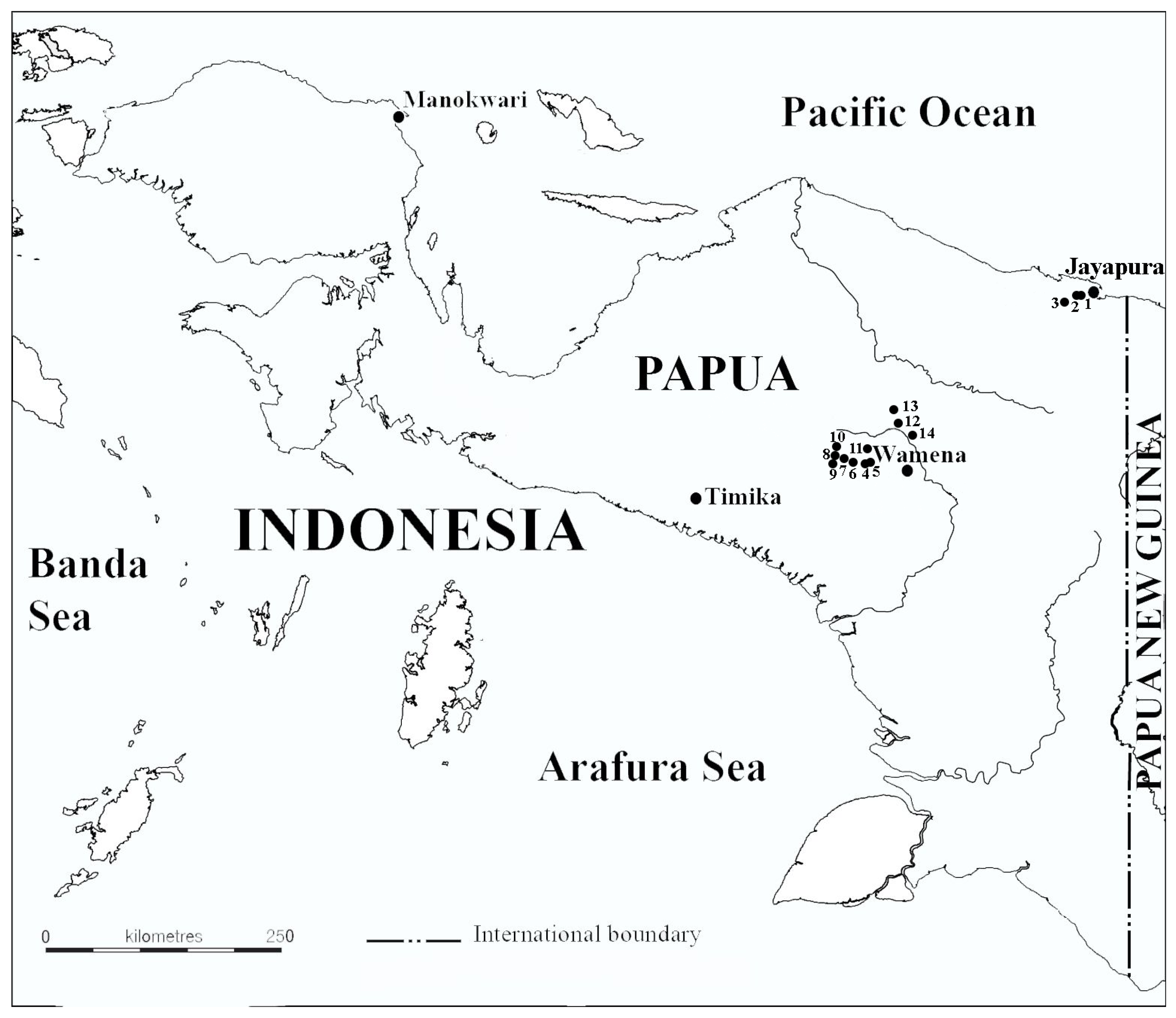
Figure 1. Map of the study area (dots indicate sampling sites).
Diversity 14 00726 g001
Figure 2. (AH) Paraphysomonas, Spiniferomonas and Chrysosphaerella taxa from Indonesian Papua, TEM. (A) Paraphysomonas cf. segmenta; (B,C) Paraphysomonas sp. (D) Paraphysomonas uniformis. (E) Spiniferomonas sp., a plate-like scale. (F) Paraphysomonas vulgaris subsp. vulgaris. (G) Paraphysomonas vulgaris subsp. brevispina. (H) Chrysosphaerella annulata, spine scale. Scale bars: (A): 1 µm, (BH): 2 µm.
Diversity 14 00726 g002
Figure 3. (AL) Chrysosphaerella and Mallomonas taxa from Indonesian Papua, TEM. (A) Chrysosphaerella annulata, plate-like scales. (B) Mallomonas cratis, a group of scales. (CE) Mallomonas cyathellata, domeless scale (C), domed scale (D) and rear scale (E) with cyathus (arrow). (F) Mallomonas cf. elongate. (G) Mallomonas furtiva. (H) Mallomonas mangofera var. foveata. (I,J) Mallomonas mangofera var. reticulata. (K) Mallomonas multisetigera. (L) Mallomonas papillosa. Scale bars: (AF,HL): 2 µm, (G): 1 µm.
Diversity 14 00726 g003
Figure 4. (AL) Mallomonas and Synura taxa from Indonesian Papua, TEM. (AC) Mallomonas solea-ferrea var. irregularis, body (A) and apical (B,C) scales. (D,E) Mallomonas striata var. serrata. (F,G) Mallomonas cf. portae-ferreae. (H) Mallomonas papuensis, a body scales. (I) Mallomonas sp. 1. (J) Mallomonas sp. 2. (K) Synura cf. longitubularis. (L) Synura mammillosa. Scale bars: (DL): 2 µm, (AC): 1 µm.
Diversity 14 00726 g004
Figure 5. (AD) Scales and bristles of Mallomonas preisigii, SEM. (A) Two scales with (in the background) or without (in the foreground) papillae. (B) A scale covered with papillae. (C) Undersurface of the scale illustrating the shallow concavity (arrow). (D) A group of scales and bristles (an arrow indicates the shallow concavity on the scale, a white arrowhead indicates the bristle tip and a black arrowhead indicates the bristle foot). Scale bars: (D): 5 µm, (AC): 1 µm.
Diversity 14 00726 g005
Figure 6. (AH) Scales of Mallomonas preisigii, TEM. (AD) Scales from site 6. (EH) Scales from site 14. Note the presence of scattered papillae on some scales. (E,F) Scale bars: 2 µm.
Diversity 14 00726 g006
Table 1. List of the sampling sites with environmental variables (T—temperature, SC—specific conductance and n/a—no parameters were measured).
Locality NameSample TypeDateCoordinatesElevation,
m a.s.l.
T, °CpHSC, µS·cm−1
1Lake SentaniPlankton14 September 20152°38′23.7″ S 140°34′04.3″ E8129.77.6230
2Lake SentaniPeriphyton14 September 20152°38′23.7″ S 140°34′04.3″ E81307.6240
3Water body along the road 1Plankton14 September 20152°39′54.72″ S 140°32′51.47″ E119n/an/an/a
4Lake HabbemaPlankton15 September 20154°07′49.5″ S 138°40′22.9″ E3331177.3322
5Lake HabbemaSqueeze from moss15 September 20154°07′49.5″ S 138°40′22.9″ E3331177.33n/a
6Peat-bog poolPlankton15 September 20154°7′40.02″ S
138°40′3.90″ E
333316.74.758
7Stream in a bogPlankton15 September 20154°7′36.30″ S
138°39′55.56″ E
3327n/a4.78n/a
8Puddle near the streamPlankton15 September 20154°7′34.68″ S
138°39′53.34″ E
3330n/an/an/a
9Peat-bog poolBenthos15 September 20154°7′36.60″ S
138°39′53.58″ E
3328n/an/an/a
10Small puddle in a bogPlankton15 September 20154°7′30.96″ S
138°39′53.88″ E
3337n/an/an/a
11Lake AnageraPlankton16 September 20153°57′07.4″ S 138°52′27.6″ E1656257.3229
12Water body along the road 2Plankton16 September 20153°54′33.4″ S 138°50′15.1″ E1661257.06234
13Small ephemeral pondPlankton16 September 20153°54′09.9″ S 138°50′17.1″ E1730276.99162
14Water body along the road 3Plankton16 September 20153°54′49.6″ S 138°51′27.4″ E1654307.0547
Plankton samples were taken with a plankton net (mesh size 20 μm). Samples from shallow peat-bog pools were obtained by squeezing the water from mosses. All samples were immediately fixed with Lugol’s solution.
Table 2. List of taxa observed at the investigated waterbodies (new taxa for Indonesia are in bold).
TaxonSite Number
1234567891011121314
Paraphysomonadales
Paraphysomonas cf. segmenta Scoble and Cavalier-Smith×
Paraphysomonas sp. ×
Paraphysomonas uniformis Scoble and Cavalier-Smith ×
Paraphysomonas vulgaris subsp. vulgaris Scoble and Cavalier-Smith×
Paraphysomonas vulgaris subsp. brevispina Scoble and Cavalier-Smith× ×
Chromulinales
Chrysosphaerella annulata Kristiansen and Tong× ×
Spiniferomonas sp. × ×
Synurales
Mallomonas cratis K. Harris and D.E. Bradley ×
Mallomonas cyathellata Wujek and Asmund ××
Mallomonas cf. elongata Reverdin ×
Mallomonas furtiva Gusev, Čertnerová, Škaloudová and Škaloud ×
Mallomonas mangofera var. foveata (Dürrschmidt) Kristiansen × ×
Mallomonas cf. mangofera var. reticulata (Cronberg) Kristiansen ×× ×
Mallomonas multisetigera Dürrschmidt ×× ×
Mallomonas papillosa K. Harris and D.E. Bradley emend. K. Harris ×
Mallomonas papuensis Kapustin, Gusev and Kulikovskiy ××
Mallomonas cf. portae-ferreae L.S. Péterfi and Asmund × ×
Mallomonas preisigii Siver × ×
Mallomonas solea-ferrea var. irregularis Němcová, Kreidlova, Pusztai and Neustupa ×
Mallomonas striata var. serrata K. Harris and D.E. Bradley × × ×
Mallomonas sp. 1 ××
Mallomonas sp. 2 × × ×
Synura cf. longitubularis Jo, Shin, Kim and Siver ×××
Synura mammillosa E. Takahashi ××××× ×
Publisher’s Note: MDPI stays neutral with regard to jurisdictional claims in published maps and institutional affiliations.

