Article

Species Diversity of Gelidium from Southern Madagascar Evaluated by an Integrative Taxonomic Approach

1
Department of Biological Sciences, Sungkyunkwan University, Suwon 16419, Korea
2
Institut de Systématique, Evolution, Biodiversité (ISYEB), Muséum National d’Histoire Naturelle, CNRS, Sorbonne Université, EPHE, 75231 Paris, France
3
Aquaculture Research Division, National Institute of Fishery Science, Gijang 46083, Korea
*
Author to whom correspondence should be addressed.
Diversity 2022, 14(10), 826; https://doi.org/10.3390/d14100826
Submission received: 11 August 2022 / Revised: 6 September 2022 / Accepted: 28 September 2022 / Published: 1 October 2022
(This article belongs to the Special Issue Algae Biodiversity: Natural and Anthropogenic Impacts)

Abstract

:
The diversity of red algae is significantly increasing; in the recent interest of climate changes, taxonomic and biogeographic studies are needed in untouched regions such as ecological important islands. We examined a collection of Gelidium specimens gathered during the 2010 Atimo Vatae expedition based on the morphology and DNA sequencing of mitochondrial cox1 and plastid rbcL. Both morphological and molecular datasets demonstrated the presence of three species in Madagascar; G. leptum G.H.Boo, L.Le Gall and H.S.Yoon, sp. nov., described here, and G. sclerophyllum W.R.Taylor and G. usmanghanii Afaq-Husain and Shameel. Gelidium leptum is distinguished by thin, slender, flattened thalli with irregular branches, compactly arranged thick-walled cells in medulla, sparse rhizoidal filaments at both sides of branches, and elongate sori of tetrasporangia. Gelidium sclerophyllum, previously considered as endemic to the Tropical Eastern Pacific, is first reported in the Indian Ocean and Hawai’i; G. usmanghanii, previously reported to occur in Pakistan and Oman, also presents a first record in the Southern Hemisphere. Phylogenies inferred from cox1 and rbcL sequences suggests that each of the three species likely diverged from different ancestors. The Madagascan Gelidium comprises two geographical elements; Madagascan endemic (G. leptum) and widespread species (G. sclerophyllum and G. usmanghanii).

1. Introduction

Gelidium J.V.Lamouroux is one of the speciose genera in red algae comprising 145 species [1]. Species are commonly found on temperate and tropical coastlines of both hemispheres [2,3,4,5]. There is a rising interest in the study of Gelidium because the biomass for agar production is in short supply globally and the price of agar has increased in the world market [6].
The genus Gelidium was established for eleven species [7]; however, the generitype was later designated as G. corneum (Hudson) J.V.Lamouroux by Schmitz [8]. Gelidium is circumscribed by plants with pinnate or simple branches, brush-like haptera, rhizoidal filaments in cortex and/or medulla, bilocular cystocarps with an ostiole on each side, and apical tetrasporangial sori [9,10,11]. Species are distinguished by plant size, branching patterns, position and shape of tetrasporangial sori, and cystocarps. Species of Gelidium have been well-studied in Australasia, Europe, northwest Pacific, North America, and western Atlantic using molecular markers [4,5,10,12,13,14,15]. However, the taxonomy of Gelidium from distantly located islands remains untouched by molecular data.
Due to the high morphology plasticity of the Gelidiales species as well as in other red algae, its species diversity was underestimated by morphology only. Since Freshwater et al. [12], molecular markers were developed in taxonomy and phylogenetic relationships in the genus Gelidium. The plastid rbcL is the most widely used marker in taxonomic studies [3,4,10,14]. The mitochondrial cox1 is a well-known DNA barcoding marker in Gelidiales [5,13,15,16]. Nuclear markers, such as internal transcribed spacer (ITS) and small and large subunit ribosomal DNAs (SSU and LSU), were also used for taxonomic studies; however, these markers have rarely been used due to its low resolution at the species level [17,18,19]. Integrating approaches, combining morphology and molecular analyses, have greatly enhanced our understanding of species identification and phylogenetic relationships in Gelidium [5,10,11,14,15].
The seaweed flora of Madagascar comprises 422 species with 29 (ca. 7%) endemics, a relatively high number of its uniqueness, which shows similar affinities to the tropical Western Indo-Pacific [20,21,22,23]. Five species of Gelidium have been listed in the Madagascan algal flora [24,25]; G. amansii (J.V.Lamouroux) J.V.Lamouroux, G. capense (S.G.Gmelin) P.C.Silva, G. crinale (Hare ex Turner) Gaillon, G. pectinatum (Montagne) Montagne (as G. corneum var. pectinatum Ardissone and Straforrello, Cormaci et al. [26]), and G. madagascariense Andriamampandry. Of these, G. madagascariense, a major agar-yielding species in Madagascar [27], has been revised based on molecular phylogeny as Orthogonacladia madagascariense (Andriamampandry) G.H.Boo and L.Le Gall [16].
Collections of the 2010 Atimo Vatae expedition to southern Madagascar by the Muséum National d’Histoire Naturelle (MNHN, Paris) have widened our knowledge on the taxonomy and biogeography of Codium (green algae), Dictyotales (brown algae), Gelidiales, and Yonagunia (red algae) [16,20,21,23,28,29,30,31,32]. However, due to its high species diversity and economic importance yielding agar, identification of Gelidium collections was challenging. We had recourse to molecular systematic tools, the mitochondrial cox1 or COI-5P and plastid rbcL sequences, which are useful for identification of species in the Gelidiales as well as in other red algae [5,12,13,16,33]. The aims of the present study were to identify those specimens and to describe new species and newly reported species in Madagascar. We included samples of G. sclerophyllum W.R.Taylor from Sri Lanka, Mexico, and Hawai’i to discuss its biogeography.

2. Materials and Methods

Specimens collected in southern Madagascar were pressed onto herbarium sheets and subsamples were dehydrated in silica gel for molecular work; housed at the cryptogam herbarium of the Muséum National d’Histoire Naturelle in Paris, France (PC). Fragments of herbarium specimens of Gelidium sclerophyllum W.R.Taylor from Sri Lanka, Hawai’i, and Mexico were available from the Ghent University macroalgal herbarium, Ghent, Belgium (GENT; collection now housed in the herbarium of Meise Botanic Garden, BR), a personal herbarium at Sherwood Lab (accession number: ARS), University of Hawai’i at Mānoa, USA, and the Natural History Museum, Chungnam National University, Korea (CNUK). Information of samples used in the present study is given in Table 1.
Morphology of specimens from Madagascar was examined under a light microscope. Microscopic observations were made by sectioning with a razor and staining with 1% aqueous aniline blue acidified with 1% HCl and mounted in 70% glycerine. Photographs were taken with a DP-71 Olympus camera (Olympus, Tokyo, Japan) attached to a BX51 Olympus microscope (Olympus, Tokyo, Japan).
DNA extraction, polymerase chain reaction amplification, and sequencing procedures followed Boo et al. [16]. The primers used for amplifying and sequencing were F7, F645, R753, and RrbcS start for plastid rbcL [12,34,35], and COXI43F and COXI1549R for mitochondrial cox1 [36]. Sequences of the forward and reverse strands were determined for all taxa, and the electropherograms were edited using MEGA version 7 [37] and checked manually. Newly generated sequences were deposited in GenBank (Table 1). Sequences were aligned using the MUSCLE algorithm in MEGA7 with default parameters and the alignment was manually adjusted. Four outgroup species in the family Gelidiaceae [16,38], Capreolia implexa Guiry and Womersley, Gelidiophycus freshwateri G.H.Boo, J.K.Park and S.M.Boo, Gelidiorariphycus loratus Iha, S.M.Guimarães, Freshwater and M.C.Oliveira, and Ptilophora spongiophila G.H.Boo, L.Le Gall, I.K.Hwang, K.A.Miller, and S.M.Boo, were included in the alignment.
Phylogenies of plastid rbcL and mitochondrial cox1 datasets were reconstructed using maximum likelihood (ML) and Bayesian inference (BI). The best-fitting nucleotide substitution model was selected using Modeltest v3.7 [39] with Akaike Information Criteria (AIC). The ML analyses were performed using the Pthreads version of RAxML v8.0.X [40] set as follows: a rapid bootstrap analysis and search for the best-scoring ML tree in one single run with 1000 bootstrap replicates under GTR+G+I model. The BI analyses were performed for individual datasets with MrBayes v3.2.1 [41] using the Metropolis-coupled Markov Chain Monte Carlo (MC3) with the GTR+G+I model. For each matrix, six million generations (four million generations for rbcL) of two independent runs were performed with four chains and sampling trees every one hundred generations. Twenty-five percent of saved trees were removed as burn-in, and the remaining trees were used to infer Bayesian posterior probabilities (BPP).
To understand relationships and geographical distribution of Gelidium sclerophyllum, haplotype network of cox1 sequences was constructed with Popart 1.7 [42] using the median-joining network (MJN).

3. Results

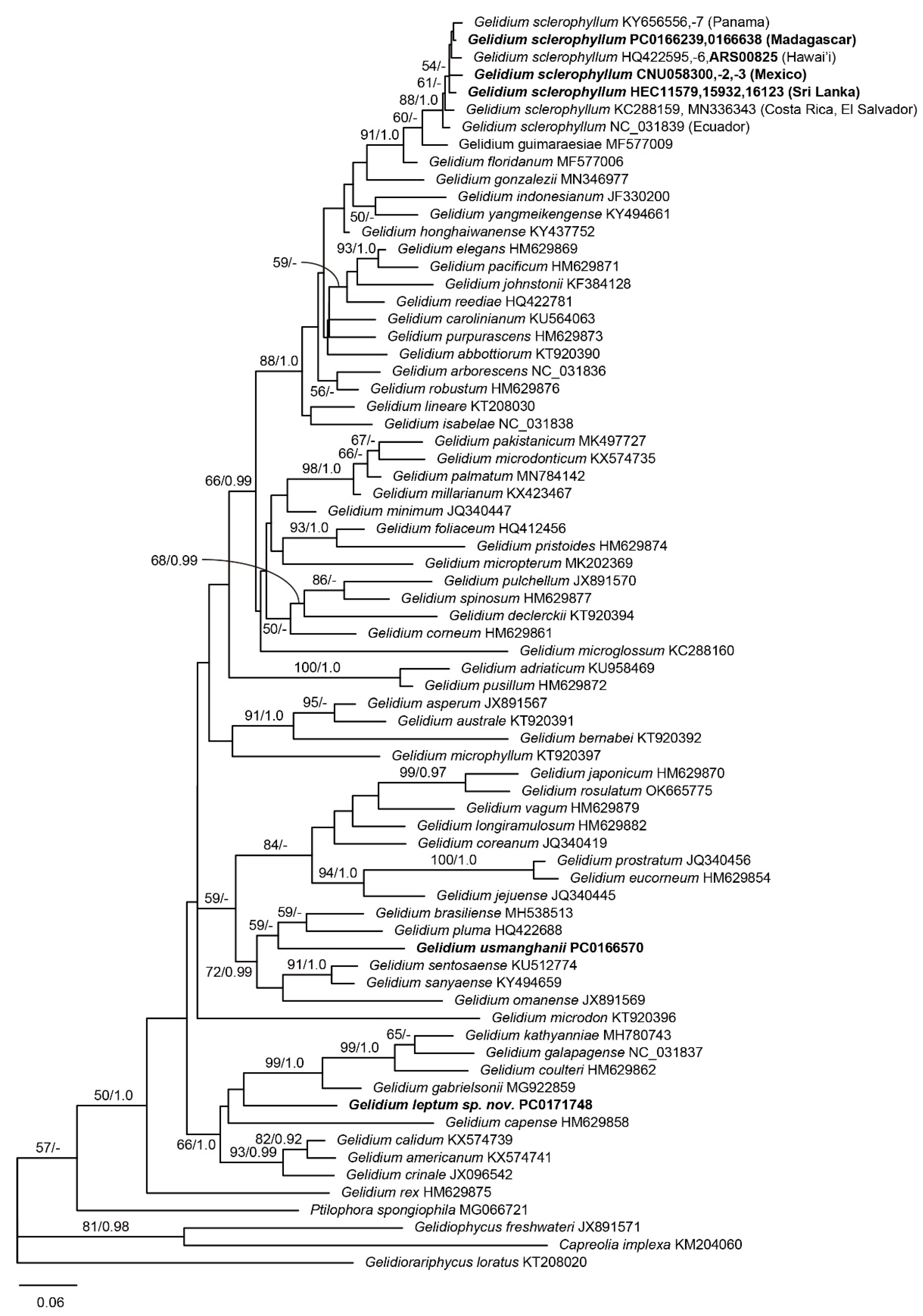
3.1. Molecular Phylogeny

Eighteen sequences (cox1: eleven, rbcL: seven) were generated from four representative specimens in the present study. A total of eighty-four rbcL sequences were aligned, including seventy-three previously published sequences of Gelidium species and four outgroups (Table S1). The rbcL phylogeny (Figure 1) confirmed the occurrence of three species, each of which was distinct from other species in the genus; Gelidium leptum, a new species described here, G. sclerophyllym, and G. usmanghanii Afaq-Husain and Shameel. Gelidium leptum was nested in a clade of Gelidium americanum (W.R.Taylor) Santelices, G. calidum Jamas, Iha, and Fujii, G. capense (S.G.Gmelin) P.C.Silva, G. coulteri Harvey, G. crinale, G. gabrielsonii Hughey and G.H.Boo, G. galapagense W.R.Taylor, and G. kathyanniae G.H.Boo and Hughey (97% ML, 1.0 BPP). The pairwise divergences of rbcL between G. leptum and other species in the clade were 3.1–4.1%. G. sclerophyllum formed a clade with the Western Atlantic species including G. floridanum W.R.Taylor and G. guimaraesiae Brunelli, Milstein, Boo, and M.T.Fujii (85% ML, 1.0 BPP), while G. usmanghanii formed a clade with G. brasiliense Brunelli, Boo, and M.T.Fujii and G. pluma Bornet ex N.H.Loomis (95% ML, 1.0 BPP).
The topology of the cox1 phylogeny (Figure 2) was largely congruent with that of rbcL. Gelidium leptum, G. sclerophyllum, and G. usmanghanii were distinct in cox1 phylogeny. Gelidium leptum formed a clade represented by G. capense (66% ML, 1.0 BPP), as seen in the rbcL topology. The pairwise divergence between G. leptum and its most closely related species (G. gabrielsonii) was 10.1%.
Haplotype network of cox1 sequences of G. sclerophyllum revealed a geographical structure. G. sclerophyllum comprised eight haplotypes from sixteen specimens. Madagascan haplotype (C8) was closely related to those from Sri Lanka (C7). Hawaiian (C5, C6) and Tropical Eastern Pacific haplotypes (C1–C4) were distantly related to each other (Figure 3).

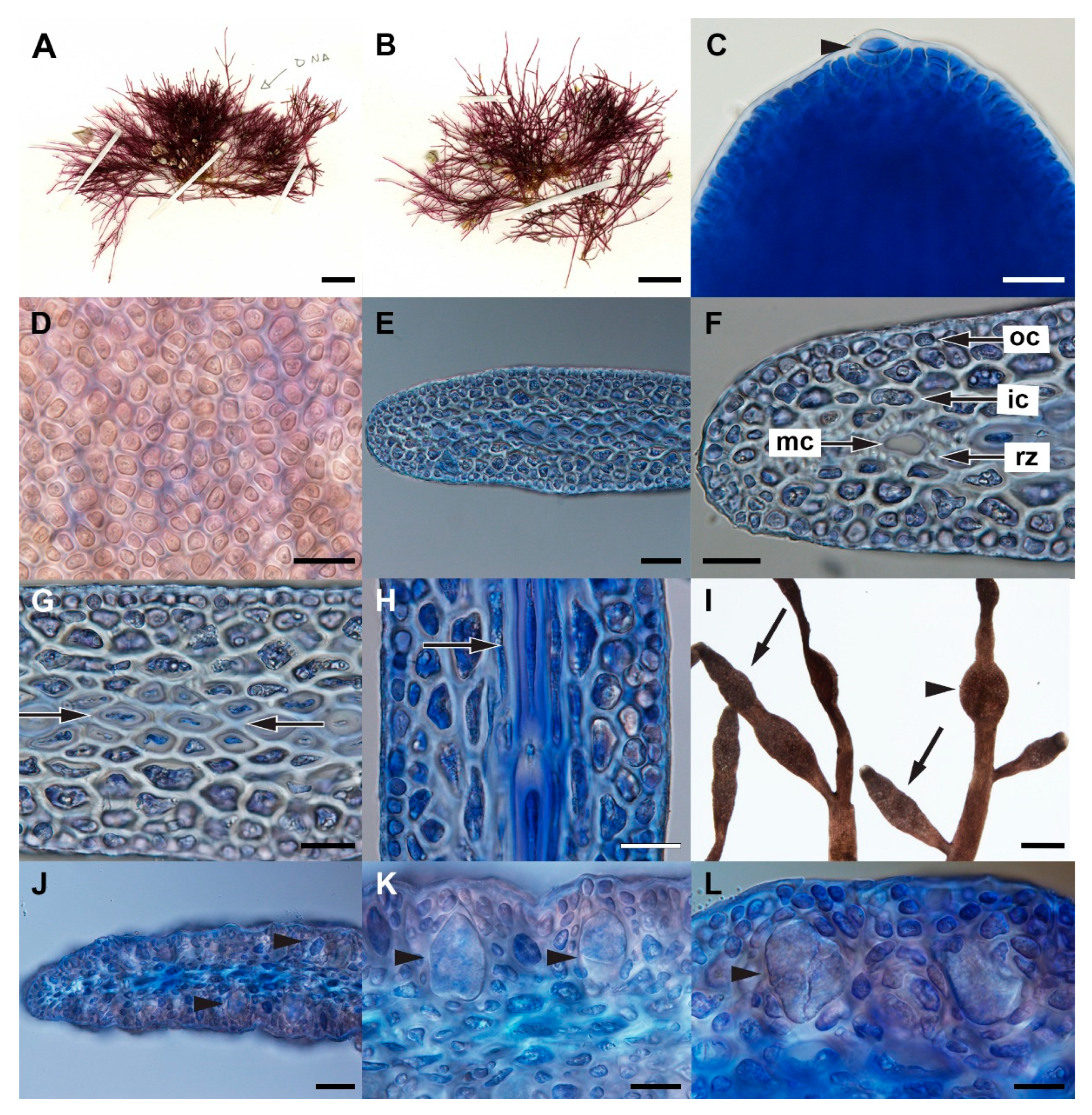
3.2. Gelidium leptum G.H.Boo, L.Le Gall, and H.S.Yoon, sp. nov.

Description: Plant (Figure 4A,B) growing in tufts, up to 5.5 cm high; erect axes arising from stolons, thin at base, slender, becoming compressed and flattened at the middle and upper portions of the branch, 102–112 μm thick. Branching rare, irregular, and sometimes trichotomous at truncated part; branches linear-lanceolate to ligulate, tapering toward acute apices (Figure 4C). Both erect and prostrate axes growing from transversely dividing, dome-shaped apical cells; surface cortical cells globose or angular (Figure 4D), 4–10 μm wide, and mostly arranged in pairs; cortex consisted of 3–4 layers of ovoid pigmented cells (Figure 4E–H); medulla consisted of 3–4 layers of rounded cells with thick-walled cells (Figure 4G,H) in transversal section, but filamentous cells in longitudinal section; rhizoidal filaments rare in the inner cortex at margin of branches (Figure 4F).
Tetrasporangial sori elongate at the tip of branches (Figure 4I), without sterile margin; tetrasporangia arranged irregularly, about 19–30 × 31–48 μm in size, arising from inner cortex (Figure 4J,K), randomly arranged along the sori, decussately divided (Figure 4K,L). Parasporangial cluster, spherical to roundish, produced at the middle of branches (Figure 4I), 371–445 μm in diameter. Female and male reproductive structures were not observed in the present study.
Holotype: PC0171748-1 (MAD1729-1) (Figure 4A), Station TA08, Baie des Galions, Madagascar (25°08′48″ S, 46°45′00″ E), collected on 15 May 2010 by E. Coppejans. Isotypes: PC0171748-2 (Figure 4B), PC0171748-3, and PC0171748-4.
Etymology: The specific epithet comes from a Greek word that means the thin, slender shape of erect and lateral branches.
Molecular sequences of holotype specimen: cox1 (OP137177) and rbcL (OP137188). Because the isotypes were duplicate specimens of the holotype as the specimen number indicates, DNA sequencing of the isotypes was not repeated.
Distribution: southern Madagascar (the present study).

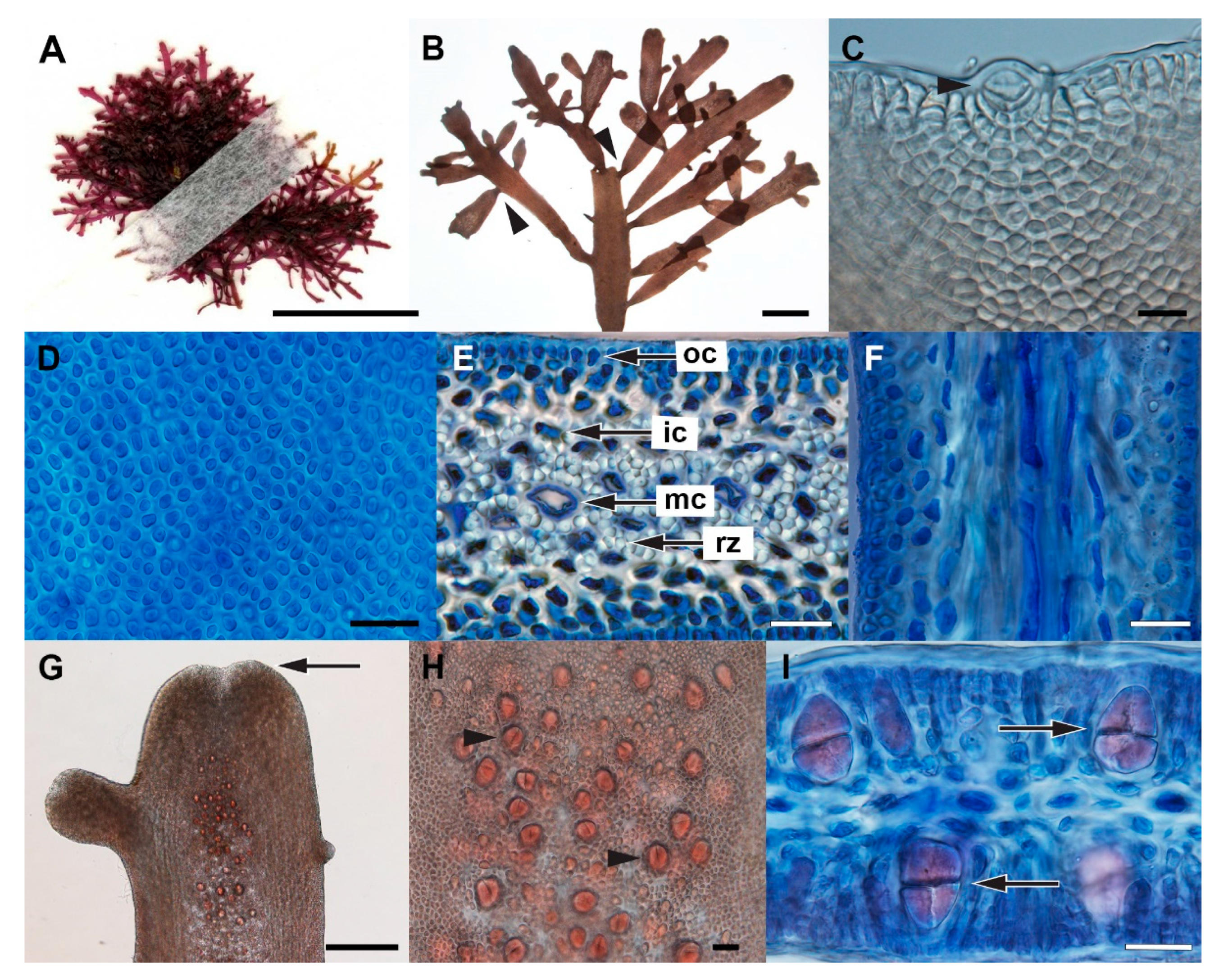
3.3. Gelidium sclerophyllum W.R.Taylor

Description: Plant (Figure 5A) consisting erect axes and stolons. Erect branches terete at the base to compressed proximally, gradually becoming flattened distally (Figure 5B), up to 2 cm long and about 477–800 μm wide, about 100 μm thick, with 2–3 orders of subopposite to alternate pinnate branchlets. Ultimate branchlets clavate, arising marginal. Main axes gradually taper toward base; shorter branches somewhat constricted basally. Vegetative axes have acute to obtuse apices with distinct apical cells (Figure 5C); surface cortical cells globose or angular (Figure 5D). Longitudinal striations present on the surfaces of branches. Cortex composed of 4–5 layers of pigmented cells with elongated to roundish surface cells. Inner cortical cells rectangular and slightly elongated longitudinally. Medulla composed of elongated, thick-walled cells with one layer of horizontally arranged roundish cells in the center. Rhizoidal filaments abundant in inner cortex and medulla (Figure 5E,F).
Tetrasporangial sori produced at the tip of flattened branches, constricted basally, elongate with obtuse to retuse apices, with wide sterile margin (Figure 5G); tetrasporangia 18–22 μm wide and 21–27 μm long, arising from the fourth or fifth cortical cells, irregularly arranged along the sori (Figure 5H), cruciately or decussately divided (Figure 5I). Female and male reproductive structures were not observed in the present study; however, Grusz and Freshwater [43] described details of development of cystocarps and spermatangia from Costa Rican specimens; cystocarps are spherical to ovoid on the apical or central parts of branches, bilocular. Spermatanial sori are strongly compressed with roundish apices of erect branches; spermatangia cut off from anticlinally elongated surface cortical cells by transverse divisions.
Specimens examined: PC0166239 (MAD0272), Station TM01, Plage Monseigneur, Fort Dauphin, Madagascar, collected on 2 May 2010 by F. Rousseau, R. Anderson, and J. Tsarahevitra. PC0166638 (MAD0671), Station TM23, Flacourt, Madagascar, collected on 13 May 2010 by F. Rousseau, R. Anderson, and J. Tsarahevitra.
Distribution: Madagascar, Mexico, and Sri Lanka (the present study), Costa Rica [39], Ecuador [43,44,45], El Salvador [46], Hawai’i (as G. crinale or G. pusillum (Stackhouse) Le Jolis; the present study and Sherwood et al. [47]), Nicaragua [48], and Panama [49].

3.4. Gelidium usmanghanii Afaq-Husain and Shameel

Description: Plant (Figure 6A) up to 7 cm high, with erect axes arising from stolons, attached to substratum by brush-type haptera. Stolons cylindrical. Erect axes cylindrical at base, abruptly turning into flattened fronds, up to 730 μm wide and 73–79 μm thick, simple at margins. Branches irregular (Figure 6B), with ultimate rameli cuneate at the base, occurring sparingly to densely along the margin. Apical cells discoid, present in a depression between cortical lobes (Figure 6C). Surface cells rounded, angular, randomly arranged, 3–6 μm in diameter (Figure 6D). Erect branches appear thick in the middle, tapering to either side into conic-obtuse ends. Cortex consisted of 3–4 layers of pigmented cells. Medulla consisted of 2–3 layers of large angular cells. Rhizoidal filaments concentrated in medulla (Figure 6E,F).
Tetrasporangial sori borne apically on unbranched or ultimate rameli (Figure 6G), with short stipe at basally, 211–440 μm in diameter, and sterile margin of about 100–140 μm on each side. Tetrasporangia arising on the third to fourth cell of the cortex (Figure 6H), 25–31 μm wide × 34–41 μm long, decussately or cruciately divided (Figure 6H,I), irregularly arranged. Although female and male reproductive structures were not observed in Madagascan specimens, Afaq-Husain and Shameel [50,51] described that cystocarp borne on branched ramuli, bilocular with 1–2 ostioles on each side. Carpospores oblongly elongate, tapering to the base, approximately 24 μm wide × 60 μm long. Male reproductive structure has not been observed in Pakistan and southern Madagascar.
Specimens examined: PC0166570-2 (MAD0603-2), PC0166570-3 (MAD0603-3), Station TR20, Sud de la baie, Lokaro, Madagascar; collected on 12 May 2010 by F. Rousseau, R. Anderson, and J. Tsarahevitra.
Molecular sequences of PC0166570-2: cox1 (OP137187) and rbcL (OP137194).
Distribution (based on molecular data): Madagascar (the present study), Pakistan [50,51,52], and Oman [52,53].
A morphological comparison of Gelidium leptum, G. sclerophyllum, and G. usmanghanii from Madagascar with other morphologically similar species is provided in Table 2.

4. Discussion

4.1. Taxonomic Implication

Our analyses of plastid rbcL and mitochondrial cox1 clearly showed the occurrence in southern Madagascar of G. sclerophyllum and G. usmanghanii as well as a new species, G. leptum. Gelidium leptum is well-distinct from about 80 species of Gelidium in both rbcL and cox1 sequences from GenBank (Figure 1 and Figure 2), and this is supported by morphological characteristics. It formed a clade with G. americanum, G. calidum, G. capense, G. coulteri, G. crinale, G. gabrielsonii, G. galapagense, and G. kathyanniae, suggesting phylogenetic relationship with species from Brazil, Eastern Pacific America, and South Africa. Gelidiun leptum is taller and slender than G. sclerophyllum and G. usmanghanii, and has thin, flattened axes and branches. Compact arrangement of thick-walled cells filled in the medulla of G. leptum is uncommon in other Gelidium species, but thick-walled cells loosely occur in medulla of G. reptans (Suhr) Kylin and Costa Rican G. sclerophyllum [43,61]. However, thick-walled cells are well-developed in the medulla and cortex of Gelidiella in the family Gelidiellaceae [9,28,31]. Tetrasporangial sori of G. sclerophyllum are elongate or flattened roundish in the apices of ultimate branchlets, as seen in other species of Gelidium [10,11,14,15,62,63]. Tetrasporangial cluster is uncommon in other species of Gelidium. They are similar to parasporangial cluster of Camyplaephora crass (Okamura) Nakamura [64]. Gelidium leptum is distinguished by thin, slender, flattened thalli with irregular branches, thick-walled cells of medulla, rhizoidal filaments rarely occurring in the margin of branches, and apical sori of tetrasporangia.
Gelidium leptum may be young or dwarf forms of G. amansii in having thin, compressed thalli with compact thick-walled cells in medulla, and apical tetrasporangia having a short stalk. However, G. amansii is much taller, terete at the lower part of erect axis, with abundant rhizoidal filaments in the inner cortex and without spherical tetrasporangial clusters (Table 2). Despite many visits to Madagascar and Mauritius, additional collections of G. amansii have not been available [22,56,57]. Several cox1 and rbcL sequences are deposited in GenBank for specimens of G. amansii from South Africa as well as Korea and China. However, G. amansii from northeast Asia is a misidentification of G. elegans Kützing [3,14,57], and rbcL sequences of G. amansii (AF308787) and G. abbottiorum Norris (EF190254) from South Africa are the same [3]. DNA sequencing from the type material of G. amansii deposited in the herbarium of the University of Caen, France is needed.
Gelidium leptum is well-separated in thallus size, branching form, and tetrasporangial sori from South African species such as G. applanatum Stegenga, Boltonm and R.J.Anderson, G. arenarium Kylin, and G. minusculum (Weber-van Bosse) R.E.Norris [59,60,61] as well as G. reptans (Table 2). All the latter species also lack DNA data.
Both cox1 and rbcL datasets demonstrate the occurrence of Gelidium sclerophyllum from southern Madagascar, which has been reported to occur in Pacific Central America solely [43,49]. Our study also confirmed the occurrence of the species from Sri Lanka and Mexico, and both cox1 sequences under the names of Hawaiian G. crinale or G. pusillum [47] are identical with those from G. sclerophyllum. Morphological characters observed on specimens from Madagascar G. sclerophyllum are generally in agreement with the description of the plants from the Pacific Central America. However, the Madagascan plants have few thick-walled cells in the medulla and lack sexual reproductive structures. The pairwise divergences (0.8–2.5% in cox1 and 0.2–0.5 % in rbcL) from Madagascar and Pacific Central America are well-matched within the genetic circumscription of G. sclerophyllum [4,13]. Gelidium sclerophyllum is thus distinguished by small, compressed thalli with branches constricted at their base, dense medulla of thick-walled cells, tetrasporangial sori borne apically on the terminal branches, with wide sterile margins, indented tips of tetrasporangial sori, and bilocular cystocarps ([43,44,54], the present study). This is the first report of G. sclerophyllum in the Indian Ocean and Hawai’i.
Our study demonstrates the occurrence of G. usmanghanii from southern Madagascar, a taxon which has for a long time been considered endemic to Pakistan [50,51]. Two rbcL sequences from unidentified Omani Gelidium [53] are identical with those of G. usmanghanii from Pakistan [52]. The Madagascan specimens match descriptions and illustrations by Afaq-Husain and Shameel [50,51]. However, coiling along the long axis and undulations toward margin of Pakistan plants, considered diagnostic features by Afaq-Husain and Shameel [50,51], are absent in Madagascan plants, suggesting that those features may be ecological responses to environmental conditions that occur in Pakistan. The morphology of G. usmanghanii is similar with that of G. sclerophyllum and G. reptans, however, G. usmanghanii has abundant rhizoidal filaments in the medulla. Gelidium usmanghanii is thus distinguished by a medium thallus size (up to 7 cm) with flattened, papery fronds, irregular branches, dense rameli on margins of the fronds, and tetrasporangial sori with wide margin ([50,51], the present study). Gelidium usmanghanii, yielding a food-grade agar [65], is of economic importance in Pakistan. Our study is the first record of G. usmanghanii from the Southern Hemisphere.
We could not find any specimens of G. amansii, G. capense, G. crinale, and G. pectinatum, previously reported in Madagascar [25], which were absent in marine algal collections of northern Madagascar [22]. However, these species, except G. amansii, are elsewhere well-studied using cox1 and/or rbcL sequences [14,62]. Further sampling along the coast of Madagascar is needed for a genuine flora of Gelidium.

4.2. Biogeographic Implication

Our study reveals that southern Madagascar includes widespread Gelidium species, although Madagascar has been considered to be an endemic hotspot or a part of South African algal flora [20,21,32]. The discovery of G. sclerophyllum in Madagascar is unexpected given that it has for a long time been considered restricted from southern Mexico to Ecuador [43,44,49,54,66]. However, G. sclerophyllum is also demonstrated to occur in Sri Lanka (the present study) as well as in Hawai’i (HQ422595-6, as G. crinale) [47]. In rbcL and cox1 phylogenies, G. sclerophyllum was consistently formed a clade of Western Atlantic species (Figure 1 and Figure 2). Given the context of phylogeny and its sister relationship mentioned above, G. sclerophyllum likely dispersed bidirectionally from a tropical Indo-Pacific ancestor to Madagascar to the west and from the Pacific Central America to the east. A haplotype network of cox1 sequences revealed a geographical structure with a close relationship between Madagascar and Sri Lanka, while the Hawaiian and Tropical Eastern Pacific haplotypes were genetically distinct from each population (Figure 3). The pan-tropical distribution of G. sclerophyllum, a small sized species, is challenging to explain based on current data, but the origin center of G. sclerophyllum is yet to be tested using additional sampling. On the other hand, G. sclerophyllum and G. floridanum W.R.Taylor were previously considered a geminate pair, diverged during the formation of the Isthmus of Panama [43].
Our study extends the range of G. usmanghanii to Madagascar in the Southern Hemisphere from Oman and Pakistan in the Northern Hemisphere [50,52,53]. The shared species between southern Madagascar and the Arabian Sea is an exceptional case in the Gelidiales. However, there are similar patterns between complex of cryptic species, with a certain degree of genetic divergence, such as Pterocladiella australafricanensis E.M.Tronchin and D.W.Freshwater, brown algae Ecklonia radiata (C.Agardh) J.Agardh and Lobophora crassa Z.Sun, P.E.Lim, and Kawai, and a green alga Codium duthieae P.C.Silva [20,21,23,67]. The range extension between the Southern and Northern Hemispheres in the Western Indian Ocean region might result from a southward dispersal from the Arabian Sea to southern Madagascar or vice versa. This hypothesis needs to be confirmed by additional sampling along the east coast of Africa.
The restriction of Gelidium leptum to Madagascar is supported by a DNA database of global species of Gelidium. The endemism in Madagascar is not uncommon in species of the Gelidiales [16,24,28,30,31,32]. Similarly, six of twenty-three molecular taxa of the Dictyotales from southern Madagascar [20] were not recorded hitherto anywhere else. It is remarkable that southern Madagascar flora of Gelidium is isolated from South Africa, where Gelidium has relatively been well-studied [61,68,69]. However, it is notable, despite a similar sampling effort in the 2010 Atimo Vatae expedition, that previous reports on the red algal genera Pterocladiella, Ptilophora, and Yonagunia, the brown alga Dictyotales, and a green algal genus Codium have shared species between those two close regions [20,21,23,29,32].
In conclusion, our study highlights the diversity and biogeography of Gelidium from southern Madagascar; a description of a new species, G. leptum, and first records of G. sclerophyllum and G. usmanghanii. Gelidium sclerophyllum links the Western Indian Ocean to the Tropical Eastern Pacific regions, an interesting issue of its historical biogeography. Additional sampling along the overall coast of Madagascar is needed to decipher the entire diversity and biogeography of Madagascan Gelidium.

Supplementary Materials

The following supporting information can be downloaded at: https://www.mdpi.com/article/10.3390/d14100826/s1, Table S1: List of published sequences for molecular analyses.

Author Contributions

Conceptualization, G.H.B., L.L.G. and H.S.Y.; resources, L.L.G. and F.R.; investigation, G.H.B. and I.K.H.; validation, G.H.B. and H.S.Y.; writing—original draft preparation, G.H.B.; writing—review and editing, G.H.B., L.L.G. and H.S.Y. All authors have read and agreed to the published version of the manuscript.

Funding

This research was supported by the Basic Science Research Program through the National Research Foundation of Korea (NRF) by the Ministry of Education (2021R1I1A1A01049542, 2022R1A2B5B03002312), the Korea Institute of Marine Science and Technology Promotion (KIMST) funded by the Ministry of Oceans and Fisheries (MOF) (20210469), and the National Institute of Fisheries Sciences (NIFS) (R2022002). Materials from PC were collected during the Atimo Vatae expedition to south Madagascar (Principal Investigator, Philippe Bouchet), part of a cluster of Mozambique–Madagascar expeditions funded by the Total Foundation, Prince Albert II of Monaco Foundation, and Stavros Niarchos Foundation under “Our Planet Reviewed”, a joint initiative of Muséum National d’Histoire Naturelle (MNHN) and Pro Natura International (PNI) in partnership with Institut d’Halieutique et des Sciences Marines, University of Toliara (IH.SM) and the Madagascar bureau of Wildlife Conservation Society (WCS). The Institut de Recherche pour le Développement (IRD) deployed its research catamaran Antéa.

Institutional Review Board Statement

Not applicable.

Data Availability Statement

Not applicable.

Acknowledgments

We thank Alison Sherwood, Eric Coppejans, and Sung Min Boo for sharing samples from Hawai’i, Sri Lanka, and Mexico.

Conflicts of Interest

The authors declare no conflict of interest.

References

  1. Guiry, M.D.; Guiry, G.M. AlgaeBase. World-Wide Electronic Publication, National University of Ireland, Galway. 2022. Available online: http://www.algaebase.org (accessed on 5 March 2022).
  2. Womersley, H.B.S.; Guiry, M.D. Order Gelidiales Kylin 1923:132. In The Marine Benthic Flora of Southern Australia. Part IIIA; Womersley, H.B.S., Ed.; Australian Biological Resources Study: Adelaide, Australia, 1994; pp. 118–142. [Google Scholar]
  3. Freshwater, D.W.; Fredericq, S.; Hommersand, M.H. A molecular phylogeny of the Gelidiales (Rhodophyta) based on analysis of plastid rbcL nucleotide sequences. J. Phycol. 1995, 31, 616–632. [Google Scholar] [CrossRef]
  4. Boo, G.H.; Kim, K.M.; Nelson, W.A.; Riosmena-Rodriguez, R.; Yoon, K.J.; Boo, S.M. Phylogenetic relationships and distribution of the selected species of the agarophyte genus Gelidium (Gelidiales, Rhodophyta). J. Appl. Phycol. 2014, 26, 1243–1251. [Google Scholar] [CrossRef]
  5. Iha, C.; Milstein, D.; Guimarães, S.M.P.B.; Freshwater, D.W.; Oliveira, M.C. DNA barcoding reveals high diversity in the Gelidiales of the Brazilian southeast coast. Bot. Mar. 2015, 58, 295–305. [Google Scholar] [CrossRef]
  6. Callaway, E. Lab staple agar hit by seaweed shortage. Nature 2015, 528, 171–172. [Google Scholar] [CrossRef] [PubMed] [Green Version]
  7. Lamouroux, J.V. Essai sur les genres de la famille des thalassiophytes non articulées. Ann. Mus. Hist. Nat. 1813, 20, 21–47, 115–139, 267–293. [Google Scholar]
  8. Schmitz, F. Systematische Übersicht der bisher bekannten Gattungen der Florideen. Flora 1889, 72, 435–456. [Google Scholar]
  9. Fan, K.C. Morphological studies of the Gelidiales. Univ. Calif. Publ. Bot. 1961, 32, 315–368. [Google Scholar]
  10. Millar, A.J.K.; Freshwater, D.W. Morphology and molecular phylogeny of the marine algal order Gelidiales (Rhodophyta) from New South Wales, including Lord Howe and Norfolk Island. Aust. Syst. Bot. 2005, 18, 215–263. [Google Scholar] [CrossRef]
  11. Boo, G.H.; Park, J.K.; Han, K.S.; Yoon, H.S. Gelidium rosulatum (Gelidiales, Rhodophyta), a new species of subtidal marine algae from Korea. Phycologia 2022, 61, 332–340. [Google Scholar] [CrossRef]
  12. Freshwater, D.W.; Rueness, J. Phylogenetic relationships of some European Gelidium (Gelidiales, Rhodophyta) species, based on rbcL nucleotide sequence analysis. Phycologia 1994, 33, 187–194. [Google Scholar] [CrossRef]
  13. Freshwater, D.W.; Tudor, K.; O’Shaughnessy, K.; Wysor, B. DNA barcoding in the red algal order Gelidiales: Comparison of COI with rbcL and verification of the “barcoding gap”. Cryptogam. Algol. 2010, 31, 435–449. [Google Scholar]
  14. Kim, K.M.; Hwang, I.K.; Yoon, H.S.; Boo, S.M. Four novel Gelidium species (Gelidiales, Rhodophyta) discovered in Korea: G. coreanum, G. jejuense, G. minimum and G. prostratum. Phycologia 2012, 51, 461–474. [Google Scholar] [CrossRef] [Green Version]
  15. Perrone, C.; Bottalico, A.; Boo, G.H.; Boo, S.M.; Miller, K.A.; Freshwater, D.W. Gelidium adriaticum sp. nov. and Gelidium carolinianum sp. nov. (Gelidiales, Rhodophyta) from the Mediterranean Sea. Phycologia 2019, 58, 359–373. [Google Scholar] [CrossRef]
  16. Boo, G.H.; Le Gall, L.; Miller, K.A.; Freshwater, D.W.; Wernberg, T.; Terada, R.; Yoon, K.J.; Boo, S.M. A novel phylogeny of the Gelidiales (Rhodophyta) based on five genes including the nuclear CesA, with descriptions of Orthogonacladia gen. nov. and Orthogonacladiaceae fam. nov. Mol. Phylogenet. Evol. 2016, 101, 359–372. [Google Scholar] [CrossRef] [PubMed] [Green Version]
  17. Bailey, J.C.; Freshwater, D.W. Molecular systematics of the Gelidiales: Inferences from separate and combined analyses of plastid rbcL and nuclear SSU gene sequences. Eur. J. Phycol. 1997, 32, 343–352. [Google Scholar] [CrossRef] [Green Version]
  18. Patwary, M.U.; Sensen, C.W.; MacKay, R.M.; van der Meer, J.P. Nucleotide sequences of small-subunit and internal transcribed spacer regions of nuclear rRNA genes support the autonomy of some genera of the Gelidiales (Rhodophyta). J. Phycol. 1998, 34, 299–305. [Google Scholar] [CrossRef]
  19. Freshwater, D.W.; Fredericq, S.; Bailey, J.C. Characteristics and utility of nuclear-encoded large-subunit ribosomal gene sequences in phylogenetic studies of red algae. Phycol. Res. 1999, 47, 33–38. [Google Scholar] [CrossRef]
  20. Steen, F.; Vieira, C.; Leliaert, F.; Payri, E.C.; De Clerck, O. Biogeographic affinities of Dictyotales from Madagascar: A phylogenetic approach. Cryptogam. Algol. 2015, 36, 129–141. [Google Scholar] [CrossRef]
  21. Verbruggen, H.; Costa, J.E. Molecular survey of Codium species diversity in southern Madagascar. Cryptogam. Algol. 2015, 36, 171–187. [Google Scholar] [CrossRef]
  22. Vieira, C.; N’Yeurt, A.D.; Rasoamanendrinka, F.; D’Hondt, S.; Thi Tran, L.-A.; Spiegel, D.V.; Kawai, H.; De Clerck, O. Marine macroalgal biodiversity of northern Madagascar: Morpho-genetic systematics and implications of anthropic impacts for conservation. Biodivers. Conserv. 2021, 30, 1501–1546. [Google Scholar] [CrossRef]
  23. Boo, G.H.; Leliaert, F.; Le Gall, L.; Coppejans, E.; De Clerck, O.; Nguyen, V.T.; Miller, K.A.; Yoon, H.S. Ancient Tethyan vicariance and long-distance dispersal drive global diversification and cryptic speciation in the red seaweed Pterocladiella. Front. Plant Sci. 2022, 13, 849476. [Google Scholar] [CrossRef] [PubMed]
  24. Andriamampandry, A.V. Beckerella pterocladioides sp. nov. et Gelidium madagascariense sp. nov. deux espèces de Gelidiales-rhodophytes de Fort-Dauphin (Madagascar). Cryptogam. Algol. 1988, 9, 243–259. [Google Scholar]
  25. Silva, P.C.; Basson, P.W.; Moe, R.L. Catalogue of the benthic marine algae of the Indian Ocean. Univ. Calif. Publ. Bot. 1996, 27, 1–1259. [Google Scholar]
  26. Cormaci, M.; Furnari, G.; Alongi, G. Benthic marine flora of the Mediterranean Sea: Rhodophyta—Rhodymeniophycidae I. Acrosymphytales, Bonnemaisoniales, Gelidiales, Gigartinales, Gracilariales. Boll. Accad. Gioenia Nat. Sci. 2020, 53, FP11–FP346. [Google Scholar] [CrossRef]
  27. Mollion, J. The seaweed resources of Madagascar. Bot. Mar. 2020, 63, 97–104. [Google Scholar] [CrossRef]
  28. Boo, G.H.; Le Gall, L.; Rousseau, F.; De Reviers, B.; Coppejans, E.; Anderson, R.; Boo, S.M. Phylogenetic relationships and morphology of Gelidiella (Gelidiales, Rhodophyta) from Madagascar with a description of Gelidiella incrassata sp. nov. Cryptogam. Algol. 2015, 36, 219–237. [Google Scholar] [CrossRef]
  29. Manghisi, A.; Morabito, M.; Boo, G.H.; Boo, S.M.; Bonillo, C.; De Clerck, O.; Le Gall, L. Two novel species of Yonagunia (Halymeniales, Rhodophyta) were uncovered in the South of Madagascar during the Atimo-Vatae expediction. Cryptogam. Algol. 2015, 36, 199–217. [Google Scholar] [CrossRef]
  30. Boo, G.H.; Le Gall, L.; Hwang, I.K.; Boo, S.M. Pterocladiella feldmannii sp. nov. and P. hamelii sp. nov. (Gelidiales, Rhodophyta), two new species uncovered in Madagascar during the Atimo Vatae Expedition. Cryptogam. Algol. 2016, 37, 179–198. [Google Scholar] [CrossRef]
  31. Boo, G.H.; Nguyen, T.V.; Kim, J.Y.; Le Gall, L.; Rico, J.M.; Bottalico, A.; Boo, S.M. A revised classification of the Gelidiellaceae (Rhodophyta) with descriptions of three new genera: Huismaniella, Millerella and Perronella. Taxon 2016, 65, 965–979. [Google Scholar] [CrossRef]
  32. Boo, G.H.; Le Gall, L.; Hwang, I.K.; Miller, K.A.; Boo, S.M. Phylogenetic relationships and biogeography of Ptilophora (Gelidiales, Rhodophyta) with descriptions of P. aureolusa, P. malagasya, and P. spongiophila from Madagascar. J. Phycol. 2018, 54, 249–263. [Google Scholar] [CrossRef]
  33. Boo, G.H.; Zubia, M.; Hughey, J.R.; Sherwood, A.R.; Fujii, M.T.; Boo, S.M.; Miller, K.A. Complete mitochondrial genomes reveal population-revel patterns in the widespread red alga Gelidiella fanii (Gelidiales, Rhodophyta). Front. Mar. Sci. 2020, 7, 583957. [Google Scholar] [CrossRef]
  34. Lin, S.-M.; Fredericq, S.; Hommersand, M.H. Systematics of the Delesseriaceae (Ceramiales, Rhodophyta) based on large subunit rDNA and rbcL sequences, including the Phycodryoideae, subfam. nov. J. Phycol. 2001, 37, 881–889. [Google Scholar] [CrossRef] [Green Version]
  35. Gavio, B.; Fredericq, S. Grateloupia turuturu (Halymeniaceae, Rhodophyta) is the correct name of the non-native species in the Atlantic known as Grateloupia doryphora. Eur. J. Phycol. 2002, 37, 349–359. [Google Scholar] [CrossRef] [Green Version]
  36. Geraldino, P.J.L.; Yang, E.C.; Boo, S.M. Morphology and molecular phylogeny of Hypnea flexicaulis (Gigartinales, Rhodophyta) from Korea. Algae 2006, 21, 417–423. [Google Scholar] [CrossRef] [Green Version]
  37. Kumar, S.; Stecher, G.; Tamura, K. MEGA7: Molecular Evolutionary Genetics Analysis version 7.0 for bigger datasets. Mol. Biol. Evol. 2016, 33, 1870–1874. [Google Scholar] [CrossRef] [PubMed] [Green Version]
  38. Iha, C.; Freshwater, D.W.; Guimarães, S.M.P.B.; Oliveira, M.C. Gelidiorariphycus gen. nov. (Gelidiales, Rhodophyta): A rare genus found in the Americas. Phycologia 2022, 61, 473–483. [Google Scholar] [CrossRef]
  39. Posada, D.; Crandall, K.A. MODELTEST: Testing the model of DNA substitution. Bioinformatics 1998, 14, 817–818. [Google Scholar] [CrossRef] [Green Version]
  40. Stamatakis, A. RAxML Version 8: A tool for phylogenetic analysis and post-analysis of large phylogenies. Bioinformatics 2014, 30, 1312–1313. [Google Scholar] [CrossRef] [Green Version]
  41. Ronquist, F.; Teslenko, M.; van der Mark, P.; Ayres, D.; Darling, A.; Hohna, S.; Larget, B.; Liu, L.; Suchard, M.A.; Huelsenbeck, J.P. MrBayes 3.2: Efficient Bayesian phylogenetic inference and model choice across a large model space. Syst. Biol. 2012, 61, 539–542. [Google Scholar] [CrossRef] [Green Version]
  42. Leigh, J.W.; Bryant, D. PopART: Full-feature software for haplotype network construction. Methods Ecol. Evol. 2015, 6, 1110–1116. [Google Scholar] [CrossRef]
  43. Grusz, A.L.; Freshwater, D.W. Studies of Costa Rican Gelidiales (Florideophyceae). II. Two Pacific taxa including Gelidium microglossum, n. sp. Pac. Sci. 2014, 68, 97–110. [Google Scholar] [CrossRef]
  44. Taylor, W.R. Pacific marine algae of the Allan Hancock expeditions to the Galapagos Islands. Allan Hancock Pac. Exped. 1945, 12, 1–528. [Google Scholar]
  45. Boo, G.H.; Hughey, J.R.; Miller, K.A.; Boo, S.M. Mitogenomes from type specimens, a genotyping tool for morphologically simple species: Ten genomes of agar-producing red algae. Sci. Rep. 2016, 6, 35337. [Google Scholar] [CrossRef] [PubMed] [Green Version]
  46. Quiroz-González, N.; Ponce-Márquez, M.A.E.; Fernández-García, C.; Rodríguez, D. Gelidium gonzalezii sp. nov. (Gelidiales, Rhodophyta) from the Mexican tropical Pacific based on molecular and morphological evidence. Phytotaxa 2020, 459, 124–138. [Google Scholar] [CrossRef]
  47. Sherwood, A.R.; Kurihara, A.; Conklin, K.Y.; Sauvage, T.; Presting, G.G. The Hawaiian Rhodophyta biodiversity survey (2006–2010): A summary of principal findings. BMC Plant Biol. 2010, 10, 258. [Google Scholar] [CrossRef] [Green Version]
  48. Fernández-García, C.; Riosmena-Rodríguez, R.; Wysor, B.; Tejada, O.L.; Cortéz, J. Checklist of the Pacific marine macroalgae of Central America. Bot. Mar. 2011, 54, 53–73. [Google Scholar] [CrossRef]
  49. Freshwater, D.W.; Idol, J.W.; Parham, S.L.; Fernández-García, C.; León, N.; Gabrielson, P.W.; Wysor, B. Molecular assisted identification reveals hidden red algae diversity from the Burica Peninsula, Pacific Panama. Diversity 2017, 9, 19. [Google Scholar] [CrossRef] [Green Version]
  50. Afaq-Husain, S.; Shameel, M. Morphology, anatomy and reproduction of the populations of a new alga, Gelidium usmanghanii (Gelidiales, Rhodophyta). Candollea 1996, 51, 433–443. [Google Scholar]
  51. Afaq-Husain, S.; Shameel, M. Further studies on Gelidium (Rhodophyta) from the coast of Pakistan. Pak. J. Bot. 1999, 31, 371–382. [Google Scholar]
  52. Freshwater, D.W.; Shahnaz, L. Phylogenetic relationships of Pakistan Gelidium (Gelidiales, Rhodophyta) species with recognition of Gelidium pakistanicum stat. nov. Bot. Mar. 2019, 62, 141–147. [Google Scholar] [CrossRef]
  53. Wynne, M.J.; Freshwater, D.W. Gelidium omanense sp. nov. (Gelidiaceae, Rhodophyta) from the Sultanate of Oman. Bot. Mar. 2004, 47, 64–72. [Google Scholar] [CrossRef]
  54. Norris, J.N. Marine Algae of the Northern Gulf of California II. Rhodophyta; Smithsonian Contributions to Botany, Number 96; Smithsonian Institution Scholarly Press: Washington, DC, USA, 2014; p. 555. [Google Scholar]
  55. Lamouroux, J.V. Dissertations sur Plusieurs Espèces de Fucus, peu Connues ou Nouvelles Avec Leur Description en Latin et en Français; de l’Imprimerie de Raymond Nouvel & Chez Treuttel et Würtz: Paris, France, 1805; p. 83. [Google Scholar]
  56. Børgesen, F. Some marine algae from Mauritius, III. Rhodophyceae, Part 2: Gelidiales, Cryptonemiales, Gigartinales. Kongelige Danske Videnskabernes Sleskab, Biologiske Meddelelser 1943, 19, 1–85. [Google Scholar]
  57. Norris, R.E. A critique on the taxonomy of an important agarophyte, Gelidium amansii. Jpn. J. Phycol. 1990, 38, 35–42. [Google Scholar]
  58. Santelices, B. A reassessment of the taxonomic status of Gelidium amansii (Lamouroux) Lamouroux. In Taxonomy of Economic Seaweeds; Abbott, I.A., Ed.; California Sea Grant College Program: La Jolla, CA, USA, 1994; Volume 4, pp. 37–53. [Google Scholar]
  59. Stegenga, H.; Bolton, J.J.; Anderson, R.J. Seaweeds of the South African West Coast; Contributions from the Bolus Herbarium: Cape Town, South Africa, 1997; pp. 1–655. [Google Scholar]
  60. Kylin, H. Verzeichnis einiger Rhodophyceen von Südafrica. Lunds Universitets Årsskrift, Ny Följd 1938, 34, 1–26. [Google Scholar]
  61. Norris, R.E. The marine red algae of Natal, South Africa: Order Gelidiales (Rhodophyta). Mem. Bot. Surv. S. Afr. 1992, 61, 1–43. [Google Scholar]
  62. Iha, C.; O’haughnessy, K.A.; Guimarães, S.M.P.B.; Oliveira, M.C.; Freshwater, D.W. Taxonomic reappraisal of Gelidium coarctatum (Gelidiales, Rhodophyta) and Gelidium lineare sp. nov. from the tropical western Atlantic. Phycologia 2016, 55, 555–563. [Google Scholar] [CrossRef]
  63. Boo, G.H.; Hughey, J.R. Phylogenomics and multigene phylogenies decipher two new cryptic marine algae from California, Gelidium gabrielsonii and G. kathyanniae (Gelidiales, Rhodophyta). J. Phycol. 2019, 55, 160–172. [Google Scholar] [CrossRef] [Green Version]
  64. Boo, S.M.; Fredriksen, S.; Rueness, J.; Lee, I.K. Field and culture studies on the life history of Camylaephora crassa (Okamura) Nakamura (Ceramiaceae, Rhodophyta). Bot. Mar. 1991, 34, 437–445. [Google Scholar] [CrossRef]
  65. Shahnaz, L.; Shehnaz, H.; Haider, A. Fourier transform infrared (FT-IR) spectroscopic investigations of four agarophytes from Northern Arabian Sea. Bangladesh J. Bot. 2019, 48, 925–932. [Google Scholar] [CrossRef]
  66. Rodríguez, D.; López, N.; González-González, J. Gelidiales (Rhodophyta) en las costas del Pacífico mexicano con énfasis en las especies tropicales. In Monografías Ficológicas; Sentíes, A., Dreckmann, K., Eds.; Universidad Autónoma Metropolitana: Mexico City, Mexico, 2008; Volume 3, pp. 27–74. [Google Scholar]
  67. Coleman, M.A.; Reddy, M.; Nimbs, M.J.; Marchell, A.; Al-Ghassani, S.A.; Bolton, J.J.; Jupp, B.P.; de Clerck, O.; Leliaert, F.; Champion, C.; et al. Loss of a globally unique kelp forest from Oman. Sci. Rep. 2022, 12, 5020. [Google Scholar] [CrossRef]
  68. Tronchin, E.M.; Freshwater, D.W.; Bolton, J.J.; Anderson, R.J. A reassessment and reclassification of species in the genera Onikusa Akatsuka and Suhria J. Agardh ex Endlicher (Gelidiales, Rhodophyta) based on molecular and morphological data. Bot. Mar. 2002, 45, 548–558. [Google Scholar] [CrossRef]
  69. Tronchin, E.M.; Freshwater, D.W. Four Gelidiales (Rhodophyta) new to southern Africa, Aphanta pachyrrhiza gen. et sp. nov., Gelidium profundum sp. nov., Pterocladiella caerulescens and P. psammophila sp. nov. Phycologia 2007, 46, 325–348. [Google Scholar] [CrossRef]
Figure 1. Maximum likelihood (ML) phylogeny of Madagascan Gelidium using plastid rbcL sequences. ML bootstrap values (≥50%) and Bayesian posterior probabilities (≥0.90) are shown at branches. Dash indicates values <50 or <0.90. Bold letter indicates newly generated sequences in the present study.
Diversity 14 00826 g001
Figure 2. Maximum likelihood (ML) phylogeny of Madagascan Gelidium using mitochondrial cox1 sequences. ML bootstrap values (≥50%) and Bayesian posterior probabilities (≥0.90) are shown at branches. Dash indicates values <50 or <0.90. Bold letter indicates newly generated sequences in the present study.
Diversity 14 00826 g002
Figure 3. Haplotype network of G. sclerophyllum based on mitochondrial cox1 sequences. Each circle denotes a single haplotype with size proportional to frequency. Each connecting line indicates one mutation step between haplotypes. Filled circle indicates inferred unsampled or extinct haplotypes. Each color represents different haplotypes.
Diversity 14 00826 g003
Figure 4. Gelidium leptum sp. nov. from southern Madagascar. (A) Holotype specimen; (B) isotype specimen; (C) obtuse apex of an erect axis with a prominent apical cell (arrowhead); (D) irregularly arranged outermost cortical cell; (E) transverse section through an erect axis showing cortex and medulla; (F) enlargement of Figure 4E showing outermost cortical cells (oc), inner cortical cells (ic), internal rhizoidal filaments (rz), and medullary cells (mc); (G) transverse section through a middle part of erect axis showing thick-walled medulla cells (arrows); (H) longitudinal section of erect axis showing thick-walled medullary cells (arrow, note the lack of internal rhizoidal filaments); (I) elongate tetrasporangial sori (arrows) at the upper branches and round parasporangial cluster (arrowheads); (J) transverse section through the tetrasporangial sorus showing tetrasporangia (arrowhead) arising from inner cortex; (K) transverse section through the tetrasporangial sorus, showing early development of tetrasporangia (arrowheads); (L) transverse section through the tetrasporangial sorus showing decussately divided tetrasporangia (arrowhead). Scale bars: 1 cm (A,B), 400 µm (I), 40 µm (E,J), and 20 µm (C,D,FH,K,L).
Diversity 14 00826 g004
Figure 5. Gelidium sclerophyllum from southern Madagascar. (A) Habit; (B) branches flattened, arising from margin of axis, having constricted base (arrowheads); (C) obtuse apex of an erect axis showing an apical cell (arrowhead); (D) irregularly arranged outermost cortical cells; (E) transverse section through a middle part of erect axis showing outermost cortical cells (oc), inner cortical cells (ic), internal rhizoidal filaments (rz), and medullary cells (mc); (F) longitudinal section of erect axis showing medulla and cortical layer; (G) tetrasporangial sori having retuse apex (arrowhead) with sterile margin. (H) Irregularly arranged tetrasporangia (arrowheads); (I) transverse section through the tetrasporangial sorus showing cruciately and decussately divided tetrasporangia (arrows) arising from inner cortex. Scale bars: 1 cm (A), 1 mm (B), 200 µm (G), 20 µm (DF,H,I), and 10 µm (C).
Diversity 14 00826 g005
Figure 6. Gelidium usmanghanii from southern Madagascar. (A) Habit; (B) branchlets arising from the margin of erect axis and second-order branches; (C) retuse apex (arrowhead); (D) irregularly arranged outermost cortical cells; (E) transverse section through a middle part of erect axis showing outermost cortical cells (oc), inner cortical cells (ic), internal rhizoidal filaments (rz), and medullary cells (mc); (F) longitudinal section of erect axis showing medullar and cortical layer; (G) tetrasporangial sori having retuse apices (arrows) with sterile margin; (H) transverse section through the tetrasporangial sorus, with a wide sterile margin, showing decussately divided tetrasporangia (arrowhead); (I) transverse section through the tetrasporangial sorus showing cruciately divided tetrasporangia arising from inner cortex (arrowhead). Scale bars: 1 cm (A), 1 mm (B), 500 µm (G), 20 µm (DF,H,I), and 10 µm (C).
Diversity 14 00826 g006
Table 1. Materials used in the present study.
SpeciesVoucher CodeCollection Informationcox1rbcL
Gelidium leptumPC0171748-1 (MAD1729-1)Baie des Galions, Madagascar (25°08′48″ S, 46°45′00″ E); 15.v.2010; E.CoppejansOP137177OP137188
G. leptumPC0171748-2 (MAD1729-2)Baie des Galions, Madagascar; 15.v.2010; E.CoppejansNANA
G. leptumPC0171748-3 (MAD1729-3)Baie des Galions, Madagascar; 15.v.2010; E.CoppejansNANA
G. sclerophyllumPC0166239 (MAD0272)Plage monseigneur, Fort Dauphin, Madagascar (25°02′09″ S, 46°59′54″ E); 2.v.2010; F.Rousseau, R.Anderson, and J.TsarahevitraOP137178OP137189
G. sclerophyllumPC0166638 (MAD0671)Flacourt, Madagascar (25°01′42″ S, 47°00′06″ E); 13.v.2010; F.Rousseau, R.Anderson, and J.TsarahevitraOP137179OP137190
G. sclerophyllumUC1884229near northern limit of Bahia San Francisco, Esmeraldas, Ecuador; 11.ii.1934; W.R.TaylorNC_031839OP137191
G. sclerophyllumCNU058300Cabo San Lucas, La Paz, Baja California Sur, Mexico; 01.iii.2014OP137180OP137192
G. sclerophyllumCNU058302Cabo San Lucas, La Paz, Baja California Sur, Mexico; 01.iii.2014OP137181NA
G. sclerophyllumCNU058303Cabo San Lucas, La Paz, Baja California Sur, Mexico; 01.iii.2014OP137182NA
G. sclerophyllumHEC11579Weligama, Sri Lanka; 07.i.1997OP137183NA
G. sclerophyllumHEC15932Polhena Beach, Matara, Sri Lanka; 16.viii.2006OP137184OP137193
G. sclerophyllumHEC16123Hikkaduwa, Sri Lanka; 02.i.2007OP137185NA
G. sclerophyllumARS00825West Beach, Ewa Beach, Oahu, Hawai’i, USA; 3.iii.1985OP137186NA
G. usmanghaniiPC0166570-2 (MAD0603-2)Sud de la baie, Lokaro, Madagascar (24°57′00″ S, 47°06′30″ E); 12.v.2010; F.Rousseau, R.Anderson, and J.TsarahevitraOP137187OP137194
G. usmanghaniiPC0166570-3 (MAD0603-3)Sud de la baie, Lokaro, Madagascar; 12.v.2010; F.Rousseau, R.Anderson, and J.TsarahevitraNANA
NA—not applicable. Bold indicates newly generated sequences in the present study.
Table 2. Characteristics of Gelidium species from southern Madagascar and morphologically similar species.
Gelidium leptum G.H.Boo, L.Le Gall, and H.S.YoonGelidium sclerophyllum W.R.TaylorGelidium usmanghanii
Afaq-Husain and Shameel
Gelidium amansii (J.V.Lamouroux) J.V.LamourouxGelidium applanatum Stegenga, Bolton, and R.J.AndersonGelidium arenarium KylinGelidium minusculum (Weber-van Bosse) R.E. NorrisGelidium reptans (Suhr) Kylin
Type localityBaie des Galions, southern Madagascarat a depth of 5.4 m near the northeast side of Ensenada de San Francisco, Esmeraldas, EcuadorBuleji, Karachi, PakistanMadagascarat a depth of 15 m, Vulcan Rock off Hout Bay, Cape Peninsula, South Africa Isipingo Beach, south of Durban, South AfricaIndonesia Cape of Good Hope, South Africa
Habitatintertidal intertidal to subtidal, on crustose coralline algae, barnacles, attached molluskson margins and vertical side of protected rock or boulderson the coastSubtidal, on Pyura stolonifera lower intertidalMvoti River region, north Durbanupper intertidal to shallow subtidal, on rocks, or on limpets or barnacles
Thallus sizeup to 5.5 cm up to 2.5 cmup to 7 cmabout 15 cmup to 0.5 cm3–6 cmabout 0.2 cm 0.5–2 cm
Erect axesthin, slender, and flattenedterete to compressed proximally, flattened distallyflattened, distorted cylindrical to compressedfoliaceous, simple to cuneate terete, slender, about 250 μm in diam.simple, flattened lanceolate, entangled flattened or partly terete, about 1 mm wide
Branchesalternate to irregular, distichous, thin and flattened2–3 orders of subopposite to alternate, constricted basallyirregular, cuneate basally, densely alternate, mostly secondary, distichousirregularly branchedirregularirregularirregular, with entire margin
Ultimate branchesirregular branchlets pinnate branchlets, clavatefoliose, without pinnate branchlets simple branchletssometimes marginal proliferations, irregularly arisingsimple to trichotomoussimple, flattenedsimple, without pinnate branchlets
Apexattenuated obtuse to retuseretuse or protruding gradually attenuatedretuse -obtuseroundish
Surface cortical cellsangular to rounded, arranged in pairs or randomly globose to cuboid, arranged randomlyangular to rounded, arranged randomly--larger than surface cells of G. crinale-angular, arranged randomly
Cortical layers3–4 layers of angular to elongate cells4–5 layers of angular to elongate cells3–4 layers of rectangular cells 5–7 layers of roundish or angular cells 3–4 layers of rectangular cells-3–4 layers of rectangular cells2–3 layers of small cells
Medulla3–4 layers of thick-walled cells transversally or filamentous cells longitudinally 2–3 layers of elongate, thick-walled cells2–3 layers of elongate cells4–5 layers of thick-walled cells3–4 layers of irregularly shaped cells -4–5 layers of various shaped cells with a row of round cell in the center4–5 layers of thick-walled filaments
Thick-walled cellscompactly arranged in inner cortex and medullararely occurring in medulla from Costa Rican specimensabsentcompactly arranged in medullaabsentabsentabsent loosely arranged in medulla
Rhizoidal filamentsrare at the margin of branches abundant in both inner cortex and medullacongested in medulla abundant in inner cortexrare in medullasparse in medullaabundant in medullain medulla
Cystocarps-bilocularbilocular, with 1-2 ostioles on opposite sides-bilocular, ostioles on opposite sides, subapically on the lobes --bilocular with ostioles on opposite side
Spermatangia-sori on ultimate branchlets----present in cultured plants-
Tetrasporangial sorielongate at the apices of branches, without sterile margin, roundish parasporangial clusterwith wide sterile marginwith wide sterile margin apiculate, up to 1 mm broad, short stalks, not geniculate- on spatulate apices with sterile margin or on trichotomous apices-on slightly widened apices, with sterile margin
Tetrasporangiairregularly arranged, 19–30 × 31–48 µm in size, decussately dividedirregularly arranged, 18–22 × 21–27 µm in size, cruciately or decussately dividedirregularly arranged, 25–31 × 34–41 µm in size, cruciately or decussately divided-- irregularly arranged on sori, about 30 µm in diam.-V-shaped arrangement
DNA sequencesrbcL, cox1 from type specimenrbcL, cox1, COI-5P, mitogenome from type specimenrbcL from topotype material, cox1 and Madagascar -----
Distributionsouthern Madagascarsouthern Madagascar, Sri Lanka, Hawai’i, Mexico to Costa RicaOman, Pakistan, southern MadagascarMadagascarSouth AfricaKenya, South AfricaAustralia, South Africa, Mediterranean Sea, IndonesiaSouth Africa, Mozambique
ReferencesThe present studyThe present study, [43,44,54]The present study, [50,51,52][7,25,55,56,57,58][59][60,61][61][60,61]
Dash indicates not applicable.
Publisher’s Note: MDPI stays neutral with regard to jurisdictional claims in published maps and institutional affiliations.

