Article

Description of a New Species of the Genus Cryptomonas (Cryptophyceae: Cryptomonadales), Isolated from Soils in a Tropical Forest

1
A.N. Severtsov Institute of Ecology and Evolution, Russian Academy of Sciences, Leninsky Prospect 33, 119071 Moscow, Russia
2
K.A. Timiryazev Institute of Plant Physiology, Russian Academy of Sciences, Botanical Street 35, 127276 Moscow, Russia
3
Joint Vietnam-Russia Tropical Science and Technology Research Center, 63 Nguyen Van Huyen, Nghia Do, Cau Giay, Hanoi 11307, Vietnam
*
Author to whom correspondence should be addressed.
Diversity 2022, 14(11), 1001; https://doi.org/10.3390/d14111001
Submission received: 14 October 2022 / Revised: 15 November 2022 / Accepted: 16 November 2022 / Published: 19 November 2022
(This article belongs to the Special Issue Biodiversity and Biogeography of Terrestrial Algae and Cyanobacteria)

Abstract

:
A new species, Cryptomonas tropica sp. nov., is described from Cat Tien National Park (Vietnam) based on morphological and molecular data. Strains of the new species were isolated from soil, which is an unusual environment for photosynthetic cryptomonads. This species has elliptical cells in ventral view and a single plastid notched into several irregular lobes without microscopically visible pyrenoids. Phylogenetic relationships inferred from nuclear-encoded SSU, LSU, ITS2 rDNA and psbA cpDNA show that the new species forms an independent branch on the phylogenetic tree of the genus Cryptomonas. In all phylogenetic analyses, this lineage was sister to clades containing other small-celled, pyrenoid-less species: Cryptomonas erosa, C. parmana, C. macilenta, C. obovoidea and C. commutata. C. tropica has been observed in two distant localities in Cat Tien National Park.

1. Introduction

Cryptomonas Ehrenberg [1] is the most diverse genus of cryptophytes, with about 100 currently accepted morphospecies [2]. Recent revisions of Cryptomonas taxa revealed that the system of morphological characters, developed in the 19th and 20th centuries, is not applicable for the correct species identification, and priority in studying the taxonomy and diversity of this genus should be given to the use of a molecular approach [3,4,5]. Although species in the genus are restricted to freshwater [3], there is also information about some of them being found in other, non-specific environments. Such unusual environments include detections of Cryptomonas species in the snow [6] and soils. Terrestrial finds of Cryptomonas are rare and confined mainly to swamps [7,8,9,10,11]. One soil strain was isolated by Hindák and examined for the production of extracellular polysaccharides [12].
Species of the genus are assumed to have cosmopolitan distribution [4]. However, molecular studies of Cryptomonas taxa have shown that the true diversity of cryptomonads has not yet been uncovered, and the study of each new area allows researchers to discover new species [5,13,14,15]. The diversity of freshwater cryptophytes, assessed by molecular methods, is studied in mostly in temperate climate zones. Some investigations are based on strains from Europe (Germany, Austria, Czech Republic, Denmark and the UK), and supplemented by several strains from large collections of algal cultures (USA, Japan, Canada, South Africa and Australia) [3,4]. Studies in South Korea show the rich cryptophyte flora present in this region [13]. As in the case of Russia, the diversity of cryptophytes was also accessed and nine new species were described and three known species were emended [5,14,15]. However, in the tropics new species of the genus have been found and described: Cryptomonas vietnamica Gusev, Podunay, Martynenko, Shkurina and Kulikovskiy [16], C. cattiensis Martynenko, Gusev, Kapustin and Guseva [17] from Vietnam and C. indica Gusev, B. Karthick, Martynenko, Shkurina et Kulikovskiy [18] from India. Vietnamese findings of new Cryptomonas species were made in the water bodies of the Cat Tien National Park. Cat Tien National Park, recognized by UNESCO in 2001, is a declared biosphere reserve and the second largest biosphere reserve in Vietnam. This area has a tropical monsoon climate with two distinct seasons: a rainy season from April to November and a dry season from December to March [19]. According to Nguyen Van Thin and Anichkin [20], the average annual temperature in Cat Tien National Park is 26 °C, the average annual precipitation is 2470 mm, and the relative humidity ranges from 66 to 100%. In the period from December to March, only 7–20 mm of precipitation occurs monthly. From April to November, i.e., for 8 months, the action of the southwest monsoons is traced, bringing the main precipitation. The peak of the wet season falls within the months of August-September. During this period, there is 400–450 mm of precipitation often leading to flooding of many forest areas. Waterlogging of territories with a constant water table can last up to three months, which is typical for the lowland forests of Vietnam (and Indochina as a whole). During the dry season, most of these areas dry up. The water regime of the studied swamps bears a pronounced seasonality: during the wet season they are covered with water, and during the dry season they gradually dry up.
This area is important for maintaining and studying the biodiversity of Southern rainforests [19]. The forests of Cat Tien National Park are monsoon semi-deciduous tropical. The forests in Cat Tien are primary (rather than secondary, which appeared at the clearing site) and represent natural communities of tropical monsoon high-trunk valley forests, rare in Vietnam [21]. Such communities can be used as a model for studying undisturbed tropical monsoon forest communities. The fauna and flora of Cat Tien are characterized by high biological diversity. Forest biocenoses of the park are a mosaic of different communities that differ from each other in terms of floristic and structural parameters, which are formed on different soils (underlain by basalts, shales, river sandy sediments) under different hydrological regimes, including long-term flooding [19,21]. There are some 3600 species that have been reported in the park: 1700 plant species, 1530 animal species, and 370 species of common fungi [22]. Studies of water bodies, located on the territory of the National Park, have revealed a rich flora of chrysophytes and diatoms [23,24,25,26,27], including rare and endemic species of microalgae, such as Mallomonas spinosa Gusev emend. Wei and Kristiansen, M. cattiensis Gusev, Doan-Nhu and Nguyen-Ngoc, M. distinguenda Gusev, Doan-Nhu, Nguyen-Ngoc and Kapustin and M. skvortsovii Gusev, Doan-Nhu, Nguyen-Ngoc and Kapustin, M. velari Gusev, Siver and Shin, M. furtiva Gusev, Certnerová, Škaloudová and Škaloud and M. lamii Gusev, Kulizin, Guseva, Shkurina and Kulikovskiy [28,29,30,31,32,33]. However, little is known about microalgae, present in the terrestrial habitats.
Here, we describe a new Cryptomonas species from Cat Tien National Park, isolated from soil, presenting an unusual environment for this group of algae.

2. Materials and Methods
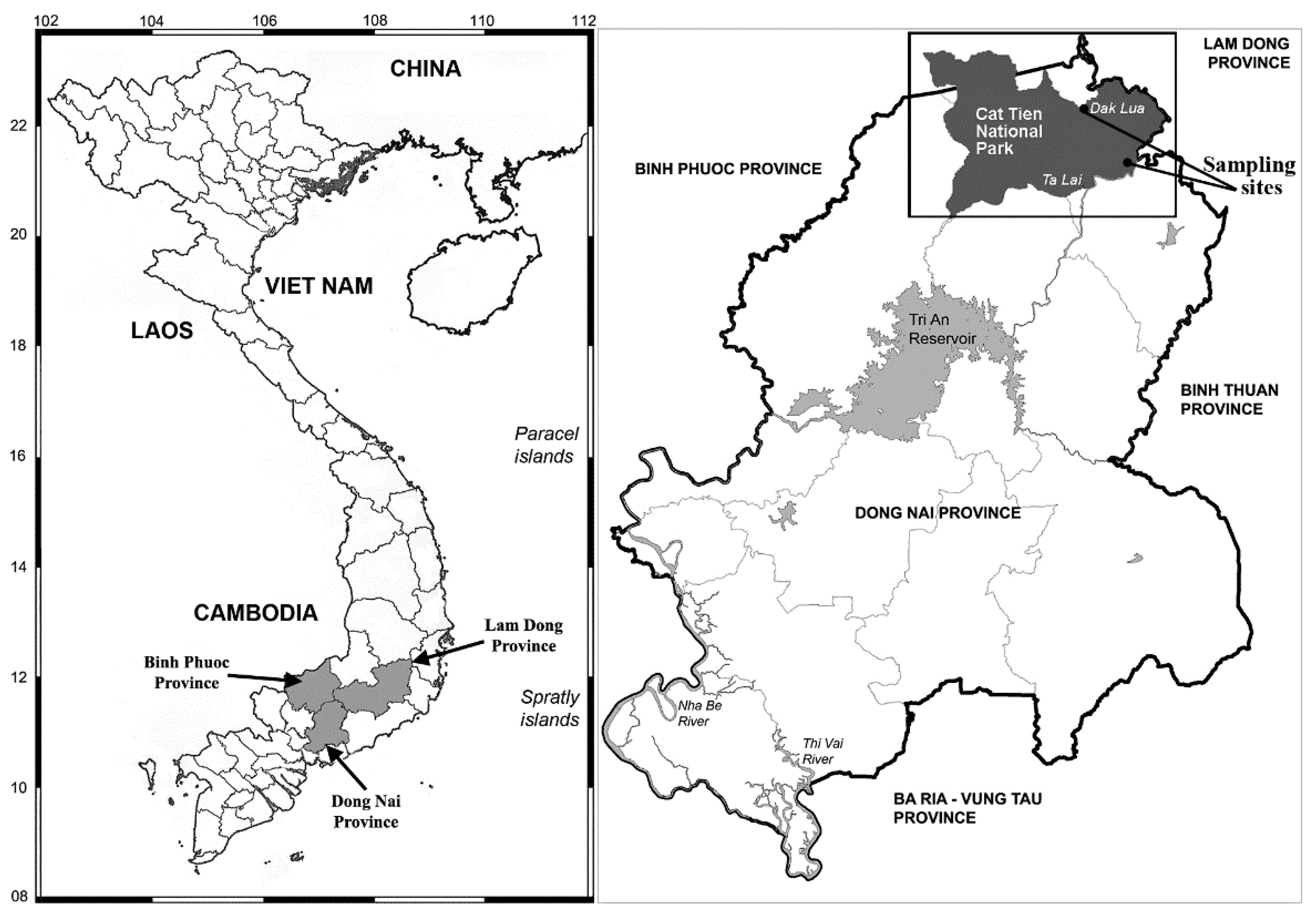
2.1. Study Area

Cat Tien National Park consists of three sections. The first is located in Dong Nai Province (Nam Cat Tien), the second is in Lam Dong Province (Cat Loc), and the third is in Binh Phuoc Province (Tay Cat Tien) (Figure 1).

2.2. Samples and Collections

Soil samples were collected from two dry grassy wetlands in Nam Cat Tien area on June 26, 2019 (N11° 30.999’ E107° 23.269’) by E. Gusev and E. Kezlya, and 20 March 2020 (N11° 24.241’ E107° 22.468’) by N. Martynenko (Figure 1). Both places stayed without water from 3 (2020) to 6 months (2019).Samples were taken during the expeditions of the Joint Vietnam-Russia Tropical Science and Technology Research Center (Ecolan 1.2 theme).
Soil samples were taken as follows: first, the surface of the sampling site was examined for the presence of evident growth of algae, then a combined sample was taken from an area of 10–30 m2. We used a metal scoop or shovel for this. Each composite sample consisted of 5–10 individual samples. After each collection, the instruments were cleaned and sterilized with alcohol. Samples were placed in plastic zip bags and labeled. Immediately after collecting, the absolute humidity was determined by the “hot drying” method in the laboratory room [34], then brought to an air-dried state and packaged. To measure pH, 30 g of soil was weighed, to which 150 mL of distilled water was added [35]. The suspension was poured into a clean glass and measured using the Hanna Piccolo 2 (HI98112) device, Hanna Instruments, Inc., Woonsocket, RI, USA. Humidity of soil samples was 49% in 2019 and 15% in 2020, and pH values were 5.1 and 5.0, respectively.

2.3. Culturing

Isolation and cultivation of algae strains were carried out at the Institute of Plant Physiology Russian Academy of Sciences. Soil from each sample was thoroughly mixed and a small amount (15–20 g) was placed into a Petri dish (diameter 60 mm), then moistened with distilled water up to 60–80% of full moisture capacity and placed into an illuminated climate chamber. After being in the chamber for 10 days, for algae detection, distilled water (3–5 mL) was added to the soil sample, shaken slightly, and then the liquid was transferred to another Petri dish and observed with an inverted microscope Zeiss Axio Vert A1. Such observations were carried out every 10–14 days. Algae cells were isolated with a micropipette, washed in 3–5 drops of sterile distilled water and placed into a 300 µL well of 96 well-plate. Non-axenic unialgal cultures were maintained in Waris-H liquid medium [36] at 22–25 °C in a growth chamber with a 12:12 h light:dark photoperiod.

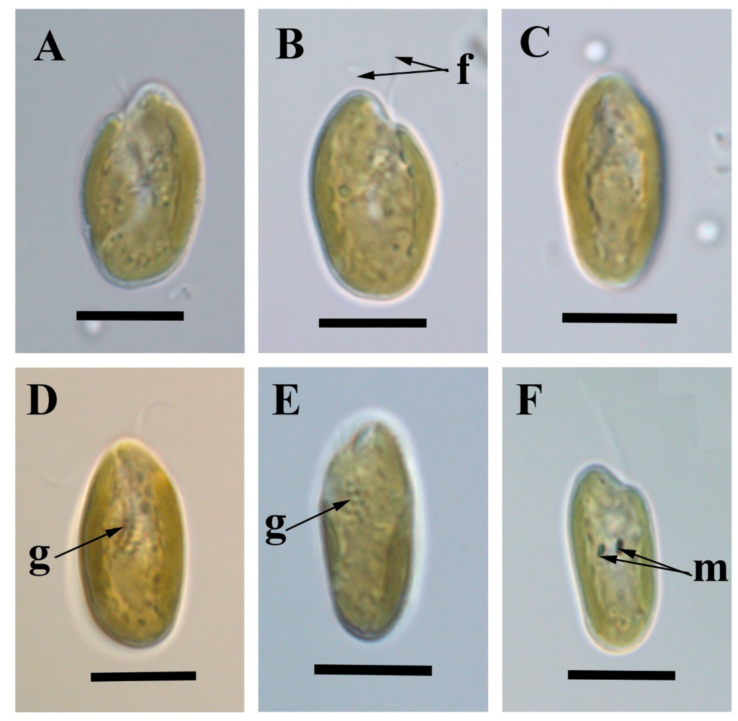
2.4. Light Microscopy

For light microscopical examinations, live cells were immobilized by embedding in ultra-low gelling agarose (Sigma-Aldrich, A4018, St. Louis, MO, USA) and examined by Nomarski differential interference contrast (DIC) with an oil immersion lens (Plan-Apochromat 100×/1.40 Oil DIC M27; microscope Zeiss AxioScope A1; Carl Zeiss AG, Oberkochem, Germany). The shape and size of cells, shape of the furrow–gullet system and cell plastids, and presence, number and position of pyrenoids were examined (n = 20 cells). For the description, we used the nomenclature proposed by Hoef-Emden and Melkonian [3], where the ventral side corresponds to the opening side of the furrow–gullet system. Calibration of magnification was carried out using a grated micrometer. Light micrographs were taken with an AxioCam ERc 5s Rev.2.

2.5. Amplification and Sequencing

The total DNA of the studied strains was extracted using InstaGeneTM Matrix as described in the manufacturer’s protocol. For amplification of targeting fragments, we chose the following primers. Nuclear SSU rDNA fragment was amplified with primers: 18S_CrN1F, 18S_826F, 18S_956R, 18S_BRK [37,38]; fragment of LSU rDNA—with crLSU_29F and crLSU_942R [4], and ITS2 rDNA—with crITS_03F and crITS_05R [39]. Amplification parameters for ribosomal genetic fragments were as follows: an initial denaturation for 5 min at 95 °C, then 35 cycles of 30 s denaturation at 94 °C, 30 s annealing at 52 °C and 50–80 s (in depending on amplicon length) extension at 72 °C, final extension of 10 min at 72 °C. The plastid psbA genetic fragment was amplified using psbA F, psbA R2 [13]. The conditions of amplification for the psbA fragments were the same as previously reported except for number of cycles (40), annealing temperature (50 °C) and time (30 s). PCR products were visualized by horizontal electrophoresis in 1.0% agarose gel stained with SYBR Safe (Life Technologies, Carlsbad, CA, USA) and purified with the ExoSAP-IT kit (Affymetrix, Santa Clara, CA, USA) according to the manufacturer’s protocol. All fragments were decoded from two sides using forward and reverse primers from PCR and the Big Dye system (Applied Biosystems, Foster City, CA, USA). Sequencing was performed using a Genetic Analyzer 3500 sequencer (Applied Biosystems, Foster City, CA, USA). Received sequences were checked manually and assembled after using BioEdit v7.1.3 and MegaX [40].

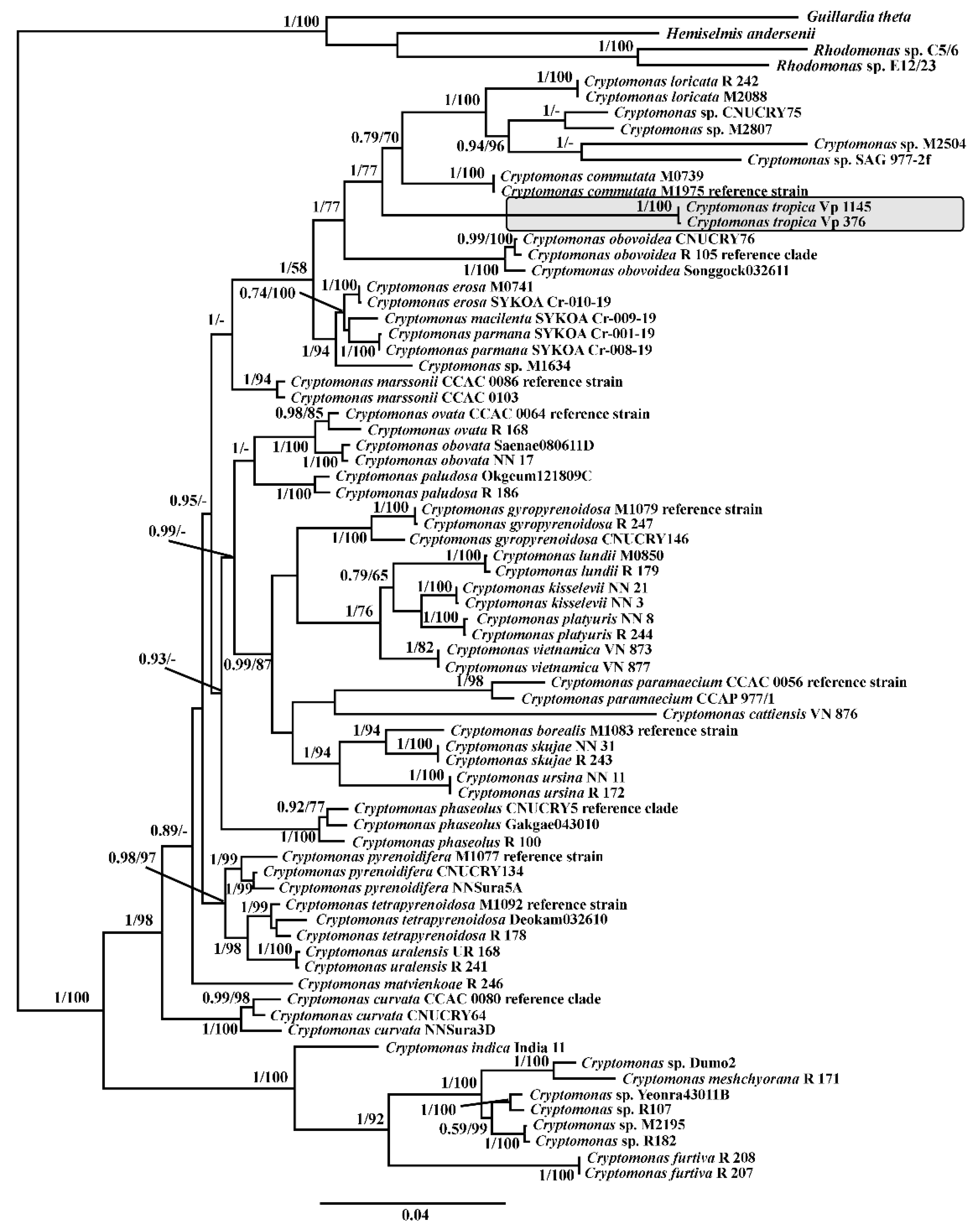
2.6. Alignment and Phylogenetic Analysis of SSU, LSU rDNA and psbA cpDNA

The newly determined sequences were aligned to other 68 Cryptomonas sequences from the GenBank database. A total of four cryptophyte taxa from the genera Rhodomonas, Guillardia and Hemiselmis were added to the dataset as outgroup taxa. Information about the analysis strains used is presented in Supplementary Table S1 (https://doi.org/10.6084/m9.figshare.21197479.v3 (accessed on 13 October 2022)). The sequences were aligned using either global SILVA alignment in the SINA v1.2.11 [41] for ribosomal fragments, or MAFFT v7 with model E-INS-i [42] for chloroplast gene fragment. The resulting alignments were manually checked for non-alignable sites, which were excluded from the analysis. To evaluate the existence of substitution saturation, we estimated the index of substitution saturation (Iss) in DAMBE 5.2.31 [43], as described by Xia and co-authors [44,45]. The results demonstrated that there was no significant saturation and therefore the sequences of these genes can be used in the phylogenetic analysis. For the protein-coding chloroplast gene, each codon position partition was tested. For Bayesian analysis, we partitioned the dataset by different genes and estimate the most appropriate substitution model for each partition separately, using the Bayesian information criterion (BIC) in the jModelTest 2.1.10 [46]. As the most fit models were selected SYM + G + I for the SSU rDNA, GTR + G + I for the LSU rDNA datasets. For each codon position of the protein-coding psbA cpDNA gene, the best model was also tested. The BIC-based model selection procedure selected the following models: GTR + G + I for the first codon position, JC for the second codon position, and GTR + G for the third codon position. Finally, we compiled the concatenated SSU rDNA + LSU rDNA + psbA cpDNA align dataset, containing 74 strains and 3347 positions (partial nuclear SSU rDNA: 1573 nt; nuclear SSU rDNA: 962 nt; plastid psbA: 812 nt). The Bayesian inference (BI) was conducted in MrBayes-3.2.5 [47] by performing three ‘hot’ and one ‘cold’ Markov chains for 15 × 106 cycles in two independent runs with the selection of each 100th generated tree. Phylogenetic tree and posterior branching probabilities were obtained after discarding the first 25% to produce estimate parameter models of nucleotide substitutions and likelihood. Files, generated by Bayesian MCMC runs, were tested using the software Tracer ver. 1.7.1. [48] for checking parameters estimated convergence, effective sample size and burn-in period. The resulting Bayesian topology was used as initial tree in Maximum Likelihood analysis (ML), performed in the program MegaX [40] with 1000 replicas. For viewing and editing trees, programs FigTree (ver 1.4.2) and Adobe Photoshop CC (19.0) were used.

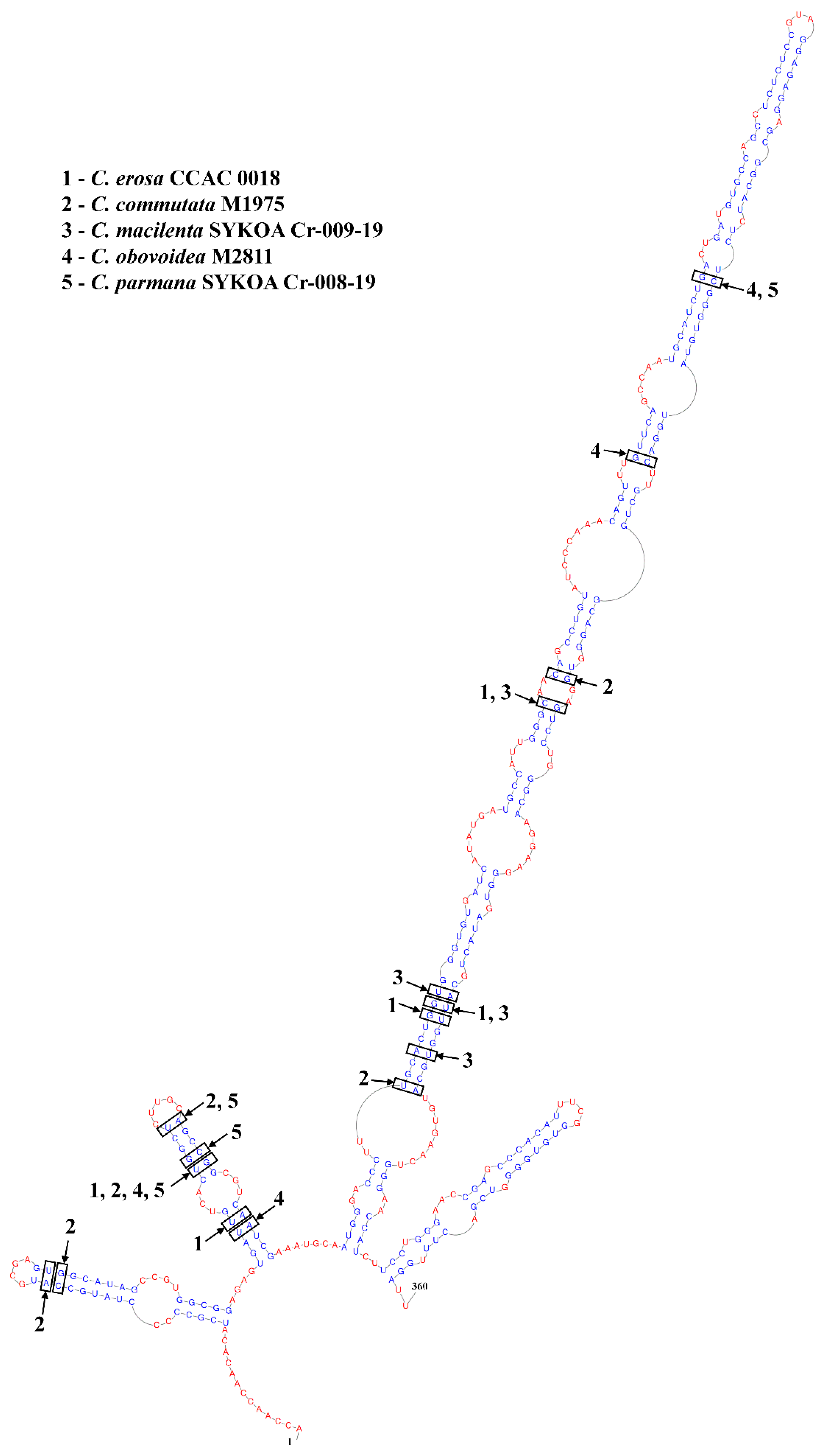
2.7. ITS2 Annotation, Secondary Structure Modeling, Alignment and Phylogeny
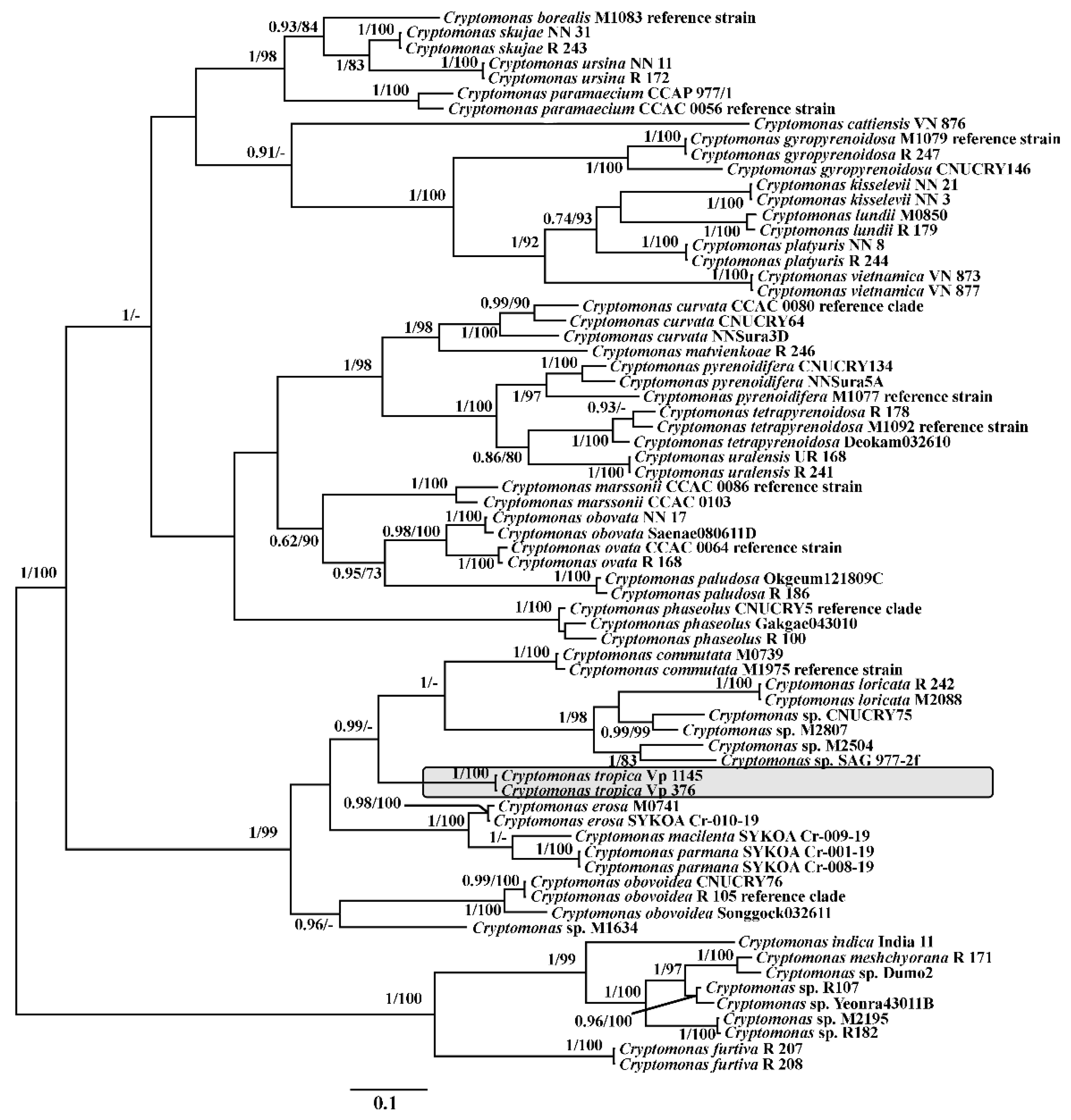
The ITS2-Annotation tool (http://its2-old.bioapps.biozentrum.uni-wuerzburg.de/cgi-bin/index.pl?annotator (accessed on 14 September 2022)) was used for the annotation of ITS2 sequences. This tool uses HMMer [49] to annotate ITS2 sequences with hidden Markov models (HMMs) [50]. The Mfold version 2.3 [51] was used for modelled secondary structures of the ITS2 rDNA region. A complete RNA secondary structure graph of the nuclear ITS2 rDNA of Cryptomonas sp. M1634, provided by Hoef-Emden [4], was used as a template and assisted in the inference of common stems, loops and bulges. The construction of the model considered the length and nucleotide composition of the spacers in the core of the model defining the helix boundaries [52], as well as the presence of the unpaired pyrimidine–pyrimidine bases in the second helix at the seventh position [53]. The secondary structure of the nuclear partial LSU rRNA was also predicted using the mfold server and the previously annotated template LSU rRNA [54]. Labelling of the helices follows as described in Wuyts et al. [55]. The resulting secondary structures were visualized in PseudoViewer3 [56]. The dataset based on ITS2 rDNA sequences was constructed with 70 Cryptomonas strains, used in previously described analysis, and contained 1119 align positions The ITS2 sequences were aligned by the ClustalW algorithm [57] with accordance to their secondary structure in 4SALE [58] and checked manually. The unrooted phylogenetic tree was recovered with the GTR + G+I substitutional model, defined by the program jModelTest 2.1.10.

3. Results

In total, two strains were isolated from soil samples taken from the dry wetlands of Cat Tien National Park in 2019 and 2020. These strains bear the same morphology and are represented by cells with a single plastid notched into several irregular lobes (Figure 2).
Phylogenetic relationships inferred from concatenated dataset of nuclear-encoded SSU, LSU rDNA, and plastid-encoded psbA using ML and BI, show that these strains have identical sequences and form an independent long branch in the tree (Figure 3).
This lineage is a sister to clades, containing strains Cryptomonas obovoidea Pascher emend. Hoef-Emden, and C. commutata (Pascher) Hoef-Emden. Both studied strains also have identical ITS2 rDNA sequences. For further analysis, the secondary structures of the ITS2 molecule were predicted. A four-helix structure could be inferred for the ITS2 sequence of C. tropica, with a long third helix. An unpaired U–U unit was found in Helix II. The total length of ITS2 was 360 nt, which is comparable with other species from the clade (Figure 4).
Phylogenetic tree of the genus Cryptomonas, based on an alignment of ITS2 rDNA sequences and their secondary structure, also revealed an independent lineage. This branch is a sister to clade, containing C. commutata and C. loricata with four unidentified Cryptomonas strains, and, in contrast to another analysis, to the clade, consisting of C. erosa Ehrenberg emend. Hoef-Emden and newly described C. macilenta Martynenko and E.S. Gusev and C. parmana Martynenko, E.S. Gusev and Sterlyagova [15] (Figure 5).
Analysis of CBCs between C. tropica and the most relative species revealed several Compensatory Base Changes (CBCs) in helices I, II, and III. We did not analyse helix IV due to its variability. At least four CBCs were present in conserved portions of helices I, II and III between C. tropica strain Vp 376 and C. obovoidea strain M2811. The number of CBCs between C. tropica and C. macilenta strain SYKOA Cr-009-19 was the same and also between C. parmana strain SYKOA Cr-008-19 (Figure 4). Number of CBCs, ITS2 length and K2p-distances between C. tropica strain Vp 376 and related species are represented in Table 1.
We describe below new species to science on the basis of molecular data.
Cryptista Adl et al.
Class Cryptophyceae Pascher emend. Schoenichen, emend. Adl et al.
Order Cryptomonadales Pascher
Genus Cryptomonas Ehrenb. emend Hoef-Emden et Melkonian
Cryptomonas tropica Martynenko, Kezlya and Gusev sp. nov. (Figure 2)
Diagnosis: Cells 15.9–21.0 µm long, 8.2–12.0 µm wide, 7.1–7.6 µm thick. Cells flattened, in ventral view elliptical to wide elliptical. In the lateral view, they are oblique apex and rounded antapex. Cells are without light microscopically visible pyrenoids and a single plastid notched into several irregular lobes. Furrow-curved and slightly narrowed at the distal end. Species differs from other species of the genus by the nucleotide sequences of nuclear ITS2, LSU, SSU rDNA, and chloroplast psbA genes. Diagnostic molecular characters, located in the nuclear LSU rRNA gene, are: CrtrLSU01_1: [GCGUGGUGGGAUUCCGGUUCGGAGAGCUUGCUCUCCAGCCUAUCAUGCCAUCACGC] [((((((((((((...((((.((((((....)))))))))).)))...)))))))))] situates in helix C1/e1 including the terminal loop; CrtrLSU01_2 encompasses the tip of the helix C1/e3: [CAAGGCUUGGAGGGUAAGGGCGUCAAACCUCUUAGCUCCUUGCGCAUG] [((..((..((((..((((((.((...)))))))).))))..))...))]; the signature CrtrLSU01_3: [CCAUUUCGCUUGUAGCCGUCCUGGAAGGACAAUUGCUGUGCUCUCUAGGCAGCUGCUUGAGAG-GUGG] [((((((((...(((((.(.((((((.(((((.....))).)).))))))).)))))....))))))))] occurs in the tip of the helix C1/e4.
The signature CrtrITS201_1 is in the 5′-part of ITS2-molecule’s helix III, including IIIa and IIIb conservative regions (reviewed by Hoef-Emden, 2007): [CAGCCUGUAUCCCAAACAGUUUGUUCAGCCAAUGCAUCUGACUGAUGUGCCAGCCUCCUCCGU-AGGAGAGGAGCGGCAUCUCUCGGGUGUAUGGACUUGCUGGCAGGGUG] [((.(((((........((((..(((((.....(((((((((..((.(((((.((.(((((((...))))))).))))))).))))))))))))))))..))))))))).))].
Holotype: a large drop of unfixed dried cells of strain VP376 on watercolor paper (hic designatus), deposited at on watercolor paper (hic designatus), deposited at MHA (Herbarium, Main Botanical Garden, Botanicheskaya Str. 4, Moscow, 127276, Russia) under the designation Cryptomonas tropica Vietnam Gusev 21-10 strain VP376. Culture was also deposited in the Culture Collection of Algae of the Institute of Biology of Komi Republic under the number SYKOA Cr-026-22. The strain is conserved as a formaldehyde-fixed sample.
Representative DNA sequences. DNA sequences of the strain VP376 were submitted to the GenBank database on 17 September 2022 under acc. nos OP456095 (nuclear ITS2 and partial LSU rDNA), OP456093 (nuclear SSU and ITS1 rDNA), OP440446 (partial psbA).
Type locality: Grassy wetland near Dac Lua, Cat Tien National Park, Dong Nai Province, Vietnam. Latitude/Longitude N11° 30.999’ E107° 23.269’, collected by E. Gusev and E. Kezlya on 26 June 2019.
Etymology: The species name “tropica” (“tropical”) was chosen because both known strains were isolated from tropical region.
Reference strain: VP376
Other strain: VP1145
Distribution: To date, Cryptomonas tropica has been observed in the type locality and other grassy wetland in Cat Tien National Park, Latitude/Longitude N11° 24.241’ E107° 22.468’.

4. Discussion

Cryptomonas tropica forms a long branch in the clade, consisting of C. obovoidea, C. commutata, C. erosa and related C. macilenta and C. parmana, C. loricata and other unidentified Cryptomonas strains: CNUCKRY75, M2807, M2504, M1634 and SAG-977-2f. This clade was previously designated “NoPyr” [3] and has since been revised with the descriptions C. erosa, C. commutata, C. obovoidea and C. loricata [4]. Later, two more species, related to C. erosa, C. macilenta and C. parmana, were described [15]. Most morphotypes of this clade are characterized by small cells, not exceeding 21 µm in length. Cells of most of these species usually have one deeply bilobed plastid without microscopically visible pyrenoids. Only C. loricata contains pyrenoids, which is a feature that distinguishes this species from other members of the clade. C. tropica, in contrast to these species, has a plastid notched into several irregular lobes. Clade also contains colorless strains of Cryptomonas—M1634 and SAG-977-2f. In rest of all, C. erosa, C. commutata, C. macilenta, C. obovoidea, C. parmana and describing here C. tropica are hardly distinguished morphologically, and the most reliable method for their discrimination is to use molecular approach.
Finds of species of the genus Cryptomonas in soils are very rare and given mainly in algae guidelines without specifying the exact location of the find. The species reported from soils included Cryptomonas tenuis Pasher [9,10], C. erosa Ehrenberg sensu lato [8,9] and C. ovata Ehrenberg sensu lato [7,9]. C. ovata sensu lato had an extensive diagnosis, so both large cells with pyrenoids as well as minute cells without pyrenoids could be referred to this species. What morphotypes were found in soils is unknown. F. Hindák isolated a soil culture of Cryptomonas from the inundation area of the river Dyje (Thaya) close to the town Lednice, Moravia [11]. On basis of light observations, P. Javornický [59] identified this strain as a small Cryptomonas phaseolus Skuja, which has small cells without pyrenoids and also can be attributed to C. ovata sensu lato. Strictly speaking, original drawings of C. ovata, made by Ehrenberg, represent large cells without pyrenoids. According to Hoef-Emden and Melkonian [3], the structures, resembling pyrenoids, correspond to Maupas ovals in position, size, and shape. Therefore, all assignments of species with pyrenoids to C. ovata can be considered misleading. Nowadays, the species C. ovata is clearly defined by including diagnosis molecular signatures as diagnostic characters [3].
As previously reported, C. tropica cannot be distinguished from C. erosa using a morphological approach, but they clearly differ from each other by their molecular features. C. tenuis is characterized by subtle cells 8–14 µm in length and 5–7 in width, which is smaller than that of C. tropica. This species is unknown from culture collections and does not yet have molecular data, which renders its emendation impossible, and its finding must be critically analyzed.
Our unpublished data indicate a very rich flora of the genus Cryptomonas in the water bodies of Cat Tien National Park. The study of about 90 strains revealed 27 phylogenetic lineages of this genus. However, Cryptomonas tropica was not found in any of the water samples. It is known that for surviving unfavorable conditions, species of the genus Cryptomonas may produce resting stages. They are represented by thick-walled cysts [3,60,61,62] or palmelloid stages enveloped by mucilage [63], to withstand adverse conditions such as desiccation. The mechanism for surviving in soil is possibly inherent in C. tropica and other cryptomonad species that inhabit soils.
In general, recent research has shown that even in well-sampled regions and habitats, the true diversity of cryptomonads has not yet been uncovered [60]. The study of soils in tropical regions can bring many interesting results and findings. The study of soil algae in Cat Tien National Park made it possible to identify new species of diatoms: Mayamaea vietnamica Glushchenko, Kezlya, Kulikovskiy and Kociolek [64], Placoneis cattiensis Glushchenko, Kezlya, Kulikovskiy and Kociolek [65], P. subundulata Kezlya, Glushchenko, Kulikovskiy and Kociolek [66]. The unique conditions of a humid, flooded rainforest may favor the formation of a unique terrestrial algae flora, both due to long periods of flooding and extremely high air humidity, which can maintain acceptable conditions for the development of organisms [67]. The discovery of Cryptomonas tropica in an uncharacteristic environment for the genus confirms this conclusion and demonstrates the prospects for research on terrestrial microalgae in tropical forests.

Supplementary Materials

The following supporting information can be downloaded at: https://www.mdpi.com/article/10.3390/d14111001/s1, Table S1. GenBank accession numbers of analyzed strains.

Author Contributions

N.M., sampling, molecular investigation, writing and drafting of the manuscript; E.K., sampling, culturing; LM investigation.; E.G., funding acquisition, project administration, supervision, writing and drafting of the manuscript. All authors have read and agreed to the published version of the manuscript.

Funding

Financial support was provided by the Russian Science Foundation (20-14-00211) for the sample processing, culture isolation, microscopy and molecular studies and identification. Analysis of environmental parameters was performed within the state assignment of the Ministry of Science and Higher Education of the Russian Federation (theme No. 122042700045-3).

Institutional Review Board Statement

Not applicable.

Informed Consent Statement

Not applicable.

Data Availability Statement

Not applicable.

Acknowledgments

The authors are grateful to the staff of the Joint Vietnam-Russia Tropical Science and Technology Research Center, for their assistance in management and sampling during the project “Ecolan 1.2.”. Authors are grateful to Julia Podunay for help in isolating strain Vp 1145. The authors also thank the three anonymous reviewers for their helpful comments resulting in a considerable improvement of the manuscript.

Conflicts of Interest

The authors declare no conflict of interest.

References

  1. Ehrenberg, C.G. Animalia Evertebrata. Symbolae physicae seu icones et descriptiones animalium evertebratorum sepositis insectis quae ex itinere per Africanum Borealem et Asiam Occidentalem Friderici Guilelmi Hemprich et Christiani Godofredi Ehrenberg medicinae et chirurgiae doctorum studio novae aut illustratae redierunt. In Symbolae Physicae; Pars Zoologica, I.V., Hemprich, P.C., Ehrenberg, C.G., Eds.; Ex Officina Academica: Mittler, Berlin, 1831; 71p. [Google Scholar]
  2. AlgaeBase. Available online: https://www.algaebase.org/search/genus/detail/?genus_id=43571 (accessed on 13 October 2022).
  3. Hoef-Emden, K.; Melkonian, M. Revision of the Genus Cryptomonas (Cryptophyceae): A Combination of Molecular Phylogeny and Morphology Provides Insights into a Long-Hidden Dimorphism. Protist 2003, 154, 371–409. [Google Scholar] [CrossRef] [PubMed]
  4. Hoef-Emden, K. Revision of the genus Cryptomonas (Cryptophyceae) II: Incongruences between classical morphospecies concept and molecular phylogeny in smaller pyrenoid-less cells. Phycologia 2007, 46, 402–428. [Google Scholar] [CrossRef]
  5. Gusev, E.; Martynenko, N.; Kulizin, P.; Kulikovskiy, M. Molecular diversity of the genus Cryptomonas (Cryptophyceae) in Russia. Eur. J. Phycol. 2022, 57, 526–550. [Google Scholar] [CrossRef]
  6. Javornický, P.; Hindák, F. Cryptomonas frigoris spec. nova (Cryptophyceae), the new cyst-forming flagellate from the snow of the High Tatras. Biologia 1970, 25, 241–250. [Google Scholar] [PubMed]
  7. Gollerbakh, M.M.; Polyansky, V.I. Identification Guide for Freshwater Algae of the USSR; Sovetskaya Nauka: Moscow, Russia, 1953; Volume 6, 212p. [Google Scholar]
  8. Gollerbach, M.M.; Shtina, E.A. Soil Algae; Nauka: Saint-Petersburg, Russia, 1969; 228p. [Google Scholar]
  9. Matvienko, O.M.; Litvinenko, R.M. Identification Guide for Freshwater Algae of the Ukrainian SSR; Naukova Dumka: Kiev, Ukraine, 1977; Volume 3, 387p. [Google Scholar]
  10. Norton, J.R.; Davis, J.S. Soil Algae from North Central Florida. Fla. Sci. 1975, 38, 77–81. Available online: https://www.jstor.org/stable/24319630 (accessed on 14 September 2022).
  11. Hindák, F. Species composition and biomass of phototrophic edaphon in the ecosystem of a floodplain forest in southern Moravia. In Floodplain Forest Ecosystem. I. Before Water Management Measures; Penka, M., Vyskot, M., Klimo, E., Vašíček, F., Eds.; Academia, Publishing House of the Czechoslovak Academy of Sciences: Prague, Czechoslovakia, 1985; pp. 239–249. ISBN 0-444-99566-8. [Google Scholar]
  12. Paulsen, B.S.; Vieira, A.A.H.; Klaveness, D. Structure of extracellular polysaccharides produced by a soil Cryptomonas sp. (cryptophyceae). J. Phycol. 1992, 28, 61–63. [Google Scholar] [CrossRef]
  13. Choi, B.; Son, M.; Kim, J.I.; Shin, W. Taxonomy and phylogeny of the genus Cryptomonas (Cryptophyceae, Cryptophyta) from Korea. Algae 2013, 28, 307–330. [Google Scholar] [CrossRef]
  14. Martynenko, N.A.; Gusev, E.S.; Kulizin, P.V.; Guseva, E.E.; McCartney, K.; Kulikovskiy, M.S. A new species of Cryptomonas (Cryptophyceae) from the Western Urals (Russia). Eur. J. Taxon. 2020, 649, 1–12. [Google Scholar] [CrossRef]
  15. Martynenko, N.A.; Gusev, E.S.; Sterlyagova, I.N.; Kulikovskiy, M.S. Revealing hidden diversity in the Cryptomonas erosa clade (Cryptophyceae), with the description of two new species from acidic habitats. Phycologia 2022, 61, 184–194. [Google Scholar] [CrossRef]
  16. Gusev, E.; Podunay, Y.; Martynenko, N.; Shkurina, N.; Kulikovskiy, M. Taxonomic studies of Cryptomonas lundii clade (Cryptophyta: Cryptophyceae) with description of a new species from Vietnam. Fottea 2020, 20, 137–143. [Google Scholar] [CrossRef]
  17. Martynenko, N.A.; Gusev, E.S.; Kapustin, D.A.; Guseva, E.E.; Kulikovskiy, M.S. Cryptomonas cattiensis sp. nov. (Cryptophyceae: Cryptomonadales), a new species described from Vietnam. Phytotaxa 2020, 454, 127–136. [Google Scholar] [CrossRef]
  18. Gusev, E.; Karthick, B.; Martynenko, N.; Shkurina, N.; Kulikovskiy, M. Cryptomonas indica sp. nov. (Cryptophyceae: Cryptomonadales), a new species described from the Western Ghats, India. Phytotaxa 2021, 518, 261–270. [Google Scholar] [CrossRef]
  19. Blanc, L.; Maury-Lechon, G.; Pascal, J.-P. Structure, floristic composition and natural regeneration in the forests of Cat Tien National Park, Vietnam: An analysis of the successional trends. J. Biogeogr. 2000, 27, 141–157. [Google Scholar] [CrossRef]
  20. Nguyen, V.T.; Anichkin, A.E. Cat Tien National Park—General information. In Structure and Functions of Soil Communities of a Monsoon Tropical Forest (Cat Tien National Park, Southern Vietnam); Tiunov, A.V., Ed.; KMK Scientific Press: Moscow, Russia, 2011; pp. 11–15. ISBN 978-5-87317-797-4. [Google Scholar]
  21. Kuznetsov, A.N.; Kuznetsova, S.P. Forest vegetation: Species composition and stand structure. In Structure and Functions of Soil Communities of a Monsoon Tropical Forest (Cat Tien National Park, Southern Vietnam); Tiunov, A.V., Ed.; KMK Scientific Press: Moscow, Russia, 2011; pp. 16–43. ISBN 978-5-87317-797-4. [Google Scholar]
  22. Cat Tien National Park. Available online: http://www.namcattien.org (accessed on 23 September 2022).
  23. Gusev, E.; Doan-Nhu, H.; Nguyen-Ngoc, L. Silica-scaled chrysophytes from Cat Tien National Park (Dong Nai Province, Vietnam). Nova Hedwig. Beih. 2017, 105, 347–364. [Google Scholar] [CrossRef]
  24. Gusev, E.S.; Guseva, E.E.; Dien, T.D.; Kulikovskiy, M.S. Flora of Silica-Scaled Chrysophytes (Chrysophyceae) of Two Provinces in Southern Vietnam. Inland Water Biol. 2022, 15, 205–216. [Google Scholar] [CrossRef]
  25. Glushchenko, A.; Genkal, S.I.; Kulikovskiy, M. Aulacoseira konstastinovii sp. nov. andAulacoseira kryloviisp. nov.—Two new centric diatoms from South-east Asia. Diatom Res. 2016, 31, 367–378. [Google Scholar] [CrossRef]
  26. Glushchenko, A.; Kulikovskiy, M.; Kociolek, J.P. New and interesting species from the genus Luticola (Bacillariophyceae) in waterbodies of Southeastern Asia. Nova Hedwig. Beih. 2017, 146, 157–173. [Google Scholar] [CrossRef]
  27. Glushchenko, A.; Kulikovskiy, M.; Kuznetsova, I.; Dorofeyuk, N.; Kociolek, J.P. New species and combinations in the genus Eunotia Ehrenberg 1837 (Bacillariophyceae: Eunotiaceae) from waterbodies of Southeastern Asia. Nova Hedwig. Beih. 2018, 147, 69–103. [Google Scholar] [CrossRef] [PubMed]
  28. Gusev, E.S. A new species of the genus Mallomonas (Synurophyceae), Mallomonas spinosa sp. nov., from Vietnam. Phytotaxa 2012, 66, 1–5. [Google Scholar] [CrossRef]
  29. Gusev, E.S.; Doan-Nhu, H.; Nguyen-Ngoc, L. Mallomonas cattiensis sp. nov. (Synurales, Chrysophyceae), a new species from Viet Nam. Phytotaxa 2015, 221, 188–192. [Google Scholar] [CrossRef]
  30. Gusev, E.S.; Doan-Nhu, H.; Nguyen-Ngoc, L.; Kapustin, D.A. Two new species of the genus Mallomonas from the Cat Tien National Park (Viet Nam): Mallomonas distinguenda and Mallomonas skvortsovii. Phytotaxa 2016, 273, 59–64. [Google Scholar] [CrossRef]
  31. Gusev, E.S.; Siver, P.A.; Shin, W. Mallomonas bronchartiana Compère Revisited: Two New Species Described from Asia. Cryptogam. Algologie 2017, 38, 3–16. [Google Scholar] [CrossRef]
  32. Gusev, E.S.; Čertnerová, D.; Škaloudová, M.; Škaloud, P. Exploring Cryptic Diversity and Distribution Patterns in the Mallomonas kalinae/rasilis Species Complex with a Description of a New Taxon—Mallomonas furtiva sp. nov. J. Eukaryot. Microbiol. 2018, 65, 38–47. [Google Scholar] [CrossRef]
  33. Gusev, E.S.; Kulizin, P.; Guseva, E.; Shkurina, N.; Kulikovskiy, M. Mallomonas lamii sp. nov. (Synurales, Chrysophyceae), a new species bearing large scales described from the tropics. Phytotaxa 2019, 423, 266–272. [Google Scholar] [CrossRef]
  34. Vadjunina, A.F.; Korchagina, Z.A. Methods of Studying the Physical Properties of Soils, 3rd ed.; Agropromizdat: Moscow, Russia, 1986; 416p. [Google Scholar]
  35. Arinushkina, E.V. Handbook for Chemical Soil Analysis, 2nd ed.; Moscow State University: Moscow, Russia, 1970; 488p. [Google Scholar]
  36. McFadden, G.I.; Melkonian, M. Use of Hepes buffer for microalgal culture media and fixation for electron microscopy. Phycologia 1986, 25, 551–557. [Google Scholar] [CrossRef]
  37. Marin, B.; Klingberg, M.; Melkonian, M. Phylogenetic Relationships among the Cryptophyta: Analyses of Nuclear-Encoded SSU rRNA Sequences Support the Monophyly of Extant Plastid-Containing Lineages. Protist 1998, 149, 265–276. [Google Scholar] [CrossRef]
  38. Kim, J.-H.; Boo, S.-M.; Shin, W.-G. Two Freshwater Cryptomonads New to Korea: Cryptomonas marssonii and C. pyrenoidifera. Algae 2007, 22, 147–152. [Google Scholar] [CrossRef]
  39. Hoef-Emden, K.; Marin, B.; Melkonian, M. Nuclear and Nucleomorph SSU rDNA Phylogeny in the Cryptophyta and the Evolution of Cryptophyte Diversity. J. Mol. Evol. 2002, 55, 161–179. [Google Scholar] [CrossRef] [PubMed]
  40. Kumar, S.; Stecher, G.; Li, M.; Knyaz, C.; Tamura, K. MEGA X: Molecular Evolutionary Genetics Analysis across Computing Platforms. Mol. Biol. Evol. 2018, 35, 1547–1549. [Google Scholar] [CrossRef]
  41. Pruesse, E.; Peplies, J.; Glöckner, F.O. SINA: Accurate high-throughput multiple sequence alignment of ribosomal RNA genes. Bioinformatics 2012, 28, 1823–1829. [Google Scholar] [CrossRef] [Green Version]
  42. Katoh, K.; Toh, H. Parallelization of the MAFFT multiple sequence alignment program. Bioinformatics 2010, 26, 1899–1900. [Google Scholar] [CrossRef] [Green Version]
  43. Xia, X.; Xie, Z. DAMBE: Software Package for Data Analysis in Molecular Biology and Evolution. J. Hered. 2001, 92, 371–373. [Google Scholar] [CrossRef] [PubMed] [Green Version]
  44. Xia, X.; Xie, Z.; Salemi, M.; Chen, L.; Wang, Y. An index of substitution saturation and its application. Mol. Phylogenetics Evol. 2002, 26, 1–7. [Google Scholar] [CrossRef]
  45. Xia, X.; Lemey, P. Assessing substitution saturation with DAMBE. In The Phylogenetic Handbook: A Practical Approach to DNA and Protein Phylogeny, 2nd ed.; Lemey, P., Salemi, M., Vandamm, A.-M., Eds.; Cambridge University Press: Cambridge, UK; pp. 615–630. [CrossRef]
  46. Darriba, D.; Taboada, G.L.; Doallo, R.; Posada, D. jModelTest 2: More models, new heuristics and parallel computing. Nat. Methods 2012, 9, 772. [Google Scholar] [CrossRef] [PubMed] [Green Version]
  47. Ronquist, F.; Huelsenbeck, J.P. MrBayes 3: Bayesian phylogenetic inference under mixed models. Bioinformatics 2003, 19, 1572–1574. [Google Scholar] [CrossRef] [Green Version]
  48. Rambaut, A.; Drummond, A.J.; Xie, D.; Baele, G.; Suchard, M.A. Posterior Summarization in Bayesian Phylogenetics Using Tracer 1.7. Syst. Biol. 2018, 67, 901–904. [Google Scholar] [CrossRef] [Green Version]
  49. Eddy, S.R. Profile hidden Markov models. Bioinformatics 1998, 14, 755–763. [Google Scholar] [CrossRef] [Green Version]
  50. Keller, A.; Schleicher, T.; Schultz, J.; Müller, T.; Dandekar, T.; Wolf, M. 5.8S-28S rRNA interaction and HMM-based ITS2 annotation. Gene 2009, 430, 50–57. [Google Scholar] [CrossRef]
  51. Zuker, M. Mfold web server for nucleic acid folding and hybridization prediction. Nucleic Acids Res. 2003, 31, 3406–3415. [Google Scholar] [CrossRef]
  52. Schultz, J.; Maisel, S.; Gerlach, D.; Müller, T.; Wolf, M. A common core of secondary structure of the internal transcribed spacer 2 (ITS2) throughout the Eukaryota. Rna 2005, 11, 361–364. [Google Scholar] [CrossRef]
  53. Caisová, L.; Marin, B.; Melkonian, M. A Consensus Secondary Structure of ITS2 in the Chlorophyta Identified by Phylogenetic Reconstruction. Protist 2013, 164, 482–496. [Google Scholar] [CrossRef] [PubMed]
  54. Gillespie, J.J.; Johnston, J.S.; Cannone, J.J.; Gutell, R.R. Characteristics of the nuclear (18S, 5.8S, 28S and 5S) and mitochondrial (12S and 16S) rRNA genes of Apis mellifera (Insecta: Hymenoptera): Structure, organization, and retrotransposable elements. Insect Mol. Biol. 2006, 15, 657–686. [Google Scholar] [CrossRef] [Green Version]
  55. Wuyts, J.; Van de Peer, Y.; De Wachter, R. Distribution of substitution rates and location of insertion sites in the tertiary structure of ribosomal RNA. Nucleic Acids Res. 2001, 29, 5017–5028. [Google Scholar] [CrossRef] [Green Version]
  56. Byun, Y.; Han, K. PseudoViewer: Web application and web service for visualizing RNA pseudoknots and secondary structures. Nucleic Acids Res. 2006, 34, W416–W422. [Google Scholar] [CrossRef] [PubMed] [Green Version]
  57. Thompson, J.D.; Higgins, D.G.; Gibson, T.J. CLUSTAL W: Improving the sensitivity of progressive multiple sequence alignment through sequence weighting, position-specific gap penalties and weight matrix choice. Nucleic Acids Res. 1994, 22, 4673–4680. [Google Scholar] [CrossRef] [PubMed] [Green Version]
  58. Seibel, P.N.; Müller, T.; Dandekar, T.; Schultz, J.; Wolf, M. 4SALE—A tool for synchronous RNA sequence and secondary structure alignment and editing. BMC Bioinform. 2006, 7, 498. [Google Scholar] [CrossRef] [Green Version]
  59. Javornický, P. Cryptomonas ovata Ehrenberg (Cryptophyceae) and some related species. Algol. Studies. 2003, 110, 43–65. [Google Scholar] [CrossRef]
  60. Hoef-Emden, K.; Archibald, J.M. Cryptophyta (Cryptomonads). In Handbook of the Protists, 2nd ed.; Archibald, J.M., Simpson, A.G.B., Slamovits, C.H., Margulis, L., Melkonian, M., Chapman, D.J., Corliss, J.O., Eds.; Springer: Cham, Switzerland, 2017; Volume 2, pp. 851–891. [Google Scholar] [CrossRef]
  61. Lichtlé, C. Effects of nitrogen deficiency and light of high intensity on Cryptomonas rufescens (Cryptophyceae) I. Cell and photosynthetic apparatus transformations and encystment. Protoplasma 1979, 101, 283–299. [Google Scholar] [CrossRef]
  62. Lichtlé, C. Effects of nitrogen deficiency and light of high intensity on Cryptomonas rufescens (Cryptophyceae) II. Excystment. Protoplasma 1980, 102, 11–19. [Google Scholar] [CrossRef]
  63. Santore, U.J. Light-and Electron-microscopic Observations of the Palmelloid) Phase in Members of the Genus Cryptomonas (Cryptophyceae). Arch. Für Protistenkd. 1978, 120, 420–435. [Google Scholar] [CrossRef]
  64. Kezlya, E.; Glushchenko, A.; Kociolek, J.P.; Maltsev, Y.; Martynenko, N.; Genkal, S.; Kulikovskiy, M. Mayamaea vietnamica sp. nov.: A new, terrestrial diatom (Bacillariophyceae) species from Vietnam. Algae 2020, 35, 325–335. [Google Scholar] [CrossRef]
  65. Kezlya, E.; Glushchenko, A.; Maltsev, Y.; Gusev, E.; Genkal, S.; Kuznetsov, A.; Kociolek, J.P.; Kulikovskiy, M. Placoneis cattiensis sp. nov.—A new, diatom (Bacillariophyceae: Cymbellales) soil species from Cát Tiên National Park (Vietnam). Phytotaxa 2020, 460, 237–248. [Google Scholar] [CrossRef]
  66. Kezlya, E.; Glushchenko, A.; Maltsev, Y.; Gusev, E.; Genkal, S.; Kociolek, J.P.; Kulikovskiy, M. Three New Species of Placoneis Mereschkowsky (Bacillariophyceae: Cymbellales) with Comments on Cryptic Diversity in the P. elginensis—Group. Water 2021, 13, 3276. [Google Scholar] [CrossRef]
  67. Foissner, W. Biogeography and dispersal of micro-organisms: A review emphasizing protists. Acta Protozool. 2006, 45, 111–136. [Google Scholar]
Figure 1. Map of the study area.
Diversity 14 01001 g001
Figure 2. Light micrographs of Cryptomonas tropica sp. nov. (strain Vp 376). (A) Ventral view; (BD) Dorsal view; (E) Lateral view, left side; (F) Lateral view, right side. Scale bar −10 µm. f—flagellas, g—furrow-gullet system, m—Maupas ovals.
Diversity 14 01001 g002
Figure 3. Molecular phylogeny of the genus Cryptomonas, based on combined data set of the partial nuclear SSU rDNA, LSU rDNA and chloroplast psbA genetic regions. Bayesian Posterior Probabilities and Maximum Likelihood bootstrap support are shown left and right of the fraction line, respectively. Scale bar represents substitutions per site. New described species are marked with a grey box.
Diversity 14 01001 g003
Figure 4. Predicted ITS2 secondary structure model of the strain Vp 376 of Cryptomonas tropica. Compensatory Base Changes between this species and some related, indicated by numbers, are marked by black boxes.
Diversity 14 01001 g004
Figure 5. Molecular phylogeny of the genus Cryptomonas, based on the nuclear internal transcribed spacer 2 (ITS2 rDNA) sequences. Bayesian Posterior Probabilities and Maximum Likelihood bootstrap support shown left and right of the fraction line, respectively. Scale bar represents substitutions per site. New described species are marked within the grey box.
Diversity 14 01001 g005
Table 1. Number of CBCs in ITS2, length and K2P-distances between C. tropica strain Vp 376 and reference strains of C. erosa, C. commutata, C. macilenta, C. obovoidea and C. parmana.
NoTaxaHelixITS2 LengthK2P-Distances
IIIIIISSULSUpsbAITS2
1C. erosa M0741 *0233640.0400.1340.0300.269
2C. commutata M19752223430.0480.1260.244
3C. macilenta SYKOA Cr-009-190044160.0400.1400.0260.313
4C. obovoidea M2811 **0223700.0540.1550.0300.273
5C. parmana SYKOA Cr-008-190314240.0410.1280.0350.326
* Epitype strain CCAC 0018 lacks some sequences, therefore, sequences of SSU, LSU and ITS2 rDNA of strain M0741 (AM051201, AJ566163) and sequence of psbA cpDNA gene of strain SYKOA Cr-010-19 (MW430149) were used for assessing parameters. ** Due to the absence of some sequences of epitype strain M2811, sequences of the SSU rDNA and psbA cpDNA genes of strain R 105 (GeneBank acc. no. MZ268240, MZ353623), belonging to the same clade as the type strain, were used for calculating parameters.
Publisher’s Note: MDPI stays neutral with regard to jurisdictional claims in published maps and institutional affiliations.

