Article

MRC-5 Human Lung Fibroblasts Alleviate the Genotoxic Effect of Fe-N Co-Doped Titanium Dioxide Nanoparticles through an OGG1/2-Dependent Reparatory Mechanism

by
Bogdan Andrei Miu
1,
Ionela Cristina Voinea
1,2,*,
Lucian Diamandescu
3 and
Anca Dinischiotu
1
1
Department of Biochemistry and Molecular Biology, Faculty of Biology, University of Bucharest, 91-95 Splaiul Independentei, 050095 Bucharest, Romania
2
Research Institute of the University of Bucharest—ICUB, University of Bucharest, 050657 Bucharest, Romania
3
National Institute of Materials Physics (NIMP), Atomistilor 405A, 077125 Magurele, Romania
*
Author to whom correspondence should be addressed.
Int. J. Mol. Sci. 2023, 24(7), 6401; https://doi.org/10.3390/ijms24076401
Submission received: 16 February 2023 / Revised: 24 March 2023 / Accepted: 26 March 2023 / Published: 29 March 2023
(This article belongs to the Special Issue Interaction of Nanomaterials with Cells and Tissues)

Abstract

:
The current study was focused on the potential of pure P25 TiO2 nanoparticles (NPs) and Fe(1%)-N co-doped P25 TiO2 NPs to induce cyto- and genotoxic effects in MRC-5 human pulmonary fibroblasts. The oxidative lesions of P25 NPs were reflected in the amount of 8-hydroxydeoxyguanosine accumulated in DNA and the lysosomal damage produced, but iron-doping partially suppressed these effects. However, neither P25 nor Fe(1%)-N co-doped P25 NPs had such a serious effect of inducing DNA fragmentation or activating apoptosis signaling. Moreover, oxo-guanine glycosylase 1/2, a key enzyme of the base excision repair mechanism, was overexpressed in response to the oxidative DNA deterioration induced by P25 and P25-Fe(1%)-N NPs.

Graphical Abstract

1. Introduction

In recent years, NPs have become a major development opportunity for biomedical [1], agricultural [2], and different industrial applications such as electronics [3], aerospace and automotive coatings [4], active food packaging [5], or environmental remediation [6]. According to some estimates, the most produced metal-based nanomaterials worldwide are made of silver, titanium, zinc, or gold [7,8]. In particular, TiO2 NPs are widely used in commercially available products or pilot applications, including cosmetics and sunscreens, paints, food products and active packaging, photoactive cement, or innovative textiles [9,10,11,12], due to their special optical and photocatalytic characteristics.
Different studies proved that TiO2 NPs, especially anatase-rutile mixtures, exhibit cyto- and genotoxicity [13,14,15], which probably derive from their ability to generate excessive ROS levels that could determine oxidative stress when the cellular antioxidant systems are overwhelmed. The toxicological response of living systems to TiO2 NPs depends on the particles’ physicochemical properties that can be modulated by various procedures [16,17]. TiO2 is present in several stoichiometric (anatase, rutile, brookite) [18,19] and non-stoichiometric crystalline structures [20,21], the toxicity of the first ones being exhaustively investigated over time. Anatase and rutile are the main crystalline forms of TiO2. Evidence shows that their toxic effects might differ when nano-dimensioned particles under ultraviolet or visible irradiation are considered [22,23]. Concerning the non-stoichiometric forms, results on their toxicological effects are nearly inexistent. To the best of our knowledge, only one study investigated the biological activity of TiO2−x, reporting eryptosis via ROS and Ca2+ signaling [24].
Within the nano-size range, smaller NPs are generally considered more cytotoxic because of their large specific surface area, which probably leads to the formation of higher amounts of ROS [25,26,27]. Agglomeration of small TiO2 NPs might enhance toxicity, while larger agglomerated NPs could lessen that effect [28]. The health risks of NPs are currently disputed, probably because of the heterogenic characteristics of the investigated NPs. However, oxidative stress is generally accepted as their main source of toxicity [29,30].
Nowadays, oxidative stress can be considered a serious threat to human health, as it is associated with the development of different metabolic chronic disorders [31,32], neurodegenerative diseases [33], or carcinogenesis [34]. Different external factors, such as UV radiation [35], an unbalanced diet [36], drugs [37,38], pesticides [39], or air pollution [40], can contribute to the excess generation of free radicals, overwhelming the enzymatic antioxidant defense mechanism of cells.
The oxidative stress might become dangerous at the cellular level when not sufficiently managed. Its negative effects emerge from the damage of biomolecules caused by excess reactive oxygen species (ROS). These affect the integrity and potential of cellular membranes, including endomembranes, by lipid peroxidation [41,42]. Moreover, ROS can impair nucleotides, leading to mutations [43] or DNA lesions [44].
The uptake of TiO2 NPs into the body tissues can occur through inhalation, ingestion, or dermal contact. Recently, the stakeholders of the European Commission agreed, based on the opinion of the European Food Safety Authority, to restrict the use of TiO2 as a food additive [45], hence preventing gastrointestinal exposure. While cutaneous application of TiO2 NPs-based sunscreens is generally considered safe [46,47], inhalation remains the main route of exposure to nanoscale particles, as they easily diffuse into the atmosphere. The International Agency for Research on Cancer raised concerns regarding the safety of nanoscale TiO2 more than 10 years ago, hence its classification in a group of substances that possibly trigger cancer through inhalation [48]. In addition, different regulations concerning cosmetic products that might lead to the inhalation of TiO2 NPs were introduced as time passed [49,50]. Different countries also established maximum occupational exposure levels to TiO2 NPs [51,52].
Genotoxicity is usually accepted as an indicator of NP-induced carcinogenesis. Different studies showed that TiO2 NPs are mainly distributed through vesicular structures and are up-taken without harming the cellular organelles [53,54,55]. In contrast, some authors claimed that NPs could interact with different organelles, including the nucleus [56,57,58]. However, experimental results suggest that the direct interaction of DNA molecules with TiO2 NPs is improbable. Only small TiO2 NPs (dimensions) could be observed in the nucleus occasionally [59,60], and therefore their amount was probably insignificant compared to the proportion of particles that were accumulated in the cytoplasm. Accordingly, diffusion of ROS into the nucleus is possibly the main mechanism causing TiO2-induced genotoxicity in the form of oxidized bases, micronuclei, and double-stranded breaks [61,62].
Considering the abovementioned regulations and the scientific data that support the negative effects of TiO2 NPs on health, researchers were forced to design strategies to attenuate the toxicity thereof. Different molecules coupled to the surface of particles during or after their synthesis might be effective in reducing ROS production. Antioxidants appear to be a straightforward solution [63], but also doping with metallic ions might be effective. Ghiazza et al. [64] found that doping TiO2 NPs with iron could alleviate their ability to induce oxidative stress in human keratinocytes. Co-doping with iron ions could provide a supplementary advantage to TiO2 NPs, as it narrows their energy band gap [65]. Therefore, less energy is required to trigger photocatalysis, a physical property exploited in some applications [66]. Normally, the photocatalytic effect of TiO2 NPs manifests stronger in UV light. On the contrary, scientific evidence indicated that iron doping could render nanoscale TiO2 become photoexcited by exposure to low visible radiation [67]. It is worth mentioning that ion doping does not always act as an inhibitor of ROS [68,69], and therefore its effect might be dependent on NPs’ characteristics, including the ion nature and concentration.
Analyzing the above information, it can be noted that the effect of iron doping on the toxicity of TiO2 NPs might be beneficial for the future of nanotechnology and needs to be further explored. Therefore, our study aimed to compare the cytotoxic and genotoxic response of MRC-5 human pulmonary fibroblasts exposed to TiO2 P25 NPs and Fe(1%)-N doped TiO2 P25 ones, respectively. Also, lysosomal modifications and DNA integrity were investigated in relation to the oxidative lesions induced by the tested NPs.

2. Results

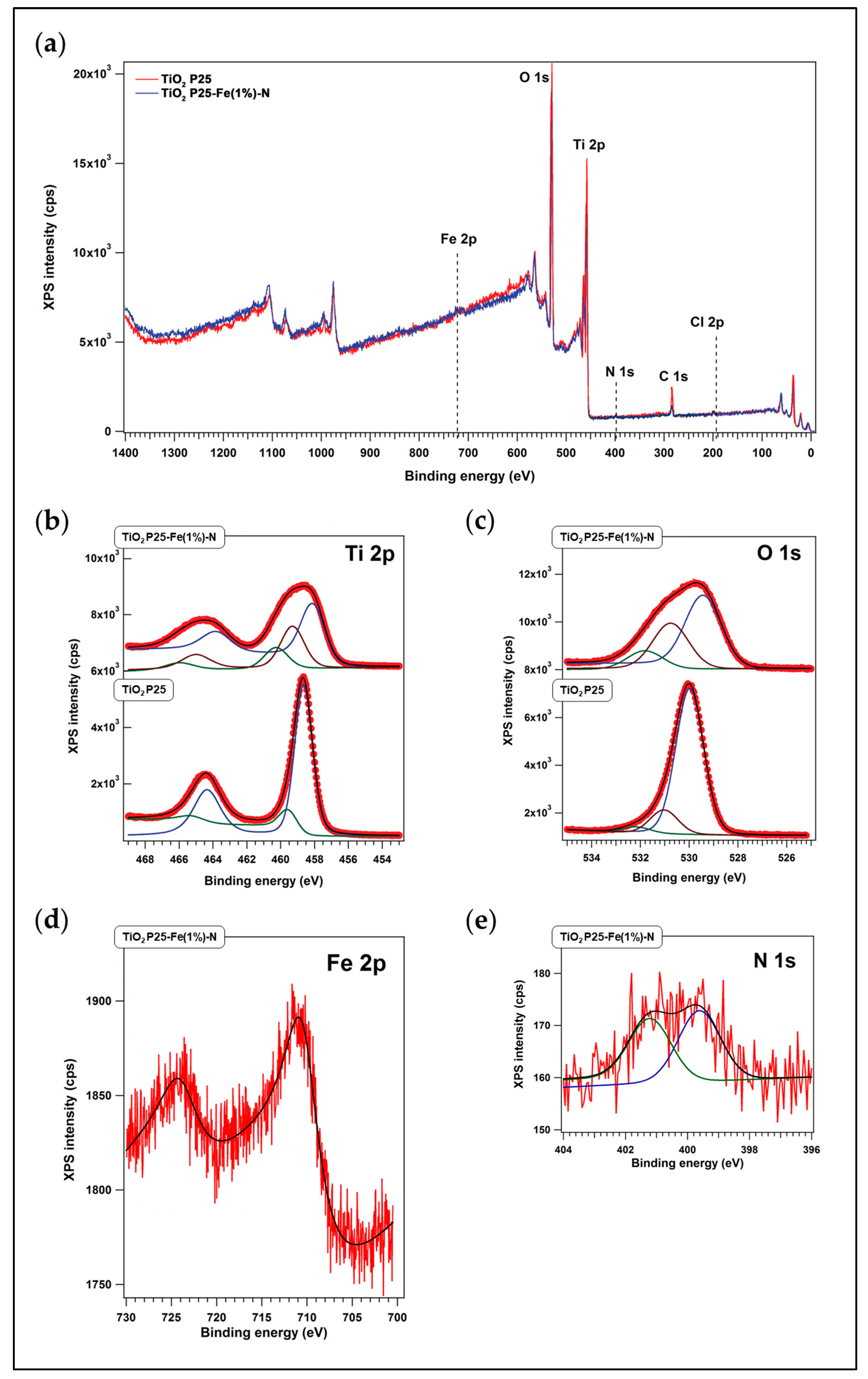
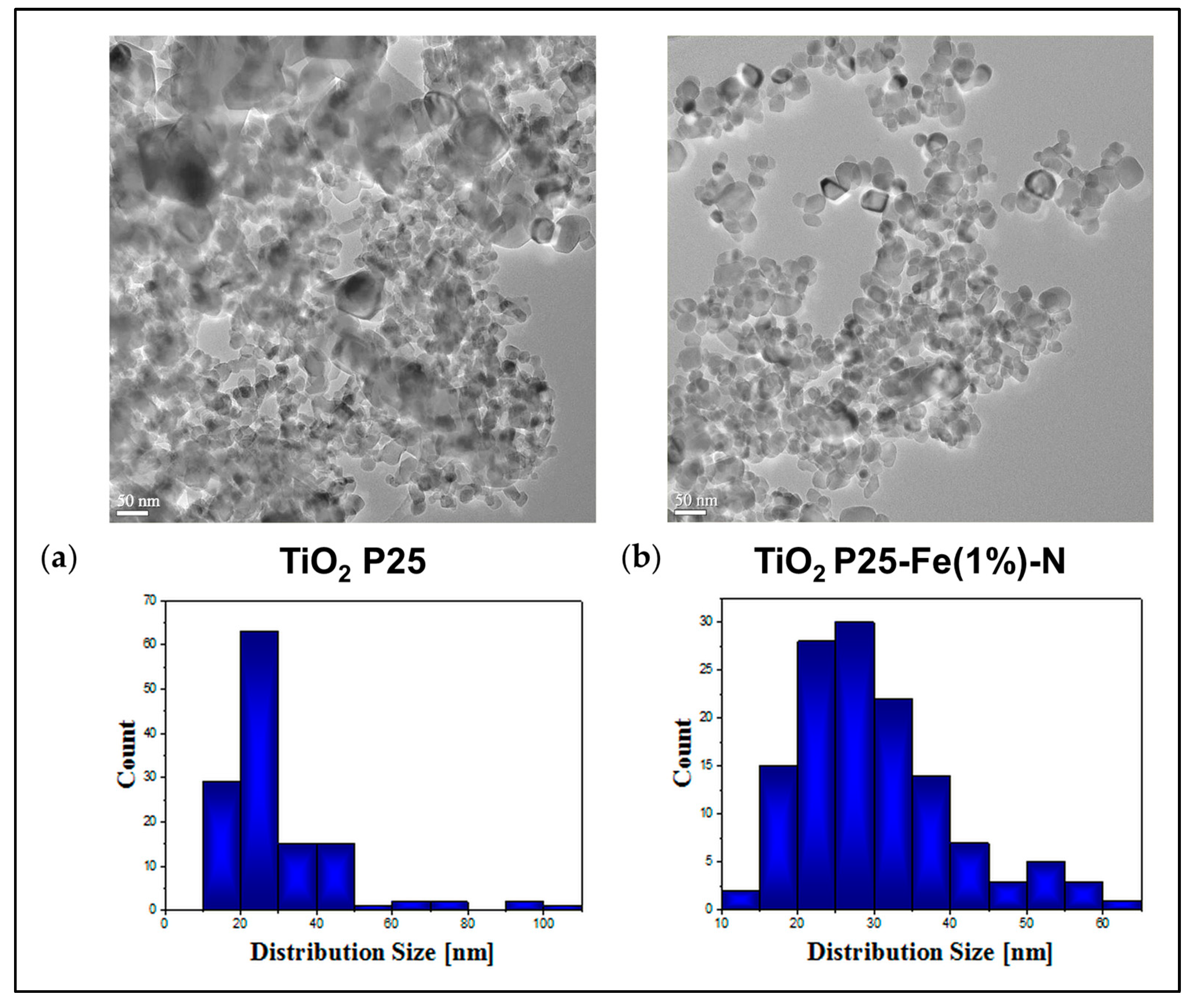
2.1. Physicochemical Characteristics of TiO2 NPs

The two types of TiO2 NPs that were used in the present work were (i) commercially available P25 NPs and (ii) the same NPs co-doped with Fe and N atoms by direct impregnation in an aqueous solution of 1% FeCl3 and in the presence of urea (P25-Fe(1%)-N) (see Section 4.1.). The concentration of FeCl3 that we chose was mainly based on a previous paper from our group where P25 NPs impregnated by dispersion in 1% FeCl3 had an enhanced photocatalytic effect in both long-wave UV (368 nm) and visible light (610 nm) [70]. Moreover, Kalantari et al. showed that co-doping with Fe and N considerably increased the TiO2 NPs’ photocatalytic activity compared to mono-doped TiO2 NPs [71].
There is evidence that a higher amount of Fe atoms on TiO2 NPs’ surface would improve the photocatalytic effect of NPs [72,73,74]. However, it was already shown that the phototoxicity of TiO2 NPs could be proportionally increased by 1% to 10% Fe-doping due to the generation of oxidizing agents via the Fenton reaction [74]. Therefore, we considered a low amount of dopant would be more appropriate for investigating the potentially toxic effects of pulmonary exposure to Fe-N doped TiO2 NPs.
The chemical content of P25 NPs and P25-Fe(1%)-N NPs was revealed by X-ray photoelectron spectroscopy (XPS) measurements (Figure 1a) and the corresponding binding energies (Table 1). The results proved the presence of Ti and O atoms in both types of NPs (Figure 1b,c). Also, Fe and N atoms were identified in the P25-Fe(1%)-N sample (Figure 1d,e). Moreover, P25 NPs were made of stoichiometrically structured TiO2, as revealed by the ratio of 2.02 between the main signals of Ti 2p3/2 (458.65 eV) and O 1s (529.98 eV). The O 1s peaks near 532 eV might be assigned to hydroxyl groups or adsorbed water molecules on the surface, and the 530–531 eV peaks to Ti-O chemical bonds, respectively. The signal at 710.40 eV is characteristic of Fe 2p3/2, revealing that P25-Fe(1%)-N NPs contained Fe3+. The peak at 399.62 eV might be assigned to oxidized nitrogen, i.e., O-Ti-N bindings. Also, the N 1s peak at 401.19 eV usually reflects interstitial nitrogen. The ratios between intensities of the XPS peaks (Fe/TiO and N/TiO) showed that P25 NPs prepared in FeCl3 had 2.1% Fe atoms and 0.5% N atoms on their surface.
As revealed in the images obtained by transmission electron microscopy (TEM), both `types of NPs generally had near polyhedral shapes with round corners (Figure 2a,b); some spheres could also be observed. The dimension of most P25 NPs was between ∼10–50 nm and had a mean particle size of 29 nm (Figure 2a). The size range of P25-Fe(1%)-N NPs was larger, with most being ∼15–60 nm. However, the mean particle size of P25-Fe(1%)-N NPs was similar to that of P25 NPs, i.e., ∼28 nm (Figure 2b). More analyses regarding the characterization of TiO2 NPs were provided in our previously published papers [70,75].

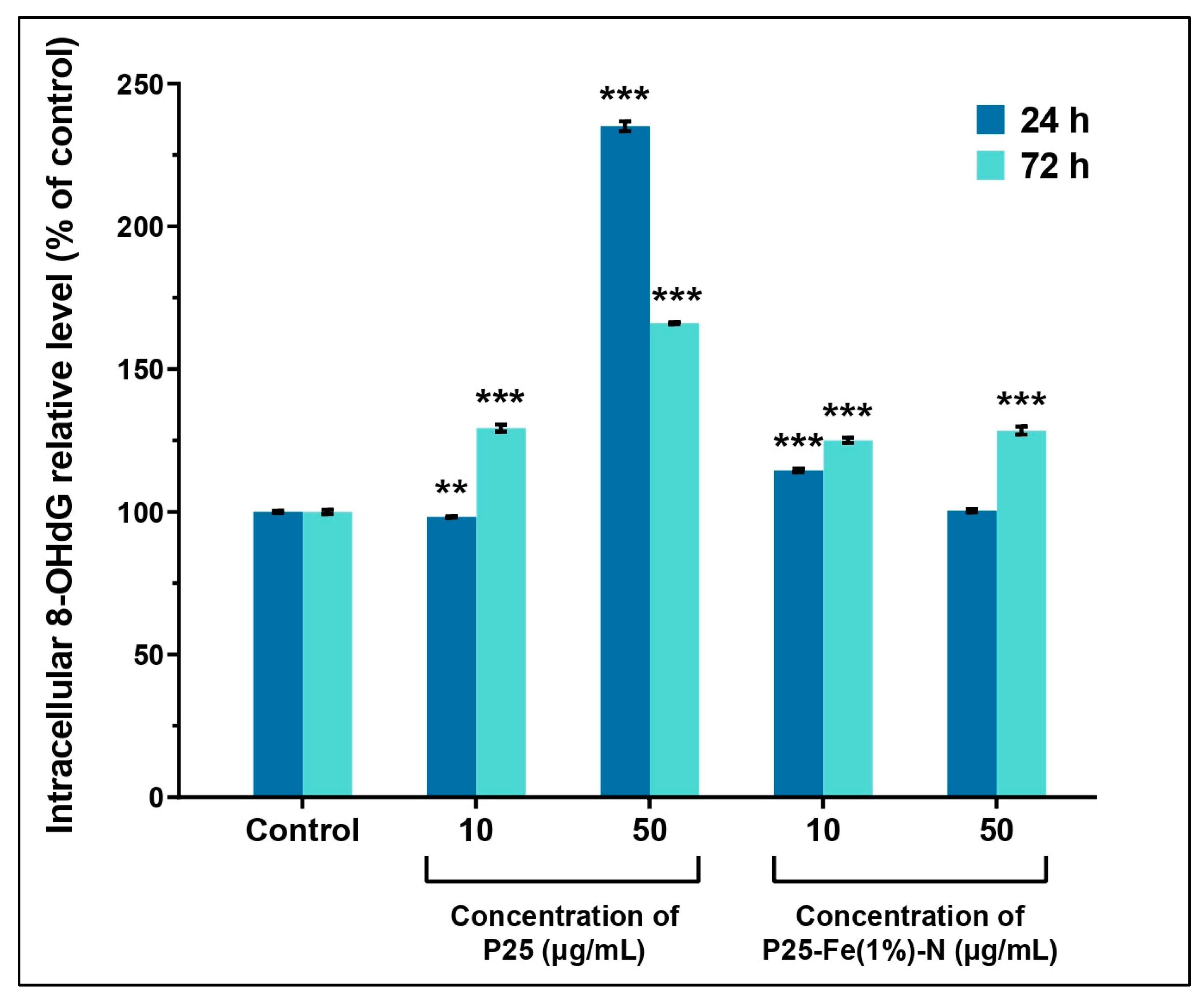
2.2. Oxidative DNA Damage Induced by TiO2 NPs in MRC-5 Cells

The concentrations of NPs, i.e., 10 µg/mL and 50 µg/mL, respectively, used by us were chosen based on our previous work [76] in which we proved that P25 NPs could cause a significant increase of oxidative stress in MRC-5 cells in a time- and dose-dependent manner while P25-Fe(1%)-N NPs had no influence on ROS level compared to the control group of cells.
ROS can damage the cell considerably by impairing the constitutive molecules of cellular structures. One of the damages induced by a high level of ROS is the oxidation of guanosine, a modification that might affect the integrity of DNA molecules. We investigated the impact of TiO2 NPs on the DNA molecules of MRC-5 cells by measuring the level of 8-hydroxydeoxyguanosine (8-OHdG), a commonly used marker for DNA oxidative lesions. Our results showed that exposure to P25-Fe(1%)-N NPs could increase the level of 8-OHdG in a time-dependent manner (Figure 3) in MRC-5 cells. The levels of 8-OHdG induced by both doses of P25-Fe(1%)-N NPs and the dose of 10 µg/mL non-doped P25 NPs were generally similar and have not exceeded 130% compared to control after 72 h of exposure. However, the higher dose of P25 NPs caused an increase of the level of 8-OHdG up to 235% compared to the control after MRC-5 cells were exposed for 24 h. Also, 8-OHdG concentration was lowered in the cells as time passed, reaching 166% compared to the control at 72 h. Interestingly, the reduction of 8-OHdG recorded at 72 h at 50 µg/mL P25 NPs contrasts with the high level of ROS measured previously by us in MRC-5 cells [76].

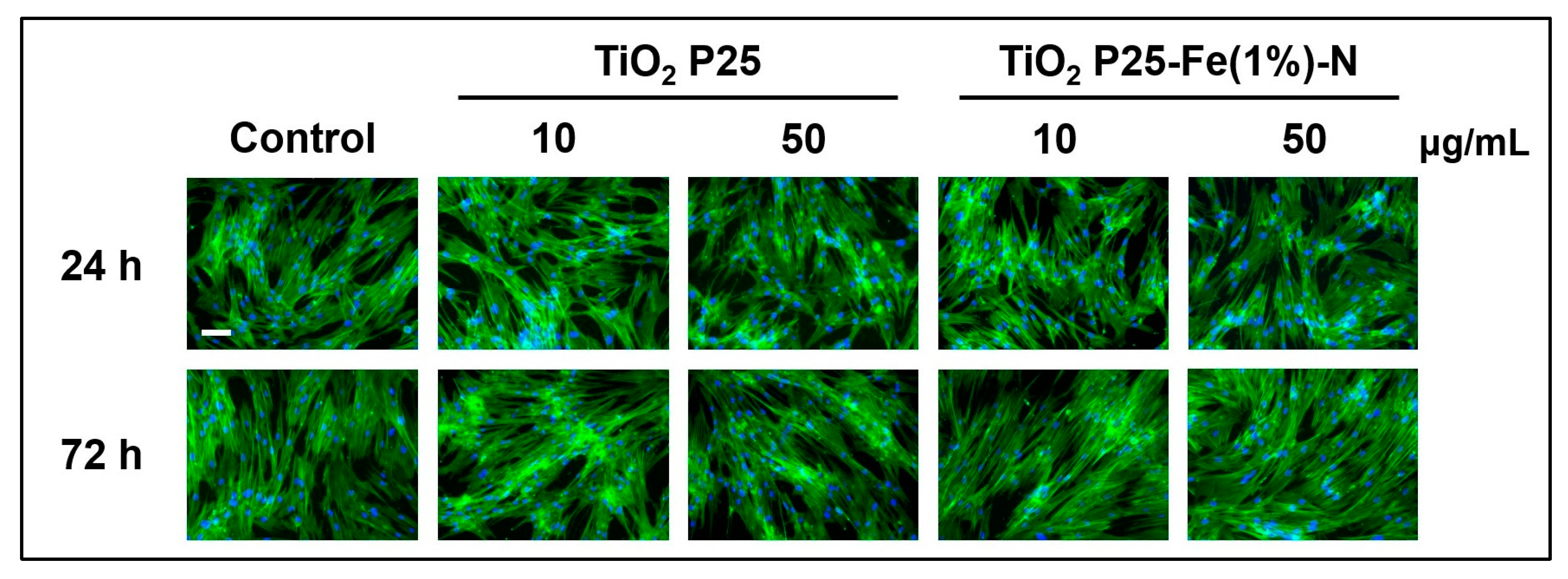
2.3. Influence of TiO2 NPs on the Morphology of MRC-5 Cells

Actin cytoskeleton plays a key role in the mechanical support of cells, also defining their morphology. Fluorescent microscopy images displayed in Figure 4 showed that TiO2 NPs had no negative impact on the MRC-5 cells’ actin cytoskeleton organization. The microscopic images suggested that MRC-5 cells maintained their fibroblast-like morphology regardless of the conditions applied in our study (type of TiO2 NPs, concentration of NPs, exposure time). Normally, these lung fibroblasts are elongated spindle-shaped bipolar cells. No disrupted filaments or cytoskeleton rearrangements were observed, while bundles of F-actin appeared very dense, indicating a high cellular density.

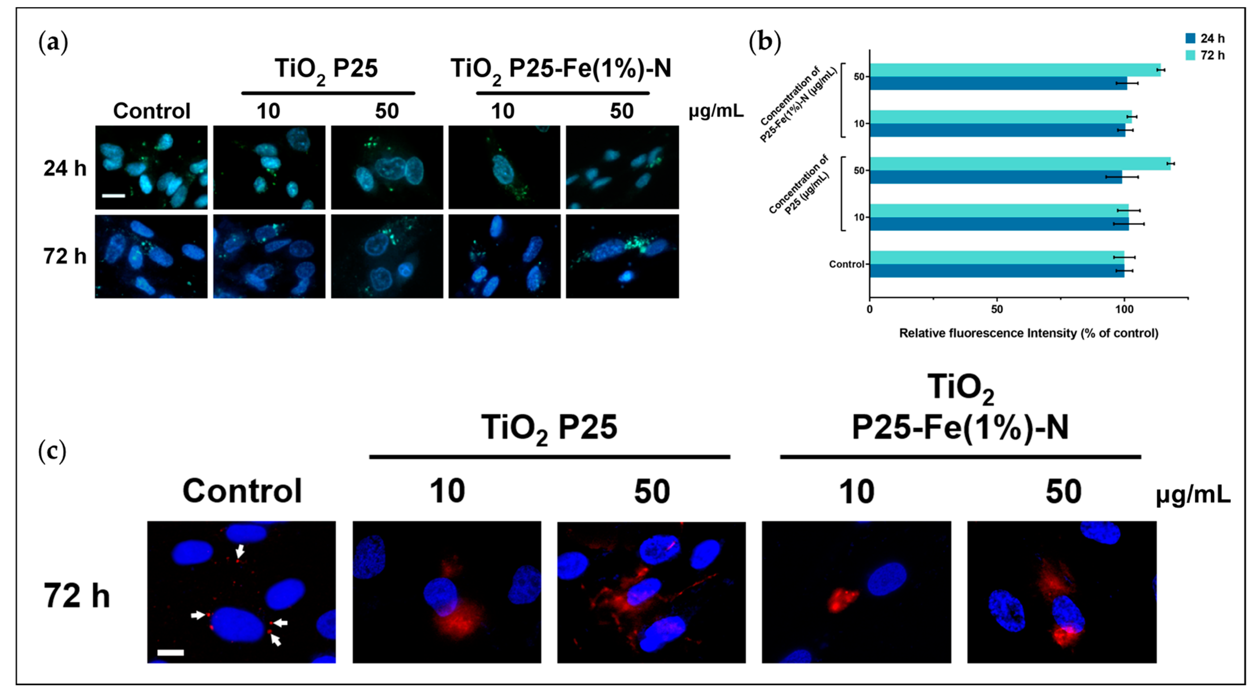
2.4. Influence of TiO2 NPs on Lysosomes’ Formation and Lysosomal Membrane Integrity in MRC-5 Cells

There are no statistically significant differences regarding the accumulation of lysosomes inside MRC-5 cells exposed to P25 and P25-Fe(1%)-N NPs (Figure 5a,b). However, we noted that the lysosome quantity increased by ∼14–18% compared to the control when the doses of 50 μg/mL at 72 h were applied. The distribution of cathepsin B suggested that the membrane of lysosomes was affected by the 72 h exposure to TiO2 NPs. Cathepsin B is a key proteolytic enzyme localized in lysosomes under physiological conditions. Therefore, when labeled with Alexa Fluor 594, cathepsin B is present in fluorescent red vesicles in healthy cells, as can be observed in our control cells (Figure 5c).
When MRC-5 cells were treated with P25 and P25-Fe(1%)-N NPs, the red signal appeared in a diffused pattern, indicating that the lysosomal membrane was permeabilized and cathepsin B was released into the cytosol. Even though permeabilization occurred in all treated pulmonary fibroblasts, images showed that the red signal is more clustered in cells exposed at P25-Fe(1%)-N NPs, suggesting their effect on lysosome integrity is less pronounced than the one of P25 NPs.

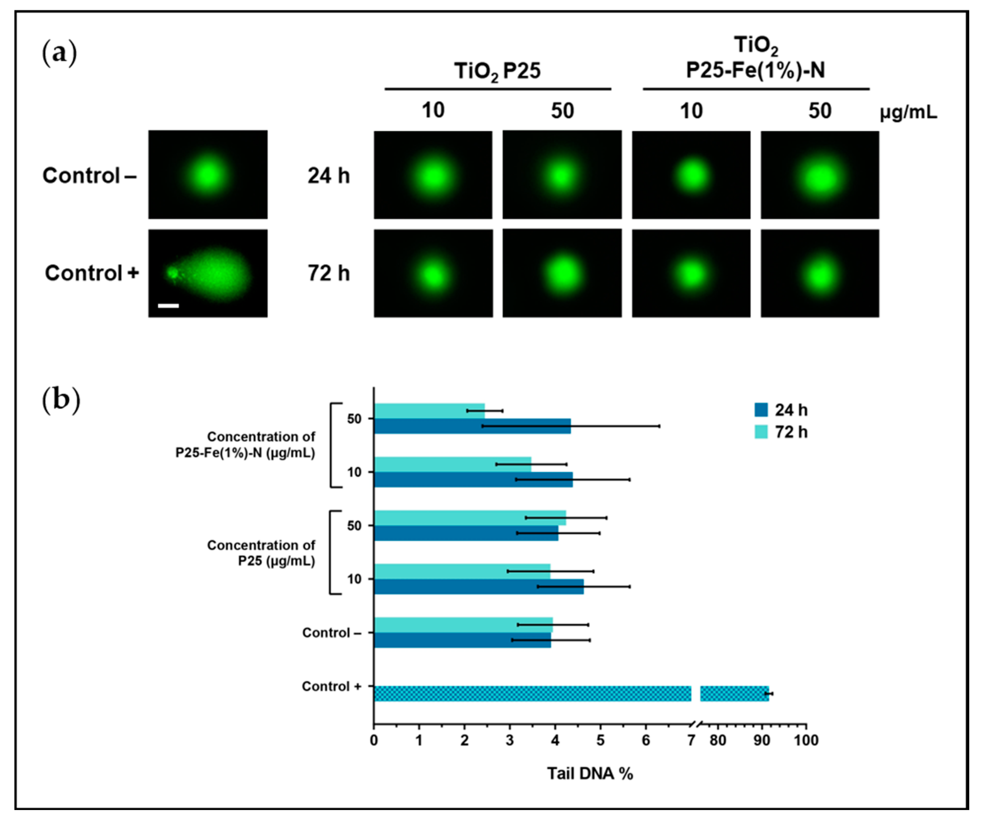
2.5. Effect of TiO2 NPs on the Integrity of DNA from MRC-5 Cells

Considering the generation of ROS and oxidative lesions induced by TiO2 NPs in MRC-5 cells, we further decided to investigate whether they critically affect the integrity of DNA molecules. Fragmentation of DNA was investigated by Comet assay that indicated no significant changes between the different conditions tested, although raised levels of DNA oxidation might be considered a marker of double-strand breaks. It can be visually observed that no small fragments of DNA detached and migrated faster, as in the case of the positive control (Figure 6a). The damage of DNA molecules was expressed in percentages of DNA in the comet tail. Based on the quantified fluorescence (Figure 6b), the tail DNA% in samples varied in the 2.4–4.6% range, while in the negative control cells, it has not exceeded 4%. These results could strengthen the evidence that a molecular mechanism ameliorates the TiO2 NPs-dependent oxidative damage of DNA within MRC-5 fibroblasts.

2.6. Cell Death Signaling in MRC-5 Cells Exposed to TiO2 NPs

To investigate whether the oxidative lesions produced by TiO2 NPs generated damages that trigger cell death signaling, protein expression of cathepsin B, p53, caspase-8, -9, and -3 were quantified using Western Blot analyses. Cathepsin B presented relatively constant levels in MRC-5 cells exposed to P25 and P25-Fe(1%)-N NPs regardless of the exposure time or dose applied (Figure 7a,b).
On the contrary, TiO2 NPs significantly changed the expression of p53 protein in MRC-5 cells in a time- and dose-dependent manner. After 24 h of exposure to 10 μg/mL of P25 and P25-Fe(1%)-N NPs, the level of p53 decreased by 3%, respectively, 12% relative to the control. The expression of p53 started to drop considerably when the highest dose of TiO2 NPs was applied. At 24 h of exposure, P25 NPs led to the diminution of p53 expression by 64%. By comparison, the effect of iron-doped TiO2 NPs was slightly milder, leading to a decreased expression by nearly 38% relative to the control. However, the results indicated that the inhibitory effect of TiO2 NPs on p53 expression was more evident as time went on. Thus, p53 expression in NP-treated MRC-5 cells exhibited a massive reduction regardless of the dose or exposure time. As can be seen in Figure 7c,d, the level of expression of p53 dropped below 10% relative to the control and was totally suppressed by the treatment with 10 μg/mL and 50 μg/mL of P25 NPs, respectively.
Both p53 and cathepsin B are involved in the initiation of programmed cell death pathways [77,78]. However, in contrast with our results, p53 normally undergoes overexpression during apoptosis [79]. In the present study, we proved that neither initiator caspases-8 and -9 nor the effector caspase-3 were activated by the TiO2 NPs applied to pulmonary fibroblasts. Based on the molecular mass, protein bands displayed on the obtained blot profiles corresponded to the uncleaved, i.e., non-activated procaspases (Figure 7e). The measured level of the apoptosis-inducing markers, i.e., cathepsin B, p53, caspase-8, -9, and -3, correlated well with the high DNA integrity revealed by the Comet assay.

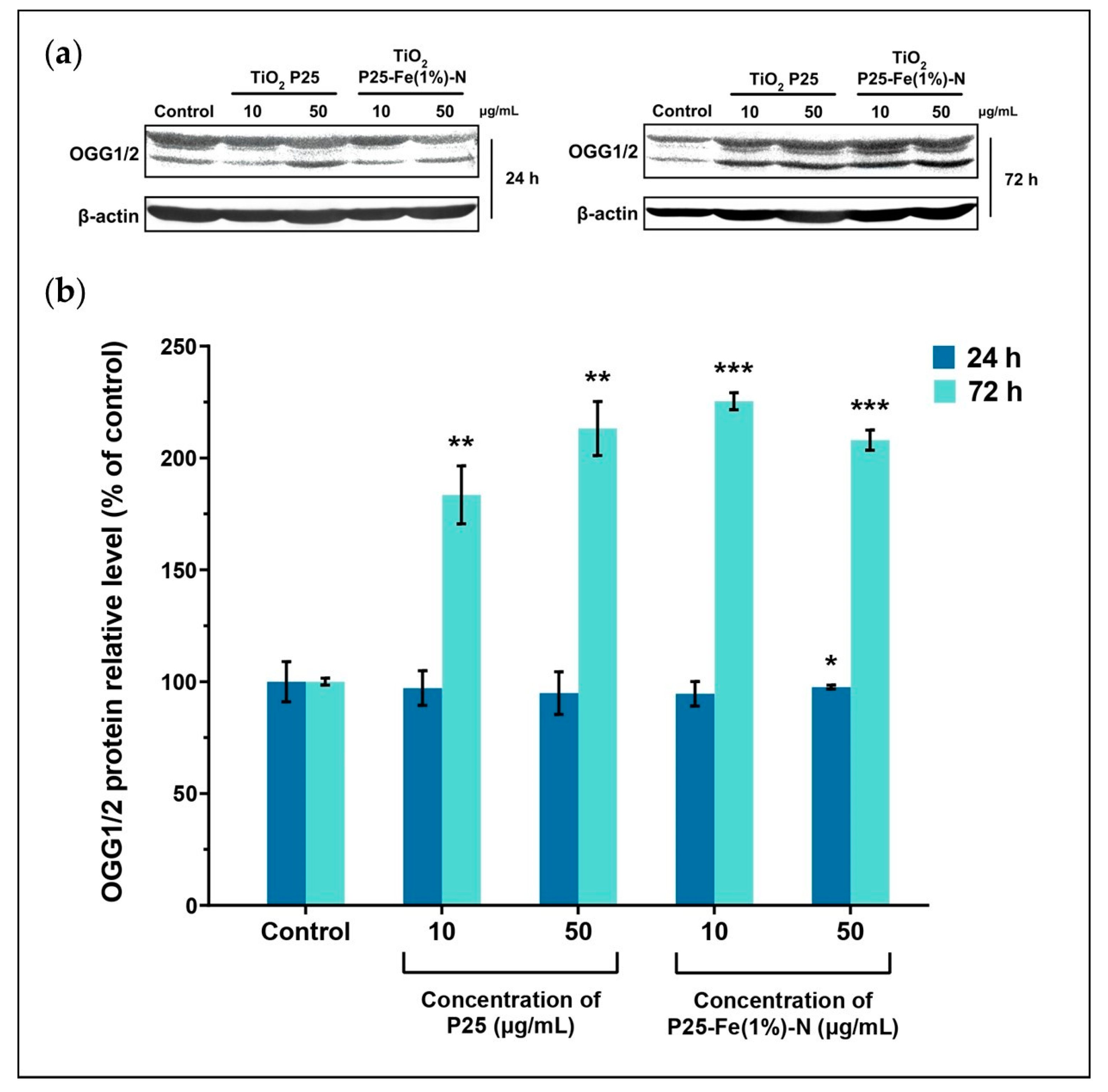
2.7. The Reparatory Role of 8-oxoguanine DNA Glycosylase in MRC-5 Cells Exposed to TiO2 NPs

As the results suggested so far, a molecular reparatory mechanism might be the reason for the low genotoxic effect of TiO2 NPs on MRC-5 cells. Therefore, we decided to investigate a key enzyme involved in the base excision repair, which can recognize oxidized guanine within the DNA, namely OGG1/2. The protein expression of OGG1/2 was assessed by Western Blot analysis, with the representative blot profiles displayed in Figure 8a. Interestingly, our results showed that the level of OGG1/2 slightly decreased when MRC-5 cells were exposed for 24 h at both doses of P25 or P25-Fe(1%)-N NPs. The expression exhibited an insignificant diminution of at most 6% relative to the control. The effect of TiO2 NPs on the level of OGG1/2 became evident at 72 h of exposure when the reparatory protein exhibited a substantial overexpression (Figure 8b). In general, P25 and P25-Fe(1%)-N NPs caused the doubling of OGG1/2 expression level, confirming that the innate base excision repair mechanism coped with the oxidative damage induced by TiO2 NPs and thus maintained the integrity of DNA molecules. The level of OGG1/2 was slightly higher in the pulmonary fibroblasts treated with P25-Fe(1%)-N NPs in comparison with the one measured in cells exposed to undoped TiO2 NPs. Differences in OGG1/2 expression were comprised between 83.5 and 125% relative to the control.

3. Discussion

Considering iron doping might intensify the use of TiO2 NPs in consumer goods, we chose to investigate the possible associated toxicological risks due to the tuning of their photocatalytic properties toward visible illuminance. Therefore, we compared the toxicity of P25 TiO2 NPs doped with iron and nitrogen with the same undoped NPs. The doses used by us were based on our previously published work [76] as well as on representative papers [80,81,82,83].
Moreover, inhalation is a major route by which TiO2 NPs enter the human body; therefore, we have chosen MRC-5 cells, which are human pulmonary fibroblasts, as the experimental model.
Previous studies stated that TiO2 NPs could also interact with the microtubules and other components of the cellular cytoskeleton [84,85]. We decided to investigate the influence of P25 and P25-Fe(1%)-N NPs on the actin cytoskeleton as they could provide valuable information regarding the morphology of treated cells. Even though some reports show that TiO2 NPs can disrupt actin filaments [86,87], we did not observe any significant changes in the organization of the cytoskeleton between exposed samples and control cells. In addition, considering the role of the actin cytoskeleton in internalization mechanisms [88], endocytosis of the P25 and P25-Fe(1%)-N NPs with sizes about 50 nm might be produced to a lesser extent in the MRC-5 cells. This fact is supported by the dimension of the large aggregates of TiO2 NPs formed [75], which would not be able to enter the cells through caveolae (with a diameter between 50nm and 80 nm) or clathrin-mediated endocytosis (with a diameter of ≈120 nm) [89]. However, Thurn et al. [90] stated that the uptake of aggregates could be possible through macropinosomes with a dimension of 500–2000 nm.
In our previous work [76], we already showed the significant difference between the ability of P25 and P25-Fe(1%)-N NPs to produce ROS. While P25 NPs could induce high levels of ROS in a time and dose-dependent manner, doping with iron ions totally suppressed the generation of oxidative stress [76]. Similarly, iron doping inhibited the production of TiO2-induced ROS in HaCaT keratinocytes [64]. In contrast, doping TiO2 NPs with copper led to higher ROS production in A549 cells [69], and doping them with zinc enhanced the oxidative stress induced in MCF-7 cells [68].
Moreover, our previous paper [76] investigated the effect of TiO2 NPs on the enzymatic antioxidant mechanism of MRC-5 cells. When the ROS level exceeded the neutralizing ability of antioxidant enzymes, some of the free radicals began to impair intracellular biomolecules. A part of the oxygen-derived free radicals produced lipid peroxidation that attacks organelles’ membranes, while others damage DNA after entering the nucleus as well as proteins [91].
Some of the most commonly studied biomarkers indicating oxidative damage on DNA molecules, are 8-hydroxylated guanine species, mainly 8-oxoguanine (8-oxoG) and its isomer, 8-OHdG. In our study, P25 NPs increased the level of 8-OHdG in MRC-5 cells in a time-dependent manner, the results being in accordance with the level of ROS produced. Interestingly, we observed an attenuated but significant increase of 8-OHdG level in pulmonary fibroblasts treated with P25-Fe(1%)-N NPs. This might be explained by the fact that iron-doped TiO2 NPs could generate some reactive species in the first hours of exposure that had probably produced their effects before initiating the antioxidant mechanisms [76]. We considered this might represent preliminary evidence that human pulmonary fibroblasts are able to counteract excessive oxidation caused by TiO2 NPs.
Some studies showed that the increased level of guanine oxidation products within cells might be linked in certain circumstances with DNA fragmentation [92,93]. On the contrary, our results indicated that the integrity of DNA molecules from MRC-5 cells was not affected by the increased level of 8-OHdG caused by exposure to TiO2 NPs. Similarly, Hackenberg et al. [94] showed that TiO2 NPs did not induce DNA fragmentation in lymphocytes obtained from the peripheral blood of human donors. Bhattacharya et al. [95] obtained the same result when they applied TiO2 NPs on both BEAS-2B (normal human bronchial epithelial cells) and IMR-90 (normal human pulmonary fibroblasts) cell cultures, showing that IMR-90 cells exhibited high levels of 8-OHdG after 24 h of exposure to TiO2 NPs. Contrariwise, the potential of TiO2 NPs to induce DNA double-stranded breaks was demonstrated in HUVEC cells. The genotoxic effect of TiO2 NPs was more pronounced as their particle size diminished, producing more DNA damage [96].
Besides damages caused by TiO2 NPs-induced oxidative stress on DNA molecules, we investigated the influence of this on the membrane of lysosomes from MRC-5 cells. So far, different studies have demonstrated that NPs could induce the permeabilization of lysosomal membranes. For example, Li et al. [97] found that the membrane of lysosomes from MRC-5 cells could be affected by Au NPs. In addition, membrane permeabilization was induced in THP-1 cells by ZnO NPs [98], in HepG2 NPs by Ag NPs [99], or in 3T3 cells by Si NPs [100]. The previously mentioned studies that investigated Au and Si NPs associated the damages caused on lysosomal membranes with an increase in the generation of ROS.
One of the roles of lysosomes is to enzymatically digest spent cellular organelles. As high levels of ROS in MRC-5 cells treated with TiO2 NPs might damage different intracellular structures, an increase in the number of lysosomes was expected. Our results suggested that the number of lysosomes was not significantly influenced by P25 and P25-Fe(1%)-N NPs exposure. However, the lysosomal membrane was significantly impaired at 72 h of exposure. The damaged membrane of lysosomes probably allowed the release of lysosomal content, especially cathepsins, enzymes that can be involved in activating caspase-dependent cell death pathways [101]. Neither the expression of the p53 protein nor those of caspase-3, -8, and -9 indicated that apoptosis was activated in MRC-5 cells by TiO2 NPs, although we observed that cathepsin B diffuses from lysosomes into the cytosol. The insignificant differences between the expression of cathepsin B validated that the diffuse red signal obtained through immunofluorescence resulted only from the lysosomal membrane permeabilization. As cathepsin B is a lysosome-resident protein, the result confirmed that TiO2 NPs did not significantly influence lysosomal formation in MRC-5 cells.
Besides the innate antioxidant defense system that acts directly on generated ROS, eukaryotic cells can cope with oxidative damage of DNA due to different reparatory mechanisms, including the base excision repair (BER) mechanism. OGG1/2 has a crucial role in the removal of oxidized guanine species, being the enzyme responsible for their recognition, hence the initiation of the BER process [102]. We found that MRC-5 cells overexpressed OGG1/2 when exposed to both P25 and P25-Fe(1%)-N TiO2 NPs for 72 h, suggesting the BER mechanism was induced. The constant level of OGG1/2 noticed after 24 h of exposure might be explained by a delay between transcription and translation processes. The first result that suggested a reparatory mechanism had been activated was the decrease of 8-OHdG level in the case of 50 μg/mL P25 NPs exposure at 72 h and further the unaffected DNA integrity revealed by Comet assay.
Du et al. [103] revealed that OGG1 is overexpressed in a dose-dependent manner in human hepatocytes L02 by a combined treatment of TiO2 NPs and lead, whereas Zijno et al. [104] showed that OGG1 level increased in human colon Caco-2 cells following treatment with TiO2 NPs. Also, Xia et al. [105] found that human kidney HEK293T cells express OGG1 in response to the oxidative damage caused by TiO2 NPs that act synergistically with CdCl2.
In contrast with our results, control of BER activity is managed by p53 through its ability to regulate the cell cycle [106]. We found that the expression of p53 was totally inhibited. Therefore, the point mutations caused by 8-OHdG in the sequence of DNA [43] might have been transmitted during cell division prior to the activation of the reparatory mechanism. However, the BER pathway can function in a p53-independent manner, as other proteins might arrest the cell cycle [107].

4. Materials and Methods

4.1. Physicochemical Characterization of TiO2 NPs

Two types of TiO2 NPs were used in this study: (i) Degussa P25 (Aeroxide® P25) purchased from Sigma Aldrich (St. Louis, MO, USA) and (ii) Degussa P25 co-doped with Fe and N atoms that were obtained experimentally by direct impregnation in 1% FeCl3 6H2O and urea. The method of impregnation of TiO2 NPs with Fe and N, as well as the characteristics of the two types of TiO2 NPs, were described in detail in the previous publications of our research group [70,75]. Briefly, powders of P25 and P25-Fe(1%)-N NPs consisted of approx. 83% anatase (with a crystallite size of around 30 nm) and approximately 17% rutile (with a crystallite size of around 50 nm) [70]. Moreover, our group showed that these types of TiO2 NPs formed large aggregates when they were suspended in MEM supplemented with 10% FBS. Zeta potential values around –10 mV also confirmed the low stability of TiO2 NPs [75].
XPS measurements provided in this work were obtained in an analysis chamber using a monochromatized Al Kα1 X-ray source (1486.74 eV). The electrons were analyzed with a 150 mm hemispherical electron energy analyzer (Phoibos, Specs Gmbh, Berlin, Germany). TEM images and measurements were performed on a JEOL 200 CX transmission electron microscope (accelerating voltage: 200 kV).

4.2. Cell Culture and Treatment with TiO2 NPs

MRC-5 human lung fibroblasts purchased from American Type Culture Collection (ATCC, catalog no. CCL-171) were cultured in vitro in Eagles minimum essential medium (MEM; Gibco/Invitrogen, Carlsbad, CA, USA) at 37 °C and in a humified atmosphere with 5% CO2. MEM containing 2 mM L-glutamine, 0.1 mM sodium pyruvate, and 4.5 g/L glucose was supplemented with 10% fetal bovine serum (FBS; Gibco/Invitrogen, Carlsbad, CA, USA). Replacement of the growth medium with a fresh one was done every two days. Sub-cultivations were performed when cells reached ~80% confluence. For sub-cultivation, MRC-5 cells were detached using a solution of 0.25% (w/v) Trypsin with 0.53 mM EDTA (Sigma Aldrich, St. Louis, MO, USA) and split into other culture flasks.
In this experiment, MRC-5 human lung fibroblasts were exposed to 10 and 50 μg/mL TiO2 NPs for 24 and 72 h. Stock suspensions of 2 mg/mL TiO2 NPs (P25 and P25-Fe(1%)-N) were prepared by adding 10 mg of each NP’s type in 5 mL of phosphate-buffered saline (PBS), pH ≈ 7.4. For improving particles’ dispersion, suspensions were sonicated for 5 min at room temperature using the ultrasonic processor UP50H (Hielscher Ultrasonics GmbH, Teltow, Germany). Then, stock suspensions were exposed for 30 min to UV light to be sterile when used. MRC-5 cells were detached as described above and seeded into 75 cm2 culture flasks. P25 and P25-Fe(1%)-N NPs were added directly into the culture medium at the abovementioned final concentrations. Cells used as the control for each assay underwent the same procedures but were grown in an NP-free culture medium.

4.3. Measurement of 8-Hydroxy-2′-Deoxyguanosine Level

The level of 8-OHdG was measured using a commercially available enzyme-linked immunosorbent assay (ELISA) kit purchased from Abcam (ab201734; Cambridge, UK). Previously, DNA from MRC-5 cells exposed to TiO2 NPs was isolated and quantified. Afterward, the DNA was digested with P1 nuclease and treated with alkaline phosphatase; thus, nucleotides were transformed into nucleosides. Further, DNA samples were processed using the 8-OHdG ELISA kit according to the manufacturer’s instructions, and finally, their absorbance was measured at 450 nm using a microplate reader (TECAN GENios, Grödig, Austria).

4.4. Fluorescence Microscopy Analysis

Fluorescent staining was used to analyze the actin cytoskeleton morphology and dynamic, lysosomes’ number and density, as well as cathepsin B localization. To observe actin filaments, MRC-5 cells cultured in flasks and exposed to TiO2 NPs were fixed with 4% paraformaldehyde for 20 min at room temperature. Then, cell membranes were permeabilized with a mixture of 0.1% Triton X-100 and 2% bovine serum albumin (BSA) for 30 min. F-actin was labeled by incubating the cells for 1 h with 20 μg/mL phalloidin-fluorescein isothiocyanate (FITC; Sigma Aldrich, St. Louis, MO, USA). The staining of cell nuclei has been done by 4′,6-diamidino-2-fenilindol (DAPI; Molecular Probes, Life Technologies, Carlsbad, CA, USA). Images of the actin cytoskeleton were acquired using the inverted fluorescence microscope Olympus IX71 (Tokyo, Japan).
The fluorescent staining of lysosomes was performed by incubating MRC-5 cells with 100 nM LysoTracker Green DND-26 (Molecular Probes, Invitrogen) for 30 min at 37 °C in a humidified atmosphere containing 5% CO2. Hoechst 33342 (Molecular Probes, Invitrogen) was used to counterstain cell nuclei. Images of stained lysosomes were taken with Olympus IX71 inverted fluorescence microscope (Tokyo, Japan). Green fluorescence intensity in different fields of view per each sample was quantified using the ImageJ 1.53u software available online at https://imagej.nih.gov/ij/ (National Institute of Health, Bethesda, MD, USA) and displayed as a mean relative to the control.
Immunofluorescent localization of cathepsin B was performed by seeding MRC-5 cells on coverslips at a density of 2 × 10⁴ cells/cm2. After fibroblasts were allowed to adhere overnight, they were exposed to TiO2 NPs, as described in Section 4.2. Further, MRC-5 cells underwent fixation and permeabilization as described above in the case of F-actin. Cathepsin B was labeled by incubating cell plates (overnight, 4 °C) with Alexa Fluor 594-coupled anti-cathepsin B antibody (Santa Cruz Biotechnology Inc., Dallas, TX, USA). The staining of cell nuclei has been done by DAPI. Labeled cathepsin B was visualized at 60x objective of the fluorescence microscope Nikon Eclipse E200 (Tokyo, Japan).

4.5. Comet Assay

Comet assay was performed using a single-cell electrophoresis kit (Cell Biolabs, INC, San Diego, CA, USA). After exposure to the two types of TiO2 NPs, MRC-5 cells were collected, resuspended in PBS, and diluted until the density of 1 × 10⁵ cells/mL was reached. A volume of 10 μL of each cellular suspension was mixed with 100 μL low melting agarose maintained at 37 °C. Further, a volume of 75 μL from this mixture was stretched uniformly in thin films on a Comet glass slide. The agarose was allowed to jellify by incubating the slides on a horizontal surface in the dark at 4 °C for 15 min. Then, cells embedded in agarose were lysed (using the lysis solution within the kit at 4 °C, 60 min) and further treated with an alkaline solution (4 °C, 30 min). Afterward, the slides were washed with deionized water and subjected for 20 min to low voltage horizontal electrophoresis migration (20V). Subsequently, the slides were washed with 70% ethanol. Finally, DNA molecules from the agarose-embedded cells were stained with the Vista Green fluorescent dye. The negative control was represented by MRC-5 cells cultivated in an NP-free growth medium. The positive control underwent the same procedure, but NP-free cultured cells embedded in agarose were exposed at 70 μM H2O2 (5 min, 4 °C). Images of the comets were acquired using the fluorescence microscope Olympus IX 71 (Tokyo, Japan). Fluorescence from representative images was quantified using the OpenComet plugin within the ImageJ 1.53u software (National Institute of Health, Bethesda, MD, USA) and displayed as a percentage of tail DNA expressed relative to the negative control.

4.6. Western Blot Analysis

Western Blot technique was used to determine the expression level of p53, cathepsin B, caspase-3, -8, -9, and OGG1/2 proteins. In advance, total protein extracts of samples were prepared, and their concentration was measured by the Bradford method. Harvested MRC-5 fibroblasts suspended in PBS were subjected to 3 cycles of 30 s ice-assisted sonication using the ultrasonic processor UP50H (Hielscher, Teltow, Germany) to disrupt the cell membranes. Obtained lysates were centrifuged at 3000× g, at 4 °C for 10 min, and then each supernatant containing the total protein extract was individually collected and stored at −80 °C until further use.
Cell lysates containing an equal amount of total protein were subjected to sodium dodecyl sulfate-polyacrylamide gel electrophoresis (SDS-PAGE) (90V, 120 min) and then transferred to a polyvinylidene fluoride membrane (PVDF; Millipore, Billerica, MA, USA) at 350 mA for 95 min within a wet transfer unit (Bio-Rad Laboratories, Hercules, CA, USA). For the detection of proteins, PVDF membranes were processed using the WesternBreeze Chromogenic Kit (Invitrogen, Grand Island, NY, USA). A blocking buffer was applied for 30 min, and then the membranes were incubated overnight with the following primary monoclonal antibodies purchased from Santa Cruz Biotechnology Inc. (Dallas, TX, USA): anti-p53, anti-cathepsin B (sc-365558), anti-β-actin (sc-517582), anti-caspase-8 (sc-5263), anti-caspase-9 (sc-56076), anti-caspase-3 (sc-7148), anti-oxoguanine glycosylase 1/2 (OGG1/2; sc-376935). Excess of the primary antibody was released, and membranes were incubated with an alkaline phosphatase-coupled anti-mouse secondary antibody. Afterward, protein bands were revealed using 5-bromo-4-chloro-3-indolyl phosphate/nitroblue tetrazolium (BCIP/NBT). Blot images were acquired with the ChemiDoc XRS+ system (Bio-Rad Laboratories, Hercules, CA, USA) and processed using Image Lab (version 6.1., Bio-Rad Laboratories, Hercules, CA, USA) software. Protein levels were quantified with GelQuant.NET software (version 1.8.2., available online at BiochemLabSolutions.com), the amount of β-actin from each sample being used to normalize protein expression. The results were expressed as percentages relative to the control cells’ protein expression.

4.7. Protein Concentration

Protein concentration was measured using the Bradford method [108]. Briefly, the optical density of the reaction product between Bradford Reagent (Sigma Aldrich, St. Louis, MO, USA) and total protein extracts was measured at 595 nm using a FlexStation 3 Spectrophotometer. Protein concentrations of all samples were calculated based on a BSA standard curve between 0 and 1.25 mg/mL (0–18.8167 μM).

4.8. Statistical Analysis

The means of three independent experiments were expressed as percentages relative to the control ± standard deviation. Statistical differences between each treatment and the control were evaluated using the Student’s two-tailed t-test. The statistical significance was displayed based on the p values as follows: * for p < 0.05; ** for p < 0.01; *** for p < 0.001. All the data were analyzed and visualized using GraphPad Prism software (version 8; GraphPad Software Inc., San Diego, CA, USA).

5. Conclusions

Our results could suggest that the oxidative lesions caused by TiO2 NPs in human pulmonary fibroblasts could be partially neutralized by co-doping them with low amounts of nitrogen and iron ions. Moreover, the toxic effects of P25-Fe(1%)-N NPs can be considered attenuated compared to the undoped P25, albeit they were not totally suppressed. The main impairments probably produced by ROS in pulmonary fibroblasts were related to the oxidation of DNA components and lysosomal membrane permeabilization that led to the leakage of lysosomes’ content into the cytoplasm. Additionally, overexpression of OGG1/2 in correlation with the integrity of DNA molecules indicated that probably the BER mechanism successfully managed the intranuclear damages induced by TiO2 NPs. Therefore, we hypothesized that MRC-5 cells might be more resilient than other cell types to the effects induced by TiO2 NPs. This conclusion could also be supported by the fact that pulmonary cells are usually more exposed to exogenous ROS-producing compounds that enter the lungs by inhalation, and their reparatory mechanisms are probably more active. However, other implications might be involved. The inhibited expression of p53 suggested that the cell cycle of pulmonary fibroblasts was not arrested during reparatory processes, as normally happens, indicating that the DNA errors, which probably occurred, might persist during cell division. In conclusion, our study showed that intracellular mechanisms of pulmonary fibroblasts could be stressed by TiO2 NPs even though cell viability was not affected. Moreover, iron doping of TiO2 NPs might be considered a suitable strategy to attenuate the effects of TiO2 NPs on MRC-5 cells. We consider that this research contributes to the knowledge regarding the interaction of doped P25 NPs with molecular mechanisms of in vitro cultured cells and might be a support for the design of safer and more efficient TiO2 NPs.

Author Contributions

Conceptualization, B.A.M.; Data curation, B.A.M. and I.C.V.; Formal analysis, B.A.M. and I.C.V.; Funding acquisition, A.D.; Investigation, B.A.M., I.C.V., and L.D.; Methodology, B.A.M. and I.C.V.; Project administration, I.C.V.; Resources, L.D.; Software, I.C.V., L.D., and A.D.; Supervision, A.D.; Visualization, B.A.M.; Writing—original draft, B.A.M.; Writing—review and editing, A.D. All authors have read and agreed to the published version of the manuscript.

Funding

B.A.M. and I.C.V. acknowledge the support of UEFISCDI within project no. 81TE/2022 (PN-IIIP1-1_1-TE-2021-1375-TRANS-NANO-BIO).

Institutional Review Board Statement

Not applicable.

Informed Consent Statement

Not applicable.

Data Availability Statement

Data are available on request from the corresponding author.

Conflicts of Interest

The authors declare no conflict of interest. The funders had no role in the study's design; in the collection, analyses, or interpretation of data; in the writing of the manuscript; or in the decision to publish the results.

References

  1. Rezic, I. Nanoparticles for Biomedical Application and Their Synthesis. Polymers 2022, 14, 4961. [Google Scholar] [CrossRef] [PubMed]
  2. Shang, Y.; Hasan, M.K.; Ahammed, G.J.; Li, M.; Yin, H.; Zhou, J. Applications of Nanotechnology in Plant Growth and Crop Protection: A Review. Molecules 2019, 24, 2558. [Google Scholar] [CrossRef] [PubMed] [Green Version]
  3. Tan, H.W.; An, J.; Chua, C.K.; Tran, T. Metallic Nanoparticle Inks for 3D Printing of Electronics. Adv. Electron. Mater. 2019, 5, 1800831. [Google Scholar] [CrossRef]
  4. Jiang, C.; Cao, Y.; Xiao, G.; Zhu, R.; Lu, Y. A review on the application of inorganic nanoparticles in chemical surface coatings on metallic substrates. RSC Adv. 2017, 7, 7531–7539. [Google Scholar] [CrossRef] [Green Version]
  5. Hoseinnejad, M.; Jafari, S.M.; Katouzian, I. Inorganic and metal nanoparticles and their antimicrobial activity in food packaging applications. Crit. Rev. Microbiol. 2018, 44, 161–181. [Google Scholar] [CrossRef] [PubMed]
  6. Guerra, F.D.; Attia, M.F.; Whitehead, D.C.; Alexis, F. Nanotechnology for Environmental Remediation: Materials and Applications. Molecules 2018, 23, 1760. [Google Scholar] [CrossRef] [Green Version]
  7. Vance, M.E.; Kuiken, T.; Vejerano, E.P.; McGinnis, S.P.; Hochella, M.F.; Rejeski, D.; Hull, M.S. Nanotechnology in the real world: Redeveloping the nanomaterial consumer products inventory. Beilstein J. Nanotechnol. 2015, 6, 1769–1780. [Google Scholar] [CrossRef] [Green Version]
  8. Piccinno, F.; Gottschalk, F.; Seeger, S.; Nowack, B. Industrial production quantities and uses of ten engineered nanomaterials in Europe and the world. J. NaNanoparticle Res. 2012, 14, 1109. [Google Scholar] [CrossRef] [Green Version]
  9. Weir, A.; Westerhoff, P.; Fabricius, L.; Hristovski, K.; von Goetz, N. Titanium dioxide nanoparticles in food and personal care products. Environ. Sci. Technol. 2012, 46, 2242–2250. [Google Scholar] [CrossRef] [Green Version]
  10. Nica, I.C.; Stan, M.S.; Dinischiotu, A.; Popa, M.; Chifiriuc, M.C.; Lazar, V.; Pircalabioru, G.G.; Bezirtzoglou, E.; Iordache, O.G.; Varzaru, E.; et al. Innovative self-cleaning and biocompatible polyester textiles nano-decorated with Fe–N-doped titanium dioxide. Nanomaterials 2016, 6, 214. [Google Scholar] [CrossRef] [Green Version]
  11. Castro-Hoyos, A.M.; Manzano, M.A.R.; Maury-Ramirez, A. Challenges and opportunities of using titanium dioxide photocatalysis on cement-based materials. Coatings 2022, 12, 968. [Google Scholar] [CrossRef]
  12. Zhang, W.; Rhim, J.W. Titanium dioxide (TiO2) for the manufacture of multifunctional active food packaging films. Food Packag. Shelf Life 2022, 31, 100806. [Google Scholar] [CrossRef]
  13. Huerta-Garcia, E.; Perez-Arizti, J.A.; Marquez-Ramirez, S.G.; Delgado-Buenrostro, N.L.; Chirino, Y.I.; Iglesias, G.G.; Lopez-Marure, R. Titanium dioxide nanoparticles induce strong oxidative stress and mitochondrial damage in glial cells. Free Radic. Biol. Med. 2014, 73, 84–94. [Google Scholar] [CrossRef] [PubMed]
  14. Pedata, P.; Ricci, G.; Malorni, L.; Venezia, A.; Cammarota, M.; Volpe, M.G.; Iannaccone, N.; Guida, V.; Schiraldi, C.; Romano, M.; et al. In vitro intestinal epithelium responses to titanium dioxide nanoparticles. Food Res. Int. 2019, 119, 634–642. [Google Scholar] [CrossRef]
  15. Ling, C.; An, H.; Li, L.; Wang, J.; Lu, T.; Wang, H.; Hu, Y.; Song, G.; Liu, S. Genotoxicity evaluation of titanium dioxide nanoparticles in vitro: A systematic review of the literature and meta-analysis. Biol. Trace Elem. Res. 2020, 199, 2057–2076. [Google Scholar] [CrossRef]
  16. Tsebriienko, T.; Popov, A.I. Effect of Poly(Titanium Oxide) on the Viscoelastic and Thermophysical Properties of Interpenetrating Polymer Networks. Crystals 2021, 11, 794. [Google Scholar] [CrossRef]
  17. Lal, M.; Sharma, P.; Ram, C. Calcination temperature effect on titanium oxide (TiO2) nanoparticles synthesis. Optik 2021, 241, 166934. [Google Scholar] [CrossRef]
  18. Dorosheva, I.B.; Valeeva, A.A.; Rempel, A.A.; Trestsova, M.A.; Utepova, I.A.; Chupakhin, O.N. Synthesis and Physicochemical Properties of Nanostructured TiO2 with Enhanced Photocatalytic Activity. Inorg. Mater. 2021, 57, 503–510. [Google Scholar] [CrossRef]
  19. Di Paola, A.; Bellardita, M.; Palmisano, L. Brookite, the Least Known TiO2 Photocatalyst. Catalysts 2013, 3, 36–73. [Google Scholar] [CrossRef] [Green Version]
  20. Manuputty, M.Y.; Xu, R.; Kraft, M. Effects of particle collection in a premixed stagnation flame synthesis of sub-stoichiometric TiO2-x nanoparticles. Chem. Eng. Sci. 2023, 265, 118155. [Google Scholar] [CrossRef]
  21. Ramanavicius, S.; Ramanavicius, A. Insights in the Application of Stoichiometric and Non-Stoichiometric Titanium Oxides for the Design of Sensors for the Determination of Gases and VOCs (TiO2−x and TinO2n−1 vs. TiO2). Sensors 2020, 20, 6833. [Google Scholar] [CrossRef]
  22. Numano, T.; Xu, J.; Futakuchi, M.; Fukamachi, K.; Alexander, D.B.; Furukawa, F.; Kanno, J.; Hirose, A.; Tsuda, H.; Suzui, M. Comparative Study of Toxic Effects of Anatase and Rutile Type Nanosized Titanium Dioxide Particles in vivo and in vitro. Asian Pac. J. Cancer Prev. 2014, 15, 929–935. [Google Scholar] [CrossRef] [PubMed] [Green Version]
  23. Iswarya, V.; Bhuvaneshwari, M.; Chandrasekaran, N.; Mukherjee, A. Individual and binary toxicity of anatase and rutile nanoparticles towards Ceriodaphnia dubia. Aquat. Toxicol. 2016, 178, 209–221. [Google Scholar] [CrossRef] [PubMed]
  24. Prokopiuk, V.; Yefimova, S.; Onishchenko, A.; Kapustnik, V.; Myasoedov, V.; Maksimchuk, P.; Butov, D.; Bespalova, I.; Tkachenko, A. Assessing the Cytotoxicity of TiO2−x Nanoparticles with a Different Ti3+(Ti2+)/Ti4+ Ratio. Biol. Trace Elem. Res. 2022. online ahead of print. [Google Scholar] [CrossRef]
  25. Xiong, S.; George, S.; Ji, Z.; Lin, S.; Yu, H.; Damoiseaux, R.; France, B.; Ng, K.W.; Loo, S.C.J. Size of TiO2 nanoparticles influences their phototoxicity: An in vitro investigation. Arch. Toxicol. 2013, 87, 99–109. [Google Scholar] [CrossRef] [Green Version]
  26. Liu, R.; Yin, L.; Pu, Y.; Liang, G.; Zhang, J.; Su, Y.; Xiao, Z.; Ye, B. Pulmonary toxicity induced by three forms of titanium dioxide nanoparticles via intra-tracheal instillation in rats. Prog. Nat. Sci. 2009, 19, 573–579. [Google Scholar] [CrossRef]
  27. Hussain, S.; Boland, S.; Baeza-Squiban, A.; Hamel, R.; Thomassen, L.C.J.; Martens, J.A.; Billon-Galland, M.A.; Fleury-Feith, J.; Moisan, F.; Pairon, J.C.; et al. Oxidative stress and proinflammatory effects of carbon black and titanium dioxide nanoparticles: Role of particle surface area and internalized amount. Toxicology 2009, 260, 142–149. [Google Scholar] [CrossRef]
  28. Murugadoss, S.; Brassinne, F.; Sebaihi, N.; Petry, J.; Cokic, S.M.; van Landuyt, K.L.; Godderis, L.; Mast, J.; Lison, D.; Hoet, P.H.; et al. Agglomeration of titanium dioxide nanoparticles increases toxicological responses in vitro and in vivo. Part. Fibre Toxicol. 2020, 17, 10. [Google Scholar] [CrossRef] [PubMed]
  29. Song, B.; Zhou, T.; Yang, W.; Liu, J.; Shao, L. Contribution of oxidative stress to TiO2 nanoparticle-induced toxicity. Environ. Toxicol. Pharmacol. 2016, 48, 130–140. [Google Scholar] [CrossRef]
  30. Horie, M.; Tabei, Y. Role of oxidative stress in nanoparticles toxicity. Free Radic. Res. 2021, 55, 331–342. [Google Scholar] [CrossRef]
  31. Chen, Z.; Tian, R.; She, Z.; Cai, J.; Li, H. Role of oxidative stress in the pathogenesis of nonalcoholic fatty liver disease. Free Radic. Biol. Med. 2020, 152, 116–141. [Google Scholar] [CrossRef]
  32. Yaribeygi, H.; Sathyapalan, T.; Atkin, S.L.; Sahebkar, A. Molecular Mechanisms Linking Oxidative Stress and Diabetes Mellitus. Oxid. Med. Cell. Longev. 2020, 2020, 8609213. [Google Scholar] [CrossRef] [Green Version]
  33. Singh, A.; Kukreti, R.; Saso, L.; Kukreti, S. Oxidative Stress: A Key Modulator in Neurodegenerative Diseases. Molecules 2019, 24, 1583. [Google Scholar] [CrossRef] [PubMed] [Green Version]
  34. Hayes, J.D.; Dinkova-Kostova, A.T.; Tew, K.D. Oxidative Stress in Cancer. Cancer Cell 2020, 38, 167–197. [Google Scholar] [CrossRef] [PubMed]
  35. Panich, U.; Sittithumcharee, G.; Rathviboon, N.; Jirawatnotai, S. Ultraviolet Radiation-Induced Skin Aging: The Role of DNA Damage and Oxidative Stress in Epidermal Stem Cell Damage Mediated Skin Aging. Stem Cells Int. 2016, 2016, 7370642. [Google Scholar] [CrossRef] [PubMed] [Green Version]
  36. Tan, B.L.; Norhaizan, M.E.; Liew, W.P.P. Nutrients and Oxidative Stress: Friend or Foe? Oxid. Med. Cell. Longev. 2018, 2018, 9719584. [Google Scholar] [CrossRef] [Green Version]
  37. Beberok, A.; Wrześniok, D.; Szlachta, M.; Rok, J.; Rzepka, Z.; Respondek, M.; Buszman, E. Lomefloxacin Induces Oxidative Stress and Apoptosis in COLO829 Melanoma Cells. Int. J. Mol. Sci. 2017, 18, 2194. [Google Scholar] [CrossRef]
  38. Wang, X.; Wu, Q.; Liu, A.; Anadon, A.; Rodriguez, J.L.; Martinez-Larranaga, M.R.; Yuan, Z.; Martinez, M.A. Paracetamol: Overdose-induced oxidative stress toxicity, metabolism, and protective effects of various compounds in vivo and in vitro. Drug Metab. Rev. 2017, 49, 395–437. [Google Scholar] [CrossRef]
  39. Jabłońska-Trypuć, A.; Wołejko, E.; Wydro, U.; Butarewicz, A. The impact of pesticides on oxidative stress level in human organism and their activity as an endocrine disruptor. J. Environ. Sci. Health B 2017, 52, 483–494. [Google Scholar] [CrossRef]
  40. Gangwar, R.S.; Bevan, G.H.; Palanivel, R.; Das, L.; Rajagopalan, S. Oxidative stress pathways of air pollution mediated toxicity: Recent insights. Redox Biol. 2020, 34, 101545. [Google Scholar] [CrossRef]
  41. Wong-ekkabut, J.; Xu, Z.; Triampo, W.; Tang, I.M.; Tieleman, D.P.; Monticelli, L. Effect of lipid peroxidation on the properties of lipid bilayers: A molecular dynamics study. Biophys. J. 2007, 93, 4225–4236. [Google Scholar] [CrossRef] [PubMed] [Green Version]
  42. Yadav, D.K.; Kumar, S.; Choi, E.H.; Chaudhary, S.; Kim, M.H. Molecular dynamic simulations of oxidized skin lipid bilayer and permeability of reactive oxygen species. Sci. Rep. 2019, 9, 4496. [Google Scholar] [CrossRef] [Green Version]
  43. Suzuki, T.; Kamiya, H. Mutations induced by 8-hydroxyguanine (8-oxo-7,8-dihydroguanine), a representative oxidized base, in mammalian cells. Genes Environ. 2017, 39, 2. [Google Scholar] [CrossRef] [Green Version]
  44. Cadet, J.; Wagner, J.R. DNA base damage by reactive oxygen species, oxidizing agents, and UV radiation. Cold Spring Harb. Perspect. Biol. 2013, 5, a012559. [Google Scholar] [CrossRef] [PubMed]
  45. Commission Regulation (EU) 2022/63 of 14 January 2022 Amending Annexes II and III to Regulation (EC) No 1333/2008 of the European Parliament and the Council as Regards the Food Additive Titanium Dioxide (E171); Official Journal of the European Union, L11/1; The European Union: Brussels, Belgium, 2022.
  46. Filipe, P.; Silva, J.N.; Silva, R.; Cirne de Castro, J.L.; Marques Gomes, M.; Alves, L.C.; Santus, R.; Pinheiro, T. Stratum corneum is an effective barrier to TiO2 and ZnO nanoparticle percutaneous absorption. Skin Pharmacol. Physiol. 2009, 22, 266–275. [Google Scholar] [CrossRef]
  47. Vujovic, M.; Kostic, E. Titanium dioxide and zinc oxide nanoparticles in sunscreens: A review of toxicological data. J. Cosmet. Sci. 2019, 70, 223–234. [Google Scholar]
  48. IARC (International Agency for Research on Cancer). Carbon Black, Titanium Dioxide, and Talc. Monographs on the Evaluation of Carcinogenic Risks to Humans; IARC: Lyon, France, 2010; Volume 93, pp. 1–452. [Google Scholar]
  49. Commission Regulation (EU) 2019/1857 of 6 November 2019 Amending Annex VI to Regulation (EC) No 1223/2009 of the European Parliament and of the Council on Cosmetic Products; Official Journal of the European Union, L286/3; The European Union: Brussels, Belgium, 2019.
  50. Commission Delegated Regulation (EU) 2020/217 of 4 October 2019 Amending, for the Purposes of Its Adaptation to Technical and Scientific Progress, Regulation (EC) No 1272/2008 of the European Parliament and of the Council on Classification, Labelling and Packaging of Substances and Mixtures and Correcting that Regulation; Official Journal of the European Union, L44/1; The European Union: Brussels, Belgium, 2020.
  51. NIOSH (National Institute for Occupational Safety and Health). Occupational exposure to titanium dioxide. Curr. Intell. Bull. 2011, 63, 120. [Google Scholar]
  52. ANSES (Agence Nationale de Securité Sanitaire de l’Alimentation, de l’Environnement et du Travail). Valeurs limites d’exposition en milieu professionnel. Le dioxyde de titane sous forme nanometrique (TiO2-NP, P25). Rapport d’expertise collective. 2020. Available online: https://www.anses.fr/fr/system/files/VSR2019SA0109Ra.pdf (accessed on 17 January 2023).
  53. Belade, E.; Armand, L.; Martinon, L.; Kheuang, L.; Fleury-Feith, J.; Baeza-Squiban, A.; Lanone, S.; Billon-Galland, M.A.; Pairon, J.C.; Boczkowski, J. A comparative transmission electron microscopy study of titanium dioxide and carbon black nanoparticles uptake in human lung epithelial and fibroblast cell lines. Toxicol. Vitr. 2012, 26, 57–66. [Google Scholar] [CrossRef] [Green Version]
  54. Janer, G.; del Molino, E.M.; Fernandez-Rosas, E.; Fernandez, A.; Vazquez-Campos, S. Cell uptake and oral absorption of titanium dioxide nanoparticles. Toxicol. Lett. 2014, 228, 103–110. [Google Scholar] [CrossRef]
  55. Huerta-Garcia, E.; Marquez-Ramirez, S.G.; Ramos-Godinez, M.P.; Lopez-Saavedra, A.; Herrera, L.A.; Parra, A.; Alfaro-Moreno, E.; Gomez, E.O.; Lopez-Marure, R. Internalization of titanium dioxide nanoparticles by glial cells is given at short times and is mainly mediated by actin reorganization-dependent endocytosis. NeuroToxicology 2015, 51, 27–37. [Google Scholar] [CrossRef]
  56. Jugan, M.L.; Barillet, S.; Simon-Deckers, A.; Herlin-Boime, N.; Sauvaigo, S.; Douki, T.; Carriere, M. Titanium dioxide nanoparticles exhibit genotoxicity and impair DNA repair activity in A549 cells. Nanotoxicology 2012, 6, 501–513. [Google Scholar] [CrossRef] [PubMed]
  57. Vignardi, C.P.; Hasue, F.M.; Sartorio, P.V.; Cardoso, C.M.; Machado, A.S.D.; Passos, M.J.A.C.R.; Santos, T.C.A.; Nucci, J.M.; Hewer, T.L.R.; Watanabe, I.S.; et al. Genotoxicity, potential cytotoxicity and cell uptake of titanium dioxide nanoparticles in the marine fish Trachinotus carolinus. Aquat. Toxicol. 2015, 158, 218–229. [Google Scholar] [CrossRef] [PubMed]
  58. Lammel, T.; Mackevica, A.; Johansson, B.R.; Sturve, J. Endocytosis, intracellular fate, accumulation, and agglomeration of titanium dioxide (TiO2) nanoparticles in the rainbow trout liver cell line RTL-W1. Environ. Sci. Pollut. Res. 2019, 26, 15354–15372. [Google Scholar] [CrossRef] [Green Version]
  59. Ribeiro, A.R.; Gemini-Piperni, S.; Travassos, R.; Lemgruber, L.; Silva, R.C.; Rossi, A.L.; Farina, M.; Anselme, K.; Shokuhfar, T.; Shahbazian-Yassar, R.; et al. Trojan-Like Internalization of Anatase Titanium Dioxide Nanoparticles by Human Osteoblast Cells. Sci. Rep. 2016, 6, 23615. [Google Scholar] [CrossRef] [PubMed]
  60. Hackenberg, S.; Friehs, G.; Froelich, K.; Ginzkey, C.; Koehler, C.; Scherzed, A.; Burghartz, M.; Hagen, R.; Kleinsasser, N. Intracellular distribution, geno- and cytotoxic effects of nanosized titanium dioxide particles in the anatase crystal phase on human nasal mucosa cells. Toxicol. Lett. 2010, 195, 9–14. [Google Scholar] [CrossRef]
  61. Trouiller, B.; Reliene, R.; Westbrook, A.; Solaimani, P.; Schiestl, R.H. Titanium dioxide nanoparticles induce DNA damage and genetic instability in vivo in mice. Cancer Res. 2009, 69, 8784–8789. [Google Scholar] [CrossRef] [Green Version]
  62. Chen, T.; Yan, J.; Li, Y. Genotoxicity of titanium dioxide nanoparticles. J. Food Drug Anal. 2014, 22, 95–104. [Google Scholar] [CrossRef]
  63. Kumar, H.; Bhardwaj, K.; Nepovimova, E.; Kuča, K.; Dhanjal, D.S.; Bhardwaj, S.; Bhatia, S.K.; Verma, R.; Kumar, D. Antioxidant functionalized nanoparticles: A combat against oxidative stress. Nanomaterials 2020, 10, 1334. [Google Scholar] [CrossRef]
  64. Ghiazza, M.; Alloa, E.; Oliaro-Bosso, S.; Viola, F.; Livraghi, S.; Rembges, D.; Capomaccio, R.; Rossi, F.; Ponti, J.; Fenoglio, I. Inhibition of the ROS-mediated cytotoxicity and genotoxicity of nano-TiO2 toward human keratinocyte cells by iron doping. J. Nanoparticle Res. 2014, 16, 2263. [Google Scholar] [CrossRef]
  65. Komaraiah, D.; Radha, E.; Kalarikkal, N.; Sivakumar, J.; Reddy, M.V.R.; Sayanna, R. Structural, optical and photoluminescence studies of sol-gel synthesized pure and iron doped TiO2 photocatalysts. Ceram. Int. 2019, 45, 25060–25068. [Google Scholar] [CrossRef]
  66. Nica, I.C.; Stan, M.S.; Dinischiotu, A. Current photocatalytic applications of nano-scaled titanium dioxide in the new era of ”smart” technologies. Rev. Biol. Biomed. Sci. 2019, 2, 1–11. [Google Scholar] [CrossRef]
  67. Sood, S.; Umar, A.; Mehta, S.K.; Kansal, S.K. Highly effective Fe-doped TiO2 nanoparticles photocatalysts for visible-light driven photocatalytic degradation of toxic organic compounds. J. Colloid Interface Sci. 2015, 450, 213–223. [Google Scholar] [CrossRef] [PubMed]
  68. Ahamed, M.; Khan, M.A.M.; Akhtar, M.J.; Alhadlaq, H.A.; Alshamsan, A. Role of Zn doping in oxidative stress mediated cytotoxicity of TiO2 nanoparticles in human breast cancer MCF-7 cells. Sci. Rep. 2016, 6, 30196. [Google Scholar] [CrossRef] [PubMed] [Green Version]
  69. Ahmad, J.; Siddiqui, M.A.; Akhtar, M.J.; Alhadlaq, H.A.; Alshamsan, A.; Khan, S.T.; Wahab, R.; Al-Khedhairy, A.A.; Al-Salim, A.; Musarrat, J.; et al. Copper doping enhanced the oxidative stress-mediated cytotoxicity of TiO2 nanoparticles in A549 cells. Hum. Exp. Toxicol. 2018, 37, 496–507. [Google Scholar] [CrossRef] [PubMed]
  70. Diamandescu, L.; Feder, M.; Vasiliu, F.; Tanase, L.; Sobetkii, A.; Dumitrescu, I.; Teodorescu, M.; Popescu, T. Hydrothermal route to (Fe, N) codoped titania photocatalysts with increased visible light activity. Ind. Textila 2017, 68, 303–308. [Google Scholar] [CrossRef]
  71. Kalantari, K.; Kalbasi, M.; Sohrabi, M.; Royaee, S.J. Enhancing the photocatalytic oxidation of dibenzothiophene using visible light responsive Fe and N co-doped TiO2 nanoparticles. Ceram. Int. 2017, 43, 973–981. [Google Scholar] [CrossRef]
  72. Khan, M.A.M.; Siwach, R.; Kumar, S.; Alhazaa, A.N. Role of Fe doping in tuning photocatalytic and photoelectrochemical properties of TiO2 for photodegradation of methylene blue. Opt. Laser Technol. 2019, 118, 170–178. [Google Scholar] [CrossRef]
  73. Matias, L.M.; Pimentel, A.; Reis-Machado, A.S.; Rodrigues, J.; Deuermeier, J.; Fortunato, E.; Martins, R.; Nunes, D. Enhanced Fe-TiO2 Solar Photocatalysts on Porous Platforms for Water Purification. Nanomaterials 2022, 12, 1005. [Google Scholar] [CrossRef]
  74. George, S.; Pokhrel, S.; Ji, Z.; Henderson, B.L.; Xia, T.; Li, L.J.; Zink, J.I.; Nel, A.E.; Madler, L. Role of Fe doping in tuning the band gap of TiO2 for photo-oxidation induced cytotoxicity paradigm. J. Am. Chem. Soc. 2011, 133, 11270–11278. [Google Scholar] [CrossRef] [Green Version]
  75. Nica, I.C.; Stan, M.S.; Popa, M.; Chifiriuc, M.C.; Lazar, V.; Pircalabioru, G.G.; Dumitrescu, I.; Ignat, M.; Feder, M.; Tanase, L.C.; et al. Interaction of new-developed TiO2-based photocatalytic nanoparticles with pathogenic microorganisms and human dermal and pulmonary fibroblasts. Int. J. Mol. Sci. 2017, 18, 249. [Google Scholar] [CrossRef] [Green Version]
  76. Nica, I.C.; Miu, B.A.; Stan, M.S.; Diamandescu, L.; Dinischiotu, A. Could Iron-Nitrogen Doping Modulate the Cytotoxicity of TiO2 Nanoparticles? Nanomaterials 2022, 12, 770. [Google Scholar] [CrossRef] [PubMed]
  77. Ranjan, A.; Iwakuma, T. Non-Canonical Cell Death Induced by p53. Int. J. Mol. Sci. 2016, 17, 2068. [Google Scholar] [CrossRef] [PubMed] [Green Version]
  78. de Castro, M.; Bunt, G.; Wouters, F. Cathepsin B launches an apoptotic exit effort upon cell death-associated disruption of lysosomes. Cell Death Discov. 2016, 2, 16012. [Google Scholar] [CrossRef] [PubMed] [Green Version]
  79. Aubrey, B.J.; Kelly, G.L.; Janic, A.; Herold, M.J.; Strasser, A. How does p53 induce apoptosis and how does this relate to p53-mediated tumour suppression? Cell Death Differ. 2018, 25, 104–113. [Google Scholar] [CrossRef] [PubMed] [Green Version]
  80. Tada-Oikawa, S.; Ichihara, G.; Fukatsu, H.; Shimanuki, Y.; Tanaka, N.; Watanabe, E.; Suzuki, Y.; Murakami, M.; Izuoka, K.; Chang, J.; et al. Titanium Dioxide Particle Type and Concentration Influence the Inflammatory Response in Caco-2 Cells. Int. J. Mol. Sci. 2016, 17, 576. [Google Scholar] [CrossRef] [Green Version]
  81. Dorier, M.; Beal, D.; Tisseyre, C.; Marie-Desvergne, C.; Dubosson, M.; Barreau, F.; Houdeau, E.; Herlin-Boime, N.; Rabilloud, T.; Carrière, M. The food additive E171 and titanium dioxide nanoparticles indirectly alter the homeostasis of human intestinal epithelial cells in vitro. Environ. Sci. Nano 2019, 6, 1549–1561. [Google Scholar] [CrossRef] [Green Version]
  82. Brandão, F.; Fernández-Bertólez, N.; Rosário, F.; Bessa, M.J.; Fraga, S.; Pásaro, E.; Teixeira, J.P.; Laffon, B.; Valdiglesias, V.; Costa, C. Genotoxicity of TiO2 Nanoparticles in Four Different Human Cell Lines (A549, HEPG2, A172 and SH-SY5Y). Nanomaterials 2020, 10, 412. [Google Scholar] [CrossRef] [Green Version]
  83. Lojk, J.; Repas, J.; Veranic, P.; Bregar, V.B.; Pavlin, M. Toxicity mechanisms of selected engineered nanoparticles on human neural cells in vitro. Toxicology 2020, 432, 152364. [Google Scholar] [CrossRef]
  84. Mao, Z.; Xu, B.; Ji, X.; Zhou, K.; Zhang, X.; Chen, M.; Han, X.; Tang, Q.; Wang, X.; Xia, Y. Titanium dioxide nanoparticles alter cellular morphology via disturbing the microtubule dynamics. Nanoscale 2015, 7, 8466. [Google Scholar] [CrossRef]
  85. Ibrahim, M.; Schoelermann, J.; Mustafa, K.; Cimpan, M.R. TiO2 nanoparticles disrupt cell adhesion and the architecture of cytoskeletal networks of human osteoblast-like cells in a size dependent manner. J. Biomed. Mater. Res. Part A 2018, 106, 2582–2593. [Google Scholar] [CrossRef]
  86. Márquez-Ramírez, S.G.; Delgado-Buenrostro, N.L.; Chirino, Y.I.; Iglesias, G.G.; López-Marure, R. Titanium dioxide nanoparticles inhibit proliferation and induce morphological changes and apoptosis in glial cells. Toxicology 2012, 302, 146–156. [Google Scholar] [CrossRef] [PubMed]
  87. Déciga-Alcaraz, A.; Delgado-Buenrostro, N.L.; Ispanixtlahuatl-Meráz, O.; Freyre-Fonseca, V.; Flores-Flores, J.O.; Ganem-Rondero, A.; Vaca-Paniagua, F.; Ramos-Godinez, M.P.; Morales-Barcenas, R.; Sánchez-Pérez, Y.; et al. Irreversible disruption of the cytoskeleton as induced by non-cytotoxic exposure to titanium dioxide nanoparticles in lung epithelial cells. Chem. Biol. Interact. 2020, 323, 109063. [Google Scholar] [CrossRef]
  88. Akamatsu, M.; Vasan, R.; Serwas, D.; Ferrin, M.A.; Rangamani, P.; Drubin, D.G. Principles of self-organization and load adaptation by the actin cytoskeleton during clathrin-mediated endocytosis. eLife 2020, 9, e49840. [Google Scholar] [CrossRef] [PubMed]
  89. Johannes, L.; Lamaze, C. Clathrin-Dependent or Not: Is It Still the Question? Traffic 2002, 3, 443–451. [Google Scholar] [CrossRef] [PubMed]
  90. Thurn, K.T.; Arora, H.; Paunesku, T.; Wu, A.; Brown, E.M.B.; Doty, C.; Kremer, J.; Woloschak, G. Endocytosis of titanium dioxide nanoparticles in prostate cancer PC-3M cells. Nanomedicine 2011, 7, 123–130. [Google Scholar] [CrossRef] [Green Version]
  91. Juan, C.A.; de la Lastra, J.M.P.; Plou, F.J.; Perez-Lebena, E. The Chemistry of Reactive Oxygen Species (ROS) Revisited: Outlining Their Role in Biological Macromolecules (DNA, Lipids and Proteins) and Induced Pathologies. Int. J. Mol. Sci. 2021, 22, 4642. [Google Scholar] [CrossRef]
  92. Santiso, R.; Tamayo, M.; Gosalvez, J.; Meseguer, M.; Garrido, N.; Fernandez, J.L. Simultaneous determination in situ of DNA fragmentation and 8-oxoguanine in human sperm. Fertil. Steril. 2010, 93, 314–318. [Google Scholar] [CrossRef]
  93. Yu, T.; Slone, J.; Liu, W.; Barnes, R.; Opresko, P.L.; Wark, L.; Mai, S.; Horvath, S.; Huang, T. Premature aging is associated with higher levels of 8-oxoguanine and increased DNA damage in the Polg mutator mouse. Aging Cell 2022, 21, e13669. [Google Scholar] [CrossRef]
  94. Hackenberg, S.; Friehs, G.; Kessler, M.; Froelich, K.; Ginzkey, C.; Koehler, C.; Scherzed, A.; Burghartz, M.; Kleinsasser, N. Nanosized titanium dioxide particles do not induce DNA damage in human peripheral blood lymphocytes. Environ. Mol. Mutagen. 2011, 52, 264–268. [Google Scholar] [CrossRef]
  95. Bhattacharya, K.; Davoren, M.; Boertz, J.; Schins, R.P.F.; Hoffmann, E.; Dopp, E. Titanium dioxide nanoparticles induce oxidative stress and DNA-adduct formation but not DNA-breakage in human lung cells. Part. Fibre Toxicol. 2009, 6, 17. [Google Scholar] [CrossRef] [Green Version]
  96. Liao, F.; Chen, L.; Liu, Y.; Zhao, D.; Peng, W.; Wang, W.; Feng, S. The size-dependent genotoxic potentials of titanium dioxide nanoparticles to endothelial cells. Environ. Toxicol. 2019, 34, 1199–1207. [Google Scholar] [CrossRef] [PubMed]
  97. Li, J.J.; Hartono, D.; Ong, C.N.; Bay, B.H.; Yung, L.Y. Autophagy and oxidative stress associated with gold nanoparticles. Biomaterials 2010, 31, 5996–6003. [Google Scholar] [CrossRef]
  98. Cho, W.S.; Duffin, R.; Howie, S.E.; Scotton, C.J.; Wallace, W.A.; Macnee, W.; Bradley, M.; Megson, I.L.; Donaldson, K. Progressive severe lung injury by zinc oxide nanoparticles; the role of Zn2+ dissolution inside lysosomes. Part. Fibre Toxicol. 2011, 8, 27. [Google Scholar] [CrossRef] [Green Version]
  99. Mishra, A.R.; Zheng, J.; Tang, X.; Goering, P.L. Silver Nanoparticle-Induced Autophagic Lysosomal Disruption and NLRP3-Inflammasome Activation in HepG2 Cells Is Size-Dependent. Toxicol. Sci. 2016, 150, 473–487. [Google Scholar] [CrossRef]
  100. Sohaebuddin, S.K.; Thevenot, P.T.; Baker, D.; Eaton, J.W.; Tang, L. Nanomaterial cytotoxicity is composition, size, and cell type dependent. Part. Fibre Toxicol. 2010, 7, 22. [Google Scholar] [CrossRef] [Green Version]
  101. Wang, F.; Gomez-Sintes, R.; Boya, P. Lysosomal membrane permeabilization and cell death. Traffic 2018, 19, 918–931. [Google Scholar] [CrossRef]
  102. D’Augustin, O.; Huet, S.; Campalans, A.; Radicella, J.P. Lost in the Crowd: How Does Human 8-Oxoguanine DNA Glycosylase 1 (OGG1) Find 8-Oxoguanine in the Genome? Int. J. Mol. Sci. 2020, 21, 8360. [Google Scholar] [CrossRef] [PubMed]
  103. Du, H.; Zhu, X.; Fan, C.; Xu, S.; Wang, Y.; Zhou, Y. Oxidative damage and OGG1 expression induced by a combined effect of titanium dioxide nanoparticles and lead acetate in human hepatocytes. Environ. Toxicol. 2012, 27, 590–597. [Google Scholar] [CrossRef] [PubMed]
  104. Zijno, A.; De Angelis, I.; De Berardis, B.; Andreoli, C.; Russo, M.T.; Pietraforte, D.; Scorza, G.; Degan, P.; Ponti, J.; Rossi, F.; et al. Different mechanisms are involved in oxidative DNA damage and genotoxicity induction by ZnO and TiO2 nanoparticles in human colon carcinoma cells. Toxicol. Vitr. 2015, 29, 1503–1512. [Google Scholar] [CrossRef]
  105. Xia, B.; Chen, J.; Zhou, Y. Cellular Oxidative Damage of HEK293T Cells Induced by Combination of CdCl2 and Nano-TiO2. J. Huazhong Univ. Sci. Technol. Med. Sci. 2011, 31, 290–294. [Google Scholar] [CrossRef]
  106. Offer, H.; Zurer, I.; Banfalvi, G.; Reha’k, M.; Falcovitz, A.; Milyavsky, M.; Goldfinger, N.; Rotter, V. p53 Modulates Base Excision Repair Activity in a Cell Cycle-specific Manner after Genotoxic Stress. Cancer Res. 2001, 61, 88–96. [Google Scholar] [PubMed]
  107. Sobol, R.W.; Kartalou, M.; Almeida, K.H.; Joyce, D.F.; Engelward, B.P.; Horton, J.K.; Prasad, R.; Samson, L.D.; Wilson, S.H. Base Excision Repair Intermediates Induce p53-independent Cytotoxic and Genotoxic Responses. J. Biol. Chem. 2003, 278, 39951–39959. [Google Scholar] [CrossRef] [PubMed] [Green Version]
  108. Bradford, M. A Rapid and Sensitive Method for the Quantitation of Microgram Quantities of Protein Utilizing the Principle of Protein-Dye Binding. Anal. Biochem. 1976, 72, 248–254. [Google Scholar] [CrossRef] [PubMed]
Figure 1. X-ray photoelectron spectroscopy (XPS) spectra of P25 NPs and Fe(1%)-N doped P25 NPs: (a) XPS survey spectrum and high-resolution XPS scan spectra over (b) Ti 2p, (c) O 1s, (d) Fe 2p and (e) N 1s peaks.
Ijms 24 06401 g001
Figure 2. Observation of TiO2 NPs size and morphology. Representative transmission electron microscopy images (above) and particle size distribution histograms (below) of (a) P25 NPs and (b) Fe(1%)-N doped P25 NPs. Scale bar: 50 nm.
Ijms 24 06401 g002
Figure 3. TiO2 NPs induced oxidative DNA damage in human lung fibroblasts. 8-OHdG levels in MRC-5 cells treated with different concentrations (10 and 50 μg/mL) of P25 NPs and Fe(1%)-N doped P25 NPs at 24 and 72 h of exposure. Each bar represents the means expressed as % relative to untreated cells ± standard deviation. Statistical significance: ** p < 0.01 and *** p < 0.001 (comparison of each treatment with the control).
Ijms 24 06401 g003
Figure 4. TiO2 NPs effect on the morphology of human lung fibroblasts. Fluorescence microscopy images of actin cytoskeleton structure in MRC-5 cells treated with P25 NPs and Fe(1%)-N doped P25 NPs (10 and 50 μg/mL) at 24 and 72 h of exposure. Bundles of F-actin (green) were labeled with phalloidin-fluorescein isothiocyanate (FITC). Nuclei (blue) were stained with DAPI. Scale bar: 100 µm.
Ijms 24 06401 g004
Figure 5. TiO2 NPs effect on lysosomes from human lung fibroblasts: (a) Representative images of lysosomes (green) labeled with LysoTracker Green and (b) quantification of green fluorescence intensity in MRC-5 cells treated with different concentrations (10 and 50 μg/mL) of P25 NPs and Fe(1%)-N doped P25 NPs at 24 and 72 h of exposure. Each bar represents the means expressed as % relative to untreated cells ± standard deviation. Nuclei (blue) were stained with Hoechst 33342. Scale bar: 50 µm; (c) Representative images of cathepsin B (red) labeled with Alexa Fluor 594 in MRC-5 cells treated with P25 NPs and Fe(1%)-N doped P25 NPs (10 and 50 μg/mL) at 72 h of exposure. White arrows indicate the vesicular disposition of cathepsin B in control cells. Nuclei (blue) were stained with DAPI. Scale bar: 20 µm.
Ijms 24 06401 g005
Figure 6. TiO2 NPs effect on DNA integrity of MRC-5 cells: (a) Representative images obtained by Comet assay (scale bar: 50 µm) and (b) quantification of the green fluorescence expressed as tail DNA percent ± standard deviation.
Ijms 24 06401 g006
Figure 7. TiO2 NPs effect on the expression of: (a,b) cathepsin B, (c,d) p53, (e) caspase-8, caspase-9, and caspase-3 in MRC-5 cells. Each bar represents the means expressed as % relative to untreated cells ± standard deviation. Statistical significance: * p < 0.05 and *** p < 0.001 (comparison of each treatment with the control).
Ijms 24 06401 g007
Figure 8. Possible initiation of the base excision repair mechanism through OGG1/2 activation by TiO2 NPs: (a) Western Blot profile and (b) quantification of the expression of OGG1/2 in MRC-5 in response to the treatment with different concentrations (10 and 50 μg/mL) of P25 NPs and Fe(1%)-N doped P25 NPs at 24 and 72 h of exposure. Each bar represents the means expressed as % relative to untreated cells ± standard deviation. Statistical significance: * p < 0.05, ** p < 0.01, and *** p < 0.001 (comparison of each treatment with the control).
Ijms 24 06401 g008
Table 1. The binding energy values extracted from the deconvolutions of the XPS spectra of P25 NPs and Fe(1%)-N doped P25 NPs.
SampleTi 2p3/2O 1sFe 2p3/2N 1s
Binding Energy (eV)
TiO2 P25458.65
459.63
529.98
531.03--
532.16
TiO2
P25-Fe(1%)-N
458.08529.42 399.62
401.19
459.30530.75710.40
460.27531.81
Disclaimer/Publisher’s Note: The statements, opinions and data contained in all publications are solely those of the individual author(s) and contributor(s) and not of MDPI and/or the editor(s). MDPI and/or the editor(s) disclaim responsibility for any injury to people or property resulting from any ideas, methods, instructions or products referred to in the content.

