Review

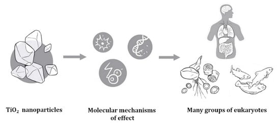
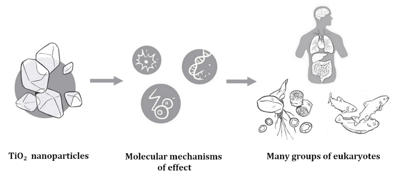
TiO2 Nanoparticles and Their Effects on Eukaryotic Cells: A Double-Edged Sword

1
Institute for Biostatistics and Medical Informatics/ELIXIR-SI Center, Faculty of Medicine, University of Ljubljana, Karantanska ulica 37, 2000 Maribor, Slovenia
2
Department of Anesthesiology, Intensive Care and Pain Management, University Medical Centre Maribor, Ljubljanska ulica 5, 2000 Maribor, Slovenia
3
Institute for Biostatistics and Medical Informatics/ELIXIR-SI Center, Faculty of Medicine, University of Ljubljana, Vrazov trg 2, 1000 Ljubljana, Slovenia
*
Author to whom correspondence should be addressed.
Int. J. Mol. Sci. 2022, 23(20), 12353; https://doi.org/10.3390/ijms232012353
Submission received: 12 September 2022 / Revised: 12 October 2022 / Accepted: 12 October 2022 / Published: 15 October 2022
(This article belongs to the Special Issue Interaction of Nanomaterials with Cells and Tissues)

Abstract

:
Nanoparticulate TiO2 (TiO2 NPs) is a widely used material, whose potential toxicity towards eukaryotic cells has been addressed by multiple studies. TiO2 NPs are considered toxic due to their production of reactive oxygen species (ROS), which can, among others, lead to cellular damage, inflammatory responses, and differences in gene expression. TiO2 NPs exhibited toxicity in multiple organs in animals, generating potential health risks also in humans, such as developing tumors or progress of preexisting cancer processes. On the other hand, the capability of TiO2 NPs to induce cell death has found application in photodynamic therapy of cancers. In aquatic environments, much has been done in understanding the impact of TiO2 on bivalves, in which an effect on hemocytes, among others, is reported. Adversities are also reported from other aquatic organisms, including primary producers. These are affected also on land and though some potential benefit might exist when it comes to agricultural plants, TiO2 can also lead to cellular damage and should be considered when it comes to transfer along the food chain towards human consumers. In general, much work still needs to be done to unravel the delicate balance between beneficial and detrimental effects of TiO2 NPs on eukaryotic cells.

Graphical Abstract

1. Introduction

Nanoparticulate titanium dioxide (TiO2 NP) is a metallic-oxide compound that is of wide use and interest in many aspects of modern society. Being semiconductive, it has high particle stability, coupled with low corrosivity and a purportedly lesser degree of toxicity than some other nanoparticles. It is also relatively cheap to produce. According to Noman et al. [1], three crystalline polymorphs exist—rutile, anatase and brookite, which differ in crystal structure. The former two possess a tetragonal structure, while the latter is rhombohedral [2]. Additionally, TiO2 NPs can also be found in amorphous forms [3].
A commonly noted property of TiO2 NPs is their photocatalytic ability, enabling them to stimulate generation of free radicals, which can then react further with other compounds [1,4]. Due to their photocatalytic and other properties, TiO2 NPs are widely used, including their application in solar cells. Other industrial uses of the compound include use in coatings, sensors and electronic devices, while probably one of the most known uses is the use of TiO2 NPs as a white pigment. The latter is applied in a broad spectrum, ranging from paints to cosmetic products, such as sunscreen or toothpaste [1,2,5]. Additionally, the material is even found in food products as an additive (E 171; [5,6,7,8]). Probably due to its wide applicability, TiO2 NPs are among the most produced nanomaterials in the world [9].
Apart from its industrial use due to favorable physical and chemical properties, TiO2 NPs can potentially even generate benefits linked to human health, such as cancer treatment [10,11,12]. Several studies also indicate its potential in the field of agriculture (e.g., [13,14,15,16]). On the other hand, TiO2 NPs can cause toxicological effects to eukaryotic cells (such as apoptosis and necrosis, see [17]), including potential risks to human well-being, as well as having detrimental environmental effects [6,18]. As put forward by Musial et al. [5] for the area of human consumption, inconsistencies in results can be found when examining various studies, dealing with the impact of TiO2 NPs.
This review is an attempt to summarize TiO2 NPs detrimental and possible beneficial effects in human and other eukaryotic cells, mainly with regards to human health and the environment.

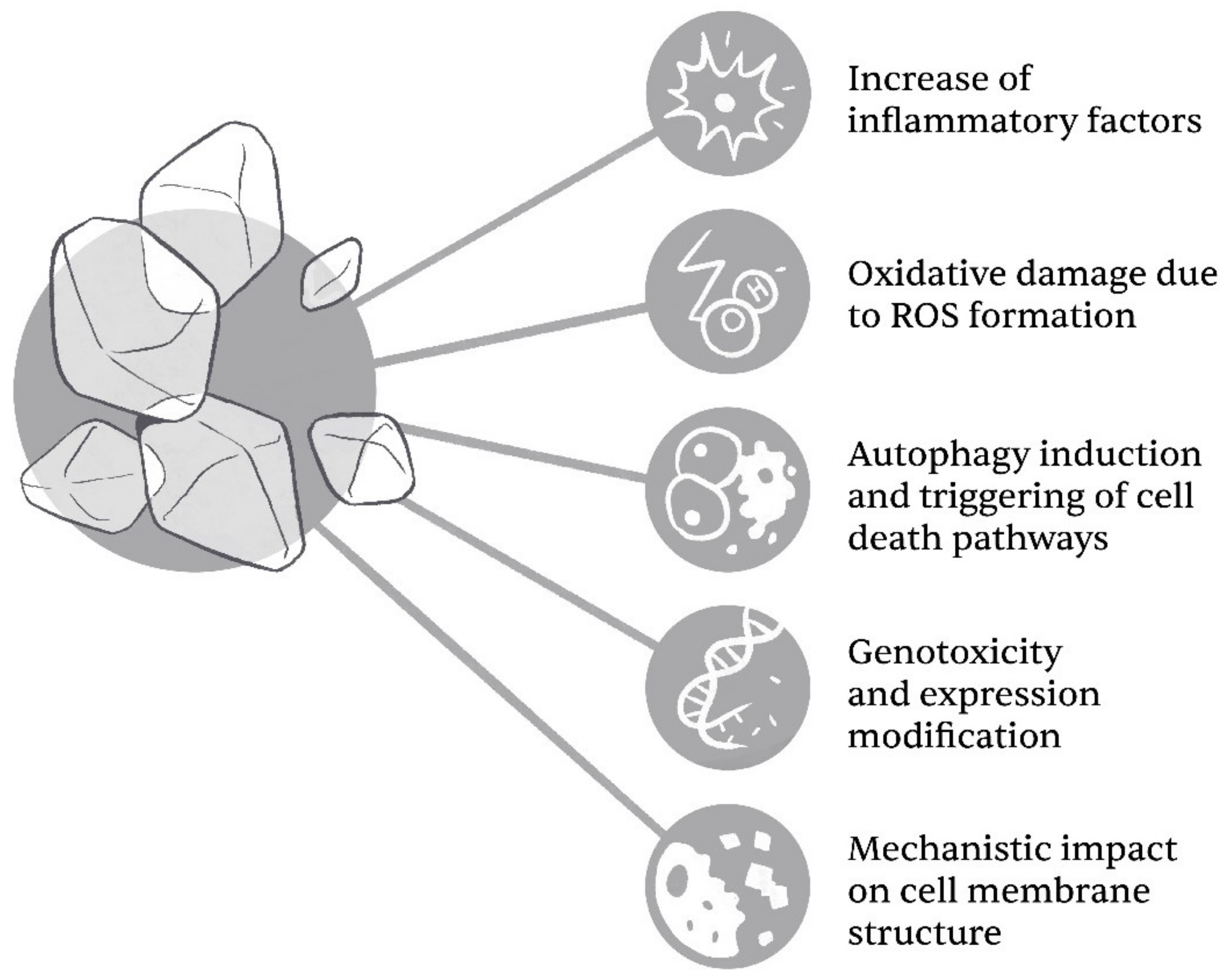
2. Molecular Mechanisms of TiO2 Toxicity

The cytotoxicity of TiO2 NPs is linked to both their size and form. Lu et al. [8] have recently demonstrated that the cytotoxicity of TiO2 seems to be inversely connected to their size—for example, by observing LDH leakage, where the author’s report an increase of the latter with a reduction of particle size. When it comes to form, both of the most common crystalline forms—anatase and rutile—seem to present cytotoxic risk, with varying results between studies [19,20,21]. The general cytotoxicity impact/mechanisms of TiO2 NPs on cellular damage are summarized in Figure 1.
TiO2 NPs induce inflammation by increasing the level of NF-κB (p65), HSP 60, p38 protein, IFN-γ, TNF-α, and nitric oxide, in a dose-dependent manner [22]. Some inflammation mediators induced by TiO2 NPs are organ-specific; for example, the brain inflammation markers IL-1β, IP-10 and CXCL1 [23]. Exposure of mice to ingested TiO2 NPs led to a stronger colonic inflammation than exposure to non-nTiO2 particles that are used in the food additive E171. TiO2 NPs significantly increased the colonic level of the pro-inflammatory cytokines IL-17 and KC/GRO, while TiO2 E171 showed no significant effects on these two cytokines. Exposure to ingested TiO2 NPs resulted in inflammation of the colonic mucosa, characterized by formation of dysplasia, distortion of crypts, widespread loss of goblet cells, and mucosal infiltration. However, both TiO2 NPs and TiO2 E171 decreased the colonic level of IL-10, an anti-inflammatory cytokine [24].
Formation of ROS is another important mechanism of toxicity of TiO2 NPs [25]. Exposure to TiO2 NPs following intranasal administration activates heme oxygenase 1 through the p38-Nrf-2 signaling pathway, leading to ROS accumulation and oxidation of lipids, proteins, and DNA. This mechanism triggered brain injury in mice [26]. Indirect DNA damage via oxidative stress results in frequent p53 mutations, elevated hepatic malondialdehyde (MDA-marker of lipid peroxidation), and decreased antioxidant defense systems. Administration of free radical scavengers such as chlorophyllin (CHL) alleviates the oxidative stress and reduces genotoxicity of TiO2 NPs [27]. H2O2 can be generated in photocatalytic reactions with TiO2 as the catalyst. H2O2 production is favored when TiO2 surface areas exceed 200 m2/g, as is the case for TiO2 NPs [28]. There are two mechanisms of H2O2 formation in TiO2 photocatalysis, and the mechanisms depend on the oxygenation rate of aqueous solutions. In oxygenated solutions, H2O2 is formed directly through reduction of molecular oxygen by conduction band electrons, while in oxygen-free solutions H2O2 can only be formed through recombination of hydroxyl radicals that originate from photocatalytic disintegration of water molecules. These mechanisms were confirmed by experiments using the radical scavenger tris(hydroxymethyl)aminomethane (Tris). H2O2 concentration increased in the oxygenated solution as Tris protected H2O2 from decomposition by hydroxyl radicals, while no H2O2 could be detected in the deoxygenated solution due to the scavenging of hydroxyl radicals by Tris, which prevented H2O2 formation [29].
There are several reports on how TiO2 NPs affect apoptosis, necrosis, necroptosis and cell cycle in human and other eukaryotic cells [17]. Astrocyte-like U87 cells treated with TiO2 NPs showed both Annexin-V staining, a marker of apoptosis, and propidium iodide (PI) staining, a marker of necrosis. Both types of the cellular staining were increased with the elevation in the treatment concentrations of TiO2 NPs [30]. Apoptosis induced by TiO2 NPs was also confirmed by measurements of mitochondrial membrane potential. Effects of TiO2 NPs on cell cycle are dependent on the crystalline phase of particles. Pure anatase TiO2 NPs arrested the cell cycle of neuronal cells at S-phase while only slight cell cycle changes were observed for the mix of anatase and rutile particles [19]. Necroptosis can also occur in lung cells exposed to TiO2 NPs. After the exposure of a primary culture of alveolar macrophages to TiO2 NPs, cell death increased in correlation with TiO2 concentration. In addition, the cytoplasm of alveolar macrophages exposed to TiO2 NPs showed expression of phosphorylated mixed lineage kinase (pMLKL) and high-mobility group box-1 (HMGB1), which are key indicators of necroptosis. Treatment of the exposed alveolar macrophages with Nec-1s (a necroptosis inhibitor) inhibited cell death [31].
TiO2 NPs-induced necroptotic pathways were also detected in A375 human melanoma cells. In smaller doses, the cells responded with a non-detrimental autophagy flux, whilst under the influence of photoactivation, autophagy is hindered, and a necroptotic reaction was triggered by production of ROS [32]. In addition, TiO2 NPs induce autophagy in normal lung cells. The extent of autophagy was determined quantitatively by the ratio of LC3 II to LC3 I, which increased significantly in cells treated with TiO2 NPs [33]. This finding is relevant since inhalation is a major absorption route for TiO2 NPs and lung cells represent the first line of contact with inhaled particles.
TiO2 NPs may cause toxic effects also through other mechanisms such as upregulation of tight junction proteins in the blood–brain barrier, as well as modulation of P-gp mRNA expression [23]. TiO2 NPs can also upregulate some enzymes; for example, alanine aminotransferase (ALT) and aspartate aminotransferase (AST) in the liver. Significant increases in ALT and AST were reported in mice [34], likely caused by the accumulation of TiO2 NPs in the liver. Furthermore, TiO2 NPs were reported to decrease the expression of dopamine receptors D1 and D2 in mice. Exposure to TiO2 NPs also decreased the expression of neurotrophins and related receptors, such as nerve growth factor (NGF) and its receptor TrkA, as well as brain-derived neurotrophic factor (BDNF) and its receptor TrkB [35].
Differential expression analysis of the entire proteome and phosphorylated proteome of primary macrophages showed that exposure to TiO2 NPs affected the expression of around 1000 proteins. TiO2 NPs had more impact on the phosphorylated proteins due to strong interaction of nanoparticles with phosphate groups in proteins. Differentially expressed proteins affected cellular component assembly, cell localization and growth, homeostasis, and tyrosine kinase signaling pathways. Differentially expressed phosphorylated proteins were involved in regulation of adaptive immune response, receptor mediated phagocytosis, and anion transmembrane transport. Exposure of macrophages to TiO2 NPs elicits inflammatory responses through up-regulation of tumour necrosis factor α (TNFα) and inducible nitric oxide synthase (iNOS). Metabolome analysis of macrophages revealed that TiO2 NPs activated the cyclooxygenase-2 (COX-2) pathway in macrophages. Quantification of metabolites derived from arachidonic acid detected a significant increase of the prostaglandins PGD2, PGE2, and 15d-PGJ2 [36].
TiO2 NPs that are orally ingested can adsorb proteins from food within the gastrointestinal tract. Formation of a protein corona around TiO2 NPs was confirmed for plant-based proteins glutenin, gliadin, zein, and soy protein that are common nutritional ingredients. A corona of adsorbed proteins can affect bioavailability of nanoparticles and the composition of the gut microbiota [37]. TiO2 particles that enter the bloodstream can adsorb serum proteins such as albumin and alter their physiological function [38].
Additionally, cytotoxic and genotoxic effects of TiO2 NPs on eukaryotic cells were observed. Hou et al. [25] highlighted the attachment of these large-surfaced particles onto cell membranes and other cellular structures, leading to cytotoxicity. Some form of cell membrane or cell wall damage might occur even when UV light is not present in conjunction with TiO2 [39], as, for example, what was observed in hamster ovary cells by Batiuskaite et al. [40]. Regarding genotoxicity of TiO2 NPs, many in vivo and in vitro studies were conducted, and overall, the studies indicating that TiO2 NPs are genotoxic outweigh the studies that state otherwise. Thus, TiO2 NPs can be earnestly treated as a potential health hazard [41].

3. TiO2 NPs Hazard for Human Health

Daily, humans are exposed to TiO2 NPs in various ways, mainly by inhalation in occupational settings, via certain consumers’ goods as food supplements (E 171), skin contact via cosmetical products and pharmaceuticals [7,42]; hence, their potential effect on the human body should not be overlooked. In vivo tests in animals revealed that inhalation, oral exposure, intraperitoneal and intravenous administration of TiO2 NPs can lead to absorption of particles in the bloodstream and accumulation of TiO2 in various organs, the liver, kidneys, spleen, lungs, brain, alimentary tract and heart, generating potential health risks (summarized in Figure 2) [42,43,44,45].

3.1. Effects of TiO2 NPs on Nervous System

To reach the brain, TiO2 NPs must cross the blood–brain barrier or migrate along afferent nerve fibers; both mechanisms have been described as feasible. Several in vivo and in vitro studies have demonstrated the neurotoxicity of TiO2 NPs through programmed cell death processes, such as apoptosis, autophagy and necroptosis depending on the duration of exposure and the dose-dependence. Although low-dose exposure could not induce any acute neurotoxicity, chronic exposure to low dose TiO2 NPs results in TiO2 accumulation and might lead to dramatic changes in the brain [43,46,47,48,49,50].
All the research objects in experiments so far only consisted of animals (such as mice and rats) or cells (such as PC12, U373, C6) [43]. In consequence, neurotoxic data of TiO2 NPs collected from those studies might be inappropriate to determine their neurotoxic effects on humans. To fully understand the neurotoxicity of TiO2 NPs, studies of human exposures to TiO2 NPs are needed [46].

3.2. Cardiovascular Effects of TiO2 NPs

Some studies showed that TiO2 NPs could be toxic and have detrimental effects on the cardiovascular system [43,50]. Rossi et al. [51] report that inhaled TiO2 NPs acutely increased cardiac excitability and promoted arrhythmogenesis in normotensive rats by a direct interaction with cardiac cells. Chen et al. [52], in their in vivo tests on rats, set forth a hypothesis that cardiac damage and inflammatory response caused by TiO2 NPs were suggested as the possible reasons for cardiovascular adverse effects. Zhao et al. [53] studied cardiopulmonary effects among workers who were exposed to TiO2 NPs. Occupational exposure by airborne TiO2 NPs was associated with oxidative stress, inflammation, lung and cardiac damage, indicated by the workers’ serum cardiopulmonary disease markers. Some workers were found to have symptoms such as shortness of breath and even suffered from pericardial effusions and hypoxemia [49]. However, most of the doses employed in studies are too high to be realistic in occupational settings [41].

3.3. Effects of TiO2 NPs on Pulmonary System

Pulmonary in vivo studies on rats support the carcinogenicity of TiO2 NPs in intratracheal and inhalation studies [41]. Mitochondrial damage, oxidative stress, and apoptotic cell death in a dose-dependent manner were involved in the toxic mechanistic pathways [47]. Lung inflammation was associated with dose-dependent increases. However, no clinical signs of obvious acute toxicity were observed [41,47]. In vitro, cell lines of human lung origin showed increased ROS generation and apoptotic death on exposure to TiO2 NPs [54]. Human lung damage (functional loss, elevated markers of oxidative stress, and inflammation) on exposure to airborne TiO2 NP has been reported [53]. However, the results from a few epidemiological studies show that there are no significant associations between TiO2 NPs exposure and risk of lung cancer. The relatively short history in production and use seems to be the main reason for the lack of human epidemiological studies for TiO2 NPs. Epidemiological studies thus far have not been able to detect an association between the occupational exposure to TiO2 NPs and an increased risk for cancer [41,55].

3.4. Effects of TiO2 NPs on Liver, Spleen and Kidneys

Mice were treated with different dosing regimens of TiO2 NPs injected intraperitoneally. In general, high doses of TiO2 NPs significantly increased the organ/body weight ratio of the liver, spleen and kidneys in mice in a dose-dependent manner [43] Intravenously applied TiO2 NPs caused massive oxidative stress, DNA damage, and mitochondria-mediated apoptosis in the kidney and liver of rats [47]. Other observations included elevated liver function biomarkers, necrosis of liver cells and apoptosis, liver fibrosis and swelling of renal glomeruli in high doses of TiO2 NPs [43,50].
As is argued in Heringa et al. (2018) and Peters et al. (2020) see [42], it can be assumed that TiO2 found in human tissues, in individuals without an occupational inhalatory exposure, almost exclusively originates from oral exposure to TiO2 particles. Analysis of postmortem tissues indicates that these particles are taken up by the intestine and subsequently transported to secondary organs such as liver, spleen and kidneys, where they can accumulate. A number of in vivo toxicity studies on rats/mice show that TiO2 NPs can trigger ROS generation, induce oxidative stress, inflammation and might lead to liver fibrosis, steatosis and edema, but it is unknown whether these events subsequently result in irreversible adverse effects in humans. With regard to kidneys and spleen, data are too limited to draw conclusions on potential health risks in humans. The level presence of TiO2 in these organs indicates that it is relevant to gain further knowledge on potential effects of TiO2 on these tissues [42].

3.5. Effects of TiO2 NPs on Alimentary Tract

The results of recent studies on rats and the human volunteer showed that TiO2 NPs were scarcely captured from the GIT and transferred into systemic circulation [43]. Available data suggest that TiO2 NPs may influence microbiota, disturb digestion and absorption of food components causing problems such as inflammatory bowel disease, obesity, or deficiencies of macro- and microelements in the body. Indeed, there are increasing concerns that its presence may lead to intestinal barrier impairment, including dysbiosis of intestinal microbiota [43,56]. Studies concerning toxicity after oral administration to rats show no obvious signs of acute toxicity [41,43]. Duan et al. demonstrated weight loss in mice after intragastric administration of TiO2 NPs. This should be explained by the reduced number of intestinal villi and the resulting loss of surface of the small intestine capable of absorbing nutrients, which consequently leads to malnutrition and weight loss [43]. Amedollia et al. also showed in their studies that after oral exposure of rats, TiO2 NPs are capable of penetrating the intestinal mucosa. The results of in vivo tests confirmed the results of in vitro studies on human intestine cell cultures. They found a decrease in the number of microvilli, resulting in a reduction of the surface area available for absorption of nutrients [43].
At the same time, there have been studies that revealed a limited influence of TiO2 NPs on human microbiota. Currently available reports therefore provide contradictory evidence in terms of the impact of TiO2 NPs on human microbiota due to the application of varying experimental models and frameworks [56].

3.6. Skin Toxicity of TiO2 NPs

Inclusion of TiO2 NPs into sunscreens has raised interesting questions regarding the potential for dermal penetration, systemic absorption, and subsequent toxicity. The in vitro and in vivo data regarding the potential of dermal absorption and/or penetration of TiO2 NPs from sunscreens exhibit controversial results. Although several articles describe the opposite, the penetration of TiO2 NPs in healthy, as well as in damaged or lesioned skin, is demonstrated in the scientific community, with damaged or lesioned skin being more susceptible to TiO2 NPs penetration. The fact that upon skin penetration, TiO2 NPs can enter the blood circulation, and then undergo translocation to various distant tissues and organs, suggests that prolonged time exposure to TiO2 NPs, may pose a health risk to consumers [57].
Extensive studies have shown that TiO2 NPs can cause cell toxicity under in vitro and in vivo conditions. However, cosmetic industries are using alternative methods to animal experimentation, such as 3D skin reconstructed models, to evaluate the irritation and corrosion of cosmetics. Sanches et al. [57] have shown that the 3D skin model can be used to test the toxicity of TiO2 NPs. The data obtained show that TiO2 NPs was considered non-irritant, non-corrosive and non-phototoxic for the 3D skin model, regardless of the crystalline type and the size of the NPs studied [57].
Melanoma is the malignancy responsible for the highest incidence of deaths from skin cancer. As is argued in Zdravković et al. [58], it can be assumed that TiO2 NPs may even influence metastatic melanoma cells’ invasiveness and aggressiveness. In practice, results obtained from the study suggest that patients with diagnosed metastatic melanoma should avoid the usage of products containing TiO2 NPs because this could increase progression and recurrence of their disease [58].

3.7. Embryotoxicity of TiO2 NPs

A great number of in vivo studies on pregnant mice/rats exposed to TiO2 NPs by various routes, including intraperitoneal and intravenous injection, revealed that those NPs could be detected in the brain of the fetus, induce an acute stress reaction on the neurons and glial cells of mouse brains and lead to CNS dysfunction, as a consequence. The authors discovered that they can even cause pregnancy complications, increase dopamine levels in the prefrontal cortex and neostriatum, alter gene expression related to brain development and enhance depressive-like behaviors in mice/rats. All the data collected from in vivo and ex vivo mouse embryo studies showed that they exhibited toxic effects on the growth and development in dose-dependent and time-dependent manners. Stemming from all above, they may affect the brain development of embryos by crossing the placental barrier and lead to CNS dysfunction later in life [46,50].

3.8. Potential Clinical Use of TiO2 NPs

Although numerous in vivo and in vitro studies have provided evidence of the toxic effects of TiO2 NPs, the capability to produce ROS that initiate programmed death has found application of targeted cytotoxicity of TiO2 NPs for photodynamic therapy (PDT) of cancers [10,11,12]. It was reported that modification of TiO2 NPs with some biomolecules that can specifically bind to cancer cells improved selectivity and efficiency of TiO2 to destroy cancer cells [59]. Wang et al. [60] report that combined TiO2 NPs exposure and UVA treatment in glioma-bearing mice led to a significant increase in the extent of necrotic areas and indices of apoptosis in established tumor grafts. Moreover, it is obvious that there is a synergistic effect of UV irradiation and TiO2 NPs on the drug uptake of target leukemia cells, which may also be utilized as a new strategy to inhibit the drug resistance of the targeted cancer cells, suggesting the feasible idea of cancer treatment using TiO2 NPs and UV irradiation with accompanying anticancer drugs [61]. Cai et al. [62] reported that photoexcited TiO2 particles effectively induced cytotoxicity against HeLa cancer cells. Some research illustrated that human colon carcinoma cells treated with photoexcited TiO2 NPs can be effectively affected and damaged [63]. Experimental exposure of TiO2 NPs was found to induce oxidative stress, genotoxicity, and apoptosis in human lung cancer cell line A549 [64].

4. TiO2 NPs and Other Eukaryotes—Toxic or Not?

The impact of TiO2 NPs on eukaryotic cells expanding far beyond human cells [25] and the flow of material within ecosystems, alongside TiO2′s ever growing application, makes it an important potential toxicant. Ranging from small, superficially simple unicellular algae to complex organisms such as vertebrates, this section will therefore strive to provide the reader with an insight into the impact that TiO2 (especially in its nanoparticulate forms) has on other eukaryotes, based on a few randomly selected recent studies. Since plenty of review literature exists, concerning environmental/ecological aspects of nanoparticulate TiO2 [18,25,65,66,67], we strive to provide only a brief insight.
The environmental impact of this nanoparticulate metal oxide should not be underestimated, not only because it can have an adverse effect on single organisms and their ecosystem functioning, but also because its potential bioaccumulation, biomagnification and transfer through the food chain may very well, in the end, impact multiple species [20,66,68,69]. The complex interconnectivity between different trophic levels within and, indeed, between different ecosystems means that next to none are immune to TiO2 contamination. TiO2 NPs enter natural environments across the globe via direct application (in, e.g., agriculture), waste and residual materials, stemming from its growing use in industrial processes. As already stated, TiO2 is a known inductor of ROS production [70,71,72,73] and, in general, the authors of various research seem to agree that the effect of TiO2 is dependent foremost on its concentration/dosage and time of exposure. We can take a good example from mice (Mus musculus) where Liu et al. [74] have reported observable organ damage after exposing mice to multiple higher doses of TiO2 in nano-anatase form. Toxicity of TiO2 NPs may be enhanced with the presence of UV light [70,71,75,76,77]. Additional factors that one needs to consider is the form in which TiO2 is dosed—bulk or nanoparticle [70,73,78], although some research seems to dispute the differential impact of separate phases [79]. The mineral form also appears to be of importance (anatase/rutile; e.g., [20]) and it is also clear that TiO2 interacts with other nanoparticles and pollutants, potentially increasing or even decreasing their toxicity by means of potentially absorbing them on the surface of TiO2 particles [80,81,82,83,84,85,86].

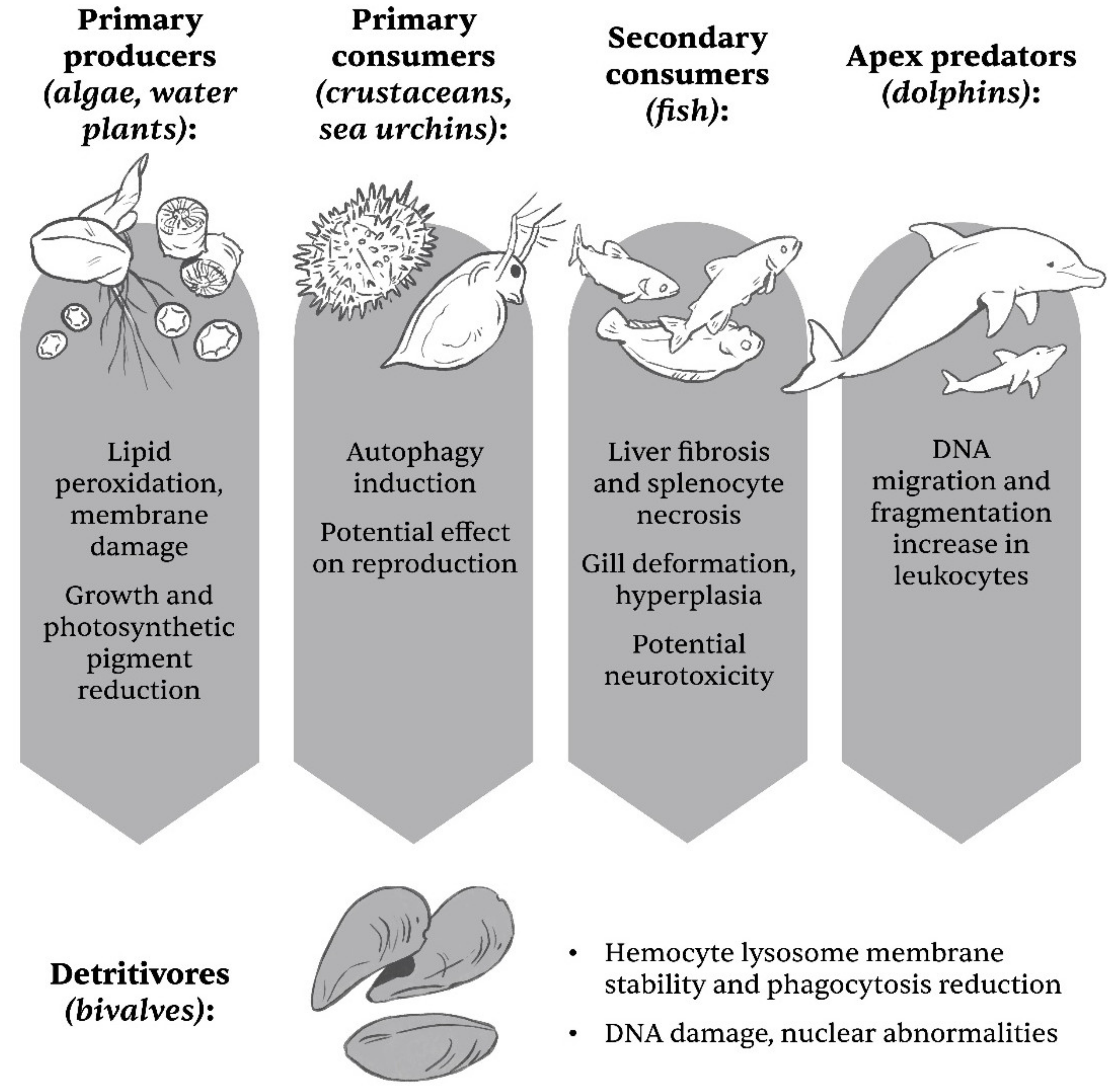
4.1. TiO2 NPs and Their Effect on Aquatic Organisms

Prone to form agglomerates/aggregates in water-based media [70,71,79,87,88,89,90,91,92], TiO2 NPs are the focus of many studies, especially in aquatic environments, where nanoparticles are indeed of general concern [18,67,93,94,95]. Additionally, the material’s agglomeration can remove microorganisms from the water column [79]. The actual effect of TiO2 remains disputed, with Dedman et al. [79] even suggesting that its effect at environmentally realistic concentrations might be disregarded under certain circumstances, at least on a microbial level, challenging the view of, e.g., Miller et al. [75], who find that TiO2 is toxic to phytoplankton even in small quantities, when exposed to UV. Another study highlights that TiO2 under UV(A) radiation does not only impact organisms themselves, but also affects extracellular enzymatic activity in heterotrophic biofilms, therefore potentially affecting the transfer of matter and energy in natural aquatic ecosystems [96].
As primary producers, aquatic plants and algae are of great importance. When exploring the effect of TiO2 (both in bulk and nanoparticulate form) whilst under exposure to visible or UV-A light, two microalgae species, Chlamydomonas reinhardtii (freshwater) and Phaeodactylum tricornutum (seawater)), reported signs of toxicity (such as membrane damage), with nanoparticles producing significantly higher ROS content. High concentrations of TiO2 also stimulated the production of exo-polymeric substances, which could be connected to a defensive response, yet their role is not entirely understood [70]. Negative toxic effects were reported also for the diatom Thalassiosira pseudonana [97], green alga Dunaliella tertiolecta [98] and Chlorella vulgaris [99]. In the latter, another environmental concern comes into effect, since ocean acidification increased nanoparticle internalization [99]. Thiagarajan et al. [76] explored the difference in sensitivity between D. salina and Chlorella sp. (the latter being more sensitive), noting a decrease in cell viability, an increase in ROS production, and lipid peroxidation. In the duckmeat species Spirodela polyrhiza, Movafeghi et al. [72] report an observable uptake into plant tissues/cells, which apparently resulted in a reduction of growth and photosynthetic pigment content in connection to concentration
Moving further upwards the food chain, a multitude of studies has focused especially on shellfish, since these organisms are often of commercial interest. Among them, bivalves (Phylum Mollusca, Class Bivalvia) have garnered considerable attention [95] since they mainly feed by filtering water and sediment and are therefore highly prone to intake of any suspended nanoparticles from the water column [88]. In the Mediterranean mussel (Mytilus galloprovincialis), a species often consumed as a food item across the Mediterranean Basin, TiO2 NPs can apparently decrease lysosomal membrane stability and alter the activity of antioxidant enzymes in the mussel’s digestive gland. Although no changes in morphology were observed, nanoparticles were noted inside lysosomal vacuoles of the cells. Decreased lysosomal membrane stability, alongside a decrease in phagocytosis was also observed in hemocytes (which are the immune cells of invertebrates) [80,89]. Phagocytosis is usually seen as an important part of immune defense in bivalves and an effect on hemocytes could potentially mean a more readily available transfer of pathogens toward human consumers [88]. Some increase in nitrite accumulation, changes in immune-related gene expression and production of extracellular ROS, DNA damage (at a chromosomal level) and potential pre-apoptotic processes (nuclear abnormalities) were also noted [80,87,89]. In general, TiO2 NPs do not seem to exhibit substantial cytotoxic effects but do have some role in triggering immune and inflammatory pathways [87]. The reduction of phagocytic activity was also observed in the Eastern oyster (Crassostrea virginica) [88] and an effect on hemocytes was also reported from three Pacific bivalve species-the Gray’s mussel Crenomytilus grayanus, the horse mussel Modiolus modiolus and Arca boucardi. TiO2 induced mortality and changes in membrane polarization in hemocytes, although with considerable variation among species [100].
In M. galloprovincialis, Canesi et al. [80] also report both antagonistic and synergistic effects of TiO2 and 2,3,7,8-tetrachlorodibenzo-p-dioxins (TCDD), depending on the cell/tissue type and molecular aspect examined. For example, an increased lysosome to cytoplasm ratio was observed when exposed to both contaminants, with the author’s highlighting that larger concentrations of bioaccumulated TCDD in the presence of TiO2 might represent a Trojan horse effect. The latter effect was also observed in another bivalve species Corbicula fluminea [82] and Shi et al. [84] report an increase in immunotoxicity towards hemocytes of the blood clam Tegillarca granosa due to a potential Trojan horse effect of TiO2 on 17β-estradiol (E2). TiO2 alone did not impact total hemocyte count of T. granosa, but hindered phagocytotic rates after 10 days of exposure. The authors also report a significant reduction of expression in tested genes, connected to the immune ability of the clam [84].
Other aquatic animals have also been explored, ranging from crustaceans [20,77], through sea urchins [101], to fish [86,90,102,103]. While testing the impact of multiple nanoparticles on Strongylocentrotus intermedius sea urchin for the purpose of using it in toxicity tests, Pikula et al. [101] noted no decrease in spermatozoan activity and only a low impact on egg fertilization. Additionally, TiO2 was among the lowest acutely toxic nanomaterials when it came to the mortality of embryos; this, however, changed by passing time, since the authors noted a time-dependent increase in toxicity. It is also very interesting to note that TiO2 NPs showed no apparent toxicity on brine shrimp Artemia salina, even under high concentration. Nonetheless, this metal oxide did have some role in the degree of toxicity of other pollutants, such as cadmium and phenanthrene [83]. Under certain circumstances, TiO2 was reported to be harmful to Daphnia magna, where TiO2 NPs in conjunction with UV light upscaled the production of the toxic -OH radical [77]. In D. magna, an impact on the digestive tract was also observed, e.g., with digestive cells producing noticeable autophagy vacuoles [20].
Fish are especially interesting when it comes to potential TiO2 NPs effects, as they often represent the higher rungs of the aquatic food chain. Consumption of prey organisms can lead to bioaccumulation as observed in turbots (Scophthalmus maximus), fed with TiO2 “contaminated” clamworms (Perinereis aibuhitensis). Such consumption among others induced tissue abnormalities, such as liver fibrosis and splenocyte necrosis after a certain time period [68]. In zebrafish (Danio rerio), a significant decrease of leukocyte count 14 days after exposure, alongside an increase in glutathione levels, when exposed to TiO2 NPs, was observed by Ramsden et al. [90]. No apparent effect on the tissue morphology was noted [90]. Zeumer et al. [103], working with rainbow trout (Oncorhynchus mykiss), report an increase in acetylcholinesterase activity under exposure to wastewater treatment plant effluent supplemented with TiO2 NPs, indicating potential chronic neurotoxic effects of TiO2 in conjunction with organic matter. Synergistic effects of TiO2 and other nanoparticles in fish are also reported. When co-examined with lead (Pb), TiO2 increased its toxicity and induced several changes, including gene expression, in D. rerio embryos [85]. In the common carp (Cyprinus carpio), the role of TiO2 NPs in enhancing acute toxicity of silver nanoparticles has been observed by Haghighat et al. [86]. The addition of only TiO2 showed less of an impact, yet nonetheless stimulated some deformities in gill tissue, including an increase in cellular proliferation (defined as hyperplasia of chloride cells [86]). A variation of chloride cell count, alongside goblet cells, was also observed, probably as a protective response, when exposing gills of the Senegalese sole (Solea senegalensis) to TiO2 NPs. Additionally, gill macrophages contained observable black metal deposits after two different TiO2 treatments [102].
Not even sea mammals are immune to the effects of TiO2 NPs. Bernardeschi et al. [104] report that both anatase and rutile form present a potential toxic element to a widespread sea mammal, the bottlenose dolphin (Tursiops truncatus). The authors noted an increase in DNA fragmentation in dolphin leukocytes, yet nonetheless note that this preliminarily observed effect seems to occur to a lesser degree when compared to human leukocytes at equal concentrations of TiO2 [104].
In summary, TiO2 NPs affect various trophic levels in aquatic biosystems, which is illustrated in Figure 3. The potential of transfer and potential bioaccumulation/magnification [20,68] should therefore not be underestimated.

4.2. TiO2 NPs and Agricultural Plants

The impact of TiO2 is not limited only to aquatic vegetation and its toxic effects have also been explored in terrestrial plants, where accumulation/uptake appears to occur via their root system in a concentration dependent manner [13,69,92]. Although detrimental effects of TiO2 should always be of concern (e.g., [73]), it is apparent that TiO2 can induce some changes that could be considered beneficial, such as an increase in chlorophyll content and connected plant weight/growth, at least at lower concentrations [13,14,15,16,105], which is particularly interesting in agriculture. However, using nanoparticles as fitomediatory means is a double-edged sword, since dietary uptake into human consumers can follow, generating health risks [13].
In rice (Oryza sativa), an increase of hydrogen peroxide (H2O2) production, membrane injury and rate of root cell death was observed at higher concentrations, likely connected to an increase in oxidative stress [15]. On the other hand, TiO2 NPs seemingly boosted chlorophyll content, biomass, and increased phosphorus and protein grain content [14,15], even when another toxicant such as cadmium was present [106]. The after-expositional increase in rice biomass and photosynthetic activity was also reported by, e.g., Zhang et al. [107]. A similar effect was observed in maize (Zea mays), where treatment of waste waters with TiO2 was explored, with Yaqoob et al. [108] reporting positive effects on plant weight and photosynthetic pigments at lower concentrations of particles. The effect of concentration was also evident when studying another very common grain plant, wheat (Triticum aestivum), where Rafique et al. [109] observed an increase of H2O2 synthesis and formation of micronuclei with increased concentration. A very similar effect to that in rice (see [15]) was noted as well, with smaller dosages of TiO2 raising protein and nutrient content and biomass production, alongside an increase of H2O2 production [110]. A cytotoxic effect (measured by lactate dehydrogenase leakage), lipid peroxidation and imbalance in antioxidative enzyme activity were observed in T. aestivum calli cells by Czyżowska and Barbasz [111].
Moving from agriculturally important grasses (fam. Poaceae) to legumes (fam. Fabaceae), TiO2 NPs showed a clear capability to cause genotoxicity in the broad bean (Vicia faba). Although it had no noticeable effect on the mitotic index, it induced chromosomal aberrations, such as breaks. Additionally, it shortened shoot length. Both of these effects were apparently concentration-independent and when compared with SiO2 nanoparticles, TiO2 NPs seem to pose a higher cytotoxic risk [112]. A similar effect was also observed in the lentil (Lens culinaris), where TiO2 NPs in anatase form caused chromosomal DNA damage and mitotic index reduction in a concentration dependent manner, with other changes signaling excessive oxidative stress. Lower concentrations, however, seemed to present a moderate, yet significantly positive effect. Additionally, the nanoparticles reduced chlorophyll content [73]. On the contrary, no adverse effects were reported for TiO2 for the Adzuki bean (Vigna angularis); however, one should among others note that particles used by Jahan et al. [92] were larger than those used in L. culinaris by Khan et al. [73], which could play a role in differential response, as TiO2 NPs were demonstrated to be more toxic when particles are smaller [8]. In V. angularis, the metal oxide improved germination (though not when combined with ZnO), stimulated growth and increased chlorophyll content. Lack of any apparent stress in the plant in question was also confirmed by no significant change in antioxidant enzymatic stress activity. Coexistence of TiO2 and ZnO apparently also reduced the uptake of both nanoparticle species into the plant [92].
Spice plant cells are no exception when it comes to (potential) effects of TiO2 NPs. Significant research was done in fenugreek (Trigonella foenum-graecum) by Missaoui et al. [16,91,113], where nanoparticles seem to induce changes in mineral/metal content, hinting at root cell membrane damage [91]. In higher doses, a chlorosis of leaves due to decrease in chlorophyll a and b had also been observed, yet interestingly half of that concentration actually elevated the chlorophyll level. The same effect was observed when examining carotenoid content, which indicates that TiO2 NPs could affect photosynthetic ability of plant cells [16]. As with other plant species, fenugreek seems to be tolerant of lower concentrations of TiO2 NPs, balancing ROS production by increased antioxidant activity. Larger concentrations diminished defensive activity and therefore presented themselves as toxic to the plant [16,113]. Similarly, at a smaller degree, TiO2 seems to boost nutrient accumulation in coriander (Coriandrum sativum), yet larger concentrations show a potentially adverse effect, apparent in the activation of oxidative stress response and wrinkling of the cell membrane [114].
A limited effect was observed when studying the effect of TiO2 on the onion (Allium cepa), mainly observed as slight alterations to the mitotic index [115]. Singh and Kumar [13] report an increase in chlorophyll content in spinach (Spinacia oleracea) after treatment with sewage sludge containing TiO2 NPs. Gordillo–Delgrado et al. [105] also report that at certain small concentrations TiO2 can be beneficial to S. oleracea, even when combined with silver nanoparticles and could be of potential agronomic use.

5. Conclusions and Perspectives

It is possible that human exposure to TiO2 NP, whether related to occupational settings, environmental pollution, or certain consumers’ goods, may contribute to aggravation of several chronic diseases of humans and hence an increase in risk of developing tumors or progress of preexisting cancer processes. To support this, we can list the recent Commission Regulation (EU) 2022/63 (Official Journal of the European Union, L11/1, 18 January 2022), which has withdrawn TiO2 (E 171) as a food additive due to safety concerns. On the contrary, numerous studies enhance our understanding on cellular and molecular behavior of human cancer cells against TiO2 NPs exposure, which reveal excellent photochemical properties and hence show their potential use in new treatment modalities of malignant tumors. Extensive studies in this area are urgently needed.
Additionally, it is clear that TiO2 does indeed possess some qualities that, under certain circumstances, make it a viable hazard to eukaryotic cells across many organismal groups. As a widely present environmental contaminant, this metal oxide can induce oxidative stress and hinder cellular function. The impact of contamination should not be underestimated, with many studies highlighting the potential of TiO2 NPs to cause environmental damage. It would seem that special interest should be pointed towards aquatic ecosystems, where TiO2 is a common pollutant, with its effects becoming more and more well studied. Nonetheless, additional work is still needed to transplant research in situ, in the actual natural environment or at least one resembling it, where multiple factors need to be considered in addition to just the presence of TiO2. Further studies, following the behavior of particles along the trophic chain at a longer time period of exposure are therefore certainly required.
In conclusion, TiO2 clearly possess several attributes, that can either produce benefits or adversities when it comes to eukaryotic cells; however, much work is still needed to examine its potential impact. Future insights should also strive to unravel the delicate influence of dosage. Even though some benefit can be observed with smaller concentrations in the field of agriculture, we wish to conclude this review with the quote of the immortal Paracelsus: Sola dosis facit venenum.

Author Contributions

Conceptualization, J.G., B.Z. and P.F.; investigation, J.G., B.Z. and M.V.; writing—original draft preparation, J.G., B.Z. and M.V.; writing—review and editing, J.G. and P.F.; visualization, J.G. and P.F.; supervision, B.L. and P.F.; project administration, B.L. and P.F.; funding acquisition, B.L. All authors have read and agreed to the published version of the manuscript.

Funding

This research received no external funding.

Institutional Review Board Statement

Not applicable.

Informed Consent Statement

Not applicable.

Data Availability Statement

Not applicable.

Acknowledgments

The research was done using the Research infrastructure ELIXIR-SI (https://elixir-slovenia.org; accessed on 1 September 2022), funded by the European Regional Development Fund, the Ministry of Science, Education and Sports and by the Slovenian Research Agency. The authors acknowledge the financial support by the Slovene Research Agency (P3-0154). We especially wish to thank Matevž Bervar who kindly provided the illustrations used in this manuscript.

Conflicts of Interest

The authors declare no conflict of interest. The funders had no role in the design of the study; in the collection, analyses, or interpretation of data; in the writing of the manuscript, or in the decision to publish the results.

References

  1. Noman, M.T.; Ashraf, M.A.; Ali, A. Synthesis and Applications of Nano-TiO2: A Review. Environ. Sci. Pollut. Res. 2019, 26, 3262–3291. [Google Scholar] [CrossRef] [PubMed]
  2. Diebold, U. The Surface Science of Titanium Dioxide. Surf. Sci. Rep. 2003, 48, 53–229. [Google Scholar] [CrossRef]
  3. Sun, S.; Song, P.; Cui, J.; Liang, S. Amorphous TiO2 Nanostructures: Synthesis, Fundamental Properties and Photocatalytic Applications. Catal. Sci. Technol. 2019, 9, 4198–4215. [Google Scholar] [CrossRef]
  4. Dahl, M.; Liu, Y.; Yin, Y. Composite Titanium Dioxide Nanomaterials. Chem. Rev. 2014, 114, 9853–9889. [Google Scholar] [CrossRef]
  5. Musial, J.; Krakowiak, R.; Mlynarczyk, D.T.; Goslinski, T.; Stanisz, B.J. Titanium Dioxide Nanoparticles in Food and Personal Care Products—What Do We Know about Their Safety? Nanomaterials 2020, 10, 1110. [Google Scholar] [CrossRef]
  6. Fiordaliso, F.; Bigini, P.; Salmona, M.; Diomede, L. Toxicological Impact of Titanium Dioxide Nanoparticles and Food-Grade Titanium Dioxide (E171) on Human and Environmental Health. Environ. Sci. Nano 2022, 9, 1199–1211. [Google Scholar] [CrossRef]
  7. Bischoff, N.S.; de Kok, T.M.; Sijm, D.T.H.M.; van Breda, S.G.; Briedé, J.J.; Castenmiller, J.J.M.; Opperhuizen, A.; Chirino, Y.I.; Dirven, H.; Gott, D.; et al. Possible Adverse Effects of Food Additive E171 (Titanium Dioxide) Related to Particle Specific Human Toxicity, Including the Immune System. Int. J. Mol. Sci. 2020, 22, E207. [Google Scholar] [CrossRef]
  8. Lu, N.; Chen, Z.; Song, J.; Weng, Y.; Yang, G.; Liu, Q.; Yang, K.; Lu, X.; Liu, Y. Size Effect of TiO2 Nanoparticles as Food Additive and Potential Toxicity. Food Biophys. 2022, 17, 75–83. [Google Scholar] [CrossRef]
  9. Vance, M.E.; Kuiken, T.; Vejerano, E.P.; McGinnis, S.P.; Jr, M.F.H.; Rejeski, D.; Hull, M.S. Nanotechnology in the Real World: Redeveloping the Nanomaterial Consumer Products Inventory. Beilstein J. Nanotechnol. 2015, 6, 1769–1780. [Google Scholar] [CrossRef] [Green Version]
  10. Raja, G.; Cao, S.; Kim, D.-H.; Kim, T.-J. Mechanoregulation of Titanium Dioxide Nanoparticles in Cancer Therapy. Mater. Sci. Eng. C 2020, 107, 110303. [Google Scholar] [CrossRef]
  11. Çeşmeli, S.; Biray Avci, C. Application of Titanium Dioxide (TiO2) Nanoparticles in Cancer Therapies. J. Drug. Target. 2019, 27, 762–766. [Google Scholar] [CrossRef] [PubMed]
  12. Ziental, D.; Czarczynska-Goslinska, B.; Mlynarczyk, D.T.; Glowacka-Sobotta, A.; Stanisz, B.; Goslinski, T.; Sobotta, L. Titanium Dioxide Nanoparticles: Prospects and Applications in Medicine. Nanomaterials 2020, 10, 387. [Google Scholar] [CrossRef] [PubMed] [Green Version]
  13. Singh, D.; Kumar, A. Binary Mixture of Nanoparticles in Sewage Sludge: Impact on Spinach Growth. Chemosphere 2020, 254, 126794. [Google Scholar] [CrossRef] [PubMed]
  14. Phothi, R.; Theerakarunwong, C.D. Enhancement of Rice (Oryza sativa L.) Physiological and Yield by Application of Nano-Titanium Dioxide. Aust. J. Crop Sci. 2020, 14, 1157–1161. [Google Scholar] [CrossRef]
  15. Waani, S.P.T.; Irum, S.; Gul, I.; Yaqoob, K.; Khalid, M.U.; Ali, M.A.; Manzoor, U.; Noor, T.; Ali, S.; Rizwan, M.; et al. TiO2 Nanoparticles Dose, Application Method and Phosphorous Levels Influence Genotoxicity in Rice (Oryza sativa L.), Soil Enzymatic Activities and Plant Growth. Ecotoxicol. Environ. Saf. 2021, 213, 111977. [Google Scholar] [CrossRef]
  16. Missaoui, T.; Smiri, M.; Chmingui, H.; Hafiane, A. Effects of Nanosized Titanium Dioxide on the Photosynthetic Metabolism of Fenugreek (Trigonella foenum-graecum L.). Comptes Rendus Biol. 2017, 340, 499–511. [Google Scholar] [CrossRef]
  17. Mohammadinejad, R.; Moosavi, M.A.; Tavakol, S.; Vardar, D.Ö.; Hosseini, A.; Rahmati, M.; Dini, L.; Hussain, S.; Mandegary, A.; Klionsky, D.J. Necrotic, Apoptotic and Autophagic Cell Fates Triggered by Nanoparticles. Autophagy 2019, 15, 4–33. [Google Scholar] [CrossRef] [Green Version]
  18. Luo, Z.; Li, Z.; Xie, Z.; Sokolova, I.M.; Song, L.; Peijnenburg, W.J.G.M.; Hu, M.; Wang, Y. Rethinking Nano-TiO2 Safety: Overview of Toxic Effects in Humans and Aquatic Animals. Small 2020, 16, 2002019. [Google Scholar] [CrossRef]
  19. Valdiglesias, V.; Costa, C.; Sharma, V.; Kiliç, G.; Pásaro, E.; Teixeira, J.P.; Dhawan, A.; Laffon, B. Comparative Study on Effects of Two Different Types of Titanium Dioxide Nanoparticles on Human Neuronal Cells. Food Chem. Toxicol. 2013, 57, 352–361. [Google Scholar] [CrossRef]
  20. Chen, X.; Zhu, Y.; Yang, K.; Zhu, L.; Lin, D. Nanoparticle TiO2 Size and Rutile Content Impact Bioconcentration and Biomagnification from Algae to Daphnia. Environ. Pollut. 2019, 247, 421–430. [Google Scholar] [CrossRef]
  21. Uboldi, C.; Urbán, P.; Gilliland, D.; Bajak, E.; Valsami-Jones, E.; Ponti, J.; Rossi, F. Role of the Crystalline Form of Titanium Dioxide Nanoparticles: Rutile, and Not Anatase, Induces Toxic Effects in Balb/3T3 Mouse Fibroblasts. Toxicol. Vitr. 2016, 31, 137–145. [Google Scholar] [CrossRef] [PubMed]
  22. Meena, R.; Kumar, S.; Paulraj, R. Titanium Oxide (TiO2) Nanoparticles in Induction of Apoptosis and Inflammatory Response in Brain. J. Nanopart. Res. 2015, 17, 49. [Google Scholar] [CrossRef]
  23. Disdier, C.; Devoy, J.; Cosnefroy, A.; Chalansonnet, M.; Herlin-Boime, N.; Brun, E.; Lund, A.; Mabondzo, A. Tissue Biodistribution of Intravenously Administrated Titanium Dioxide Nanoparticles Revealed Blood-Brain Barrier Clearance and Brain Inflammation in Rat. Part. Fibre Toxicol. 2015, 12, 27. [Google Scholar] [CrossRef] [PubMed] [Green Version]
  24. Cao, X.; Han, Y.; Gu, M.; Du, H.; Song, M.; Zhu, X.; Ma, G.; Pan, C.; Wang, W.; Zhao, E.; et al. Foodborne Titanium Dioxide Nanoparticles Induce Stronger Adverse Effects in Obese Mice than Non-Obese Mice: Gut Microbiota Dysbiosis, Colonic Inflammation, and Proteome Alterations. Small 2020, 16, e2001858. [Google Scholar] [CrossRef]
  25. Hou, J.; Wang, L.; Wang, C.; Zhang, S.; Liu, H.; Li, S.; Wang, X. Toxicity and Mechanisms of Action of Titanium Dioxide Nanoparticles in Living Organisms. J. Environ. Sci. 2019, 75, 40–53. [Google Scholar] [CrossRef]
  26. Ze, Y.; Zheng, L.; Zhao, X.; Gui, S.; Sang, X.; Su, J.; Guan, N.; Zhu, L.; Sheng, L.; Hu, R.; et al. Molecular Mechanism of Titanium Dioxide Nanoparticles-Induced Oxidative Injury in the Brain of Mice. Chemosphere 2013, 92, 1183–1189. [Google Scholar] [CrossRef]
  27. El-Ghor, A.A.; Noshy, M.M.; Galal, A.; Mohamed, H.R.H. Normalization of Nano-Sized TiO2-Induced Clastogenicity, Genotoxicity and Mutagenicity by Chlorophyllin Administration in Mice Brain, Liver, and Bone Marrow Cells. Toxicol. Sci. 2014, 142, 21–32. [Google Scholar] [CrossRef]
  28. Sahel, K.; Elsellami, L.; Mirali, I.; Dappozze, F.; Bouhent, M.; Guillard, C. Hydrogen Peroxide and Photocatalysis. Appl. Catal. B 2016, 188, 106–112. [Google Scholar] [CrossRef]
  29. Diesen, V.; Jonsson, M. Formation of H2O2 in TiO2 Photocatalysis of Oxygenated and Deoxygenated Aqueous Systems: A Probe for Photocatalytically Produced Hydroxyl Radicals. J. Phys. Chem. C 2014, 118, 10083–10087. [Google Scholar] [CrossRef]
  30. Lai, J.C.K.; Lai, M.B.; Jandhyam, S.; Dukhande, V.V.; Bhushan, A.; Daniels, C.K.; Leung, S.W. Exposure to Titanium Dioxide and Other Metallic Oxide Nanoparticles Induces Cytotoxicity on Human Neural Cells and Fibroblasts. Int. J. Nanomed. 2008, 3, 533–545. [Google Scholar] [CrossRef]
  31. Sagawa, T.; Honda, A.; Ishikawa, R.; Miyasaka, N.; Nagao, M.; Akaji, S.; Kida, T.; Tsujikawa, T.; Yoshida, T.; Kawahito, Y.; et al. Role of Necroptosis of Alveolar Macrophages in Acute Lung Inflammation of Mice Exposed to Titanium Dioxide Nanoparticles. Nanotoxicology 2021, 15, 1312–1330. [Google Scholar] [CrossRef] [PubMed]
  32. Mohammadalipour, Z.; Rahmati, M.; Khataee, A.; Moosavi, M.A. Differential Effects of N-TiO2 Nanoparticle and its Photo-Activated Form on Autophagy and Necroptosis in Human Melanoma A375 Cells. J. Cell. Physiol. 2020, 235, 8246–8259. [Google Scholar] [CrossRef] [PubMed]
  33. Yu, K.-N.; Chang, S.-H.; Park, S.J.; Lim, J.; Lee, J.; Yoon, T.-J.; Kim, J.-S.; Cho, M.-H. Titanium Dioxide Nanoparticles Induce Endoplasmic Reticulum Stress-Mediated Autophagic Cell Death via Mitochondria-Associated Endoplasmic Reticulum Membrane Disruption in Normal Lung Cells. PLoS ONE 2015, 10, e0131208. [Google Scholar] [CrossRef] [PubMed] [Green Version]
  34. Yin, J.; Kang, C.; Li, Y.; Li, Q.; Zhang, X.; Li, W. Aerosol Inhalation Exposure Study of Respiratory Toxicity Induced by 20 Nm Anatase Titanium Dioxide Nanoparticles. Toxicol. Res. 2014, 3, 367–374. [Google Scholar] [CrossRef]
  35. Su, M.; Sheng, L.; Zhao, X.; Wang, L.; Yu, X.; Hong, J.; Xu, B.; Liu, D.; Jiang, H.; Ze, X.; et al. Involvement of Neurotrophins and Related Signaling Genes in TiO2 Nanoparticle—Induced Inflammation in the Hippocampus of Mice. Toxicol. Res. 2015, 4, 344–350. [Google Scholar] [CrossRef]
  36. Chen, Q.; Wang, N.; Zhu, M.; Lu, J.; Zhong, H.; Xue, X.; Guo, S.; Li, M.; Wei, X.; Tao, Y.; et al. TiO2 Nanoparticles Cause Mitochondrial Dysfunction, Activate Inflammatory Responses, and Attenuate Phagocytosis in Macrophages: A Proteomic and Metabolomic Insight. Redox Biol. 2018, 15, 266–276. [Google Scholar] [CrossRef]
  37. Bing, J.; Xiao, X.; McClements, D.J.; Biao, Y.; Chongjiang, C. Protein Corona Formation around Inorganic Nanoparticles: Food Plant Proteins-TiO2 Nanoparticle Interactions. Food Hydrocoll. 2021, 115, 106594. [Google Scholar] [CrossRef]
  38. Yang, Y.; Knust, S.; Schwiderek, S.; Qin, Q.; Yun, Q.; Grundmeier, G.; Keller, A. Protein Adsorption at Nanorough Titanium Oxide Surfaces: The Importance of Surface Statistical Parameters beyond Surface Roughness. Nanomaterials 2021, 11, 357. [Google Scholar] [CrossRef]
  39. Liu, P.; Duan, W.; Wang, Q.; Li, X. The Damage of Outer Membrane of Escherichia Coli in the Presence of TiO2 Combined with UV Light. Colloids Surf. B Biointerfaces 2010, 78, 171–176. [Google Scholar] [CrossRef]
  40. Batiuskaite, D.; Bruzaite, I.; Snitka, V.; Ramanavicius, A. Assessment of TiO2 Nanoparticle Impact on Surface Morphology of Chinese Hamster Ovary Cells. Materials 2022, 15, 4570. [Google Scholar] [CrossRef]
  41. Shi, H.; Magaye, R.; Castranova, V.; Zhao, J. Titanium Dioxide Nanoparticles: A Review of Current Toxicological Data. Part. Fibre Toxicol. 2013, 10, 15. [Google Scholar] [CrossRef] [PubMed] [Green Version]
  42. Brand, W.; Peters, R.J.B.; Braakhuis, H.M.; Maślankiewicz, L.; Oomen, A.G. Possible Effects of Titanium Dioxide Particles on Human Liver, Intestinal Tissue, Spleen and Kidney after Oral Exposure. Nanotoxicology 2020, 14, 985–1007. [Google Scholar] [CrossRef] [PubMed]
  43. Baranowska-Wójcik, E.; Szwajgier, D.; Oleszczuk, P.; Winiarska-Mieczan, A. Effects of Titanium Dioxide Nanoparticles Exposure on Human Health—A Review. Biol. Trace Elem. Res. 2020, 193, 118–129. [Google Scholar] [CrossRef] [Green Version]
  44. Morita, K.; Suzuki, T.; Nishimura, Y.; Matsumoto, K.; Numako, C.; Sato, K.; Nakayama, M.; Sasaki, R.; Ogino, C.; Kondo, A. In Vivo Tissue Distribution and Safety of Polyacrylic Acid-Modified Titanium Peroxide Nanoparticles as Novel Radiosensitizers. J. Biosci. Bioeng. 2018, 126, 119–125. [Google Scholar] [CrossRef]
  45. Geraets, L.; Oomen, A.G.; Krystek, P.; Jacobsen, N.R.; Wallin, H.; Laurentie, M.; Verharen, H.W.; Brandon, E.F.; de Jong, W.H. Tissue Distribution and Elimination after Oral and Intravenous Administration of Different Titanium Dioxide Nanoparticles in Rats. Part. Fibre Toxicol. 2014, 11, 30. [Google Scholar] [CrossRef] [PubMed] [Green Version]
  46. Song, B.; Liu, J.; Feng, X.; Wei, L.; Shao, L. A Review on Potential Neurotoxicity of Titanium Dioxide Nanoparticles. Nanoscale Res. Lett. 2015, 10, 1042. [Google Scholar] [CrossRef] [PubMed] [Green Version]
  47. Papp, A.; Horváth, T.; Igaz, N.; Gopisetty, M.K.; Kiricsi, M.; Berkesi, D.S.; Kozma, G.; Kónya, Z.; Wilhelm, I.; Patai, R.; et al. Presence of Titanium and Toxic Effects Observed in Rat Lungs, Kidneys, and Central Nervous System in vivo and in Cultured Astrocytes in vitro on Exposure by Titanium Dioxide Nanorods. Int. J. Nanomed. 2020, 15, 9939–9960. [Google Scholar] [CrossRef]
  48. Zhou, T.; Huang, W.-K.; Xu, Q.-Y.; Zhou, X.; Wang, Y.; Yue, Z.-H.; Song, B. Nec-1 Attenuates Neurotoxicity Induced by Titanium Dioxide Nanomaterials on Sh-Sy5y Cells Through RIP1. Nanoscale Res. Lett. 2020, 15, 65. [Google Scholar] [CrossRef]
  49. Shabbir, S.; Kulyar, M.F.-E.-A.; Bhutta, Z.A.; Boruah, P.; Asif, M. Toxicological Consequences of Titanium Dioxide Nanoparticles (TiO2NPs) and Their Jeopardy to Human Population. Bionanoscience 2021, 11, 621–632. [Google Scholar] [CrossRef]
  50. Jia, X.; Wang, S.; Zhou, L.; Sun, L. The Potential Liver, Brain, and Embryo Toxicity of Titanium Dioxide Nanoparticles on Mice. Nanoscale Res. Lett. 2017, 12, 478. [Google Scholar] [CrossRef]
  51. Rossi, S.; Savi, M.; Mazzola, M.; Pinelli, S.; Alinovi, R.; Gennaccaro, L.; Pagliaro, A.; Meraviglia, V.; Galetti, M.; Lozano-Garcia, O.; et al. Subchronic Exposure to Titanium Dioxide Nanoparticles Modifies Cardiac Structure and Performance in Spontaneously Hypertensive Rats. Part. Fibre Toxicol. 2019, 16, 25. [Google Scholar] [CrossRef] [PubMed] [Green Version]
  52. Chen, Z.; Wang, Y.; Zhuo, L.; Chen, S.; Zhao, L.; Luan, X.; Wang, H.; Jia, G. Effect of Titanium Dioxide Nanoparticles on the Cardiovascular System after Oral Administration. Toxicol. Lett. 2015, 239, 123–130. [Google Scholar] [CrossRef] [PubMed]
  53. Zhao, L.; Zhu, Y.; Chen, Z.; Xu, H.; Zhou, J.; Tang, S.; Xu, Z.; Kong, F.; Li, X.; Zhang, Y.; et al. Cardiopulmonary Effects Induced by Occupational Exposure to Titanium Dioxide Nanoparticles. Nanotoxicology 2018, 12, 169–184. [Google Scholar] [CrossRef] [PubMed]
  54. Hanot-Roy, M.; Tubeuf, E.; Guilbert, A.; Bado-Nilles, A.; Vigneron, P.; Trouiller, B.; Braun, A.; Lacroix, G. Oxidative Stress Pathways Involved in Cytotoxicity and Genotoxicity of Titanium Dioxide (TiO2) Nanoparticles on Cells Constitutive of Alveolo-Capillary Barrier in Vitro. Toxicol. Vitr. 2016, 33, 125–135. [Google Scholar] [CrossRef]
  55. Guseva Canu, I.; Fraize-Frontier, S.; Michel, C.; Charles, S. Weight of Epidemiological Evidence for Titanium Dioxide Risk Assessment: Current State and Further Needs. J. Expo. Sci. Environ. Epidemiol. 2020, 30, 430–435. [Google Scholar] [CrossRef]
  56. Baranowska-Wójcik, E. Factors Conditioning the Potential Effects TiO2 NPs Exposure on Human Microbiota: A Mini-Review. Biol. Trace Elem. Res. 2021, 199, 4458–4465. [Google Scholar] [CrossRef]
  57. Sanches, P.L.; Geaquinto, L.R.D.O.; Cruz, R.; Schuck, D.C.; Lorencini, M.; Granjeiro, J.M.; Ribeiro, A.R.L. Toxicity Evaluation of TiO2 Nanoparticles on the 3D Skin Model: A Systematic Review. Front. Bioeng. Biotechnol. 2020, 8, 575. [Google Scholar] [CrossRef]
  58. Zdravkovic, B.; Zdravkovic, T.P.; Zdravkovic, M.; Strukelj, B.; Ferk, P. The Influence of Nano-TiO2 on Metabolic Activity, Cytotoxicity and ABCB5 MRNA Expression in WM-266-4 Human Metastatic Melanoma Cell Line. J. BUON 2019, 24, 338–346. [Google Scholar]
  59. Xu, J.; Sun, Y.; Huang, J.; Chen, C.; Liu, G.; Jiang, Y.; Zhao, Y.; Jiang, Z. Photokilling Cancer Cells Using Highly Cell-Specific Antibody-TiO2 Bioconjugates and Electroporation. Bioelectrochemistry 2007, 71, 217–222. [Google Scholar] [CrossRef]
  60. Wang, C.; Cao, S.; Tie, X.; Qiu, B.; Wu, A.; Zheng, Z. Induction of Cytotoxicity by Photoexcitation of TiO2 Can Prolong Survival in Glioma-Bearing Mice. Mol. Biol. Rep. 2011, 38, 523–530. [Google Scholar] [CrossRef]
  61. Song, M.; Zhang, R.; Dai, Y.; Gao, F.; Chi, H.; Lv, G.; Chen, B.; Wang, X. The in vitro Inhibition of Multidrug Resistance by Combined Nanoparticulate Titanium Dioxide and UV Irradition. Biomaterials 2006, 27, 4230–4238. [Google Scholar] [CrossRef] [PubMed]
  62. Cai, R.; Kubota, Y.; Shuin, T.; Sakai, H.; Hashimoto, K.; Fujishima, A. Induction of Cytotoxicity by Photoexcited TiO2 Particles. Cancer Res. 1992, 52, 2346–2348. [Google Scholar] [PubMed]
  63. Zhang, A.-P.; Sun, Y.-P. Photocatalytic Killing Effect of TiO2 Nanoparticles on Ls-174-t Human Colon Carcinoma Cells. World J. Gastroenterol. 2004, 10, 3191–3193. [Google Scholar] [CrossRef] [PubMed]
  64. Srivastava, R.K.; Rahman, Q.; Kashyap, M.P.; Singh, A.K.; Jain, G.; Jahan, S.; Lohani, M.; Lantow, M.; Pant, A.B. Nano-Titanium Dioxide Induces Genotoxicity and Apoptosis in Human Lung Cancer Cell Line, A549. Hum. Exp. Toxicol. 2013, 32, 153–166. [Google Scholar] [CrossRef] [PubMed]
  65. Menard, A.; Drobne, D.; Jemec, A. Ecotoxicity of Nanosized TiO2. Review of in vivo Data. Environ. Pollut. 2011, 159, 677–684. [Google Scholar] [CrossRef]
  66. Maurer-Jones, M.A.; Gunsolus, I.L.; Murphy, C.J.; Haynes, C.L. Toxicity of Engineered Nanoparticles in the Environment. Anal. Chem. 2013, 85, 3036–3049. [Google Scholar] [CrossRef] [Green Version]
  67. Zhu, Y.; Liu, X.; Hu, Y.; Wang, R.; Chen, M.; Wu, J.; Wang, Y.; Kang, S.; Sun, Y.; Zhu, M. Behavior, Remediation Effect and Toxicity of Nanomaterials in Water Environments. Environ. Res. 2019, 174, 54–60. [Google Scholar] [CrossRef]
  68. Wang, Z.; Yin, L.; Zhao, J.; Xing, B. Trophic Transfer and Accumulation of TiO2 Nanoparticles from Clamworm (Perinereis aibuhitensis) to Juvenile Turbot (Scophthalmus maximus) along a Marine Benthic Food Chain. Water Res. 2016, 95, 250–259. [Google Scholar] [CrossRef] [Green Version]
  69. Wu, J.; Bosker, T.; Vijver, M.G.; Peijnenburg, W.J.G.M. Trophic Transfer and Toxicity of (Mixtures of) Ag and TiO2 Nanoparticles in the Lettuce–Terrestrial Snail Food Chain. Environ. Sci. Technol. 2021, 55, 16563–16572. [Google Scholar] [CrossRef]
  70. Sendra, M.; Moreno-Garrido, I.; Yeste, M.P.; Gatica, J.M.; Blasco, J. Toxicity of TiO2, in Nanoparticle or Bulk Form to Freshwater and Marine Microalgae under Visible Light and UV-A Radiation. Environ. Pollut. 2017, 227, 39–48. [Google Scholar] [CrossRef]
  71. Chen, X.; Gao, Y.; Liu, P. Effects of Nano-TiO2 Mediated Photocatalysis on Microcystis Aeruginosa Cells. Appl. Sci. 2018, 8, 2073. [Google Scholar] [CrossRef]
  72. Movafeghi, A.; Khataee, A.; Abedi, M.; Tarrahi, R.; Dadpour, M.; Vafaei, F. Effects of TiO2 Nanoparticles on the Aquatic Plant Spirodela Polyrrhiza: Evaluation of Growth Parameters, Pigment Contents and Antioxidant Enzyme Activities. J. Environ. Sci. 2018, 64, 130–138. [Google Scholar] [CrossRef] [PubMed]
  73. Khan, Z.; Shahwar, D.; Ansari, M.K.Y.; Chandel, R. Toxicity Assessment of Anatase (TiO2) Nanoparticles: A Pilot Study on Stress Response Alterations and DNA Damage Studies in Lens Culinaris Medik. Heliyon 2019, 5, e02069. [Google Scholar] [CrossRef] [Green Version]
  74. Liu, H.; Ma, L.; Zhao, J.; Liu, J.; Yan, J.; Ruan, J.; Hong, F. Biochemical Toxicity of Nano-Anatase TiO2 Particles in Mice. Biol. Trace. Elem. Res. 2009, 129, 170–180. [Google Scholar] [CrossRef] [PubMed]
  75. Miller, R.J.; Bennett, S.; Keller, A.A.; Pease, S.; Lenihan, H.S. TiO2 Nanoparticles are Phototoxic to Marine Phytoplankton. PLoS ONE 2012, 7, e30321. [Google Scholar] [CrossRef] [Green Version]
  76. Thiagarajan, V.; Ramasubbu, S.; Natarajan, C.; Mukherjee, A. Differential Sensitivity of Marine Algae Dunaliella Salina and Chlorella Sp. to P25 TiO2 NPs. Environ. Sci. Pollut. Res. 2019, 26, 21394–21403. [Google Scholar] [CrossRef]
  77. Farner, J.M.; Cheong, R.S.; Mahé, E.; Anand, H.; Tufenkji, N. Comparing TiO2 Nanoparticle Formulations: Stability and Photoreactivity Are Key Factors in Acute Toxicity to Daphnia Magna. Environ. Sci. Nano 2019, 6, 2532–2543. [Google Scholar] [CrossRef]
  78. Chowdhury, M.A.; Shuvho, M.B.A.; Hossain, M.I.; Ali, M.O.; Kchaou, M.; Rahman, A.; Yeasmin, N.; Khan, A.S.; Rahman, M.A.; Mofijur, M. Multiphysical Analysis of Nanoparticles and Their Effects on Plants. Biotechnol. Appl. Biochem. 2021, 68, 1257–1270. [Google Scholar] [CrossRef]
  79. Dedman, C.J.; King, A.M.; Christie-Oleza, J.A.; Davies, G.-L. Environmentally Relevant Concentrations of Titanium Dioxide Nanoparticles Pose Negligible Risk to Marine Microbes. Environ. Sci. Nano 2021, 8, 1236–1255. [Google Scholar] [CrossRef]
  80. Canesi, L.; Frenzilli, G.; Balbi, T.; Bernardeschi, M.; Ciacci, C.; Corsolini, S.; Della Torre, C.; Fabbri, R.; Faleri, C.; Focardi, S.; et al. Interactive Effects of N-TiO2 and 2,3,7,8-TCDD on the Marine Bivalve Mytilus galloprovincialis. Aquat. Toxicol. 2014, 153, 53–65. [Google Scholar] [CrossRef]
  81. Li, L.; Sillanpää, M.; Schultz, E. Influence of Titanium Dioxide Nanoparticles on Cadmium and Lead Bioaccumulations and Toxicities to Daphnia Magna. J. Nanopart. Res. 2017, 19, 223. [Google Scholar] [CrossRef]
  82. Fan, X.; Wang, C.; Wang, P.; Hu, B.; Wang, X. TiO2 Nanoparticles in Sediments: Effect on the Bioavailability of Heavy Metals in the Freshwater Bivalve Corbicula Fluminea. J. Hazard. Mater. 2018, 342, 41–50. [Google Scholar] [CrossRef] [PubMed]
  83. Lu, J.; Tian, S.; Lv, X.; Chen, Z.; Chen, B.; Zhu, X.; Cai, Z. TiO2 Nanoparticles in the Marine Environment: Impact on the Toxicity of Phenanthrene and Cd2+ to Marine Zooplankton Artemia salina. Sci. Total. Environ. 2018, 615, 375–380. [Google Scholar] [CrossRef] [PubMed]
  84. Shi, W.; Guan, X.; Han, Y.; Zha, S.; Fang, J.; Xiao, G.; Yan, M.; Liu, G. The Synergic Impacts of TiO2 Nanoparticles and 17β-Estradiol (E2) on the Immune Responses, E2 Accumulation, and Expression of Immune-Related Genes of the Blood Clam, Tegillarca granosa. Fish Shellfish Immunol. 2018, 81, 29–36. [Google Scholar] [CrossRef] [PubMed]
  85. Hu, S.; Han, J.; Yang, L.; Li, S.; Guo, Y.; Zhou, B.; Wu, H. Impact of Co-Exposure to Titanium Dioxide Nanoparticles and Pb on Zebrafish Embryos. Chemosphere 2019, 233, 579–589. [Google Scholar] [CrossRef]
  86. Haghighat, F.; Kim, Y.; Sourinejad, I.; Yu, I.J.; Johari, S.A. Titanium Dioxide Nanoparticles Affect the Toxicity of Silver Nanoparticles in Common Carp (Cyprinus carpio). Chemosphere 2021, 262, 127805. [Google Scholar] [CrossRef]
  87. Canesi, L.; Ciacci, C.; Vallotto, D.; Gallo, G.; Marcomini, A.; Pojana, G. In Vitro Effects of Suspensions of Selected Nanoparticles (C60 Fullerene, TiO2, SiO2) on Mytilus Hemocytes. Aquat. Toxicol. 2010, 96, 151–158. [Google Scholar] [CrossRef]
  88. Abbott Chalew, T.E.; Galloway, J.F.; Graczyk, T.K. Pilot Study on Effects of Nanoparticle Exposure on Crassostrea Virginica Hemocyte Phagocytosis. Mar. Pollut. Bull. 2012, 64, 2251–2253. [Google Scholar] [CrossRef]
  89. Barmo, C.; Ciacci, C.; Canonico, B.; Fabbri, R.; Cortese, K.; Balbi, T.; Marcomini, A.; Pojana, G.; Gallo, G.; Canesi, L. In Vivo Effects of N-TiO2 on Digestive Gland and Immune Function of the Marine Bivalve Mytilus galloprovincialis. Aquat. Toxicol. 2013, 132–133, 9–18. [Google Scholar] [CrossRef]
  90. Ramsden, C.S.; Henry, T.B.; Handy, R.D. Sub-Lethal Effects of Titanium Dioxide Nanoparticles on the Physiology and Reproduction of Zebrafish. Aquat. Toxicol. 2013, 126, 404–413. [Google Scholar] [CrossRef]
  91. Missaoui, T.; Smiri, M.; Chemingui, H.; Alhalili, Z.; Hafiane, A. Disturbance in Mineral Nutrition of Fenugreek Grown in Water Polluted with Nanosized Titanium Dioxide. Bull. Environ. Contam. Toxicol. 2021, 106, 327–333. [Google Scholar] [CrossRef] [PubMed]
  92. Jahan, S.; Alias, Y.B.; Bakar, A.F.B.A.; Yusoff, I.B. Toxicity Evaluation of ZnO and TiO2 Nanomaterials in Hydroponic Red Bean (Vigna angularis) Plant: Physiology, Biochemistry and Kinetic Transport. J. Environ. Sci. 2018, 72, 140–152. [Google Scholar] [CrossRef] [PubMed]
  93. Moore, M.N. Do Nanoparticles Present Ecotoxicological Risks for the Health of the Aquatic Environment? Environ. Int. 2006, 32, 967–976. [Google Scholar] [CrossRef] [PubMed]
  94. Gottschalk, F.; Sonderer, T.; Scholz, R.W.; Nowack, B. Modeled Environmental Concentrations of Engineered Nanomaterials (TiO2, ZnO, Ag, CNT, Fullerenes) for Different Regions. Environ. Sci. Technol. 2009, 43, 9216–9222. [Google Scholar] [CrossRef]
  95. Li, Z.; Hu, M.; Song, H.; Lin, D.; Wang, Y. Toxic Effects of Nano-TiO2 in Bivalves—A Synthesis of Meta-Analysis and Bibliometric Analysis. J. Environ. Sci. 2021, 104, 188–203. [Google Scholar] [CrossRef]
  96. Schug, H.; Isaacson, C.W.; Sigg, L.; Ammann, A.A.; Schirmer, K. Effect of TiO2 Nanoparticles and UV Radiation on Extracellular Enzyme Activity of Intact Heterotrophic Biofilms. Environ. Sci. Technol. 2014, 48, 11620–11628. [Google Scholar] [CrossRef]
  97. Galletti, A.; Seo, S.; Joo, S.H.; Su, C.; Blackwelder, P. Effects of Titanium Dioxide Nanoparticles Derived from Consumer Products on the Marine Diatom Thalassiosira Pseudonana. Environ. Sci. Pollut. Res. Int. 2016, 23, 21113–21122. [Google Scholar] [CrossRef]
  98. Schiavo, S.; Oliviero, M.; Philippe, A.; Manzo, S. Nanoparticles Based Sunscreens Provoke Adverse Effects on Marine Microalgae Dunaliella tertiolecta. Environ. Sci. Nano 2018, 5, 3011–3022. [Google Scholar] [CrossRef]
  99. Xia, B.; Sui, Q.; Sun, X.; Han, Q.; Chen, B.; Zhu, L.; Qu, K. Ocean Acidification Increases the Toxic Effects of TiO2 Nanoparticles on the Marine Microalga Chlorella vulgaris. J. Hazard. Mater. 2018, 346, 1–9. [Google Scholar] [CrossRef]
  100. Pikula, K.; Chaika, V.; Zakharenko, A.; Savelyeva, A.; Kirsanova, I.; Anisimova, A.; Golokhvast, K. Toxicity of Carbon, Silicon, and Metal-Based Nanoparticles to the Hemocytes of Three Marine Bivalves. Animals 2020, 10, 827. [Google Scholar] [CrossRef]
  101. Pikula, K.; Zakharenko, A.; Chaika, V.; Em, I.; Nikitina, A.; Avtomonov, E.; Tregubenko, A.; Agoshkov, A.; Mishakov, I.; Kuznetsov, V.; et al. Toxicity of Carbon, Silicon, and Metal-Based Nanoparticles to Sea Urchin Strongylocentrotus Intermedius. Nanomaterials 2020, 10, 1825. [Google Scholar] [CrossRef] [PubMed]
  102. Mieiro, C.L.; Martins, M.; da Silva, M.; Coelho, J.P.; Lopes, C.B.; da Silva, A.A.; Alves, J.; Pereira, E.; Pardal, M.; Costa, M.H.; et al. Advances on Assessing Nanotoxicity in Marine Fish—The Pros and Cons of Combining an Ex Vivo Approach and Histopathological Analysis in Gills. Aquat. Toxicol. 2019, 217, 105322. [Google Scholar] [CrossRef] [PubMed]
  103. Zeumer, R.; Galhano, V.; Monteiro, M.S.; Kuehr, S.; Knopf, B.; Meisterjahn, B.; Soares, A.M.V.M.; Loureiro, S.; Lopes, I.; Schlechtriem, C. Chronic Effects of Wastewater-Borne Silver and Titanium Dioxide Nanoparticles on the Rainbow Trout (Oncorhynchus mykiss). Sci. Total. Environ. 2020, 723, 137974. [Google Scholar] [CrossRef] [PubMed]
  104. Bernardeschi, M.; Guidi, P.; Scarcelli, V.; Frenzilli, G.; Nigro, M. Genotoxic Potential of TiO2 on Bottlenose Dolphin Leukocytes. Anal. Bioanal. Chem. 2010, 396, 619–623. [Google Scholar] [CrossRef]
  105. Gordillo-Delgado, F.; Zuluaga-Acosta, J.; Restrepo-Guerrero, G. Effect of the Suspension of Ag-Incorporated TiO2 Nanoparticles (Ag-TiO2 NPs) on Certain Growth, Physiology and Phytotoxicity Parameters in Spinach Seedlings. PLoS ONE 2020, 15, e0244511. [Google Scholar] [CrossRef]
  106. Zhang, W.; Long, J.; Geng, J.; Li, J.; Wei, Z. Impact of Titanium Dioxide Nanoparticles on Cd Phytotoxicity and Bioaccumulation in Rice (Oryza sativa L.). Int. J. Environ. Res. Public Health 2020, 17, E2979. [Google Scholar] [CrossRef]
  107. Zhang, Y.; Liu, N.; Wang, W.; Sun, J.; Zhu, L. Photosynthesis and Related Metabolic Mechanism of Promoted Rice (Oryza sativa L.) Growth by TiO2 Nanoparticles. Front. Environ. Sci. Eng. 2020, 14, 103. [Google Scholar] [CrossRef]
  108. Yaqoob, S.; Ullah, F.; Mehmood, S.; Mahmood, T.; Ullah, M.; Khattak, A.; Zeb, M.A. Effect of Waste Water Treated with TiO2 Nanoparticles on Early Seedling Growth of Zea mays L. J. Water Reuse Desalin. 2018, 8, 424–431. [Google Scholar] [CrossRef] [Green Version]
  109. Rafique, R.; Zahra, Z.; Virk, N.; Shahid, M.; Pinelli, E.; Park, T.J.; Kallerhoff, J.; Arshad, M. Dose-Dependent Physiological Responses of Triticum aestivum L. to Soil Applied TiO2 Nanoparticles: Alterations in Chlorophyll Content, H2O2 Production, and Genotoxicity. Agric. Ecosyst. Environ. 2018, 255, 95–101. [Google Scholar] [CrossRef]
  110. Ullah, S.; Adeel, M.; Zain, M.; Rizwan, M.; Irshad, M.K.; Jilani, G.; Hameed, A.; Khan, A.; Arshad, M.; Raza, A.; et al. Physiological and Biochemical Response of Wheat (Triticum aestivum) to TiO2 Nanoparticles in Phosphorous Amended Soil: A Full Life Cycle Study. J. Environ. Manage. 2020, 263, 110365. [Google Scholar] [CrossRef]
  111. Czyżowska, A.; Barbasz, A. Effect of ZnO, TiO2, Al2O3 and ZrO2 Nanoparticles on Wheat Callus Cells. Acta Biochim. Pol. 2019, 66, 365–370. [Google Scholar] [CrossRef] [PubMed]
  112. Thabet, A.F.; Galal, O.A.; El-Samahy, M.F.M.; Tuda, M. Higher Toxicity of Nano-Scale TiO2 and Dose-Dependent Genotoxicity of Nano-Scale SiO2 on the Cytology and Seedling Development of Broad Bean Vicia faba. SN Appl. Sci. 2019, 1, 956. [Google Scholar] [CrossRef] [Green Version]
  113. Missaoui, T.; Smiri, M.; Chemingui, H.; Jbira, E.; Hafiane, A. Regulation of Mitochondrial and Cytosol Antioxidant Systems of Fenugreek (Trigonella foenum graecum L.) Exposed to Nanosized Titanium Dioxide. Bull. Environ. Contam. Toxicol. 2018, 101, 326–337. [Google Scholar] [CrossRef]
  114. Hu, J.; Wu, X.; Wu, F.; Chen, W.; White, J.C.; Yang, Y.; Wang, B.; Xing, B.; Tao, S.; Wang, X. Potential Application of Titanium Dioxide Nanoparticles to Improve the Nutritional Quality of Coriander (Coriandrum sativum L.). J. Hazard. Mater. 2020, 389, 121837. [Google Scholar] [CrossRef] [PubMed]
  115. Klančnik, K.; Drobne, D.; Valant, J.; Dolenc Koce, J. Use of a Modified Allium Test with NanoTiO2. Ecotoxicol. Environ. Saf. 2011, 74, 85–92. [Google Scholar] [CrossRef] [PubMed]
Figure 1. The general influences of cytotoxicity of TiO2 NPs on eukaryotic cells. Mechanisms, presented herein, are often interconnected (Illustrator: Matevž Bervar).
Ijms 23 12353 g001
Figure 2. TiO2 NPs can potentially cause adverse effects on a cellular level in a variety of organ systems within the human body. Apart from nervous, respiratory, cardiovascular and digestive systems, there is some report of toxicity towards the skin and of potential impacts on embryonal development (Illustrator: Matevž Bervar).
Ijms 23 12353 g002
Figure 3. Some of the effects of TiO2 NPs on various organisms consisting different trophic levels in aquatic ecosystems. Apart from bioaccumulation and biomagnification, one should not ignore the impact of potential biomass reduction from one of the rungs due to TiO2 NPs toxicity, which could in turn affect all other levels (Illustrator: Matevž Bervar).
Ijms 23 12353 g003
Publisher’s Note: MDPI stays neutral with regard to jurisdictional claims in published maps and institutional affiliations.

