Next Issue
Volume 22, March-2
Previous Issue
Volume 22, February-2
 
 
ijms-logo

Journal Browser

Journal Browser

Int. J. Mol. Sci., Volume 22, Issue 5 (March-1 2021) – 555 articles

Cover Story (view full-size image): 
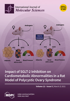
Polycystic ovary syndrome (PCOS) is the most common endocrine disorder in reproductive-age women. PCOS is characterized by hyperandrogenism and ovulatory dysfunction. Women with PCOS have a high prevalence of obesity, insulin resistance (IR), increased blood pressure (BP), and activation of the renin angiotensin system (RAS). We hypothesized that hyperandrogenemia upregulates renal sodium-glucose cotransporter-2 (SGLT2) expression and that the SGLT2 inhibitor Empagliflozin ameliorates cardiometabolic complications in a hyperandrogenemic PCOS model. Androgens upregulated renal SGLT2 expression. Empagliflozin attenuated androgen-mediated increases in fat mass, plasma leptin, intra-renal RAS, and BP but failed to decrease plasma insulin, HbA1c, or albuminuria. In summary, SGLT2 inhibition proved beneficial in adiposity and BP reduction in the PCOS model; however, additional therapies may be needed to [...] Read more.
Order results
Result details
Section
Select all
Export citation of selected articles as:
13 pages, 11062 KiB  
Article
Hyaluronic Acid and a Short Peptide Improve the Performance of a PCL Electrospun Fibrous Scaffold Designed for Bone Tissue Engineering Applications
by Dana Rachmiel, Inbar Anconina, Safra Rudnick-Glick, Michal Halperin-Sternfeld, Lihi Adler-Abramovich and Amit Sitt
Int. J. Mol. Sci. 2021, 22(5), 2425; https://doi.org/10.3390/ijms22052425 - 12 Mar 2021
Cited by 20 | Viewed by 4466
Abstract
Bone tissue engineering is a rapidly developing, minimally invasive technique for regenerating lost bone with the aid of biomaterial scaffolds that mimic the structure and function of the extracellular matrix (ECM). Recently, scaffolds made of electrospun fibers have aroused interest due to their [...] Read more.
Bone tissue engineering is a rapidly developing, minimally invasive technique for regenerating lost bone with the aid of biomaterial scaffolds that mimic the structure and function of the extracellular matrix (ECM). Recently, scaffolds made of electrospun fibers have aroused interest due to their similarity to the ECM, and high porosity. Hyaluronic acid (HA) is an abundant component of the ECM and an attractive material for use in regenerative medicine; however, its processability by electrospinning is poor, and it must be used in combination with another polymer. Here, we used electrospinning to fabricate a composite scaffold with a core/shell morphology composed of polycaprolactone (PCL) polymer and HA and incorporating a short self-assembling peptide. The peptide includes the arginine-glycine-aspartic acid (RGD) motif and supports cellular attachment based on molecular recognition. Electron microscopy imaging demonstrated that the fibrous network of the scaffold resembles the ECM structure. In vitro biocompatibility assays revealed that MC3T3-E1 preosteoblasts adhered well to the scaffold and proliferated, with significant osteogenic differentiation and calcium mineralization. Our work emphasizes the potential of this multi-component approach by which electrospinning, molecular self-assembly, and molecular recognition motifs are combined, to generate a leading candidate to serve as a scaffold for bone tissue engineering. Full article
(This article belongs to the Special Issue Molecular Recognition in Biological and Bioengineered Systems)
Show Figures

Figure 1

27 pages, 2435 KiB  
Review
TRP Channels as Cellular Targets of Particulate Matter
by Alina Milici and Karel Talavera
Int. J. Mol. Sci. 2021, 22(5), 2783; https://doi.org/10.3390/ijms22052783 - 09 Mar 2021
Cited by 18 | Viewed by 4546
Abstract
Particulate matter (PM) is constituted by particles with sizes in the nanometer to micrometer scales. PM can be generated from natural sources such as sandstorms and wildfires, and from human activities, including combustion of fuels, manufacturing and construction or specially engineered for applications [...] Read more.
Particulate matter (PM) is constituted by particles with sizes in the nanometer to micrometer scales. PM can be generated from natural sources such as sandstorms and wildfires, and from human activities, including combustion of fuels, manufacturing and construction or specially engineered for applications in biotechnology, food industry, cosmetics, electronics, etc. Due to their small size PM can penetrate biological tissues, interact with cellular components and induce noxious effects such as disruptions of the cytoskeleton and membranes and the generation of reactive oxygen species. Here, we provide an overview on the actions of PM on transient receptor potential (TRP) proteins, a superfamily of cation-permeable channels with crucial roles in cell signaling. Their expression in epithelial cells and sensory innervation and their high sensitivity to chemical, thermal and mechanical stimuli makes TRP channels prime targets in the major entry routes of noxious PM, which may result in respiratory, metabolic and cardiovascular disorders. On the other hand, the interactions between TRP channel and engineered nanoparticles may be used for targeted drug delivery. We emphasize in that much further research is required to fully characterize the mechanisms underlying PM-TRP channel interactions and their relevance for PM toxicology and biomedical applications. Full article
Show Figures

Figure 1

26 pages, 5689 KiB  
Article
The Leukotriene Receptor Antagonist Montelukast Attenuates Neuroinflammation and Affects Cognition in Transgenic 5xFAD Mice
by Johanna Michael, Julia Zirknitzer, Michael Stefan Unger, Rodolphe Poupardin, Tanja Rieß, Nadine Paiement, Horst Zerbe, Birgit Hutter-Paier, Herbert Reitsamer and Ludwig Aigner
Int. J. Mol. Sci. 2021, 22(5), 2782; https://doi.org/10.3390/ijms22052782 - 09 Mar 2021
Cited by 16 | Viewed by 4449
Abstract
Alzheimer’s disease (AD) is the most common form of dementia. In particular, neuroinflammation, mediated by microglia cells but also through CD8+ T-cells, actively contributes to disease pathology. Leukotrienes are involved in neuroinflammation and in the pathological hallmarks of AD. In consequence, leukotriene signaling—more [...] Read more.
Alzheimer’s disease (AD) is the most common form of dementia. In particular, neuroinflammation, mediated by microglia cells but also through CD8+ T-cells, actively contributes to disease pathology. Leukotrienes are involved in neuroinflammation and in the pathological hallmarks of AD. In consequence, leukotriene signaling—more specifically, the leukotriene receptors—has been recognized as a potential drug target to ameliorate AD pathology. Here, we analyzed the effects of the leukotriene receptor antagonist montelukast (MTK) on hippocampal gene expression in 5xFAD mice, a commonly used transgenic AD mouse model. We identified glial activation and neuroinflammation as the main pathways modulated by MTK. The treatment increased the number of Tmem119+ microglia and downregulated genes related to AD-associated microglia and to lipid droplet-accumulating microglia, suggesting that the MTK treatment targets and modulates microglia phenotypes in the disease model compared to the vehicle. MTK treatment further reduced infiltration of CD8+T-cells into the brain parenchyma. Finally, MTK treatment resulted in improved cognitive functions. In summary, we provide a proof of concept for MTK to be a potential drug candidate for AD and provide novel modes of action via modulation of microglia and CD8+ T-cells. Of note, 5xFAD females showed a more severe pathology, and in consequence, MTK treatment had a more pronounced effect in the females compared to the males. The effects on neuroinflammation, i.e., microglia and CD8+ T-cells, as well as the effects on cognitive outcome, were dose-dependent, therefore arguing for the use of higher doses of MTK in AD clinical trials compared to the approved asthma dose. Full article
(This article belongs to the Special Issue Molecular, Cellular and Systemic Signature of Microglia)
Show Figures

Figure 1

24 pages, 3128 KiB  
Article
Vitamin D Compounds PRI-2191 and PRI-2205 Enhance Anastrozole Activity in Human Breast Cancer Models
by Beata Filip-Psurska, Mateusz Psurski, Artur Anisiewicz, Patrycja Libako, Ewa Zbrojewicz, Magdalena Maciejewska, Michał Chodyński, Andrzej Kutner and Joanna Wietrzyk
Int. J. Mol. Sci. 2021, 22(5), 2781; https://doi.org/10.3390/ijms22052781 - 09 Mar 2021
Cited by 9 | Viewed by 3475
Abstract
1,25-Dihydroxycholecalciferol, the hormonally active vitamin D3 metabolite, is known to exhibit therapeutic effects against breast cancer, mainly by lowering the expression of estrogen receptors and aromatase activity. Previously, the safety of the vitamin D active metabolite (24R)-1,24-dihydroxycholecalciferol (PRI-2191) and 1,25(OH) [...] Read more.
1,25-Dihydroxycholecalciferol, the hormonally active vitamin D3 metabolite, is known to exhibit therapeutic effects against breast cancer, mainly by lowering the expression of estrogen receptors and aromatase activity. Previously, the safety of the vitamin D active metabolite (24R)-1,24-dihydroxycholecalciferol (PRI-2191) and 1,25(OH)2D3 analog PRI-2205 was tested, and the in vitro activity of these analogs against different cancer cell lines was studied. We determined the effect of the two vitamin D compounds on anastrozole (An) activity against breast cancer based on antiproliferative activity, ELISA, flow cytometry, enzyme inhibition potency, PCR, and xenograft study. Both the vitamin D active metabolite and synthetic analog regulated the growth of not only estrogen receptor-positive cells (T47D and MCF-7, in vitro and in vivo), but also hormone-independent cancer cells such as SKBR-3 (HER-2-positive) and MDA-MB-231 (triple-negative), despite their relatively low VDR expression. Combined with An, PRI-2191 and PRI-2205 significantly inhibited the tumor growth of MCF-7 cells. Potentiation of the antitumor activity in combined treatment of MCF-7 tumor-bearing mice is related to the reduced activity of aromatase by both An (enzyme inhibition) and vitamin D compounds (switched off/decreased aromatase gene expression, decreased expression of other genes related to estrogen signaling) and by regulation of the expression of the estrogen receptor ERα and VDR. Full article
Show Figures

Figure 1

21 pages, 4452 KiB  
Article
Microglial Heterogeneity and Its Potential Role in Driving Phenotypic Diversity of Alzheimer’s Disease
by Stefano Sorrentino, Roberto Ascari, Emanuela Maderna, Marcella Catania, Bernardino Ghetti, Fabrizio Tagliavini, Giorgio Giaccone and Giuseppe Di Fede
Int. J. Mol. Sci. 2021, 22(5), 2780; https://doi.org/10.3390/ijms22052780 - 09 Mar 2021
Cited by 11 | Viewed by 3429
Abstract
Alzheimer’s disease (AD) is increasingly recognized as a highly heterogeneous disorder occurring under distinct clinical and neuropathological phenotypes. Despite the molecular determinants of such variability not being well defined yet, microglial cells may play a key role in this process by releasing distinct [...] Read more.
Alzheimer’s disease (AD) is increasingly recognized as a highly heterogeneous disorder occurring under distinct clinical and neuropathological phenotypes. Despite the molecular determinants of such variability not being well defined yet, microglial cells may play a key role in this process by releasing distinct pro- and/or anti-inflammatory cytokines, potentially affecting the expression of the disease. We carried out a neuropathological and biochemical analysis on a series of AD brain samples, gathering evidence about the heterogeneous involvement of microglia in AD. The neuropathological studies showed differences concerning morphology, density and distribution of microglial cells among AD brains. Biochemical investigations showed increased brain levels of IL-4, IL-6, IL-13, CCL17, MMP-7 and CXCL13 in AD in comparison with control subjects. The molecular profiling achieved by measuring the brain levels of 25 inflammatory factors known to be involved in neuroinflammation allowed a stratification of the AD patients in three distinct “neuroinflammatory clusters”. These findings strengthen the relevance of neuroinflammation in AD pathogenesis suggesting, in particular, that the differential involvement of neuroinflammatory molecules released by microglial cells during the development of the disease may contribute to modulate the characteristics and the severity of the neuropathological changes, driving—at least in part—the AD phenotypic diversity. Full article
(This article belongs to the Special Issue Microglia Heterogeneity and Its Relevance for Translational Research)
Show Figures

Graphical abstract

19 pages, 2328 KiB  
Article
Plumbagin, a Biomolecule with (Anti)Osteoclastic Properties
by Sevinj Sultanli, Soni Ghumnani, Richa Ashma and Katharina F. Kubatzky
Int. J. Mol. Sci. 2021, 22(5), 2779; https://doi.org/10.3390/ijms22052779 - 09 Mar 2021
Cited by 10 | Viewed by 3483
Abstract
Plumbagin is a plant-derived naphthoquinone that is widely used in traditional Asian medicine due to its anti-inflammatory and anti-microbial properties. Additionally, plumbagin is cytotoxic for cancer cells due to its ability to trigger reactive oxygen species (ROS) formation and subsequent apoptosis. Since it [...] Read more.
Plumbagin is a plant-derived naphthoquinone that is widely used in traditional Asian medicine due to its anti-inflammatory and anti-microbial properties. Additionally, plumbagin is cytotoxic for cancer cells due to its ability to trigger reactive oxygen species (ROS) formation and subsequent apoptosis. Since it was reported that plumbagin may inhibit the differentiation of bone resorbing osteoclasts in cancer-related models, we wanted to elucidate whether plumbagin interferes with cytokine-induced osteoclastogenesis. Using C57BL/6 mice, we unexpectedly found that plumbagin treatment enhanced osteoclast formation and that this effect was most pronounced when cells were pre-treated for 24 h with plumbagin before subsequent M-CSF/RANKL stimulation. Plumbagin caused a fast induction of NFATc1 signalling and mTOR-dependent activation of p70S6 kinase which resulted in the initiation of protein translation. In line with this finding, we observed an increase in RANK surface expression after Plumbagin stimulation that enhanced the responsiveness for subsequent RANKL treatment. However, in Balb/c mice and Balb/c-derived RAW264.7 macrophages, these findings could not be corroborated and osteoclastogenesis was inhibited. Our results suggest that the effects of plumbagin depend on the model system used and can therefore either trigger or inhibit osteoclast formation. Full article
Show Figures

Figure 1

23 pages, 1663 KiB  
Review
Prominent Role of Histone Modifications in the Regulation of Tumor Metastasis
by Mariam Markouli, Dimitrios Strepkos, Efthimia K. Basdra, Athanasios G. Papavassiliou and Christina Piperi
Int. J. Mol. Sci. 2021, 22(5), 2778; https://doi.org/10.3390/ijms22052778 - 09 Mar 2021
Cited by 17 | Viewed by 4011
Abstract
Tumor aggressiveness and progression is highly dependent on the process of metastasis, regulated by the coordinated interplay of genetic and epigenetic mechanisms. Metastasis involves several steps of epithelial to mesenchymal transition (EMT), anoikis resistance, intra- and extravasation, and new tissue colonization. EMT is [...] Read more.
Tumor aggressiveness and progression is highly dependent on the process of metastasis, regulated by the coordinated interplay of genetic and epigenetic mechanisms. Metastasis involves several steps of epithelial to mesenchymal transition (EMT), anoikis resistance, intra- and extravasation, and new tissue colonization. EMT is considered as the most critical process allowing cancer cells to switch their epithelial characteristics and acquire mesenchymal properties. Emerging evidence demonstrates that epigenetics mechanisms, DNA methylation, histone modifications, and non-coding RNAs participate in the widespread changes of gene expression that characterize the metastatic phenotype. At the chromatin level, active and repressive histone post-translational modifications (PTM) in association with pleiotropic transcription factors regulate pivotal genes involved in the initiation of the EMT process as well as in intravasation and anoikis resistance, playing a central role in the progression of tumors. Herein, we discuss the main epigenetic mechanisms associated with the different steps of metastatic process, focusing in particular on the prominent role of histone modifications and the modifying enzymes that mediate transcriptional regulation of genes associated with tumor progression. We further discuss the development of novel treatment strategies targeting the reversibility of histone modifications and highlight their importance in the future of cancer therapy. Full article
Show Figures

Figure 1

17 pages, 1757 KiB  
Review
Bcl-xL: A Focus on Melanoma Pathobiology
by Anna Maria Lucianò, Ana B. Pérez-Oliva, Victoriano Mulero and Donatella Del Bufalo
Int. J. Mol. Sci. 2021, 22(5), 2777; https://doi.org/10.3390/ijms22052777 - 09 Mar 2021
Cited by 16 | Viewed by 3468
Abstract
Apoptosis is the main mechanism by which multicellular organisms eliminate damaged or unwanted cells. To regulate this process, a balance between pro-survival and pro-apoptotic proteins is necessary in order to avoid impaired apoptosis, which is the cause of several pathologies, including cancer. Among [...] Read more.
Apoptosis is the main mechanism by which multicellular organisms eliminate damaged or unwanted cells. To regulate this process, a balance between pro-survival and pro-apoptotic proteins is necessary in order to avoid impaired apoptosis, which is the cause of several pathologies, including cancer. Among the anti-apoptotic proteins, Bcl-xL exhibits a high conformational flexibility, whose regulation is strictly controlled by alternative splicing and post-transcriptional regulation mediated by transcription factors or microRNAs. It shows relevant functions in different forms of cancer, including melanoma. In melanoma, Bcl-xL contributes to both canonical roles, such as pro-survival, protection from apoptosis and induction of drug resistance, and non-canonical functions, including promotion of cell migration and invasion, and angiogenesis. Growing evidence indicates that Bcl-xL inhibition can be helpful for cancer patients, but at present, effective and safe therapies targeting Bcl-xL are lacking due to toxicity to platelets. In this review, we summarized findings describing the mechanisms of Bcl-xL regulation, and the role that Bcl-xL plays in melanoma pathobiology and response to therapy. From these findings, it emerged that even if Bcl-xL plays a crucial role in melanoma pathobiology, we need further studies aimed at evaluating the involvement of Bcl-xL and other members of the Bcl-2 family in the progression of melanoma and at identifying new non-toxic Bcl-xL inhibitors. Full article
(This article belongs to the Special Issue Advances in Bcl-xL Research 2.0)
Show Figures

Figure 1

18 pages, 1921 KiB  
Review
Interactions of Lipid Droplets with the Intracellular Transport Machinery
by Selma Yilmaz Dejgaard and John F. Presley
Int. J. Mol. Sci. 2021, 22(5), 2776; https://doi.org/10.3390/ijms22052776 - 09 Mar 2021
Cited by 11 | Viewed by 6289
Abstract
Historically, studies of intracellular membrane trafficking have focused on the secretory and endocytic pathways and their major organelles. However, these pathways are also directly implicated in the biogenesis and function of other important intracellular organelles, the best studied of which are peroxisomes and [...] Read more.
Historically, studies of intracellular membrane trafficking have focused on the secretory and endocytic pathways and their major organelles. However, these pathways are also directly implicated in the biogenesis and function of other important intracellular organelles, the best studied of which are peroxisomes and lipid droplets. There is a large recent body of work on these organelles, which have resulted in the introduction of new paradigms regarding the roles of membrane trafficking organelles. In this review, we discuss the roles of membrane trafficking in the life cycle of lipid droplets. This includes the complementary roles of lipid phase separation and proteins in the biogenesis of lipid droplets from endoplasmic reticulum (ER) membranes, and the attachment of mature lipid droplets to membranes by lipidic bridges and by more conventional protein tethers. We also discuss the catabolism of neutral lipids, which in part results from the interaction of lipid droplets with cytosolic molecules, but with important roles for both macroautophagy and microautophagy. Finally, we address their eventual demise, which involves interactions with the autophagocytotic machinery. We pay particular attention to the roles of small GTPases, particularly Rab18, in these processes. Full article
(This article belongs to the Special Issue Intracellular Membrane Transport: Models and Machines)
Show Figures

Figure 1

15 pages, 2842 KiB  
Article
Deletion of Irf4 in T Cells Suppressed Autoimmune Uveitis and Dysregulated Transcriptional Programs Linked to CD4+ T Cell Differentiation and Metabolism
by Minkyung Kang, Hyun-Su Lee, Jin Kyeong Choi, Cheng-Rong Yu and Charles E. Egwuagu
Int. J. Mol. Sci. 2021, 22(5), 2775; https://doi.org/10.3390/ijms22052775 - 09 Mar 2021
Cited by 3 | Viewed by 2747
Abstract
Interferon regulatory factor-4 (IRF4) and IRF8 regulate differentiation, growth and functions of lymphoid and myeloid cells. Targeted deletion of irf8 in T cells (CD4-IRF8KO) has been shown to exacerbate colitis and experimental autoimmune uveitis (EAU), a mouse model of human uveitis. We therefore [...] Read more.
Interferon regulatory factor-4 (IRF4) and IRF8 regulate differentiation, growth and functions of lymphoid and myeloid cells. Targeted deletion of irf8 in T cells (CD4-IRF8KO) has been shown to exacerbate colitis and experimental autoimmune uveitis (EAU), a mouse model of human uveitis. We therefore generated mice lacking irf4 in T cells (CD4-IRF4KO) and investigated whether expression of IRF4 by T cells is also required for regulating T cells that suppress autoimmune diseases. Surprisingly, we found that CD4-IRF4KO mice are resistant to EAU. Suppression of EAU derived in part from inhibiting pathogenic responses of Th17 cells while inducing expansion of regulatory lymphocytes that secrete IL-10 and/or IL-35 in the eye and peripheral lymphoid tissues. Furthermore, CD4-IRF4KO T cells exhibit alterations in cell metabolism and are defective in the expression of two Ikaros zinc-finger (IKZF) transcription factors (Ikaros, Aiolos) that are required for lymphocyte differentiation, metabolism and cell-fate decisions. Thus, synergistic effects of IRF4 and IkZFs might induce metabolic reprogramming of differentiating lymphocytes and thereby dynamically regulate relative abundance of T and B lymphocyte subsets that mediate immunopathogenic mechanisms during uveitis. Moreover, the diametrically opposite effects of IRF4 and IRF8 during EAU suggests that intrinsic function of IRF4 in T cells might be activating proinflammatory responses while IRF8 promotes expansion of immune-suppressive mechanisms. Full article
(This article belongs to the Section Molecular Immunology)
Show Figures

Figure 1

25 pages, 8426 KiB  
Article
Synthesis and Pharmacological In Vitro Investigations of Novel Shikonin Derivatives with a Special Focus on Cyclopropane Bearing Derivatives
by Nadine Kretschmer, Antje Hufner, Christin Durchschein, Katrin Popodi, Beate Rinner, Birgit Lohberger and Rudolf Bauer
Int. J. Mol. Sci. 2021, 22(5), 2774; https://doi.org/10.3390/ijms22052774 - 09 Mar 2021
Cited by 9 | Viewed by 2631
Abstract
Melanoma is the deadliest form of skin cancer and accounts for about three quarters of all skin cancer deaths. Especially at an advanced stage, its treatment is challenging, and survival rates are very low. In previous studies, we showed that the constituents of [...] Read more.
Melanoma is the deadliest form of skin cancer and accounts for about three quarters of all skin cancer deaths. Especially at an advanced stage, its treatment is challenging, and survival rates are very low. In previous studies, we showed that the constituents of the roots of Onosma paniculata as well as a synthetic derivative of the most active constituent showed promising results in metastatic melanoma cell lines. In the current study, we address the question whether we can generate further derivatives with optimized activity by synthesis. Therefore, we prepared 31, mainly novel shikonin derivatives and screened them in different melanoma cell lines (WM9, WM164, and MUG-Mel2 cells) using the XTT viability assay. We identified (R)-1-(1,4-dihydro-5,8-dihydroxy-1,4-dioxonaphthalen-2-yl)-4-methylpent-3-enyl 2-cyclopropyl-2-oxoacetate as a novel derivative with even higher activity. Furthermore, pharmacological investigations including the ApoToxGloTM Triplex assay, LDH assay, and cell cycle measurements revealed that this compound induced apoptosis and reduced cells in the G1 phase accompanied by an increase of cells in the G2/M phase. Moreover, it showed hardly any effects on the cell membrane integrity. However, it also exhibited cytotoxicity against non-tumorigenic cells. Nevertheless, in summary, we could show that shikonin derivatives might be promising drug leads in the treatment of melanoma. Full article
(This article belongs to the Special Issue Skin Inflammation Aging and Diseases)
Show Figures

Graphical abstract

20 pages, 4204 KiB  
Article
Stress and Nasal Allergy: Corticotropin-Releasing Hormone Stimulates Mast Cell Degranulation and Proliferation in Human Nasal Mucosa
by Mika Yamanaka-Takaichi, Yukari Mizukami, Koji Sugawara, Kishiko Sunami, Yuichi Teranishi, Yukimi Kira, Ralf Paus and Daisuke Tsuruta
Int. J. Mol. Sci. 2021, 22(5), 2773; https://doi.org/10.3390/ijms22052773 - 09 Mar 2021
Cited by 7 | Viewed by 9967
Abstract
Psychological stress exacerbates mast cell (MC)-dependent inflammation, including nasal allergy, but the underlying mechanisms are not thoroughly understood. Because the key stress-mediating neurohormone, corticotropin-releasing hormone (CRH), induces human skin MC degranulation, we hypothesized that CRH may be a key player in stress-aggravated nasal [...] Read more.
Psychological stress exacerbates mast cell (MC)-dependent inflammation, including nasal allergy, but the underlying mechanisms are not thoroughly understood. Because the key stress-mediating neurohormone, corticotropin-releasing hormone (CRH), induces human skin MC degranulation, we hypothesized that CRH may be a key player in stress-aggravated nasal allergy. In the current study, we probed this hypothesis in human nasal mucosa MCs (hM-MCs) in situ using nasal polyp organ culture and tested whether CRH is required for murine M-MC activation by perceived stress in vivo. CRH stimulation significantly increased the number of hM-MCs, stimulated both their degranulation and proliferation ex vivo, and increased stem cell factor (SCF) expression in human nasal mucosa epithelium. CRH also sensitized hM-MCs to further CRH stimulation and promoted a pro-inflammatory hM-MC phenotype. The CRH-induced increase in hM-MCs was mitigated by co-administration of CRH receptor type 1 (CRH-R1)-specific antagonist antalarmin, CRH-R1 small interfering RNA (siRNA), or SCF-neutralizing antibody. In vivo, restraint stress significantly increased the number and degranulation of murine M-MCs compared with sham-stressed mice. This effect was mitigated by intranasal antalarmin. Our data suggest that CRH is a major activator of hM-MC in nasal mucosa, in part via promoting SCF production, and that CRH-R1 antagonists such as antalarmin are promising candidate therapeutics for nasal mucosa neuroinflammation induced by perceived stress. Full article
(This article belongs to the Section Molecular Biology)
Show Figures

Graphical abstract

43 pages, 8002 KiB  
Article
Design, Synthesis, and Evaluation of Novel 3-Carboranyl-1,8-Naphthalimide Derivatives as Potential Anticancer Agents
by Sebastian Rykowski, Dorota Gurda-Woźna, Marta Orlicka-Płocka, Agnieszka Fedoruk-Wyszomirska, Małgorzata Giel-Pietraszuk, Eliza Wyszko, Aleksandra Kowalczyk, Paweł Stączek, Andrzej Bak, Agnieszka Kiliszek, Wojciech Rypniewski and Agnieszka B. Olejniczak
Int. J. Mol. Sci. 2021, 22(5), 2772; https://doi.org/10.3390/ijms22052772 - 09 Mar 2021
Cited by 18 | Viewed by 3411
Abstract
We synthesized a series of novel 3-carboranyl-1,8-naphthalimide derivatives, mitonafide and pinafide analogs, using click chemistry, reductive amination and amidation reactions and investigated their in vitro effects on cytotoxicity, cell death, cell cycle, and the production of reactive oxygen species in a HepG2 cancer [...] Read more.
We synthesized a series of novel 3-carboranyl-1,8-naphthalimide derivatives, mitonafide and pinafide analogs, using click chemistry, reductive amination and amidation reactions and investigated their in vitro effects on cytotoxicity, cell death, cell cycle, and the production of reactive oxygen species in a HepG2 cancer cell line. The analyses showed that modified naphthalic anhydrides and naphthalimides bearing ortho- or meta-carboranes exhibited diversified activity. Naphthalimides were more cytotoxic than naphthalic anhydrides, with the highest IC50 value determined for compound 9 (3.10 µM). These compounds were capable of inducing cell cycle arrest at G0/G1 or G2M phase and promoting apoptosis, autophagy or ferroptosis. The most promising conjugate 35 caused strong apoptosis and induced ROS production, which was proven by the increased level of 2′-deoxy-8-oxoguanosine in DNA. The tested conjugates were found to be weak topoisomerase II inhibitors and classical DNA intercalators. Compounds 33, 34, and 36 fluorescently stained lysosomes in HepG2 cells. Additionally, we performed a similarity-based assessment of the property profile of the conjugates using the principal component analysis. The creation of an inhibitory profile and descriptor-based plane allowed forming a structure–activity landscape. Finally, a ligand-based comparative molecular field analysis was carried out to specify the (un)favorable structural modifications (pharmacophoric pattern) that are potentially important for the quantitative structure–activity relationship modeling of the carborane–naphthalimide conjugates. Full article
(This article belongs to the Section Biochemistry)
Show Figures

Graphical abstract

16 pages, 3351 KiB  
Article
Combined Application of Pan-AKT Inhibitor MK-2206 and BCL-2 Antagonist Venetoclax in B-Cell Precursor Acute Lymphoblastic Leukemia
by Anna Richter, Elisabeth Fischer, Clemens Holz, Julia Schulze, Sandra Lange, Anett Sekora, Gudrun Knuebel, Larissa Henze, Catrin Roolf, Hugo Murua Escobar and Christian Junghanss
Int. J. Mol. Sci. 2021, 22(5), 2771; https://doi.org/10.3390/ijms22052771 - 09 Mar 2021
Cited by 8 | Viewed by 2843
Abstract
Aberrant PI3K/AKT signaling is a hallmark of acute B-lymphoblastic leukemia (B-ALL) resulting in increased tumor cell proliferation and apoptosis deficiency. While previous AKT inhibitors struggled with selectivity, MK-2206 promises meticulous pan-AKT targeting with proven anti-tumor activity. We herein, characterize the effect of MK-2206 [...] Read more.
Aberrant PI3K/AKT signaling is a hallmark of acute B-lymphoblastic leukemia (B-ALL) resulting in increased tumor cell proliferation and apoptosis deficiency. While previous AKT inhibitors struggled with selectivity, MK-2206 promises meticulous pan-AKT targeting with proven anti-tumor activity. We herein, characterize the effect of MK-2206 on B-ALL cell lines and primary samples and investigate potential synergistic effects with BCL-2 inhibitor venetoclax to overcome limitations in apoptosis induction. MK-2206 incubation reduced AKT phosphorylation and influenced downstream signaling activity. Interestingly, after MK-2206 mono application tumor cell proliferation and metabolic activity were diminished significantly independently of basal AKT phosphorylation. Morphological changes but no induction of apoptosis was detected in the observed cell lines. In contrast, primary samples cultivated in a protective microenvironment showed a decrease in vital cells. Combined MK-2206 and venetoclax incubation resulted in partially synergistic anti-proliferative effects independently of application sequence in SEM and RS4;11 cell lines. Venetoclax-mediated apoptosis was not intensified by addition of MK-2206. Functional assessment of BCL-2 inhibition via Bax translocation assay revealed slightly increased pro-apoptotic signaling after combined MK-2206 and venetoclax incubation. In summary, we demonstrate that the pan-AKT inhibitor MK-2206 potently blocks B-ALL cell proliferation and for the first time characterize the synergistic effect of combined MK-2206 and venetoclax treatment in B-ALL. Full article
(This article belongs to the Special Issue Novel Agents and Mechanisms in Acute Leukemias)
Show Figures

Figure 1

20 pages, 2920 KiB  
Article
Preclinical Evaluation of 99mTc-ZHER2:41071, a Second-Generation Affibody-Based HER2-Visualizing Imaging Probe with a Low Renal Uptake
by Maryam Oroujeni, Sara S. Rinne, Anzhelika Vorobyeva, Annika Loftenius, Joachim Feldwisch, Per Jonasson, Vladimir Chernov, Anna Orlova, Fredrik Y. Frejd and Vladimir Tolmachev
Int. J. Mol. Sci. 2021, 22(5), 2770; https://doi.org/10.3390/ijms22052770 - 09 Mar 2021
Cited by 17 | Viewed by 3560
Abstract
Radionuclide imaging of HER2 expression in tumours may enable stratification of patients with breast, ovarian, and gastroesophageal cancers for HER2-targeting therapies. A first-generation HER2-binding affibody molecule [99mTc]Tc-ZHER2:V2 demonstrated favorable imaging properties in preclinical studies. Thereafter, the affibody scaffold has been extensively [...] Read more.
Radionuclide imaging of HER2 expression in tumours may enable stratification of patients with breast, ovarian, and gastroesophageal cancers for HER2-targeting therapies. A first-generation HER2-binding affibody molecule [99mTc]Tc-ZHER2:V2 demonstrated favorable imaging properties in preclinical studies. Thereafter, the affibody scaffold has been extensively modified, which increased its melting point, improved storage stability, and increased hydrophilicity of the surface. In this study, a second-generation affibody molecule (designated ZHER2:41071) with a new improved scaffold has been prepared and characterized. HER2-binding, biodistribution, and tumour-targeting properties of [99mTc]Tc-labelled ZHER2:41071 were investigated. These properties were compared with properties of the first-generation affibody molecules, [99mTc]Tc-ZHER2:V2 and [99mTc]Tc-ZHER2:2395. [99mTc]Tc-ZHER2:41071 bound specifically to HER2 expressing cells with an affinity of 58 ± 2 pM. The renal uptake for [99mTc]Tc-ZHER2:41071 and [99mTc]Tc-ZHER2:V2 was 25–30 fold lower when compared with [99mTc]Tc-ZHER2:2395. The uptake in tumour and kidney for [99mTc]Tc-ZHER2:41071 and [99mTc]Tc-ZHER2:V2 in SKOV-3 xenografts was similar. In conclusion, an extensive re-engineering of the scaffold did not compromise imaging properties of the affibody molecule labelled with 99mTc using a GGGC chelator. The new probe, [99mTc]Tc-ZHER2:41071 provided the best tumour-to-blood ratio compared to HER2-imaging probes for single photon emission computed tomography (SPECT) described in the literature so far. [99mTc]Tc-ZHER2:41071 is a promising candidate for further clinical translation studies. Full article
(This article belongs to the Special Issue Cancer Molecular Imaging 2.0)
Show Figures

Figure 1

48 pages, 22172 KiB  
Review
A Review of the Pharmacological Activities and Recent Synthetic Advances of γ-Butyrolactones
by Joonseong Hur, Jaebong Jang and Jaehoon Sim
Int. J. Mol. Sci. 2021, 22(5), 2769; https://doi.org/10.3390/ijms22052769 - 09 Mar 2021
Cited by 33 | Viewed by 6393
Abstract
γ-Butyrolactone, a five-membered lactone moiety, is one of the privileged structures of diverse natural products and biologically active small molecules. Because of their broad spectrum of biological and pharmacological activities, synthetic methods for γ-butyrolactones have received significant attention from synthetic and [...] Read more.
γ-Butyrolactone, a five-membered lactone moiety, is one of the privileged structures of diverse natural products and biologically active small molecules. Because of their broad spectrum of biological and pharmacological activities, synthetic methods for γ-butyrolactones have received significant attention from synthetic and medicinal chemists for decades. Recently, new developments and improvements in traditional methods have been reported by considering synthetic efficiency, feasibility, and green chemistry. In this review, the pharmacological activities of natural and synthetic γ-butyrolactones are described, including their structures and bioassay methods. Mainly, we summarize recent advances, occurring during the past decade, in the construction of γ-butyrolactone classified based on the bond formation in γ-butyrolactone between (i) C5-O1 bond, (ii) C4-C5 and C2-O1 bonds, (iii) C3-C4 and C2-O1 bonds, (iv) C3-C4 and C5-O1 bonds, (v) C2-C3 and C2-O1 bonds, (vi) C3-C4 bond, and (vii) C2-O1 bond. In addition, the application to the total synthesis of natural products bearing γ-butyrolactone scaffolds is described. Full article
Show Figures

Figure 1

16 pages, 4271 KiB  
Article
Rational Design of Adenylate Kinase Thermostability through Coevolution and Sequence Divergence Analysis
by Jian Chang, Chengxin Zhang, Huaqiang Cheng and Yan-Wen Tan
Int. J. Mol. Sci. 2021, 22(5), 2768; https://doi.org/10.3390/ijms22052768 - 09 Mar 2021
Cited by 6 | Viewed by 2448
Abstract
Protein engineering is actively pursued in industrial and laboratory settings for high thermostability. Among the many protein engineering methods, rational design by bioinformatics provides theoretical guidance without time-consuming experimental screenings. However, most rational design methods either rely on protein tertiary structure information or [...] Read more.
Protein engineering is actively pursued in industrial and laboratory settings for high thermostability. Among the many protein engineering methods, rational design by bioinformatics provides theoretical guidance without time-consuming experimental screenings. However, most rational design methods either rely on protein tertiary structure information or have limited accuracies. We proposed a primary-sequence-based algorithm for increasing the heat resistance of a protein while maintaining its functions. Using adenylate kinase (ADK) family as a model system, this method identified a series of amino acid sites closely related to thermostability. Single- and double-point mutants constructed based on this method increase the thermal denaturation temperature of the mesophilic Escherichia coli (E. coli) ADK by 5.5 and 8.3 °C, respectively, while preserving most of the catalytic function at ambient temperatures. Additionally, the constructed mutants have improved enzymatic activity at higher temperature. Full article
(This article belongs to the Special Issue Enzymes as Biocatalysts: Current Research Trends and Applications)
Show Figures

Figure 1

14 pages, 2574 KiB  
Article
Comparative Analysis of CREB3 and CREB3L2 Protein Expression in HEK293 Cells
by Kentaro Oh-hashi, Ayumi Yamamoto, Ryoichi Murase and Yoko Hirata
Int. J. Mol. Sci. 2021, 22(5), 2767; https://doi.org/10.3390/ijms22052767 - 09 Mar 2021
Cited by 4 | Viewed by 2705
Abstract
We performed a comparative analysis of two ER-resident CREB3 family proteins, CREB3 and CREB3L2, in HEK293 cells using pharmacological and genome editing approaches and identified several differences between the two. Treatment with brefeldin A (BFA) and monensin induced the cleavage of full-length CREB3 [...] Read more.
We performed a comparative analysis of two ER-resident CREB3 family proteins, CREB3 and CREB3L2, in HEK293 cells using pharmacological and genome editing approaches and identified several differences between the two. Treatment with brefeldin A (BFA) and monensin induced the cleavage of full-length CREB3 and CREB3L2; however, the level of the full-length CREB3 protein, but not CREB3L2 protein, was not noticeably reduced by the monensin treatment. On the other hand, treatment with tunicamycin (Tm) shifted the molecular weight of the full-length CREB3L2 protein downward but abolished CREB3 protein expression. Thapsigargin (Tg) significantly increased the expression of only full-length CREB3L2 protein concomitant with a slight increase in the level of its cleaved form. Treatment with cycloheximide and MG132 revealed that both endogenous CREB3 and CREB3L2 are proteasome substrates. In addition, kifunensine, an α-mannosidase inhibitor, significantly increased the levels of both full-length forms. Consistent with these findings, cells lacking SEL1L, a crucial ER-associated protein degradation (ERAD) component, showed increased expression of both full-length CREB3 and CREB3L2; however, cycloheximide treatment downregulated full-length CREB3L2 protein expression more rapidly in SEL1L-deficient cells than the full-length CREB3 protein. Finally, we investigated the induction of the expression of several CREB3 and CREB3L2 target genes by Tg and BFA treatments and SEL1L deficiency. In conclusion, this study suggests that both endogenous full-length CREB3 and CREB3L2 are substrates for ER-associated protein degradation but are partially regulated by distinct mechanisms, each of which contributes to unique cellular responses that are distinct from canonical ER signals. Full article
(This article belongs to the Special Issue Physio-Pathological Role of ERAD and Its Pharmacological Modulation)
Show Figures

Figure 1

23 pages, 7888 KiB  
Article
Gene Expression-Related Changes in Morphologies of Organelles and Cellular Component Organization in Mucopolysaccharidoses
by Lidia Gaffke, Karolina Pierzynowska, Estera Rintz, Zuzanna Cyske, Izabela Giecewicz and Grzegorz Węgrzyn
Int. J. Mol. Sci. 2021, 22(5), 2766; https://doi.org/10.3390/ijms22052766 - 09 Mar 2021
Cited by 20 | Viewed by 2706
Abstract
Mucopolysaccharidoses (MPS) are inherited metabolic diseases characterized by accumulation of incompletely degraded glycosaminoglycans (GAGs) in lysosomes. Although primary causes of these diseases are mutations in genes coding for enzymes involved in lysosomal GAG degradation, it was demonstrated that storage of these complex carbohydrates [...] Read more.
Mucopolysaccharidoses (MPS) are inherited metabolic diseases characterized by accumulation of incompletely degraded glycosaminoglycans (GAGs) in lysosomes. Although primary causes of these diseases are mutations in genes coding for enzymes involved in lysosomal GAG degradation, it was demonstrated that storage of these complex carbohydrates provokes a cascade of secondary and tertiary changes affecting cellular functions. Potentially, this might lead to appearance of cellular disorders which could not be corrected even if the primary cause of the disease is removed. In this work, we studied changes in cellular organelles in MPS fibroblasts relative to control cells. All 11 types and subtypes of MPS were included into this study to obtain a complex picture of changes in organelles in this group of diseases. Two experimental approaches were employed, transcriptomic analyses and electron microscopic assessment of morphology of organelles. We analyzed levels of transcripts of genes grouped into two terms included into the QuickGO database, ‘Cellular component organization’ (GO:0016043) and ‘Cellular anatomical entity’ (GO:0110165), to find that number of transcripts with significantly changed levels in MPS fibroblasts vs. controls ranged from 109 to 322 (depending on MPS type) in GO:0016043, and from 70 to 208 in GO:0110165. This dysregulation of expression of genes crucial for proper structures and functions of various organelles was accompanied by severe changes in morphologies of lysosomes, nuclei, mitochondria, Golgi apparatus, and endoplasmic reticulum. Interestingly, some observed changes occurred in all/most MPS types while others were specific to particular disease types/subtypes. We suggest that severe changes in organelles in MPS cells might arise from dysregulation of expression of a battery of genes involved in organelles’ structures and functions. Intriguingly, normalization of GAG levels by using recombinant human enzymes specific to different MPS types corrected morphologies of some, but not all, organelles, while it failed to improve regulation of expression of selected genes. These results might suggest reasons for inability of enzyme replacement therapy to correct all MPS symptoms, particularly if initiated at advanced stages of the disease. Full article
(This article belongs to the Special Issue Genetic and Metabolic Molecular Research of Lysosomal Storage Disease)
Show Figures

Figure 1

21 pages, 4376 KiB  
Article
Butyrate Improves Skin/Lung Fibrosis and Intestinal Dysbiosis in Bleomycin-Induced Mouse Models
by Hee Jin Park, Ok-Yi Jeong, Sung Hak Chun, Yun Hong Cheon, Mingyo Kim, Suhee Kim and Sang-Il Lee
Int. J. Mol. Sci. 2021, 22(5), 2765; https://doi.org/10.3390/ijms22052765 - 09 Mar 2021
Cited by 24 | Viewed by 4808
Abstract
Systemic sclerosis (SSc) is an autoimmune disorder characterized by fibrosis of the skin and internal organs. Despite several studies on SSc treatments, effective treatments for SSc are still lacking. Since evidence suggests an association between intestinal microbiota and SSc, we focused on butyrate, [...] Read more.
Systemic sclerosis (SSc) is an autoimmune disorder characterized by fibrosis of the skin and internal organs. Despite several studies on SSc treatments, effective treatments for SSc are still lacking. Since evidence suggests an association between intestinal microbiota and SSc, we focused on butyrate, which has beneficial effects in autoimmune diseases as a bacterial metabolite. Here, we investigated the therapeutic potential of sodium butyrate (SB) using a bleomycin-induced fibrosis mouse model of SSc and human dermal fibroblasts (HDFs). SB attenuated bleomycin-induced dermal and lung fibrosis in mice. SB influenced fecal microbiota composition (phyla Actinobacteria and Bacteroidetes, genera Bifidobacterium and Ruminococcus_g2). SB controlled macrophage differentiation in mesenteric lymph nodes, spleen, and bronchoalveolar lavage cells of mice with bleomycin-induced skin fibrosis. Profibrotic and proinflammatory gene expression was suppressed by SB administration in skin. Furthermore, SB inhibited transforming growth factor β1-responsive proinflammatory expression with increased acetylation of histone 3 in HDFs. Subcutaneous SB application had antifibrogenic effects on the skin. Butyrate ameliorated skin and lung fibrosis by improving anti-inflammatory activity in a mouse model of SSc. Butyrate may exhibit indirect and direct anti-fibrogenic action on fibroblasts by regulating macrophage differentiation and inhibition of histone deacetylase 3. These findings suggest butyrate as an SSc treatment. Full article
(This article belongs to the Section Molecular Immunology)
Show Figures

Figure 1

17 pages, 3399 KiB  
Article
Reduction of Amyloid Burden by Proliferated Homeostatic Microglia in Toxoplasma gondii-Infected Alzheimer’s Disease Model Mice
by Ji-Hun Shin, Young Sang Hwang, Bong-Kwang Jung, Seung-Hwan Seo, Do-Won Ham and Eun-Hee Shin
Int. J. Mol. Sci. 2021, 22(5), 2764; https://doi.org/10.3390/ijms22052764 - 09 Mar 2021
Cited by 5 | Viewed by 3423
Abstract
In this study, we confirmed that the number of resident homeostatic microglia increases during chronic Toxoplasma gondii infection. Given that the progression of Alzheimer’s disease (AD) worsens with the accumulation of amyloid β (Aβ) plaques, which are eliminated through microglial phagocytosis, we hypothesized [...] Read more.
In this study, we confirmed that the number of resident homeostatic microglia increases during chronic Toxoplasma gondii infection. Given that the progression of Alzheimer’s disease (AD) worsens with the accumulation of amyloid β (Aβ) plaques, which are eliminated through microglial phagocytosis, we hypothesized that T. gondii-induced microglial proliferation would reduce AD progression. Therefore, we investigated the association between microglial proliferation and Aβ plaque burden using brain tissues isolated from 5XFAD AD mice (AD group) and T. gondii-infected AD mice (AD + Toxo group). In the AD + Toxo group, amyloid plaque burden significantly decreased compared with the AD group; conversely, homeostatic microglial proliferation, and number of plaque-associated microglia significantly increased. As most plaque-associated microglia shifted to the disease-associated microglia (DAM) phenotype in both AD and AD + Toxo groups and underwent apoptosis after the lysosomal degradation of phagocytosed Aβ plaques, this indicates that a sustained supply of homeostatic microglia is required for alleviating Aβ plaque burden. Thus, chronic T. gondii infection can induce microglial proliferation in the brains of mice with progressed AD; a sustained supply of homeostatic microglia is a promising prospect for AD treatment. Full article
(This article belongs to the Section Molecular Neurobiology)
Show Figures

Graphical abstract

17 pages, 972 KiB  
Review
Dysbiosis in the Development of Type I Diabetes and Associated Complications: From Mechanisms to Targeted Gut Microbes Manipulation Therapies
by Gratiela Gradisteanu Pircalabioru, Nicolae Corcionivoschi, Ozan Gundogdu, Mariana-Carmen Chifiriuc, Luminita Gabriela Marutescu, Bogdan Ispas and Octavian Savu
Int. J. Mol. Sci. 2021, 22(5), 2763; https://doi.org/10.3390/ijms22052763 - 09 Mar 2021
Cited by 11 | Viewed by 4186
Abstract
Globally, we are facing a worrying increase in type 1 diabetes mellitus (T1DM) incidence, with onset at younger age shedding light on the need to better understand the mechanisms of disease and step-up prevention. Given its implication in immune system development and regulation [...] Read more.
Globally, we are facing a worrying increase in type 1 diabetes mellitus (T1DM) incidence, with onset at younger age shedding light on the need to better understand the mechanisms of disease and step-up prevention. Given its implication in immune system development and regulation of metabolism, there is no surprise that the gut microbiota is a possible culprit behind T1DM pathogenesis. Additionally, microbiota manipulation by probiotics, prebiotics, dietary factors and microbiota transplantation can all modulate early host–microbiota interactions by enabling beneficial microbes with protective potential for individuals with T1DM or at high risk of developing T1DM. In this review, we discuss the challenges and perspectives of translating microbiome data into clinical practice. Nevertheless, this progress will only be possible if we focus our interest on developing numerous longitudinal, multicenter, interventional and double-blind randomized clinical trials to confirm their efficacy and safety of these therapeutic approaches. Full article
(This article belongs to the Special Issue Gut Microbiota and Immunity)
Show Figures

Figure 1

12 pages, 1136 KiB  
Review
Bone Marrow Aspirate Matrix: A Convenient Ally in Regenerative Medicine
by José Fábio Lana, Lucas Furtado da Fonseca, Gabriel Azzini, Gabriel Santos, Marcelo Braga, Alvaro Motta Cardoso Junior, William D. Murrell, Alberto Gobbi, Joseph Purita and Marco Antonio Percope de Andrade
Int. J. Mol. Sci. 2021, 22(5), 2762; https://doi.org/10.3390/ijms22052762 - 09 Mar 2021
Cited by 7 | Viewed by 3140
Abstract
The rise in musculoskeletal disorders has prompted medical experts to devise novel effective alternatives to treat complicated orthopedic conditions. The ever-expanding field of regenerative medicine has allowed researchers to appreciate the therapeutic value of bone marrow-derived biological products, such as the bone marrow [...] Read more.
The rise in musculoskeletal disorders has prompted medical experts to devise novel effective alternatives to treat complicated orthopedic conditions. The ever-expanding field of regenerative medicine has allowed researchers to appreciate the therapeutic value of bone marrow-derived biological products, such as the bone marrow aspirate (BMA) clot, a potent orthobiologic which has often been dismissed and regarded as a technical complication. Numerous in vitro and in vivo studies have contributed to the expansion of medical knowledge, revealing optimistic results concerning the application of autologous bone marrow towards various impactful disorders. The bone marrow accommodates a diverse family of cell populations and a rich secretome; therefore, autologous BMA-derived products such as the “BMA Matrix”, may represent a safe and viable approach, able to reduce the costs and some drawbacks linked to the expansion of bone marrow. BMA provides —it eliminates many hurdles associated with its preparation, especially in regards to regulatory compliance. The BMA Matrix represents a suitable alternative, indicated for the enhancement of tissue repair mechanisms by modulating inflammation and acting as a natural biological scaffold as well as a reservoir of cytokines and growth factors that support cell activity. Although promising, more clinical studies are warranted in order to further clarify the efficacy of this strategy. Full article
(This article belongs to the Section Molecular Biology)
Show Figures

Figure 1

12 pages, 265 KiB  
Review
Machine Learning and Novel Biomarkers for the Diagnosis of Alzheimer’s Disease
by Chun-Hung Chang, Chieh-Hsin Lin and Hsien-Yuan Lane
Int. J. Mol. Sci. 2021, 22(5), 2761; https://doi.org/10.3390/ijms22052761 - 09 Mar 2021
Cited by 80 | Viewed by 9234
Abstract
Background: Alzheimer’s disease (AD) is a complex and severe neurodegenerative disease that still lacks effective methods of diagnosis. The current diagnostic methods of AD rely on cognitive tests, imaging techniques and cerebrospinal fluid (CSF) levels of amyloid-β1-42 (Aβ42), total tau protein and hyperphosphorylated [...] Read more.
Background: Alzheimer’s disease (AD) is a complex and severe neurodegenerative disease that still lacks effective methods of diagnosis. The current diagnostic methods of AD rely on cognitive tests, imaging techniques and cerebrospinal fluid (CSF) levels of amyloid-β1-42 (Aβ42), total tau protein and hyperphosphorylated tau (p-tau). However, the available methods are expensive and relatively invasive. Artificial intelligence techniques like machine learning tools have being increasingly used in precision diagnosis. Methods: We conducted a meta-analysis to investigate the machine learning and novel biomarkers for the diagnosis of AD. Methods: We searched PubMed, the Cochrane Central Register of Controlled Trials, and the Cochrane Database of Systematic Reviews for reviews and trials that investigated the machine learning and novel biomarkers in diagnosis of AD. Results: In additional to Aβ and tau-related biomarkers, biomarkers according to other mechanisms of AD pathology have been investigated. Neuronal injury biomarker includes neurofiliament light (NFL). Biomarkers about synaptic dysfunction and/or loss includes neurogranin, BACE1, synaptotagmin, SNAP-25, GAP-43, synaptophysin. Biomarkers about neuroinflammation includes sTREM2, and YKL-40. Besides, d-glutamate is one of coagonists at the NMDARs. Several machine learning algorithms including support vector machine, logistic regression, random forest, and naïve Bayes) to build an optimal predictive model to distinguish patients with AD from healthy controls. Conclusions: Our results revealed machine learning with novel biomarkers and multiple variables may increase the sensitivity and specificity in diagnosis of AD. Rapid and cost-effective HPLC for biomarkers and machine learning algorithms may assist physicians in diagnosing AD in outpatient clinics. Full article
20 pages, 2299 KiB  
Article
Anti-Apoptotic Effect of Apelin in Human Placenta: Studies on BeWo Cells and Villous Explants from Third-Trimester Human Pregnancy
by Ewa Mlyczyńska, Małgorzata Myszka, Patrycja Kurowska, Monika Dawid, Tomasz Milewicz, Marta Bałajewicz-Nowak, Paweł Kowalczyk and Agnieszka Rak
Int. J. Mol. Sci. 2021, 22(5), 2760; https://doi.org/10.3390/ijms22052760 - 09 Mar 2021
Cited by 16 | Viewed by 2518
Abstract
Previously, we demonstrated the expression of apelin and G-protein-coupled receptor APJ in human placenta cell lines as well as its direct action on placenta cell proliferation and endocrinology. The objective of this study was to examine the effect of apelin on placenta apoptosis [...] Read more.
Previously, we demonstrated the expression of apelin and G-protein-coupled receptor APJ in human placenta cell lines as well as its direct action on placenta cell proliferation and endocrinology. The objective of this study was to examine the effect of apelin on placenta apoptosis in BeWo cells and villous explants from the human third trimester of pregnancy. The BeWo cells and villous explants were incubated with apelin (2 and 20 ng/mL) alone or with staurosporine for 24 to 72 h. First, we analysed the dose- and time-dependent effect of apelin on the expression of apoptotic factors on the mRNA level by real-time PCR and on the protein level using Western blot. Next, we checked caspase 3 and 7 activity by Caspase-Glo 3/7, DNA fragmentation by the Cell Death Detection ELISA kit and oxygen consumption by the MitoXpress-Xtra Oxygen Consumption assay. We found that apelin increased the expression of pro-survival and decreased proapoptotic factors on mRNA and protein levels in both BeWo cells and villous explants. Additionally, apelin inhibited caspase 3 and 7 activity and DNA fragmentation in staurosporine-induced apoptosis as also attenuated oxidative stress by increasing extracellular oxygen consumption. The antiapoptotic effect of apelin in BeWo cells was mediated by the APJ receptor and mitogen-activated protein kinase (ERK1/2/MAP3/1) and protein kinase B (AKT). The obtained results showed the antiapoptotic effect of apelin on trophoblast cells, suggesting its participation in the development of the placenta. Full article
(This article belongs to the Special Issue Adipokines in Health and Diseases)
Show Figures

Figure 1

16 pages, 3740 KiB  
Article
Increased Presence of Complement Factors and Mast Cells in Alveolar Bone and Tooth Resorption
by Kathrin Luntzer, Ina Lackner, Birte Weber, Yvonne Mödinger, Anita Ignatius, Florian Gebhard, Susann-Yvonne Mihaljevic, Melanie Haffner-Luntzer and Miriam Kalbitz
Int. J. Mol. Sci. 2021, 22(5), 2759; https://doi.org/10.3390/ijms22052759 - 09 Mar 2021
Cited by 3 | Viewed by 2325
Abstract
Periodontitis is the inflammatory destruction of the tooth-surrounding and -supporting tissue, resulting at worst in tooth loss. Another locally aggressive disease of the oral cavity is tooth resorption (TR). This is associated with the destruction of the dental mineralized tissue. However, the underlying [...] Read more.
Periodontitis is the inflammatory destruction of the tooth-surrounding and -supporting tissue, resulting at worst in tooth loss. Another locally aggressive disease of the oral cavity is tooth resorption (TR). This is associated with the destruction of the dental mineralized tissue. However, the underlying pathomechanisms remain unknown. The complement system, as well as mast cells (MCs), are known to be involved in osteoclastogenesis and bone loss. The complement factors C3 and C5 were previously identified as key players in periodontal disease. Therefore, we hypothesize that complement factors and MCs might play a role in alveolar bone and tooth resorption. To investigate this, we used the cat as a model because of the naturally occurring high prevalence of both these disorders in this species. Teeth, gingiva samples and serum were collected from domestic cats, which had an appointment for dental treatment under anesthesia, as well as from healthy cats. Histological analyses, immunohistochemical staining and the CH-50 and AH-50 assays revealed increased numbers of osteoclasts and MCs, as well as complement activity in cats with TR. Calcifications score in the gingiva was highest in animals that suffer from TR. This indicates that MCs and the complement system are involved in the destruction of the mineralized tissue in this condition. Full article
(This article belongs to the Special Issue Osteoclastogenesis and Osteogenesis)
Show Figures

Figure 1

17 pages, 1125 KiB  
Review
Assessing Plasmin Generation in Health and Disease
by Adam Miszta, Dana Huskens, Demy Donkervoort, Molly J. M. Roberts, Alisa S. Wolberg and Bas de Laat
Int. J. Mol. Sci. 2021, 22(5), 2758; https://doi.org/10.3390/ijms22052758 - 09 Mar 2021
Cited by 21 | Viewed by 5587
Abstract
Fibrinolysis is an important process in hemostasis responsible for dissolving the clot during wound healing. Plasmin is a central enzyme in this process via its capacity to cleave fibrin. The kinetics of plasmin generation (PG) and inhibition during fibrinolysis have been poorly understood [...] Read more.
Fibrinolysis is an important process in hemostasis responsible for dissolving the clot during wound healing. Plasmin is a central enzyme in this process via its capacity to cleave fibrin. The kinetics of plasmin generation (PG) and inhibition during fibrinolysis have been poorly understood until the recent development of assays to quantify these metrics. The assessment of plasmin kinetics allows for the identification of fibrinolytic dysfunction and better understanding of the relationships between abnormal fibrin dissolution and disease pathogenesis. Additionally, direct measurement of the inhibition of PG by antifibrinolytic medications, such as tranexamic acid, can be a useful tool to assess the risks and effectiveness of antifibrinolytic therapy in hemorrhagic diseases. This review provides an overview of available PG assays to directly measure the kinetics of plasmin formation and inhibition in human and mouse plasmas and focuses on their applications in defining the role of plasmin in diseases, including angioedema, hemophilia, rare bleeding disorders, COVID-19, or diet-induced obesity. Moreover, this review introduces the PG assay as a promising clinical and research method to monitor antifibrinolytic medications and screen for genetic or acquired fibrinolytic disorders. Full article
(This article belongs to the Special Issue Fibrinogen/Fibrin, Factor XIII and Fibrinolysis in Diseases)
Show Figures

Figure 1

15 pages, 23641 KiB  
Review
Diabetes, Obesity, and Inflammation: Impact on Clinical and Radiographic Features of Breast Cancer
by Braden Miller, Hunter Chalfant, Alexandra Thomas, Elizabeth Wellberg, Christina Henson, Molly W. McNally, William E. Grizzle, Ajay Jain and Lacey R. McNally
Int. J. Mol. Sci. 2021, 22(5), 2757; https://doi.org/10.3390/ijms22052757 - 09 Mar 2021
Cited by 17 | Viewed by 3996
Abstract
Obesity, diabetes, and inflammation increase the risk of breast cancer, the most common malignancy in women. One of the mainstays of breast cancer treatment and improving outcomes is early detection through imaging-based screening. There may be a role for individualized imaging strategies for [...] Read more.
Obesity, diabetes, and inflammation increase the risk of breast cancer, the most common malignancy in women. One of the mainstays of breast cancer treatment and improving outcomes is early detection through imaging-based screening. There may be a role for individualized imaging strategies for patients with certain co-morbidities. Herein, we review the literature regarding the accuracy of conventional imaging modalities in obese and diabetic women, the potential role of anti-inflammatory agents to improve detection, and the novel molecular imaging techniques that may have a role for breast cancer screening in these patients. We demonstrate that with conventional imaging modalities, increased sensitivity often comes with a loss of specificity, resulting in unnecessary biopsies and overtreatment. Obese women have body size limitations that impair image quality, and diabetes increases the risk for dense breast tis-sue. Increased density is known to obscure the diagnosis of cancer on routine screening mammography. Novel molecu-lar imaging agents with targets such as estrogen receptor, human epidermal growth factor receptor 2 (HER2), pyrimi-dine analogues, and ligand-targeted receptor probes, among others, have potential to reduce false positive results. They can also improve detection rates with increased resolution and inform therapeutic decision making. These emerg-ing imaging techniques promise to improve breast cancer diagnosis in obese patients with diabetes who have dense breasts, but more work is needed to validate their clinical application. Full article
(This article belongs to the Special Issue Molecular Imaging in Diabetes, Obesity, and Infections 2.0)
Show Figures

Figure 1

33 pages, 727 KiB  
Review
Omics Approaches in Adipose Tissue and Skeletal Muscle Addressing the Role of Extracellular Matrix in Obesity and Metabolic Dysfunction
by Augusto Anguita-Ruiz, Mireia Bustos-Aibar, Julio Plaza-Díaz, Andrea Mendez-Gutierrez, Jesús Alcalá-Fdez, Concepción María Aguilera and Francisco Javier Ruiz-Ojeda
Int. J. Mol. Sci. 2021, 22(5), 2756; https://doi.org/10.3390/ijms22052756 - 09 Mar 2021
Cited by 16 | Viewed by 4933
Abstract
Extracellular matrix (ECM) remodeling plays important roles in both white adipose tissue (WAT) and the skeletal muscle (SM) metabolism. Excessive adipocyte hypertrophy causes fibrosis, inflammation, and metabolic dysfunction in adipose tissue, as well as impaired adipogenesis. Similarly, disturbed ECM remodeling in SM has [...] Read more.
Extracellular matrix (ECM) remodeling plays important roles in both white adipose tissue (WAT) and the skeletal muscle (SM) metabolism. Excessive adipocyte hypertrophy causes fibrosis, inflammation, and metabolic dysfunction in adipose tissue, as well as impaired adipogenesis. Similarly, disturbed ECM remodeling in SM has metabolic consequences such as decreased insulin sensitivity. Most of described ECM molecular alterations have been associated with DNA sequence variation, alterations in gene expression patterns, and epigenetic modifications. Among others, the most important epigenetic mechanism by which cells are able to modulate their gene expression is DNA methylation. Epigenome-Wide Association Studies (EWAS) have become a powerful approach to identify DNA methylation variation associated with biological traits in humans. Likewise, Genome-Wide Association Studies (GWAS) and gene expression microarrays have allowed the study of whole-genome genetics and transcriptomics patterns in obesity and metabolic diseases. The aim of this review is to explore the molecular basis of ECM in WAT and SM remodeling in obesity and the consequences of metabolic complications. For that purpose, we reviewed scientific literature including all omics approaches reporting genetic, epigenetic, and transcriptomic (GWAS, EWAS, and RNA-seq or cDNA arrays) ECM-related alterations in WAT and SM as associated with metabolic dysfunction and obesity. Full article
(This article belongs to the Special Issue Proteolysis of Extracellular Matrix in Human Disease)
Show Figures

Figure 1

16 pages, 2647 KiB  
Article
Epigenetic Changes Governing Scn5a Expression in Denervated Skeletal Muscle
by David Carreras, Rebecca Martinez-Moreno, Mel·lina Pinsach-Abuin, Manel M. Santafe, Pol Gomà, Ramon Brugada, Fabiana S. Scornik, Guillermo J. Pérez and Sara Pagans
Int. J. Mol. Sci. 2021, 22(5), 2755; https://doi.org/10.3390/ijms22052755 - 09 Mar 2021
Cited by 4 | Viewed by 2515
Abstract
The SCN5A gene encodes the α-subunit of the voltage-gated cardiac sodium channel (NaV1.5), a key player in cardiac action potential depolarization. Genetic variants in protein-coding regions of the human SCN5A have been largely associated with inherited cardiac arrhythmias. Increasing evidence also [...] Read more.
The SCN5A gene encodes the α-subunit of the voltage-gated cardiac sodium channel (NaV1.5), a key player in cardiac action potential depolarization. Genetic variants in protein-coding regions of the human SCN5A have been largely associated with inherited cardiac arrhythmias. Increasing evidence also suggests that aberrant expression of the SCN5A gene could increase susceptibility to arrhythmogenic diseases, but the mechanisms governing SCN5A expression are not yet well understood. To gain insights into the molecular basis of SCN5A gene regulation, we used rat gastrocnemius muscle four days following denervation, a process well known to stimulate Scn5a expression. Our results show that denervation of rat skeletal muscle induces the expression of the adult cardiac Scn5a isoform. RNA-seq experiments reveal that denervation leads to significant changes in the transcriptome, with Scn5a amongst the fifty top upregulated genes. Consistent with this increase in expression, ChIP-qPCR assays show enrichment of H3K27ac and H3K4me3 and binding of the transcription factor Gata4 near the Scn5a promoter region. Also, Gata4 mRNA levels are significantly induced upon denervation. Genome-wide analysis of H3K27ac by ChIP-seq suggest that a super enhancer recently described to regulate Scn5a in cardiac tissue is activated in response to denervation. Altogether, our experiments reveal that similar mechanisms regulate the expression of Scn5a in denervated muscle and cardiac tissue, suggesting a conserved pathway for SCN5A expression among striated muscles. Full article
(This article belongs to the Special Issue Epigenetic Mechanisms of Cardiac Disease)
Show Figures

Figure 1

Previous Issue
Next Issue
Back to TopTop