From Cell to Embryo: A Theme Issue Honoring Professor Dr. Roberto Mayor

A special issue of Journal of Developmental Biology (ISSN 2221-3759).

Deadline for manuscript submissions: closed (20 December 2022) | Viewed by 20511

Special Issue Editors


E-Mail Website
Guest Editor
Department of Anatomy and Cell Biology, George Washington University School of Medicine and Health Sciences, Washington, DC, USA
Interests: neural fate specification; Craniofacial development; cranial sensory placodes; Six1; neural gene regulatory networks
Special Issues, Collections and Topics in MDPI journals

E-Mail Website
Guest Editor
Department of Craniofacial Biology, School of Dental Medicine, University of Colorado Anschutz Medical Campus, Aurora, CO 80045, USA
Interests: neural crest development; craniofacial development; zebrafish
Special Issues, Collections and Topics in MDPI journals

Special Issue Information

Dear Colleagues,

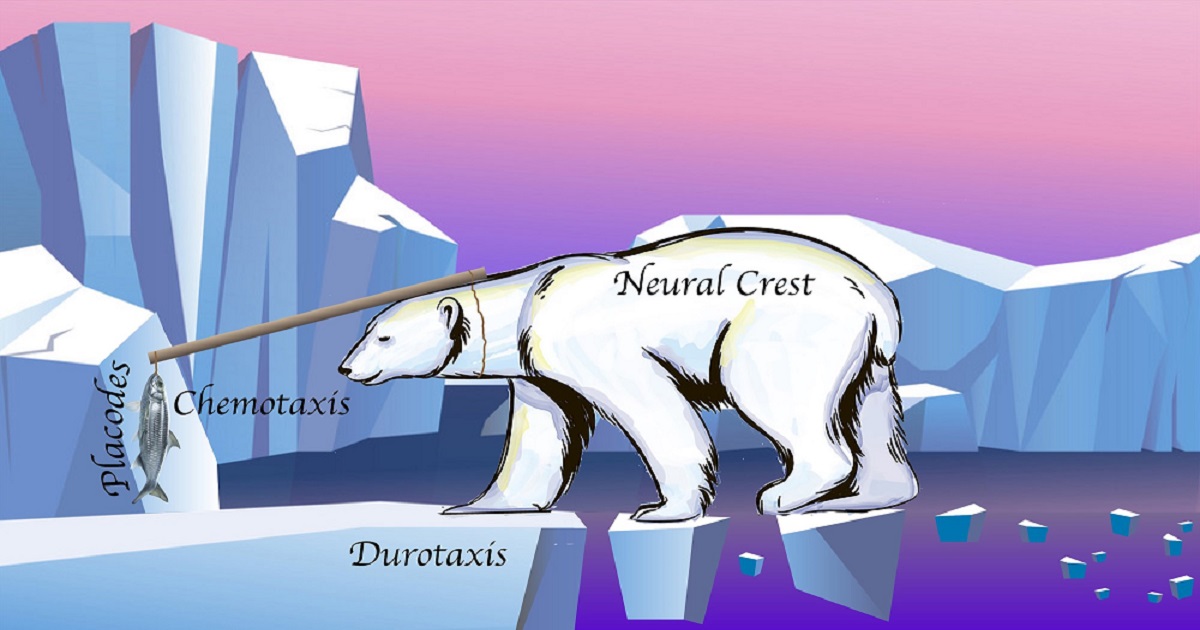
Multicellular organisms begin as a single fertilized egg cell that then divides into a large number of cells that undergo morphogenesis requiring cell–cell interactions and migration in order to create the complex tissues and organs that form the mature organism. During development, embryonic cells acquire a variety of specialized cell fates, communicate with neighbors or distant tissues, migrate as single cells or as a collective to new positions, and form sheets, tubes or blocks of cells that undergo involution, branching or even more complex morphogenetic movements. For over 100 years, classical embryological approaches have been critical for revealing the steps that specify different cell fates, which cells interact when germ layers and tissues are induced, and the process of morphogenetic movements. However, only in the past few decades have the cell biological bases of these developmental processes been elucidated by advances in molecular genetics, reagents for tracking subcellular compartments and organelles and high resolution microscopy. For over 30 years, Professor Roberto Mayor has been a leader in applying the most advanced cell biological techniques to understanding vertebrate development, beginning with his first publication in 1989 on the role of the cytoskeleton in the process of compaction during mammalian blastocyst formation. When early in his career his attention turned to the neural crest cells, he had the opportunity to combine molecular and cell biological approaches to ask how these cells are induced and what controls their directional migration in the embryo. He has had the foresight to investigate both the chemical and the mechanical signals that regulate directed cell movement, and to consider migration from a “supracellular” organization.

This Special Issue is focused on the cell biology of how the single fertilized egg is transformed into a completed individual organism. Many of these processes underlie a variety of congenital defects and thus impact our understanding of childhood disease. The Special Issue will include a short review of Professor Mayor’s contributions to the field, and other original research articles and reviews on all aspects of the cellular processes that regulate developmental events.

Prof. Dr. Sally A. Moody
Prof. Dr. Kristin Bruk Artinger
Guest Editors

Manuscript Submission Information

Manuscripts should be submitted online at www.mdpi.com by registering and logging in to this website. Once you are registered, click here to go to the submission form. Manuscripts can be submitted until the deadline. All submissions that pass pre-check are peer-reviewed. Accepted papers will be published continuously in the journal (as soon as accepted) and will be listed together on the special issue website. Research articles, review articles as well as short communications are invited. For planned papers, a title and short abstract (about 100 words) can be sent to the Editorial Office for announcement on this website.

Submitted manuscripts should not have been published previously, nor be under consideration for publication elsewhere (except conference proceedings papers). All manuscripts are thoroughly refereed through a single-blind peer-review process. A guide for authors and other relevant information for submission of manuscripts is available on the Instructions for Authors page. Journal of Developmental Biology is an international peer-reviewed open access quarterly journal published by MDPI.

Please visit the Instructions for Authors page before submitting a manuscript. The Article Processing Charge (APC) for publication in this open access journal is 1800 CHF (Swiss Francs). Submitted papers should be well formatted and use good English. Authors may use MDPI's English editing service prior to publication or during author revisions.

Keywords

  • cell adhesion
  • signaling pathways
  • cell fate determination
  • morphogenetic movements
  • extracellular matrix
  • cell migration
  • cell polarity

Published Papers (8 papers)

Order results
Result details
Select all
Export citation of selected articles as:

Research

Jump to: Review

15 pages, 4717 KiB  
Article
The Tumor Suppressor Adenomatous Polyposis Coli (apc) Is Required for Neural Crest-Dependent Craniofacial Development in Zebrafish
by Xiaolei Liu, William D. Jones, Mathieu Quesnel-Vallières, Sudhish A. Devadiga, Kristin Lorent, Alexander J. Valvezan, Rebecca L. Myers, Ning Li, Christopher J. Lengner, Yoseph Barash, Michael Pack and Peter S. Klein
J. Dev. Biol. 2023, 11(3), 29; https://doi.org/10.3390/jdb11030029 - 29 Jun 2023
Viewed by 1630
Abstract
Neural crest (NC) is a unique vertebrate cell type arising from the border of the neural plate and epidermis that gives rise to diverse tissues along the entire body axis. Roberto Mayor and colleagues have made major contributions to our understanding of NC [...] Read more.
Neural crest (NC) is a unique vertebrate cell type arising from the border of the neural plate and epidermis that gives rise to diverse tissues along the entire body axis. Roberto Mayor and colleagues have made major contributions to our understanding of NC induction, delamination, and migration. We report that a truncating mutation of the classical tumor suppressor Adenomatous Polyposis Coli (apc) disrupts craniofacial development in zebrafish larvae, with a marked reduction in the cranial neural crest (CNC) cells that contribute to mandibular and hyoid pharyngeal arches. While the mechanism is not yet clear, the altered expression of signaling molecules that guide CNC migration could underlie this phenotype. For example, apcmcr/mcr larvae express substantially higher levels of complement c3, which Mayor and colleagues showed impairs CNC cell migration when overexpressed. However, we also observe reduction in stroma-derived factor 1 (sdf1/cxcl12), which is required for CNC migration into the head. Consistent with our previous work showing that APC directly enhances the activity of glycogen synthase kinase 3 (GSK-3) and, independently, that GSK-3 phosphorylates multiple core mRNA splicing factors, we identify 340 mRNA splicing variations in apc mutant zebrafish, including a splice variant that deletes a conserved domain in semaphorin 3f (sema3f), an axonal guidance molecule and a known regulator of CNC migration. Here, we discuss potential roles for apc in CNC development in the context of some of the seminal findings of Mayor and colleagues. Full article
Show Figures

Graphical abstract

16 pages, 6428 KiB  
Article
Neurogenin 2 and Neuronal Differentiation 1 Control Proper Development of the Chick Trigeminal Ganglion and Its Nerve Branches
by Parinaz Bina, Margaret A. Hines, Johena Sanyal and Lisa A. Taneyhill
J. Dev. Biol. 2023, 11(1), 8; https://doi.org/10.3390/jdb11010008 - 19 Feb 2023
Viewed by 1941
Abstract
The trigeminal ganglion contains the cell bodies of sensory neurons comprising cranial nerve V, which relays information related to pain, touch, and temperature from the face and head to the brain. Like other cranial ganglia, the trigeminal ganglion is composed of neuronal derivatives [...] Read more.
The trigeminal ganglion contains the cell bodies of sensory neurons comprising cranial nerve V, which relays information related to pain, touch, and temperature from the face and head to the brain. Like other cranial ganglia, the trigeminal ganglion is composed of neuronal derivatives of two critical embryonic cell types, neural crest and placode cells. Neurogenesis within the cranial ganglia is promoted by Neurogenin 2 (Neurog2), which is expressed in trigeminal placode cells and their neuronal derivatives, and transcriptionally activates neuronal differentiation genes such as Neuronal Differentiation 1 (NeuroD1). Little is known, however, about the role of Neurog2 and NeuroD1 during chick trigeminal gangliogenesis. To address this, we depleted Neurog2 and NeuroD1 from trigeminal placode cells with morpholinos and demonstrated that Neurog2 and NeuroD1 influence trigeminal ganglion development. While knockdown of both Neurog2 and NeuroD1 affected innervation of the eye, Neurog2 and NeuroD1 had opposite effects on ophthalmic nerve branch organization. Taken together, our results highlight, for the first time, functional roles for Neurog2 and NeuroD1 during chick trigeminal gangliogenesis. These studies shed new light on the molecular mechanisms underlying trigeminal ganglion formation and may also provide insight into general cranial gangliogenesis and diseases of the peripheral nervous system. Full article
Show Figures

Figure 1

20 pages, 10348 KiB  
Article
Zebrafish Slit2 and Slit3 Act Together to Regulate Retinal Axon Crossing at the Midline
by Camila Davison, Gabriela Bedó and Flavio R. Zolessi
J. Dev. Biol. 2022, 10(4), 41; https://doi.org/10.3390/jdb10040041 - 23 Sep 2022
Viewed by 2309
Abstract
Slit-Robo signaling regulates midline crossing of commissural axons in different systems. In zebrafish, all retinofugal axons cross at the optic chiasm to innervate the contralateral tectum. Here, the mutant for the Robo2 receptor presents severe axon guidance defects, which were not completely reproduced [...] Read more.
Slit-Robo signaling regulates midline crossing of commissural axons in different systems. In zebrafish, all retinofugal axons cross at the optic chiasm to innervate the contralateral tectum. Here, the mutant for the Robo2 receptor presents severe axon guidance defects, which were not completely reproduced in a Slit2 ligand null mutant. Since slit3 is also expressed around this area at the stage of axon crossing, we decided to analyze the possibility that it collaborates with Slit2 in this process. We found that the disruption of slit3 expression by sgRNA-Cas9 injection caused similar, albeit slightly milder, defects than those of the slit2 mutant, while the same treatment in the slit2−/−mz background caused much more severe defects, comparable to those observed in robo2 mutants. Tracking analysis of in vivo time-lapse experiments indicated differential but complementary functions of these secreted factors in the correction of axon turn errors around the optic chiasm. Interestingly, RT-qPCR analysis showed a mild increase in slit2 expression in slit3-deficient embryos, but not the opposite. Our observations support the previously proposed “repulsive channel” model for Slit-Robo action at the optic chiasm, with both Slits acting in different manners, most probably relating to their different spatial expression patterns. Full article
Show Figures

Graphical abstract

14 pages, 2327 KiB  
Article
The Core Splicing Factors EFTUD2, SNRPB and TXNL4A Are Essential for Neural Crest and Craniofacial Development
by Byung-Yong Park, Melanie Tachi-Duprat, Chibuike Ihewulezi, Arun Devotta and Jean-Pierre Saint-Jeannet
J. Dev. Biol. 2022, 10(3), 29; https://doi.org/10.3390/jdb10030029 - 08 Jul 2022
Cited by 6 | Viewed by 2667
Abstract
Mandibulofacial dysostosis (MFD) is a human congenital disorder characterized by hypoplastic neural-crest-derived craniofacial bones often associated with outer and middle ear defects. There is growing evidence that mutations in components of the spliceosome are a major cause for MFD. Genetic variants affecting the [...] Read more.
Mandibulofacial dysostosis (MFD) is a human congenital disorder characterized by hypoplastic neural-crest-derived craniofacial bones often associated with outer and middle ear defects. There is growing evidence that mutations in components of the spliceosome are a major cause for MFD. Genetic variants affecting the function of several core splicing factors, namely SF3B4, SF3B2, EFTUD2, SNRPB and TXNL4A, are responsible for MFD in five related but distinct syndromes known as Nager and Rodriguez syndromes (NRS), craniofacial microsomia (CFM), mandibulofacial dysostosis with microcephaly (MFDM), cerebro-costo-mandibular syndrome (CCMS) and Burn–McKeown syndrome (BMKS), respectively. Animal models of NRS and MFDM indicate that MFD results from an early depletion of neural crest progenitors through a mechanism that involves apoptosis. Here we characterize the knockdown phenotype of Eftud2, Snrpb and Txnl4a in Xenopus embryos at different stages of neural crest and craniofacial development. Our results point to defects in cranial neural crest cell formation as the likely culprit for MFD associated with EFTUD2, SNRPB and TXNL4A haploinsufficiency, and suggest a commonality in the etiology of these craniofacial spliceosomopathies. Full article
Show Figures

Figure 1

Review

Jump to: Research

15 pages, 546 KiB  
Review
Quantitative Experimental Embryology: A Modern Classical Approach
by Lara Busby, Dillan Saunders, Guillermo Serrano Nájera and Benjamin Steventon
J. Dev. Biol. 2022, 10(4), 44; https://doi.org/10.3390/jdb10040044 - 18 Oct 2022
Cited by 2 | Viewed by 2944
Abstract
Experimental Embryology is often referred to as a classical approach of developmental biology that has been to some extent replaced by the introduction of molecular biology and genetic techniques to the field. Inspired by the combination of this approach with advanced techniques to [...] Read more.
Experimental Embryology is often referred to as a classical approach of developmental biology that has been to some extent replaced by the introduction of molecular biology and genetic techniques to the field. Inspired by the combination of this approach with advanced techniques to uncover core principles of neural crest development by the laboratory of Roberto Mayor, we review key quantitative examples of experimental embryology from recent work in a broad range of developmental biology questions. We propose that quantitative experimental embryology offers essential ways to explore the reaction of cells and tissues to targeted cell addition, removal, and confinement. In doing so, it is an essential methodology to uncover principles of development that remain elusive such as pattern regulation, scaling, and self-organisation. Full article
Show Figures

Figure 1

18 pages, 358 KiB  
Review
Extracellular Vesicles and Membrane Protrusions in Developmental Signaling
by Callie M. Gustafson and Laura S. Gammill
J. Dev. Biol. 2022, 10(4), 39; https://doi.org/10.3390/jdb10040039 - 21 Sep 2022
Cited by 3 | Viewed by 2574
Abstract
During embryonic development, cells communicate with each other to determine cell fate, guide migration, and shape morphogenesis. While the relevant secreted factors and their downstream target genes have been characterized extensively, how these signals travel between embryonic cells is still emerging. Evidence is [...] Read more.
During embryonic development, cells communicate with each other to determine cell fate, guide migration, and shape morphogenesis. While the relevant secreted factors and their downstream target genes have been characterized extensively, how these signals travel between embryonic cells is still emerging. Evidence is accumulating that extracellular vesicles (EVs), which are well defined in cell culture and cancer, offer a crucial means of communication in embryos. Moreover, the release and/or reception of EVs is often facilitated by fine cellular protrusions, which have a history of study in development. However, due in part to the complexities of identifying fragile nanometer-scale extracellular structures within the three-dimensional embryonic environment, the nomenclature of developmental EVs and protrusions can be ambiguous, confounding progress. In this review, we provide a robust guide to categorizing these structures in order to enable comparisons between developmental systems and stages. Then, we discuss existing evidence supporting a role for EVs and fine cellular protrusions throughout development. Full article
18 pages, 3881 KiB  
Review
Sculpting an Embryo: The Interplay between Mechanical Force and Cell Division
by Nawseen Tarannum, Rohan Singh and Sarah Woolner
J. Dev. Biol. 2022, 10(3), 37; https://doi.org/10.3390/jdb10030037 - 01 Sep 2022
Cited by 2 | Viewed by 2804
Abstract
The journey from a single fertilised cell to a multicellular organism is, at the most fundamental level, orchestrated by mitotic cell divisions. Both the rate and the orientation of cell divisions are important in ensuring the proper development of an embryo. Simultaneous with [...] Read more.
The journey from a single fertilised cell to a multicellular organism is, at the most fundamental level, orchestrated by mitotic cell divisions. Both the rate and the orientation of cell divisions are important in ensuring the proper development of an embryo. Simultaneous with cell proliferation, embryonic cells constantly experience a wide range of mechanical forces from their surrounding tissue environment. Cells must be able to read and respond correctly to these forces since they are known to affect a multitude of biological functions, including cell divisions. The interplay between the mechanical environment and cell divisions is particularly crucial during embryogenesis when tissues undergo dynamic changes in their shape, architecture, and overall organisation to generate functional tissues and organs. Here we review our current understanding of the cellular mechanisms by which mechanical force regulates cell division and place this knowledge within the context of embryogenesis and tissue morphogenesis. Full article
Show Figures

Figure 1

14 pages, 2217 KiB  
Review
Feedback Regulation of Signaling Pathways for Precise Pre-Placodal Ectoderm Formation in Vertebrate Embryos
by Tatsuo Michiue and Kohei Tsukano
J. Dev. Biol. 2022, 10(3), 35; https://doi.org/10.3390/jdb10030035 - 26 Aug 2022
Cited by 1 | Viewed by 2261
Abstract
Intracellular signaling pathways are essential to establish embryonic patterning, including embryonic axis formation. Ectodermal patterning is also governed by a series of morphogens. Four ectodermal regions are thought to be controlled by morphogen gradients, but some perturbations are expected to occur during dynamic [...] Read more.
Intracellular signaling pathways are essential to establish embryonic patterning, including embryonic axis formation. Ectodermal patterning is also governed by a series of morphogens. Four ectodermal regions are thought to be controlled by morphogen gradients, but some perturbations are expected to occur during dynamic morphogenetic movement. Therefore, a mechanism to define areas precisely and reproducibly in embryos, including feedback regulation of signaling pathways, is necessary. In this review, we outline ectoderm pattern formation and signaling pathways involved in the establishment of the pre-placodal ectoderm (PPE). We also provide an example of feedback regulation of signaling pathways for robust formation of the PPE, showing the importance of this regulation. Full article
Show Figures

Figure 1

Back to TopTop