


Diagnostics 2024, 14(1), 18; https://doi.org/10.3390/diagnostics14010018 - 21 Dec 2023
Viewed by 800
Abstract
►
Show Figures
The pathways through which mature blood cells in the bone marrow (BM) enter the blood stream and exit the BM, hematopoietic stem cells in the peripheral blood return to the BM, and other substances exit the BM are referred to as the marrow–blood
[...] Read more.
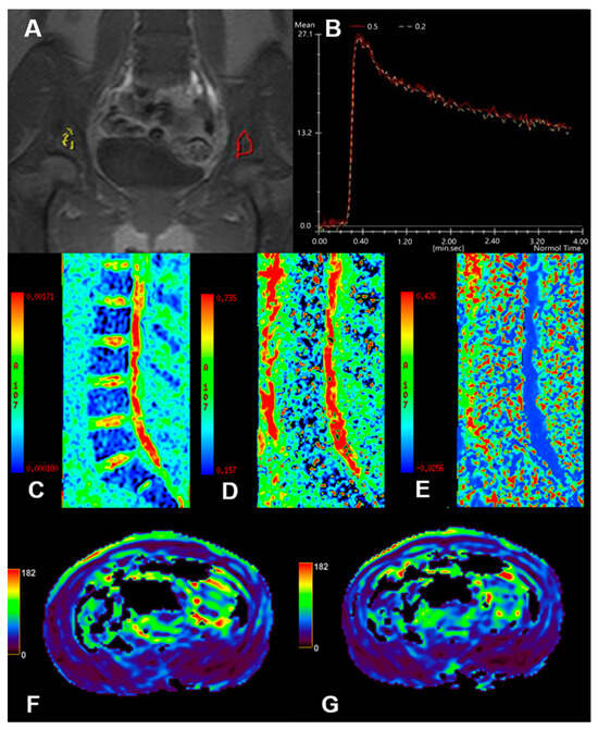
The pathways through which mature blood cells in the bone marrow (BM) enter the blood stream and exit the BM, hematopoietic stem cells in the peripheral blood return to the BM, and other substances exit the BM are referred to as the marrow–blood barrier (MBB). This barrier plays an important role in the restrictive sequestration of blood cells, the release of mature blood cells, and the entry and exit of particulate matter. In some blood diseases and tumors, the presence of immature cells in the blood suggests that the MBB is damaged, mainly manifesting as increased permeability, especially in angiogenesis. Some imaging methods have been used to monitor the integrity and permeability of the MBB, such as DCE-MRI, IVIM, ASL, BOLD-MRI, and microfluidic devices, which contribute to understanding the process of related diseases and developing appropriate treatment options. In this review, we briefly introduce the theory of MBB imaging modalities along with their clinical applications.
Full article

Figure 1
