


Cells 2022, 11(18), 2854; https://doi.org/10.3390/cells11182854 - 13 Sep 2022
Cited by 1 | Viewed by 4077
Abstract
►
Show Figures
Accumulated experimental and clinical evidence supports the development of human allogeneic liver-derived progenitor cells (HALPCs) to treat fibro-inflammatory liver diseases. The aim of the present study was to evaluate their therapeutic effect in a non-alcoholic steatohepatitis (NASH)-STAM mouse model. The immune signaling characteristics
[...] Read more.
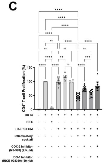
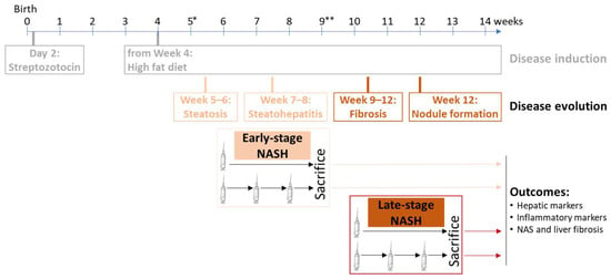
Accumulated experimental and clinical evidence supports the development of human allogeneic liver-derived progenitor cells (HALPCs) to treat fibro-inflammatory liver diseases. The aim of the present study was to evaluate their therapeutic effect in a non-alcoholic steatohepatitis (NASH)-STAM mouse model. The immune signaling characteristics of HALPCs were first assessed in vitro. Upon inflammation treatment, HALPCs secreted large amounts of potent bioactive prostaglandin E2 and indoleamine 2,3-dioxygenase, which significantly reduced CD4+ T-lymphocyte proliferation and secretion of proinflammatory cytokines. In vivo, HALPCs were intravenously administered as single or triple shots (of a dose of 12.5 × 10⁶ cells/kg BW) in STAM mice. Transplantation of HALPCs was associated with a significant decrease in the NAFLD activity score at an early stage and in both inflammation and hepatocyte ballooning scores in late-stage NASH. Sirius red staining analyses revealed decreased collagen deposition in the pericentral region at both stages of NASH. Altogether, these findings showed the anti-inflammatory and anti-fibrotic features of HALPCs in an in vivo NASH model, which suggests their potential to reverse the progression of this chronic fibro-inflammatory disease.
Full article

Figure 1
