Role of Stromal Cells in Determining Tumor and Cancer Stem Cell Behaviors and Therapeutic Response

A topical collection in Cancers (ISSN 2072-6694). This collection belongs to the section "Molecular Cancer Biology".

Viewed by 46213

Editors


E-Mail Website
Collection Editor
Department of Biosciences, Biotechnology and Biopharmaceutics, University of Bari, Via E. Orabona 4, 70126 Bari, Italy
Interests: cancer pathophysiology; tumor microenvironment; pH regulation; invasion; vasculogenesis; hypoxia; breast; pancreatic; prostate

E-Mail Website
Collection Editor
Department of Bioscience, Biotechnology and Biopharmaceutics, University of Bari Aldo Moro, Bari, Italy
Interests: role of tumor microenvironment (TME) in driving PDAC; mechanisms of PDAC metastasis; mechanisms of PDAC therapy resistance; novel chemo- and immunotherapies for PDAC
Special Issues, Collections and Topics in MDPI journals

Topical Collection Information

Dear Colleagues,

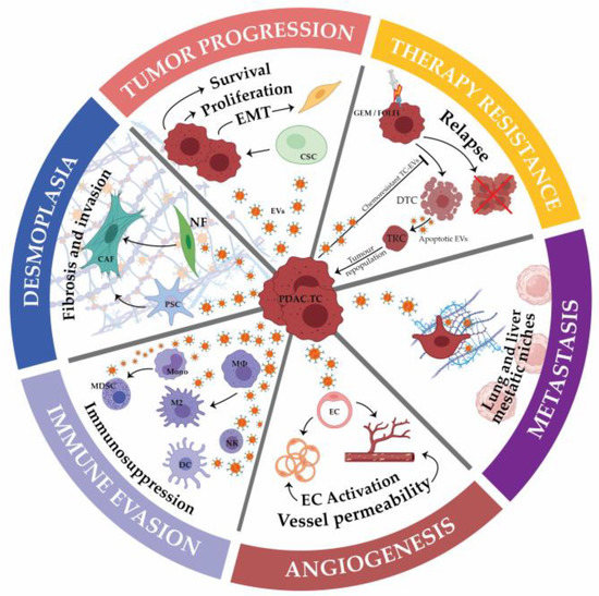
The tumor microenvironment (TME) is composed of stromal extracellular matrix (ECM) components (i.e. laminins, fibronectin, collagens, proteoglycans, elastin) and stromal cells (fibroblasts, endothelial cells, macrophages and lymphocytes). Most of these cellular components produce tumor-supportive ECM and secrete growth factors and chemokines that further alter theECM and generate oncogenic signals, thus playing key roles in tumor transformation, cell proliferation and tissue invasion. Tumor ECM is altered during malignant progression, and its interaction with cancer cells plays an essential role in tumor metabolism, development, progression, recruitment and metabolic reprograming of tumor and stromal cells and treatment response. The TME also includes the tumor metabolic microenvironment (TMM), which is characterized by dynamic, interacting areas of hypoxia, low extracellular pH (pHe) and low nutrients. This adverse pathophysiological TMM is formed by vascular abnormalities, inadequate microcirculation, high vascular permeability and increased interstitial fluid pressure. Beyond hypoxia and acidic pHe, this TMM leads to the upregulation of glycolytic capacity (Warburg effect), lactate accumulation and energy depletion. Of these conditions, the best characterized is hypoxia, which contributes, among other effects, to mutagenesis, suppression of apoptosis, epithelial-to-mesenchymal transition and cancer stem cell (CSC) selection. Highly acidic TME (pHe 6.4-6.8) is the result of elevated metabolic rates in highly proliferative cancer cells, in conjunction with often greatly increased rates of net cellular acid extrusion.

Indeed, the acidic tumor microenvironment is an important contributing factor to the metastasis of cancer cells, via a combination of toxicity to adjacent normal cells, degradation of the ECM through the induced secretion and activation of proteases, increased cancer cell motility and invasion, reduced immunological defenses and stromal cells activation, and/or recruitment to drive malignant progression.

The reciprocal interactions of stromal cell types with neoplastic cells (the bulk parenchymal tumor cells and CSCs), the ECM and the metabolic microenvironment are highly complex and most likely vary between tumor types and stages. Given the highly complex nature of the tumor microenvironment and its components, it is likely that many microenvironmental factors contribute to cancer growth and progression, and that they interact and co-evolve in very complex ways.

The challenge, therefore, is to identify the important aspects of these interactions in the context of metastatic progression. This Topical Collection will present research papers and reviews, exploring the complexities and roles of stromal cells in determining the heterogeneity of tumor properties and behaviors, to improve our understanding of metastatic progression and therapeutic resistance.

Dr. Stephan Joel Reshkin
Dr. Rosa Angela Cardone
Collection Editors

Manuscript Submission Information

Manuscripts should be submitted online at www.mdpi.com by registering and logging in to this website. Once you are registered, click here to go to the submission form. Manuscripts can be submitted until the deadline. All submissions that pass pre-check are peer-reviewed. Accepted papers will be published continuously in the journal (as soon as accepted) and will be listed together on the collection website. Research articles, review articles as well as short communications are invited. For planned papers, a title and short abstract (about 100 words) can be sent to the Editorial Office for announcement on this website.

Submitted manuscripts should not have been published previously, nor be under consideration for publication elsewhere (except conference proceedings papers). All manuscripts are thoroughly refereed through a single-blind peer-review process. A guide for authors and other relevant information for submission of manuscripts is available on the Instructions for Authors page. Cancers is an international peer-reviewed open access semimonthly journal published by MDPI.

Please visit the Instructions for Authors page before submitting a manuscript. The Article Processing Charge (APC) for publication in this open access journal is 2900 CHF (Swiss Francs). Submitted papers should be well formatted and use good English. Authors may use MDPI's English editing service prior to publication or during author revisions.

Keywords

  • stromal cells
  • cancer stem cells
  • tumor stromal and biological microenvironments
  • spatial intratumoral pH
  • pO2
  • and nutrient concentrations

Published Papers (17 papers)

2024

Jump to: 2023, 2022, 2021, 2020

16 pages, 4281 KiB  
Article
Oscillatory Hypoxia Can Induce Senescence of Adipose-Derived Mesenchymal Stromal Cells Potentiating Invasive Transformation of Breast Epithelial Cells
by Ashkan Novin, Khadija Wali, Aditya Pant, Shaofei Liu, Wenqiang Du, Yamin Liu, Lichao Wang, Ming Xu, Binsheng Wang, Yasir Suhail and Kshitiz
Cancers 2024, 16(5), 969; https://doi.org/10.3390/cancers16050969 - 28 Feb 2024
Viewed by 798
Abstract
Obesity is strongly associated with occurrence, metastasis, and resistance to therapy in breast cancers, which also exhibit high adipose content in the tumor microenvironment. Adipose tissue-derived mesenchymal stromal cells (ASCs) are recruited to breast cancer by many mechanisms, including hypoxia, and contribute to [...] Read more.
Obesity is strongly associated with occurrence, metastasis, and resistance to therapy in breast cancers, which also exhibit high adipose content in the tumor microenvironment. Adipose tissue-derived mesenchymal stromal cells (ASCs) are recruited to breast cancer by many mechanisms, including hypoxia, and contribute to metastatic transition of the cancer. Breast cancers are characterized by regions of hypoxia, which can be temporally unstable owing to a mismatch between oxygen supply and consumption. Using a high-sensitivity nanopatterned stromal invasion assay, we found that ASCs could promote stromal invasion of not only breast cancer cell lines but also MCF10A1, a cell line derived from untransformed breast epithelium. RNA sequencing of MCF10A1 cells conditioned with medium from ASCs revealed upregulation of genes associated with increased cell migration, chemotaxis, and metastasis. Furthermore, we found that fluctuating or oscillating hypoxia could induce senescence in ASCs, which could result in an increased invasive potential in the treated MCF10A1 cells. These findings highlight the complex interplay within the breast cancer microenvironment, hypoxia, and the role of ASCs in transforming even non-cancerous breast epithelium toward an invasive phenotype, providing insights into early metastatic events. Full article
Show Figures

Figure 1

2023

Jump to: 2024, 2022, 2021, 2020

18 pages, 1905 KiB  
Article
Extracellular Matrix Collagen I Differentially Regulates the Metabolic Plasticity of Pancreatic Ductal Adenocarcinoma Parenchymal Cell and Cancer Stem Cell
by Diana Tavares-Valente, Stefania Cannone, Maria Raffaella Greco, Tiago Miguel Amaral Carvalho, Fátima Baltazar, Odília Queirós, Gennaro Agrimi, Stephan J. Reshkin and Rosa Angela Cardone
Cancers 2023, 15(15), 3868; https://doi.org/10.3390/cancers15153868 - 29 Jul 2023
Cited by 1 | Viewed by 1406
Abstract
Pancreatic ductal adenocarcinoma (PDAC) has a 5-year survival rate of less than 10 percent largely due to the intense fibrotic desmoplastic reaction, characterized by high levels of extracellular matrix (ECM) collagen I that constitutes a niche for a subset of cancer cells, the [...] Read more.
Pancreatic ductal adenocarcinoma (PDAC) has a 5-year survival rate of less than 10 percent largely due to the intense fibrotic desmoplastic reaction, characterized by high levels of extracellular matrix (ECM) collagen I that constitutes a niche for a subset of cancer cells, the cancer stem cells (CSCs). Cancer cells undergo a complex metabolic adaptation characterized by changes in metabolic pathways and biosynthetic processes. The use of the 3D organotypic model in this study allowed us to manipulate the ECM constituents and mimic the progression of PDAC from an early tumor to an ever more advanced tumor stage. To understand the role of desmoplasia on the metabolism of PDAC parenchymal (CPC) and CSC populations, we studied their basic metabolic parameters in organotypic cultures of increasing collagen content to mimic in vivo conditions. We further measured the ability of the bioenergetic modulators (BMs), 2-deoxyglucose, dichloroacetate and phenformin, to modify their metabolic dependence and the therapeutic activity of paclitaxel albumin nanoparticles (NAB-PTX). While all the BMs decreased cell viability and increased cell death in all ECM types, a distinct, collagen I-dependent profile was observed in CSCs. As ECM collagen I content increased (e.g., more aggressive conditions), the CSCs switched from glucose to mostly glutamine metabolism. All three BMs synergistically potentiated the cytotoxicity of NAB-PTX in both cell lines, which, in CSCs, was collagen I-dependent and the strongest when treated with phenformin + NAB-PTX. Metabolic disruption in PDAC can be useful both as monotherapy or combined with conventional drugs to more efficiently block tumor growth. Full article
Show Figures

Figure 1

2022

Jump to: 2024, 2023, 2021, 2020

12 pages, 2076 KiB  
Article
Cancer Associated Fibroblast (CAF) Regulation of PDAC Parenchymal (CPC) and CSC Phenotypes Is Modulated by ECM Composition
by Stefania Cannone, Maria Raffaella Greco, Tiago M. A. Carvalho, Helene Guizouarn, Olivier Soriani, Daria Di Molfetta, Richard Tomasini, Katrine Zeeberg, Stephan Joel Reshkin and Rosa Angela Cardone
Cancers 2022, 14(15), 3737; https://doi.org/10.3390/cancers14153737 - 31 Jul 2022
Cited by 7 | Viewed by 2197
Abstract
Background: Pancreatic ductal adenocarcinoma (PDAC) is one of the deadliest of all cancers, having one of the lowest five-year survival rates. One of its hallmarks is a dense desmoplastic stroma consisting in the abnormal accumulation of extracellular matrix (ECM) components, especially Collagen I. [...] Read more.
Background: Pancreatic ductal adenocarcinoma (PDAC) is one of the deadliest of all cancers, having one of the lowest five-year survival rates. One of its hallmarks is a dense desmoplastic stroma consisting in the abnormal accumulation of extracellular matrix (ECM) components, especially Collagen I. This highly fibrotic stroma embeds the bulk cancer (parenchymal) cells (CPCs), cancer stem cells (CSCs) and the main producers of the stromal reaction, the Cancer Associated Fibroblasts (CAFs). Little is known about the role of the acellular ECM in the interplay of the CAFs with the different tumor cell types in determining their phenotypic plasticity and eventual cell fate. Methods: Here, we analyzed the role of ECM collagen I in modulating the effect of CAF-derived signals by incubating PDAC CPCs and CSCs grown on ECM mimicking early (low collagen I levels) and late (high collagen I levels) stage PDAC stroma with conditioned medium from primary cultured CAFs derived from patients with PDAC in a previously described three-dimensional (3D) organotypic model of PDAC. Results: We found that CAFs (1) reduced CPC growth while favoring CSC growth independently of the ECM; (2) increased the invasive capacity of only CPCs on the ECM mimicking the early tumor; and (3) favored vasculogenic mimicry (VM) especially of the CSCs on the ECM mimicking an early tumor. Conclusions: We conclude that the CAFs and acellular stromal components interact to modulate the tumor behaviors of the PDAC CPC and CSC cell types and drive metastatic progression by stimulating the phenotypic characteristics of each tumor cell type that contribute to metastasis. Full article
Show Figures

Graphical abstract

23 pages, 4725 KiB  
Article
Metastatic Transition of Pancreatic Ductal Cell Adenocarcinoma Is Accompanied by the Emergence of Pro-Invasive Cancer-Associated Fibroblasts
by Shaofei Liu, Yasir Suhail, Ashkan Novin, Lorrie Perpetua and Kshitiz
Cancers 2022, 14(9), 2197; https://doi.org/10.3390/cancers14092197 - 28 Apr 2022
Cited by 8 | Viewed by 2647
Abstract
Cancer-associated fibroblasts (CAFs) are now appreciated as key regulators of cancer metastasis, particularly in cancers with high stromal content, e.g., pancreatic ductal cell carcinoma (PDAC). However, it is not yet well understood if fibroblasts are always primed to be cooperative in PDAC transition [...] Read more.
Cancer-associated fibroblasts (CAFs) are now appreciated as key regulators of cancer metastasis, particularly in cancers with high stromal content, e.g., pancreatic ductal cell carcinoma (PDAC). However, it is not yet well understood if fibroblasts are always primed to be cooperative in PDAC transition to metastasis, if they undergo transformation which ensures their cooperativity, and if such transformations are cancer-driven or intrinsic to fibroblasts. We performed a fibroblast-centric analysis of PDAC cancer, as it transitioned from the primary site to trespass stromal compartment reaching the lymph node using published single-cell RNA sequencing data by Peng et al. We have characterized the change in fibroblast response to cancer from a normal wound healing response in the initial stages to the emergence of subclasses with myofibroblast and inflammatory fibroblasts such as signatures. We have previously posited “Evolved Levels of Invasibility (ELI)”, a framework describing the evolution of stromal invasability as a selected phenotype, which explains the large and correlated reduction in stromal invasion by placental trophoblasts and cancer cells in certain mammals. Within PDAC samples, we found large changes in fibroblast subclasses at succeeding stages of PDAC progression, with the emergence of specific subclasses when cancer trespasses stroma to metastasize to proximal lymph nodes (stage IIA to IIB). Surprisingly, we found that the initial metastatic transition is accompanied by downregulation of ELI-predicted pro-resistive genes, and the emergence of a subclass of fibroblasts with ELI-predicted increased invasibility. Interestingly, this trend was also observed in stellate cells. Using a larger cohort of bulk RNAseq data from The Cancer Genome Atlas for PDAC cancers, we confirmed that genes describing this emergent fibroblast subclass are also correlated with lymph node metastasis of cancer cells. Experimental testing of selected genes characterizing pro-resistive and pro-invasive fibroblast clusters confirmed their contribution in regulating stromal invasability as a phenotype. Our data confirm that the complexity of stromal response to cancer is really a function of stage-wise emergence of distinct fibroblast clusters, characterized by distinct gene sets which confer initially a predominantly pro-resistive and then a pro-invasive property to the stroma. Stromal response therefore transitions from being tumor-limiting to a pro-metastatic state, facilitating stromal trespass and the onset of metastasis. Full article
Show Figures

Figure 1

19 pages, 1020 KiB  
Review
Caveolae-Associated Molecules, Tumor Stroma, and Cancer Drug Resistance: Current Findings and Future Perspectives
by Jin-Yih Low and Marikki Laiho
Cancers 2022, 14(3), 589; https://doi.org/10.3390/cancers14030589 - 25 Jan 2022
Cited by 7 | Viewed by 3443
Abstract
The discovery of small, “cave-like” invaginations at the plasma membrane, called caveola, has opened up a new and exciting research area in health and diseases revolving around this cellular ultrastructure. Caveolae are rich in cholesterol and orchestrate cellular signaling events. Within caveola, the [...] Read more.
The discovery of small, “cave-like” invaginations at the plasma membrane, called caveola, has opened up a new and exciting research area in health and diseases revolving around this cellular ultrastructure. Caveolae are rich in cholesterol and orchestrate cellular signaling events. Within caveola, the caveola-associated proteins, caveolins and cavins, are critical components for the formation of these lipid rafts, their dynamics, and cellular pathophysiology. Their alterations underlie human diseases such as lipodystrophy, muscular dystrophy, cardiovascular disease, and diabetes. The expression of caveolins and cavins is modulated in tumors and in tumor stroma, and their alterations are connected with cancer progression and treatment resistance. To date, although substantial breakthroughs in cancer drug development have been made, drug resistance remains a problem leading to treatment failures and challenging translation and bench-to-bedside research. Here, we summarize the current progress in understanding cancer drug resistance in the context of caveola-associated molecules and tumor stroma and discuss how we can potentially design therapeutic avenues to target these molecules in order to overcome treatment resistance. Full article
Show Figures

Figure 1

18 pages, 1861 KiB  
Review
The Obscure Potential of AHNAK2
by Mohamed Zardab, Konstantinos Stasinos, Richard P. Grose and Hemant M. Kocher
Cancers 2022, 14(3), 528; https://doi.org/10.3390/cancers14030528 - 21 Jan 2022
Cited by 10 | Viewed by 3344
Abstract
AHNAK2 is a protein discovered in 2004, with a strong association with oncogenesis in various epithelial cancers. It has a large 616 kDa tripartite structure and is thought to take part in the formation of large multi-protein complexes. High expression is found in [...] Read more.
AHNAK2 is a protein discovered in 2004, with a strong association with oncogenesis in various epithelial cancers. It has a large 616 kDa tripartite structure and is thought to take part in the formation of large multi-protein complexes. High expression is found in clear cell renal carcinoma, pancreatic ductal adenocarcinoma, uveal melanoma, and lung adenocarcinoma, with a relation to poor prognosis. Little work has been done in exploring the function and relation AHNAK2 has with cancer, with early studies showing promising potential as a future biomarker and therapeutic target. Full article
Show Figures

Figure 1

2021

Jump to: 2024, 2023, 2022, 2020

17 pages, 6617 KiB  
Article
The J2-Immortalized Murine Macrophage Cell Line Displays Phenotypical and Metabolic Features of Primary BMDMs in Their M1 and M2 Polarization State
by Iolanda Spera, Ricardo Sánchez-Rodríguez, Maria Favia, Alessio Menga, Francisca C. Venegas, Roberta Angioni, Fabio Munari, Martina Lanza, Annalisa Campanella, Ciro L. Pierri, Marcella Canton and Alessandra Castegna
Cancers 2021, 13(21), 5478; https://doi.org/10.3390/cancers13215478 - 31 Oct 2021
Cited by 6 | Viewed by 3432
Abstract
Macrophages are immune cells that are important for the development of the defensive front line of the innate immune system. Following signal recognition, macrophages undergo activation toward specific functional states, consisting not only in the acquisition of specific features but also of peculiar [...] Read more.
Macrophages are immune cells that are important for the development of the defensive front line of the innate immune system. Following signal recognition, macrophages undergo activation toward specific functional states, consisting not only in the acquisition of specific features but also of peculiar metabolic programs associated with each function. For these reasons, macrophages are often isolated from mice to perform cellular assays to study the mechanisms mediating immune cell activation. This requires expensive and time-consuming breeding and housing of mice strains. To overcome this issue, we analyzed an in-house J2-generated immortalized macrophage cell line from BMDMs, both from a functional and metabolic point of view. By assaying the intracellular and extracellular metabolism coupled with the phenotypic features of immortalized versus primary BMDMs, we concluded that classically and alternatively immortalized macrophages display similar phenotypical, metabolic and functional features compared to primary cells polarized in the same way. Our study validates the use of this immortalized cell line as a suitable model with which to evaluate in vitro how perturbations can influence the phenotypical and functional features of murine macrophages. Full article
Show Figures

Figure 1

12 pages, 1905 KiB  
Article
Bisphenol A Alters the Energy Metabolism of Stromal Cells and Could Promote Bladder Cancer Progression
by Ève Pellerin, Stéphane Chabaud, Frédéric Pouliot, Martin Pelletier and Stéphane Bolduc
Cancers 2021, 13(21), 5461; https://doi.org/10.3390/cancers13215461 - 30 Oct 2021
Cited by 10 | Viewed by 2135
Abstract
Bisphenol A (BPA) is an endocrine-disrupting molecule used in plastics. Through its release in food and the environment, BPA can be found in humans and is mostly excreted in urine. The bladder is therefore continuously exposed to this compound. BPA can bind to [...] Read more.
Bisphenol A (BPA) is an endocrine-disrupting molecule used in plastics. Through its release in food and the environment, BPA can be found in humans and is mostly excreted in urine. The bladder is therefore continuously exposed to this compound. BPA can bind to multiple cell receptors involved in proliferation, migration and invasion pathways, and exposure to BPA is associated with cancer progression. Considering the physiological concentrations of BPA in urine, we tested the effect of nanomolar concentrations of BPA on the metabolism of bladder fibroblasts and cancer-associated fibroblasts (CAFs). Our results show that BPA led to a decreased metabolism in fibroblasts, which could alter the extracellular matrix. Furthermore, CAF induction triggered a metabolic switch, similar to the Warburg effect described in cancer cells. Additionally, we demonstrated that nanomolar concentrations of BPA could exacerbate this metabolic switch observed in CAFs via an increased glycolytic metabolism, leading to greater acidification of the extracellular environment. These findings suggest that chronic exposure to BPA could promote cancer progression through an alteration of the metabolism of stromal cells. Full article
Show Figures

Figure 1

23 pages, 2241 KiB  
Review
Emerging Bone Marrow Microenvironment-Driven Mechanisms of Drug Resistance in Acute Myeloid Leukemia: Tangle or Chance?
by Marilena Ciciarello, Giulia Corradi, Dorian Forte, Michele Cavo and Antonio Curti
Cancers 2021, 13(21), 5319; https://doi.org/10.3390/cancers13215319 - 22 Oct 2021
Cited by 15 | Viewed by 3037
Abstract
Acute myeloid leukemia (AML) has been considered for a long time exclusively driven by critical mutations in hematopoietic stem cells. Recently, the contribution of further players, such as stromal and immune bone marrow (BM) microenvironment components, to AML onset and progression has been [...] Read more.
Acute myeloid leukemia (AML) has been considered for a long time exclusively driven by critical mutations in hematopoietic stem cells. Recently, the contribution of further players, such as stromal and immune bone marrow (BM) microenvironment components, to AML onset and progression has been pointed out. In particular, mesenchymal stromal cells (MSCs) steadily remodel the leukemic niche, not only favoring leukemic cell growth and development but also tuning their responsiveness to treatments. The list of mechanisms driven by MSCs to promote a leukemia drug-resistant phenotype has progressively expanded. Moreover, the relative proportion and the activation status of immune cells in the BM leukemic microenvironment may vary by influencing their reactivity against leukemic cells. In that, the capacity of the stroma to re-program immune cells, thus promoting and/or hampering therapeutic efficacy, is emerging as a crucial aspect in AML biology, adding an extra layer of complexity. Current treatments for AML have mainly focused on eradicating leukemia cells, with little consideration for the leukemia-damaged BM niche. Increasing evidence on the contribution of stromal and immune cells in response to therapy underscores the need to hold the mutual interplay, which takes place in the BM. A careful dissection of these interactions will help provide novel applications for drugs already under experimentation and open a wide array of opportunities for new drug discovery. Full article
Show Figures

Figure 1

15 pages, 1999 KiB  
Article
A Transcriptomic Approach Reveals Selective Ribosomal Remodelling in the Tumour Versus the Stromal Compartment of Metastatic Colorectal Cancer
by Elena Lastraioli, Federico Alessandro Ruffinatti, Francesco Di Costanzo, Cesare Sala, Luca Munaron and Annarosa Arcangeli
Cancers 2021, 13(16), 4188; https://doi.org/10.3390/cancers13164188 - 20 Aug 2021
Cited by 4 | Viewed by 2142
Abstract
Because of its high incidence and poor prognosis, colorectal cancer (CRC) represents an important health issue in several countries. As with other carcinomas, the so-called tumour microenvironment (TME) has been shown to play key roles in CRC progression and related therapeutical outcomes, even [...] Read more.
Because of its high incidence and poor prognosis, colorectal cancer (CRC) represents an important health issue in several countries. As with other carcinomas, the so-called tumour microenvironment (TME) has been shown to play key roles in CRC progression and related therapeutical outcomes, even though a deeper understanding of the underlying molecular mechanisms is needed to devise new treatment strategies. For some years now, omics technologies and consolidated bioinformatics pipelines have allowed scientists to access large amounts of biologically relevant information, even when starting from small tissue samples; thus, in order to shed new light upon the role of the TME in CRC, we compared the gene expression profiles of 6 independent tumour tissues (all progressed towards metastatic disease) to the expression profile of the surrounding stromata. To do this, paraffin-embedded whole tissues were first microdissected to obtain samples enriched with tumour and stromal cells, respectively. Afterwards, RNA was extracted and analysed using a microarray-based approach. A thorough bioinformatics analysis was then carried out to identify transcripts differentially expressed between the two groups and possibly enriched functional terms. Overall, 193 genes were found to be significantly downregulated in tumours compared to the paired stromata. The functional analysis of the downregulated gene list revealed three principal macro areas of interest: the extracellular matrix, cell migration, and angiogenesis. Conversely, among the upregulated genes, the main alterations detected by the functional annotation were related to the ribosomal proteins (rProteins) of both the large (60S) and small (40S) subunits of the cytosolic ribosomes. Subsequent gene set enrichment analysis (GSEA) confirmed the massive overexpression of most cytosolic—but not mitochondrial—ribosome rProteins. Full article
Show Figures

Graphical abstract

25 pages, 1400 KiB  
Review
Cancer-Associated Fibroblasts: Implications for Cancer Therapy
by Ana Maia and Stefan Wiemann
Cancers 2021, 13(14), 3526; https://doi.org/10.3390/cancers13143526 - 14 Jul 2021
Cited by 24 | Viewed by 4505
Abstract
Tumour cells do not exist as an isolated entity. Instead, they are surrounded by and closely interact with cells of the environment they are emerged in. The tumour microenvironment (TME) is not static and several factors, including cancer cells and therapies, have been [...] Read more.
Tumour cells do not exist as an isolated entity. Instead, they are surrounded by and closely interact with cells of the environment they are emerged in. The tumour microenvironment (TME) is not static and several factors, including cancer cells and therapies, have been described to modulate several of its components. Fibroblasts are key elements of the TME with the capacity to influence tumour progression, invasion and response to therapy, which makes them attractive targets in cancer treatment. In this review, we focus on fibroblasts and their numerous roles in the TME with a special attention to recent findings describing their heterogeneity and role in therapy response. Furthermore, we explore how different therapies can impact these cells and their communication with cancer cells. Finally, we highlight potential strategies targeting this cell type that can be employed for improving patient outcome. Full article
Show Figures

Figure 1

17 pages, 2283 KiB  
Review
Hampering Stromal Cells in the Tumor Microenvironment as a Therapeutic Strategy to Destem Cancer Stem Cells
by Katherine Po Sin Chung, Rainbow Wing Hei Leung and Terence Kin Wah Lee
Cancers 2021, 13(13), 3191; https://doi.org/10.3390/cancers13133191 - 25 Jun 2021
Cited by 7 | Viewed by 2364
Abstract
Cancer stem cells (CSCs) within the tumor bulk play crucial roles in tumor initiation, recurrence and therapeutic resistance. In addition to intrinsic regulation, a growing body of evidence suggests that the phenotypes of CSCs are also regulated extrinsically by stromal cells in the [...] Read more.
Cancer stem cells (CSCs) within the tumor bulk play crucial roles in tumor initiation, recurrence and therapeutic resistance. In addition to intrinsic regulation, a growing body of evidence suggests that the phenotypes of CSCs are also regulated extrinsically by stromal cells in the tumor microenvironment (TME). Here, we discuss the current knowledge of the interplay between stromal cells and cancer cells with a special focus on how stromal cells drive the stemness of cancer cells and immune evasive mechanisms of CSCs. Knowledge gained from the interaction between CSCs and stromal cells will provide a mechanistic basis for the development of novel therapeutic strategies for the treatment of cancers. Full article
Show Figures

Figure 1

22 pages, 1650 KiB  
Review
Cancer-Associated Fibroblasts in Breast Cancer Treatment Response and Metastasis
by Patricia Fernández-Nogueira, Gemma Fuster, Álvaro Gutierrez-Uzquiza, Pere Gascón, Neus Carbó and Paloma Bragado
Cancers 2021, 13(13), 3146; https://doi.org/10.3390/cancers13133146 - 23 Jun 2021
Cited by 27 | Viewed by 3980
Abstract
Breast cancer (BrCa) is the leading cause of death among women worldwide, with about one million new cases diagnosed each year. In spite of the improvements in diagnosis, early detection and treatment, there is still a high incidence of mortality and failure to [...] Read more.
Breast cancer (BrCa) is the leading cause of death among women worldwide, with about one million new cases diagnosed each year. In spite of the improvements in diagnosis, early detection and treatment, there is still a high incidence of mortality and failure to respond to current therapies. With the use of several well-established biomarkers, such as hormone receptors and human epidermal growth factor receptor-2 (HER2), as well as genetic analysis, BrCa patients can be categorized into multiple subgroups: Luminal A, Luminal B, HER2-enriched, and Basal-like, with specific treatment strategies. Although chemotherapy and targeted therapies have greatly improved the survival of patients with BrCa, there is still a large number of patients who relapse or who fail to respond. The role of the tumor microenvironment in BrCa progression is becoming increasingly understood. Cancer-associated fibroblasts (CAFs) are the principal population of stromal cells in breast tumors. In this review, we discuss the current understanding of CAFs’ role in altering the tumor response to therapeutic agents as well as in fostering metastasis in BrCa. In addition, we also review the available CAFs-directed molecular therapies and their potential implications for BrCa management. Full article
Show Figures

Figure 1

30 pages, 794 KiB  
Article
The Cellular and Biological Impact of Extracellular Vesicles in Pancreatic Cancer
by Zainab Hussain, Jeremy Nigri and Richard Tomasini
Cancers 2021, 13(12), 3040; https://doi.org/10.3390/cancers13123040 - 18 Jun 2021
Cited by 5 | Viewed by 2776
Abstract
Deciphering the interactions between tumor and stromal cells is a growing field of research to improve pancreatic cancer-associated therapies and patients’ care. Indeed, while accounting for 50 to 90% of the tumor mass, many pieces of evidence reported that beyond their structural role, [...] Read more.
Deciphering the interactions between tumor and stromal cells is a growing field of research to improve pancreatic cancer-associated therapies and patients’ care. Indeed, while accounting for 50 to 90% of the tumor mass, many pieces of evidence reported that beyond their structural role, the non-tumoral cells composing the intra-tumoral microenvironment influence tumor cells’ proliferation, metabolism, cell death and resistance to therapies, among others. Simultaneously, tumor cells can influence non-tumoral neighboring or distant cells in order to shape a tumor-supportive and immunosuppressive environment as well as influencing the formation of metastatic niches. Among intercellular modes of communication, extracellular vesicles can simultaneously transfer the largest variety of signals and were recently reported as key effectors of cell–cell communication in pancreatic cancer, from its development to its evolution as well as its ability to resist available treatments. This review focuses on extracellular vesicles-mediated communication between different cellular components of pancreatic tumors, from the modulation of cellular activities and abilities to their biological and physiological relevance. Taking into consideration the intra-tumoral microenvironment and its extracellular-mediated crosstalk as main drivers of pancreatic cancer development should open up new therapeutic windows. Full article
Show Figures

Figure 1

19 pages, 676 KiB  
Review
Race as a Contributor to Stromal Modulation of Tumor Progression
by Mamatha Kakarla, Sathyavathi ChallaSivaKanaka, Simon W. Hayward and Omar E. Franco
Cancers 2021, 13(11), 2656; https://doi.org/10.3390/cancers13112656 - 28 May 2021
Cited by 10 | Viewed by 2907
Abstract
Stromal cells play crucial roles in tumor development and are increasingly attractive targets for therapy. There are considerable racial disparities in the incidence and progression of many tumors, reflecting both environmental exposure and genetic differences existing between races. Tumorigenesis and tumor progression are [...] Read more.
Stromal cells play crucial roles in tumor development and are increasingly attractive targets for therapy. There are considerable racial disparities in the incidence and progression of many tumors, reflecting both environmental exposure and genetic differences existing between races. Tumorigenesis and tumor progression are linked to both the propensity to suffer an initiating event and the host response to such an event once it occurs, contributing to incidence and outcomes. In this review, we focused on racial disparities in the tumor microenvironment (TME) of different cancers as potential modulators of growth, metastasis, and response to treatment. Several studies suggest that the TME in AA has a distinct tumor biology and may facilitate both early onset and aggressive tumor growth while inhibiting anti-tumorigenic properties. The TME of AA patients often exhibits an immunosuppressive microenvironment with a substantial enrichment of immune inflammatory pathways and genes. As a result, AA patients can potentially benefit more from treatment strategies that modulate the immune system. Focusing on TME components for diagnostic and therapeutic purposes to address racial disparities is a promising area of investigation. Future basic and clinical research studies on personalized cancer diagnosis and treatment should acknowledge the significance of TME in racial disparities. Full article
Show Figures

Figure 1

16 pages, 2206 KiB  
Article
Wound Fluid from Breast Cancer Patients Undergoing Intraoperative Radiotherapy Exhibits an Altered Cytokine Profile and Impairs Mesenchymal Stromal Cell Function
by Anne Wuhrer, Stefanie Uhlig, Benjamin Tuschy, Sebastian Berlit, Elena Sperk, Karen Bieback and Marc Sütterlin
Cancers 2021, 13(9), 2140; https://doi.org/10.3390/cancers13092140 - 29 Apr 2021
Cited by 8 | Viewed by 2151
Abstract
Intraoperative radiotherapy (IORT) displays an increasingly used treatment option for early breast cancer. It exhibits non-inferiority concerning the risk of recurrence compared to conventional external irradiation (EBRT) in suitable patients with early breast cancer. Since most relapses occur in direct proximity of the [...] Read more.
Intraoperative radiotherapy (IORT) displays an increasingly used treatment option for early breast cancer. It exhibits non-inferiority concerning the risk of recurrence compared to conventional external irradiation (EBRT) in suitable patients with early breast cancer. Since most relapses occur in direct proximity of the former tumor site, the reduction of the risk of local recurrence effected by radiotherapy might partially be due to an alteration of the irradiated tumor bed’s micromilieu. Our aim was to investigate if IORT affects the local micromilieu, especially immune cells with concomitant cytokine profile, and if it has an impact on growth conditions for breast cancer cells as well as mammary mesenchymal stromal cells (MSC), the latter considered as a model of the tumor bed stroma.42 breast cancer patients with breast-conserving surgery were included, of whom 21 received IORT (IORT group) and 21 underwent surgery without IORT (control group). Drainage wound fluid (WF) was collected from both groups 24 h after surgery for flow cytometric analysis of immune cell subset counts and potential apoptosis and for multiplex cytokine analyses (cytokine array and ELISA). It served further as a supplement in cultures of MDA-MB 231 breast cancer cells and mammary MSC for functional analyses, including proliferation, wound healing and migration. Furthermore, the cytokine profile within conditioned media from WF-treated MSC cultures was assessed. Flow cytometric analysis showed no group-related changes of cell count, activation state and apoptosis rates of myeloid, lymphoid leucocytes and regulatory T cells in the WF. Multiplex cytokine analysis of the WF revealed group-related differences in the expression levels of several cytokines, e.g., oncostatin-M, leptin and IL-1β. The application of WF in MDA-MB 231 cultures did not show a group-related difference in proliferation, wound healing and chemotactic migration. However, WF from IORT-treated patients significantly inhibited mammary MSC proliferation, wound healing and migration compared to WF from the control group. The conditioned media collected from WF-treated MSC-cultures also exhibited altered concentrations of VEGF, RANTES and GROα. IORT causes significant changes in the cytokine profile and MSC growth behavior. These changes in the tumor bed could potentially contribute to the beneficial oncological outcome entailed by this technique. The consideration whether this alteration also affects MSC interaction with other stroma components presents a promising gateway for future investigations. Full article
Show Figures

Graphical abstract

2020

Jump to: 2024, 2023, 2022, 2021

3 pages, 169 KiB  
Editorial
Role of Stromal Cells in Determining Tumor and Cancer Stem Cell Behaviors and Therapeutic Response
by Stephan J. Reshkin and Rosa Angela Cardone
Cancers 2020, 12(11), 3162; https://doi.org/10.3390/cancers12113162 - 28 Oct 2020
Cited by 1 | Viewed by 1260
Abstract
While research previously focused extensively on the tumor cells, over the last two decades, the tumor microenvironment (TME) has received increasing attention with a particular emphasis in its role in tumor development, metabolism, progression, and treatment response [...] Full article
Back to TopTop