


Biosensors 2023, 13(10), 948; https://doi.org/10.3390/bios13100948 - 23 Oct 2023
Viewed by 2903
Abstract
►
Show Figures
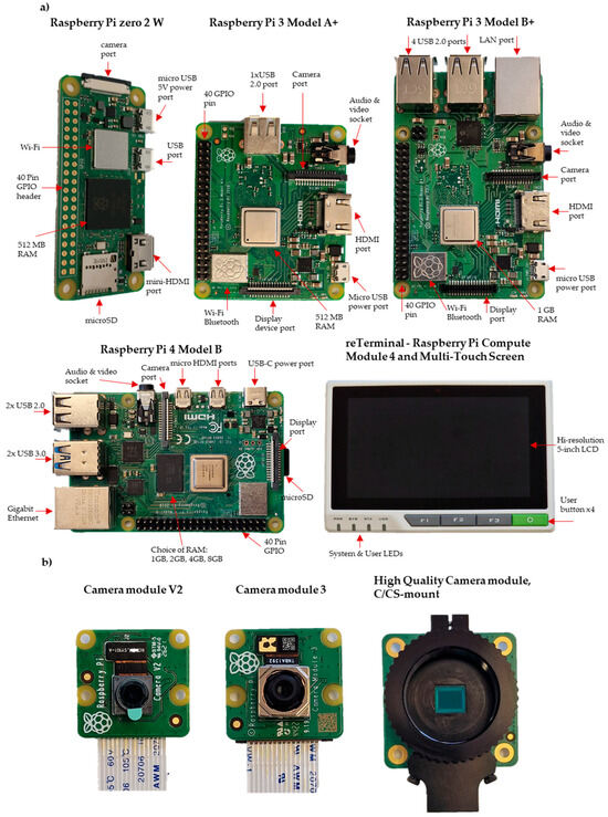
The integration of Raspberry Pi miniature computer systems with microfluidics has revolutionised the development of low-cost and customizable analytical systems in life science laboratories. This review explores the applications of Raspberry Pi in microfluidics, with a focus on imaging, including microscopy and automated
[...] Read more.
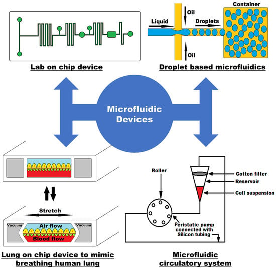
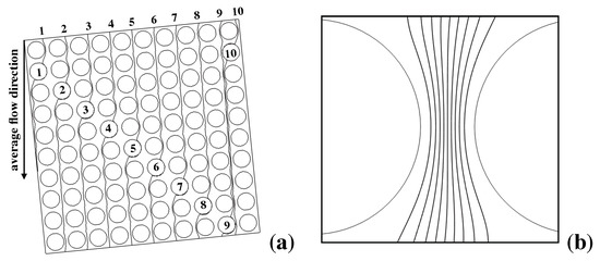
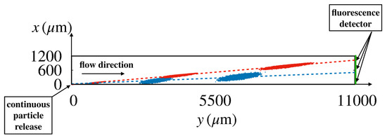
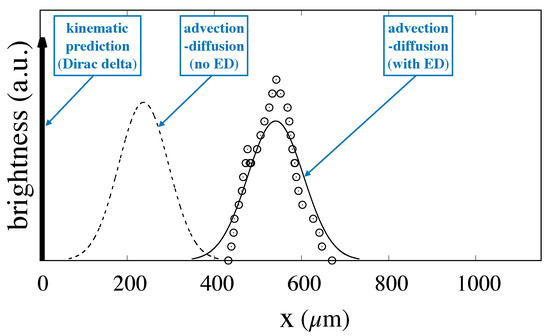
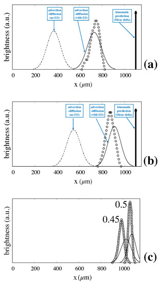
The integration of Raspberry Pi miniature computer systems with microfluidics has revolutionised the development of low-cost and customizable analytical systems in life science laboratories. This review explores the applications of Raspberry Pi in microfluidics, with a focus on imaging, including microscopy and automated image capture. By leveraging the low cost, flexibility and accessibility of Raspberry Pi components, high-resolution imaging and analysis have been achieved in direct mammalian and bacterial cellular imaging and a plethora of image-based biochemical and molecular assays, from immunoassays, through microbial growth, to nucleic acid methods such as real-time-qPCR. The control of image capture permitted by Raspberry Pi hardware can also be combined with onboard image analysis. Open-source hardware offers an opportunity to develop complex laboratory instrumentation systems at a fraction of the cost of commercial equipment and, importantly, offers an opportunity for complete customisation to meet the users’ needs. However, these benefits come with a trade-off: challenges remain for those wishing to incorporate open-source hardware equipment in their own work, including requirements for construction and operator skill, the need for good documentation and the availability of rapid prototyping such as 3D printing plus other components. These advances in open-source hardware have the potential to improve the efficiency, accessibility, and cost-effectiveness of microfluidic-based experiments and applications.
Full article
Figure 1

