Oral Regenerative Medicine: Current and Future

A special issue of Biomolecules (ISSN 2218-273X). This special issue belongs to the section "Biological Factors".

Deadline for manuscript submissions: closed (31 May 2021) | Viewed by 36028

Special Issue Editors


E-Mail Website
Guest Editor
Department of Dentistry and Oral Surgery, Keio University School of Medicine, Shinjuku-ku, Tokyo, Japan
Interests: tissue engineering; biomaterials; oral diseases
Special Issues, Collections and Topics in MDPI journals

E-Mail Website
Guest Editor
1. Developmental Biology, Harvard School of Dental Medicine, 188 Longwood Avenue, Boston, MA, USA
2. Department of Dentistry and Oral Surgery, Keio University School of Medicine, Shinjuku-ku, Tokyo, Japan
Interests: regenerative medicine; stem cells; developmental biology
Special Issues, Collections and Topics in MDPI journals

Special Issue Information

Dear Colleagues,

Some oral tissues maintain their homeostasis by regeneration. This biological regulation leads to appropriate oral function and provides systemic healthy condition by ideal digestion and intake of nutrition. Even if some tissues break down, general dental treatment or growth factors can recover them. However, huge disruption of oral tissues due to tooth loss, malignant diseases, and severe trauma can cause irreversible tissue loss in oral cavity. To compensate for this, artificial material, i.e., prosthesis and dental implantation, supports the improvement of human quality of life. While biomaterials have developed and applied for patients in recent decades, cellular treatment is still limited because of some problematic issues.

Which is the most significant factor for researchers to solve these problems?

The oral cavity consists of several tissue complexes with oral microbiota. In addition, oral tissues are under daily stresses, such as mastication and exposure to extraoral space. This unchangeable natural fact would make us think deeper on how oral regenerative medicine coexists with unavoidable factors.

In this collection, we will present and share the current oral regenerative medicine frontiers and tasks, and we also discuss attractive tools, methods, and techniques that will be useful for future oral basic and clinical sciences.

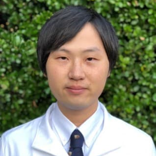
Dr. Taneaki Nakagawa
Dr. Takehito Ouchi
Guest Editors

Manuscript Submission Information

Manuscripts should be submitted online at www.mdpi.com by registering and logging in to this website. Once you are registered, click here to go to the submission form. Manuscripts can be submitted until the deadline. All submissions that pass pre-check are peer-reviewed. Accepted papers will be published continuously in the journal (as soon as accepted) and will be listed together on the special issue website. Research articles, review articles as well as short communications are invited. For planned papers, a title and short abstract (about 100 words) can be sent to the Editorial Office for announcement on this website.

Submitted manuscripts should not have been published previously, nor be under consideration for publication elsewhere (except conference proceedings papers). All manuscripts are thoroughly refereed through a single-blind peer-review process. A guide for authors and other relevant information for submission of manuscripts is available on the Instructions for Authors page. Biomolecules is an international peer-reviewed open access monthly journal published by MDPI.

Please visit the Instructions for Authors page before submitting a manuscript. The Article Processing Charge (APC) for publication in this open access journal is 2700 CHF (Swiss Francs). Submitted papers should be well formatted and use good English. Authors may use MDPI's English editing service prior to publication or during author revisions.

Keywords

  • Tooth regeneration
  • Stem cells
  • Tissue engineering
  • Regenerative therapy
  • Growth factor
  • Biomaterial
  • Gene regulation
  • Organoid
  • Cancer biology

Published Papers (9 papers)

Order results
Result details
Select all
Export citation of selected articles as:

Editorial

Jump to: Research, Review

3 pages, 163 KiB  
Editorial
Tissue Regeneration and Physiological Functional Recovery in Dental and Craniofacial Fields
by Takehito Ouchi and Taneaki Nakagawa
Biomolecules 2021, 11(11), 1644; https://doi.org/10.3390/biom11111644 - 06 Nov 2021
Cited by 3 | Viewed by 1879
Abstract
Dental and oral tissues maintain homeostasis through potential reparative or regenerative processes [...] Full article
(This article belongs to the Special Issue Oral Regenerative Medicine: Current and Future)

Research

Jump to: Editorial, Review

17 pages, 2523 KiB  
Article
Plasma Membrane Ca2+–ATPase in Rat and Human Odontoblasts Mediates Dentin Mineralization
by Maki Kimura, Hiroyuki Mochizuki, Ryouichi Satou, Miyu Iwasaki, Eitoyo Kokubu, Kyosuke Kono, Sachie Nomura, Takeshi Sakurai, Hidetaka Kuroda and Yoshiyuki Shibukawa
Biomolecules 2021, 11(7), 1010; https://doi.org/10.3390/biom11071010 - 10 Jul 2021
Cited by 6 | Viewed by 2254
Abstract
Intracellular Ca2+ signaling engendered by Ca2+ influx and mobilization in odontoblasts is critical for dentinogenesis induced by multiple stimuli at the dentin surface. Increased Ca2+ is exported by the Na+–Ca2+ exchanger (NCX) and plasma membrane Ca2+ [...] Read more.
Intracellular Ca2+ signaling engendered by Ca2+ influx and mobilization in odontoblasts is critical for dentinogenesis induced by multiple stimuli at the dentin surface. Increased Ca2+ is exported by the Na+–Ca2+ exchanger (NCX) and plasma membrane Ca2+–ATPase (PMCA) to maintain Ca2+ homeostasis. We previously demonstrated a functional coupling between Ca2+ extrusion by NCX and its influx through transient receptor potential channels in odontoblasts. Although the presence of PMCA in odontoblasts has been previously described, steady-state levels of mRNA-encoding PMCA subtypes, pharmacological properties, and other cellular functions remain unclear. Thus, we investigated PMCA mRNA levels and their contribution to mineralization under physiological conditions. We also examined the role of PMCA in the Ca2+ extrusion pathway during hypotonic and alkaline stimulation-induced increases in intracellular free Ca2+ concentration ([Ca2+]i). We performed RT-PCR and mineralization assays in human odontoblasts. [Ca2+]i was measured using fura-2 fluorescence measurements in odontoblasts isolated from newborn Wistar rat incisor teeth and human odontoblasts. We detected mRNA encoding PMCA1–4 in human odontoblasts. The application of hypotonic or alkaline solutions transiently increased [Ca2+]i in odontoblasts in both rat and human odontoblasts. The Ca2+ extrusion efficiency during the hypotonic or alkaline solution-induced [Ca2+]i increase was decreased by PMCA inhibitors in both cell types. Alizarin red and von Kossa staining showed that PMCA inhibition suppressed mineralization. In addition, alkaline stimulation (not hypotonic stimulation) to human odontoblasts upregulated the mRNA levels of dentin matrix protein-1 (DMP-1) and dentin sialophosphoprotein (DSPP). The PMCA inhibitor did not affect DMP-1 or DSPP mRNA levels at pH 7.4–8.8 and under isotonic and hypotonic conditions, respectively. We also observed PMCA1 immunoreactivity using immunofluorescence analysis. These findings indicate that PMCA participates in maintaining [Ca2+]i homeostasis in odontoblasts by Ca2+ extrusion following [Ca2+]i elevation. In addition, PMCA participates in dentinogenesis by transporting Ca2+ to the mineralizing front (which is independent of non-collagenous dentin matrix protein secretion) under physiological and pathological conditions following mechanical stimulation by hydrodynamic force inside dentinal tubules, or direct alkaline stimulation by the application of high-pH dental materials. Full article
(This article belongs to the Special Issue Oral Regenerative Medicine: Current and Future)
Show Figures

Figure 1

14 pages, 2217 KiB  
Article
In Vitro Compression Model for Orthodontic Tooth Movement Modulates Human Periodontal Ligament Fibroblast Proliferation, Apoptosis and Cell Cycle
by Julia Brockhaus, Rogerio B. Craveiro, Irma Azraq, Christian Niederau, Sarah K. Schröder, Ralf Weiskirchen, Joachim Jankowski and Michael Wolf
Biomolecules 2021, 11(7), 932; https://doi.org/10.3390/biom11070932 - 23 Jun 2021
Cited by 15 | Viewed by 2846
Abstract
Human Periodontal Ligament Fibroblasts (hPDLF), as part of the periodontal apparatus, modulate inflammation, regeneration and bone remodeling. Interferences are clinically manifested as attachment loss, tooth loosening and root resorption. During orthodontic tooth movement (OTM), remodeling and adaptation of the periodontium is required in [...] Read more.
Human Periodontal Ligament Fibroblasts (hPDLF), as part of the periodontal apparatus, modulate inflammation, regeneration and bone remodeling. Interferences are clinically manifested as attachment loss, tooth loosening and root resorption. During orthodontic tooth movement (OTM), remodeling and adaptation of the periodontium is required in order to enable tooth movement. hPDLF involvement in the early phase-OTM compression side was investigated for a 72-h period through a well-studied in vitro model. Changes in the morphology, cell proliferation and cell death were analyzed. Specific markers of the cell cycle were investigated by RT-qPCR and Western blot. The study showed that the morphology of hPDLF changes towards more unstructured, unsorted filaments under mechanical compression. The total cell numbers were significantly reduced with a higher cell death rate over the whole observation period. hPDLF started to recover to pretreatment conditions after 48 h. Furthermore, key molecules involved in the cell cycle were significantly reduced under compressive force at the gene expression and protein levels. These findings revealed important information for a better understanding of the preservation and remodeling processes within the periodontium through Periodontal Ligament Fibroblasts during orthodontic tooth movement. OTM initially decelerates the hPDLF cell cycle and proliferation. After adapting to environmental changes, human Periodontal Ligament Fibroblasts can regain homeostasis of the periodontium, affecting its reorganization. Full article
(This article belongs to the Special Issue Oral Regenerative Medicine: Current and Future)
Show Figures

Figure 1

14 pages, 3390 KiB  
Article
The Effect of Cultivation Passaging on the Relative Telomere Length and Proliferation Capacity of Dental Pulp Stem Cells
by Nela Pilbauerova, Tomas Soukup, Tereza Suchankova Kleplova, Jan Schmidt and Jakub Suchanek
Biomolecules 2021, 11(3), 464; https://doi.org/10.3390/biom11030464 - 20 Mar 2021
Cited by 9 | Viewed by 2346
Abstract
Telomeres are repetitive nucleoprotein DNA sequences that shorten with each cell division. The stem cells activate telomerase to compensate for the telomere loss. This study aimed to evaluate the effect of cultivation passaging on the relative telomere length and proliferation capacity of dental [...] Read more.
Telomeres are repetitive nucleoprotein DNA sequences that shorten with each cell division. The stem cells activate telomerase to compensate for the telomere loss. This study aimed to evaluate the effect of cultivation passaging on the relative telomere length and proliferation capacity of dental pulp stem cells. We used ten dental pulp stem cell (DPSC) lineages stored for 12 months using uncontrolled-rate freezing to reach the study’s goal. We analyzed their proliferation rate, phenotype using flow cytometry, multipotency, and relative telomere length using a qPCR analysis. We determined the relative telomere length in the added study by performing analysis after one, two, and three weeks of cultivation with no passaging. We documented the telomere attrition with increasing passaging. The shorter the relative telomere length, the lower reached population doublings, and longer population doubling time were observed at the end of the cultivation. We observed the telomere prolongation in DPSCs cultivated for two weeks with no passaging in the added subsequent study. We concluded that excessive proliferation demands on DPSCs during in vitro cultivation result in telomere attrition. We opened the theory that the telomerase might be more efficient during cell cultivation with no passaging. This observation could help in preserving the telomere length during ex vivo DPSC expansion. Full article
(This article belongs to the Special Issue Oral Regenerative Medicine: Current and Future)
Show Figures

Figure 1

8 pages, 1488 KiB  
Article
Prepubertal Periodontitis in a Patient with Combined Classical and Periodontal Ehlers–Danlos Syndrome
by Friedrich Stock, Marcel Hanisch, Sarah Lechner, Saskia Biskup, Axel Bohring, Johannes Zschocke and Ines Kapferer-Seebacher
Biomolecules 2021, 11(2), 149; https://doi.org/10.3390/biom11020149 - 24 Jan 2021
Cited by 7 | Viewed by 3578
Abstract
We report an extremely rare case of combined classical and periodontal Ehlers−Danlos syndrome (EDS) with early severe periodontitis and a generalized lack of attached gingiva. A German family with classical EDS was investigated by physical and dental evaluation and exome and Sanger sequencing. [...] Read more.
We report an extremely rare case of combined classical and periodontal Ehlers−Danlos syndrome (EDS) with early severe periodontitis and a generalized lack of attached gingiva. A German family with classical EDS was investigated by physical and dental evaluation and exome and Sanger sequencing. Due to the specific periodontal phenotype in the affected child, an additional diagnosis of periodontal EDS was suspected. Physical and genetic examination of two affected and three unaffected family members revealed a family diagnosis of classical EDS with a heterozygous mutation in COL5A1 (c.1502del; p.Pro501Leufs*57). Additional to the major clinical criteria for classical EDS—generalized joint hypermobility, hyperelastic skin, and atrophic scarring —the child aged four years presented with generalized alveolar bone loss up to 80%, early loss of two lower incisors, severe gingival recession, and generalized lack of attached gingiva. Due to these clinical findings, an additional diagnosis of periodontal EDS was suspected. Further genetic analysis revealed the novel missense mutation c.658T>G (p.Cys220Gly) in C1R in a heterozygous state. Early severe periodontitis in association with generalized lack of attached gingiva is pathognomonic for periodontal EDS and led to the right clinical and genetic diagnosis in the present case. Full article
(This article belongs to the Special Issue Oral Regenerative Medicine: Current and Future)
Show Figures

Figure 1

16 pages, 1967 KiB  
Article
Low Molecular Weight Hyaluronic Acid Effect on Dental Pulp Stem Cells In Vitro
by Jan Schmidt, Nela Pilbauerova, Tomas Soukup, Tereza Suchankova-Kleplova and Jakub Suchanek
Biomolecules 2021, 11(1), 22; https://doi.org/10.3390/biom11010022 - 28 Dec 2020
Cited by 16 | Viewed by 2892
Abstract
Hyaluronic acid (HA) and dental pulp stem cells (DPSCs) are attractive research topics, and their combined use in the field of tissue engineering seems to be very promising. HA is a natural extracellular biopolymer found in various tissues, including dental pulp, and due [...] Read more.
Hyaluronic acid (HA) and dental pulp stem cells (DPSCs) are attractive research topics, and their combined use in the field of tissue engineering seems to be very promising. HA is a natural extracellular biopolymer found in various tissues, including dental pulp, and due to its biocompatibility and biodegradability, it is also a suitable scaffold material. However, low molecular weight (LMW) fragments, produced by enzymatic cleavage of HA, have different bioactive properties to high molecular weight (HMW) HA. Thus, the impact of HA must be assessed separately for each molecular weight fraction. In this study, we present the effect of three LMW-HA fragments (800, 1600, and 15,000 Da) on DPSCs in vitro. Discrete biological parameters such as DPSC viability, morphology, and cell surface marker expression were determined. Following treatment with LMW-HA, DPSCs initially presented with an acute reduction in proliferation (p < 0.0016) and soon recovered in subsequent passages. They displayed significant size reduction (p = 0.0078, p = 0.0019, p = 0.0098) while maintaining high expression of DPSC markers (CD29, CD44, CD73, CD90). However, in contrast to controls, a significant phenotypic shift (p < 0.05; CD29, CD34, CD90, CD106, CD117, CD146, CD166) of surface markers was observed. These findings provide a basis for further detailed investigations and present a strong argument for the importance of HA scaffold degradation kinetics analysis. Full article
(This article belongs to the Special Issue Oral Regenerative Medicine: Current and Future)
Show Figures

Graphical abstract

Review

Jump to: Editorial, Research

14 pages, 2167 KiB  
Review
Cranial Suture Mesenchymal Stem Cells: Insights and Advances
by Bo Li, Yigan Wang, Yi Fan, Takehito Ouchi, Zhihe Zhao and Longjiang Li
Biomolecules 2021, 11(8), 1129; https://doi.org/10.3390/biom11081129 - 31 Jul 2021
Cited by 20 | Viewed by 6867
Abstract
The cranial bones constitute the protective structures of the skull, which surround and protect the brain. Due to the limited repair capacity, the reconstruction and regeneration of skull defects are considered as an unmet clinical need and challenge. Previously, it has been proposed [...] Read more.
The cranial bones constitute the protective structures of the skull, which surround and protect the brain. Due to the limited repair capacity, the reconstruction and regeneration of skull defects are considered as an unmet clinical need and challenge. Previously, it has been proposed that the periosteum and dura mater provide reparative progenitors for cranial bones homeostasis and injury repair. In addition, it has also been speculated that the cranial mesenchymal stem cells reside in the perivascular niche of the diploe, namely, the soft spongy cancellous bone between the interior and exterior layers of cortical bone of the skull, which resembles the skeletal stem cells’ distribution pattern of the long bone within the bone marrow. Not until recent years have several studies unraveled and validated that the major mesenchymal stem cell population of the cranial region is primarily located within the suture mesenchyme of the skull, and hence, they are termed suture mesenchymal stem cells (SuSCs). Here, we summarized the characteristics of SuSCs, this newly discovered stem cell population of cranial bones, including the temporospatial distribution pattern, self-renewal, and multipotent properties, contribution to injury repair, as well as the signaling pathways and molecular mechanisms associated with the regulation of SuSCs. Full article
(This article belongs to the Special Issue Oral Regenerative Medicine: Current and Future)
Show Figures

Figure 1

18 pages, 715 KiB  
Review
Function of Dental Follicle Progenitor/Stem Cells and Their Potential in Regenerative Medicine: From Mechanisms to Applications
by Ruiye Bi, Ping Lyu, Yiming Song, Peiran Li, Dongzhe Song, Chen Cui and Yi Fan
Biomolecules 2021, 11(7), 997; https://doi.org/10.3390/biom11070997 - 07 Jul 2021
Cited by 28 | Viewed by 6948
Abstract
Dental follicle progenitor/stem cells (DFPCs) are a group of dental mesenchyme stem cells that lie in the dental follicle and play a critical role in tooth development and maintaining function. Originating from neural crest, DFPCs harbor a multipotential differentiation capacity. More importantly, they [...] Read more.
Dental follicle progenitor/stem cells (DFPCs) are a group of dental mesenchyme stem cells that lie in the dental follicle and play a critical role in tooth development and maintaining function. Originating from neural crest, DFPCs harbor a multipotential differentiation capacity. More importantly, they have superiorities, including the easy accessibility and abundant sources, active self-renewal ability and noncontroversial sources compared with other stem cells, making them an attractive candidate in the field of tissue engineering. Recent advances highlight the excellent properties of DFPCs in regeneration of orofacial tissues, including alveolar bone repair, periodontium regeneration and bio-root complex formation. Furthermore, they play a unique role in maintaining a favorable microenvironment for stem cells, immunomodulation and nervous related tissue regeneration. This review is intended to summarize the current knowledge of DFPCs, including their stem cell properties, physiological functions and clinical application potential. A deep understanding of DFPCs can thus inspire novel perspectives in regenerative medicine in the future. Full article
(This article belongs to the Special Issue Oral Regenerative Medicine: Current and Future)
Show Figures

Figure 1

42 pages, 26897 KiB  
Review
From the Matrix to the Nucleus and Back: Mechanobiology in the Light of Health, Pathologies, and Regeneration of Oral Periodontal Tissues
by Martin Philipp Dieterle, Ayman Husari, Thorsten Steinberg, Xiaoling Wang, Imke Ramminger and Pascal Tomakidi
Biomolecules 2021, 11(6), 824; https://doi.org/10.3390/biom11060824 - 31 May 2021
Cited by 11 | Viewed by 5022
Abstract
Among oral tissues, the periodontium is permanently subjected to mechanical forces resulting from chewing, mastication, or orthodontic appliances. Molecularly, these movements induce a series of subsequent signaling processes, which are embedded in the biological concept of cellular mechanotransduction (MT). Cell and tissue structures, [...] Read more.
Among oral tissues, the periodontium is permanently subjected to mechanical forces resulting from chewing, mastication, or orthodontic appliances. Molecularly, these movements induce a series of subsequent signaling processes, which are embedded in the biological concept of cellular mechanotransduction (MT). Cell and tissue structures, ranging from the extracellular matrix (ECM) to the plasma membrane, the cytosol and the nucleus, are involved in MT. Dysregulation of the diverse, fine-tuned interaction of molecular players responsible for transmitting biophysical environmental information into the cell’s inner milieu can lead to and promote serious diseases, such as periodontitis or oral squamous cell carcinoma (OSCC). Therefore, periodontal integrity and regeneration is highly dependent on the proper integration and regulation of mechanobiological signals in the context of cell behavior. Recent experimental findings have increased the understanding of classical cellular mechanosensing mechanisms by both integrating exogenic factors such as bacterial gingipain proteases and newly discovered cell-inherent functions of mechanoresponsive co-transcriptional regulators such as the Yes-associated protein 1 (YAP1) or the nuclear cytoskeleton. Regarding periodontal MT research, this review offers insights into the current trends and open aspects. Concerning oral regenerative medicine or weakening of periodontal tissue diseases, perspectives on future applications of mechanobiological principles are discussed. Full article
(This article belongs to the Special Issue Oral Regenerative Medicine: Current and Future)
Show Figures

Figure 1

Back to TopTop