


Animals 2023, 13(16), 2601; https://doi.org/10.3390/ani13162601 - 12 Aug 2023
Cited by 1 | Viewed by 1027
Abstract
►
Show Figures
Two main classes of perivascular multipotent populations have been described: the microvascular pericytes and the vascular wall mesenchymal stem cells (VW-MSCs). VW-MSCs are isolated from large vessels in many species and they participate in vascular remodeling together with other cellular components such as
[...] Read more.
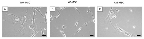
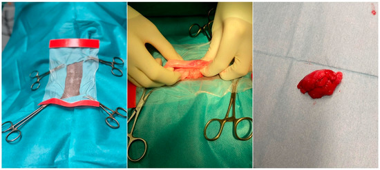
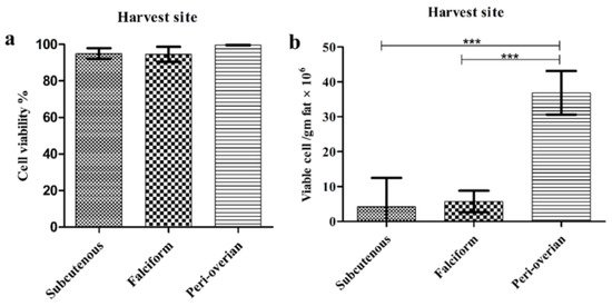
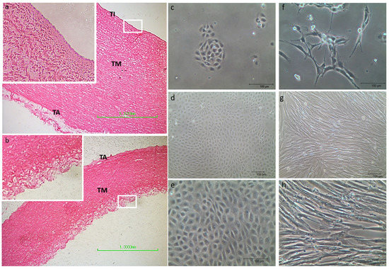
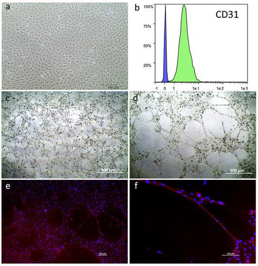
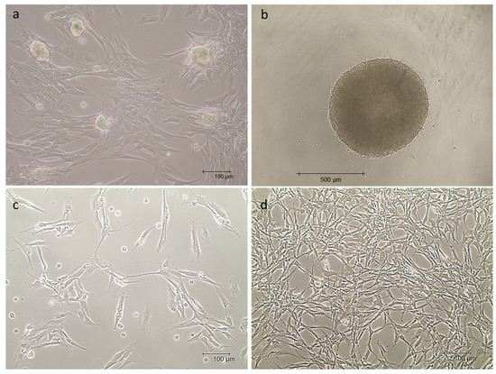
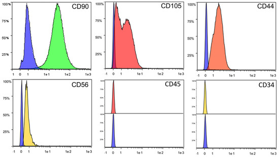
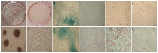
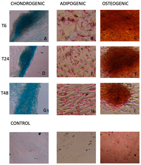
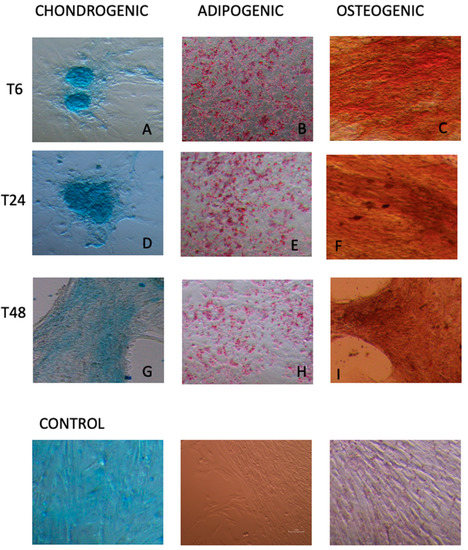
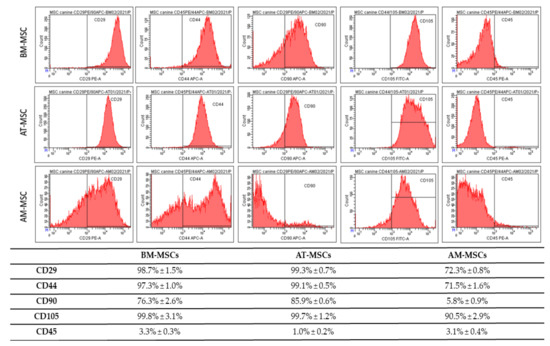
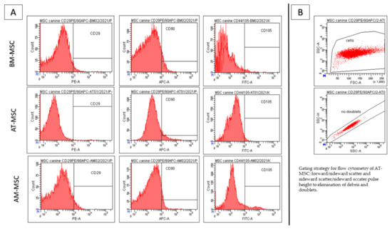
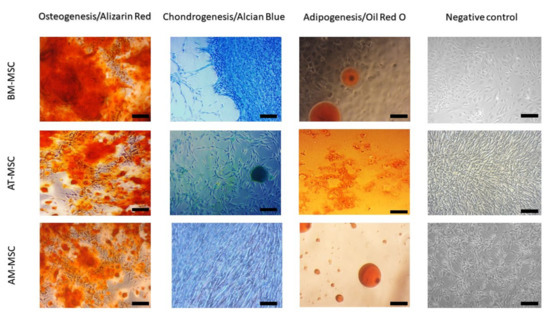
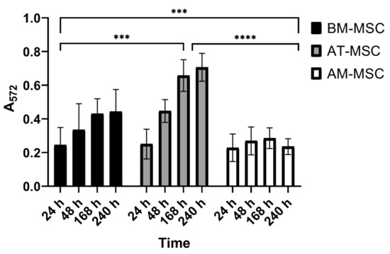
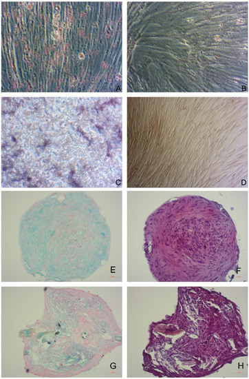
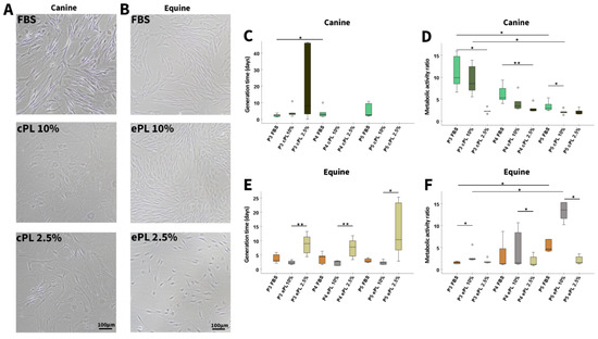
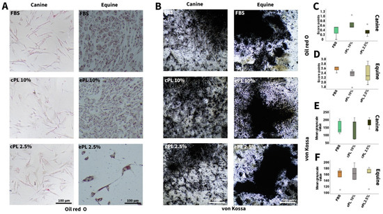
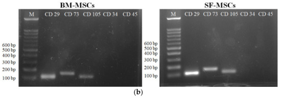
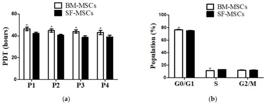
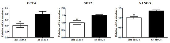
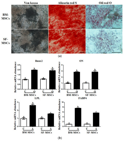
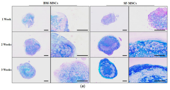
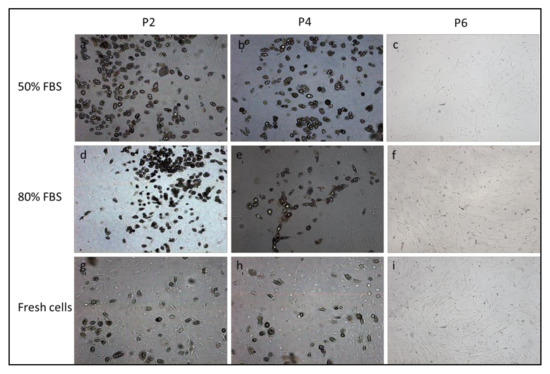
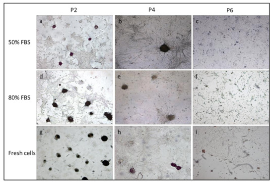
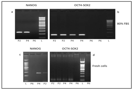
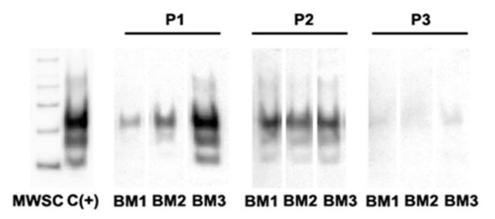
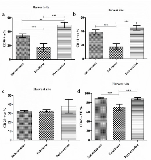
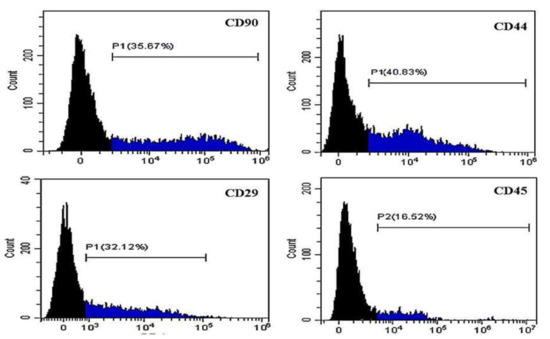
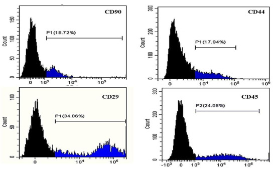
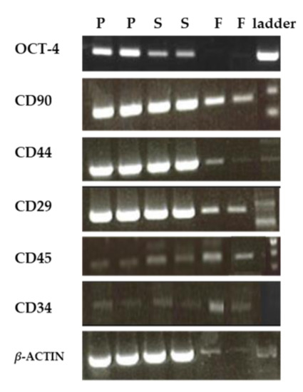
Two main classes of perivascular multipotent populations have been described: the microvascular pericytes and the vascular wall mesenchymal stem cells (VW-MSCs). VW-MSCs are isolated from large vessels in many species and they participate in vascular remodeling together with other cellular components such as endothelial cells. Considering that the Göttingen Minipigs are widely used in Europe as a translational model in the field of cardiovascular diseases, the aim of the present research was to isolate VW-MSCs from the adult aorta of Göttingen Minipigs while preserving and also collecting endothelial cells. The results obtained in the present research demonstrated that this new protocol allows us to obtain a pure population of VW-MSCs and endothelial cells. VW-MSCs from Göttingen Minipigs responded fully to the MSC minima international criteria, being positive to CD105, CD90, and CD44 and negative to CD45 and CD34. Moreover, VW-MSCs presented a differentiative potential towards osteogenic, chondrogenic, and adipogenic lineages. Overall, the present protocol, preserving the viability and phenotypic features of the two isolated populations, opens future possibilities of using minipig VW-MSCs and endothelial cells in in vitro vascular remodeling studies.
Full article
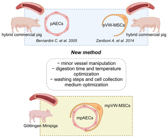
Figure 1

