

Analysis of Muscle Strength and Electromyographic Activity during Different Deadlift Positions

 , , ,
, , ,  , , ,
, , ,  and
and 
Abstract
:1. Introduction
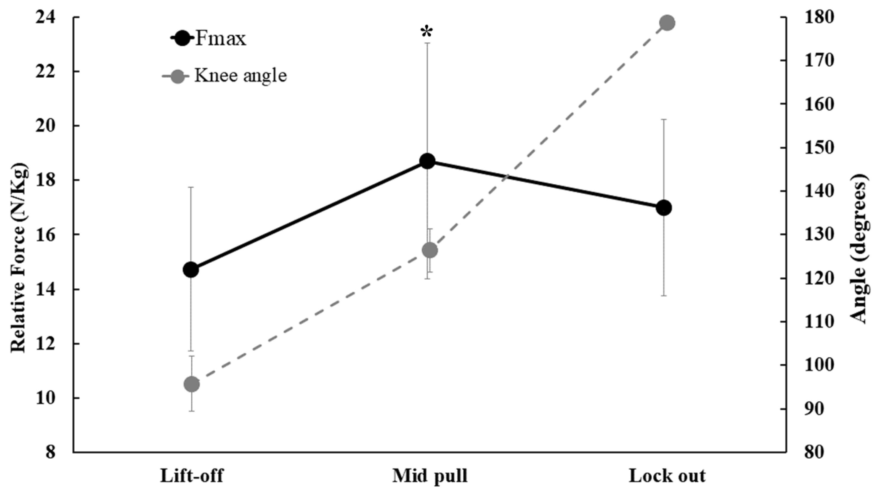
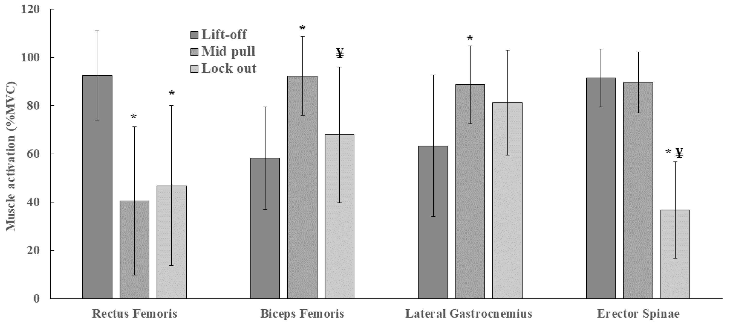
2. Results
3. Discussion
4. Materials and Methods
4.1. Participants
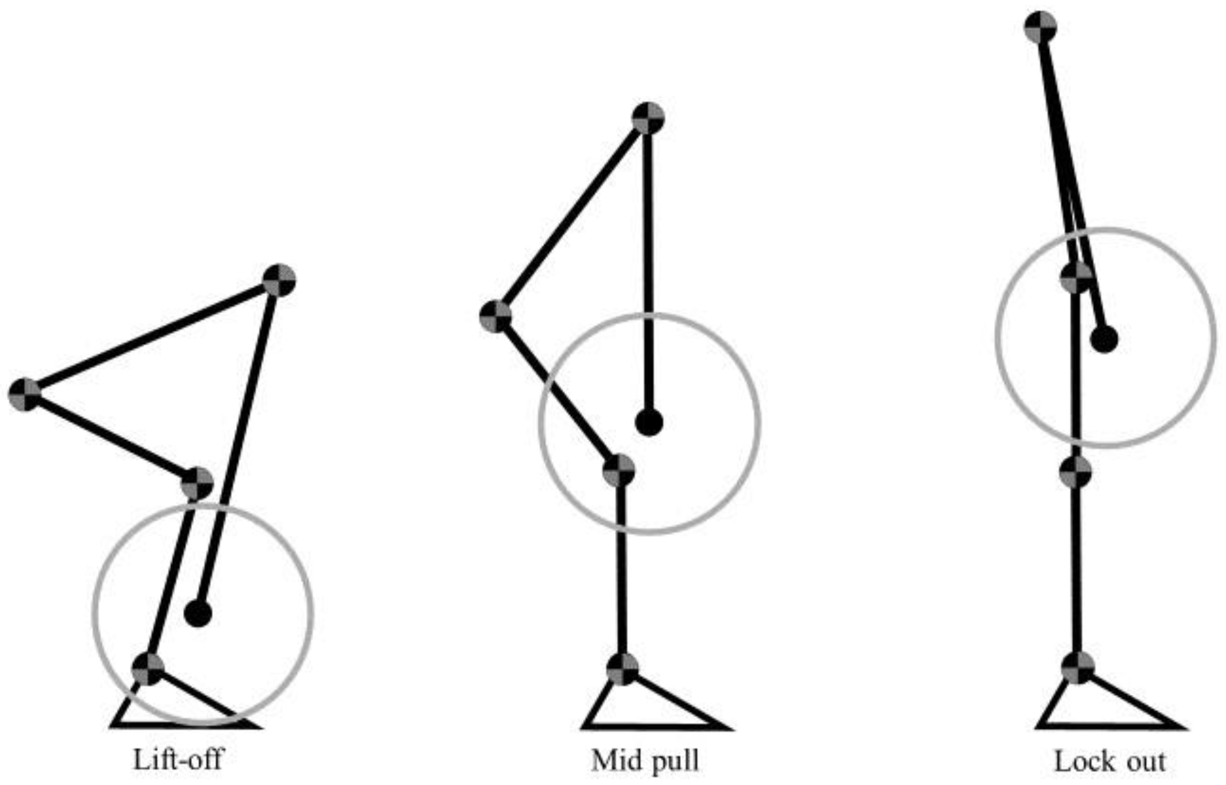
4.2. Experimental Protocol
4.3. Isometric Strength
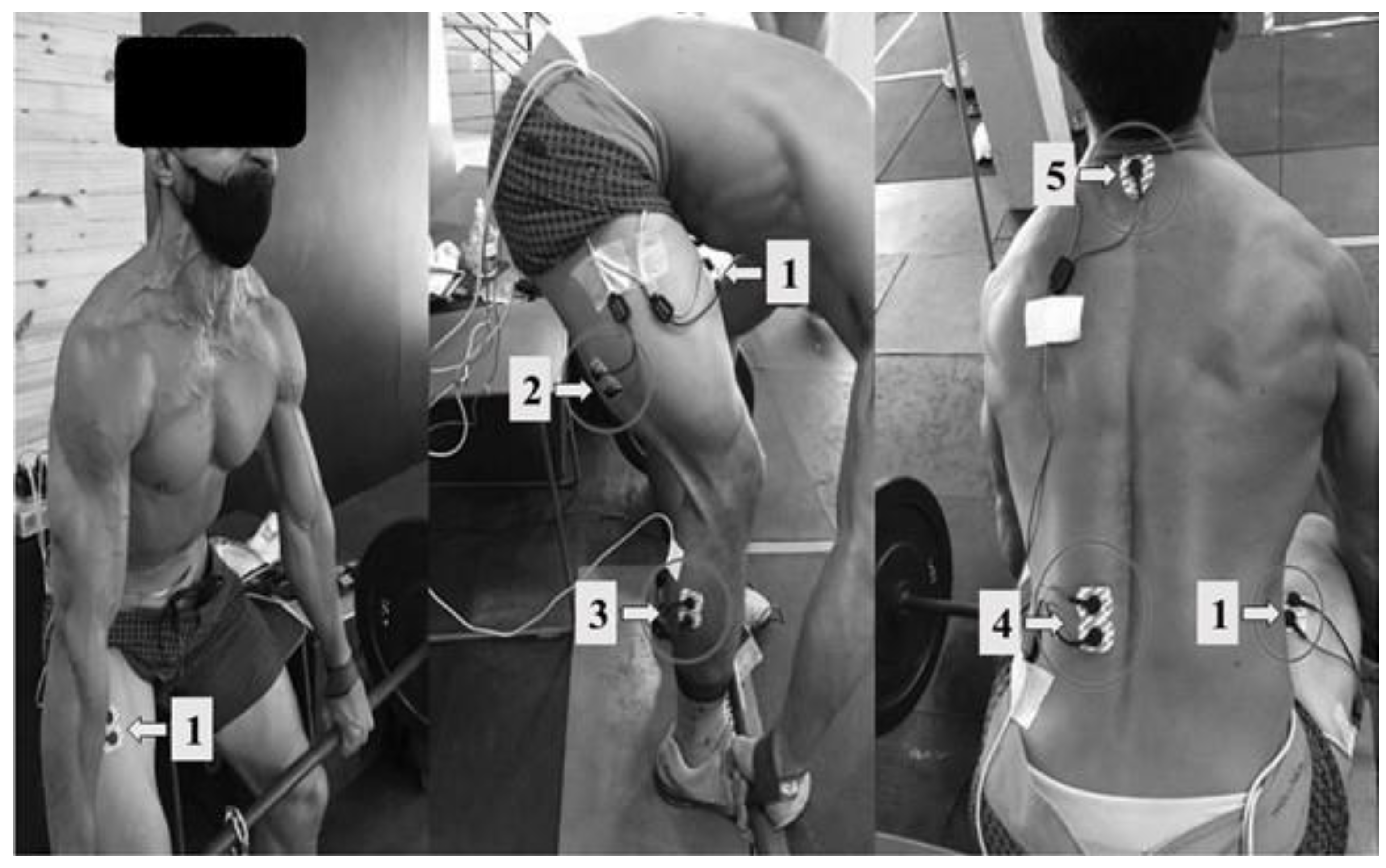
4.4. Electromyographic Data
4.5. Statistical Analysis
Author Contributions
Funding
Institutional Review Board Statement
Informed Consent Statement
Data Availability Statement
Conflicts of Interest
References
- O’Reilly, M.A.; Whelan, D.F.; Ward, T.E.; Delahunt, E.; Caulfield, B.M. Classification of deadlift biomechanics with wearable inertial measurement units. J. Biomech. 2017, 58, 155–161. [Google Scholar] [CrossRef] [PubMed]
- Choe, K.H.; Coburn, J.W.; Costa, P.B.; Pamukoff, D.N. Hip and Knee Kinetics During a Back Squat and Deadlift. J. Strength Cond. Res. 2021, 35, 1364–1371. [Google Scholar] [CrossRef] [PubMed]
- McGuigan, M.; Wilson, B.; McGuigan, M.W. Biomechanical Analysis of the Deadlift. J. Strength Cond. Res. 1996, 10, 250–255. [Google Scholar]
- Lee, S.; Schultz, J.; Timgren, J.; Staelgraeve, K.; Miller, M.; Liu, Y. An electromyographic and kinetic comparison of conventional and Romanian deadlifts. J. Exerc. Sci. Fit. 2018, 16, 87–93. [Google Scholar] [CrossRef]
- Mohamed, O.; Perry, J.; Hislop, H. Relationship between wire EMG activity, muscle length; and torque of the hamstrings. Clin. Biomech. 2002, 17, 569–579. [Google Scholar] [CrossRef]
- Kompf, J.; Arandjelović, O. The Sticking Point in the Bench Press; the Squat; and the Deadlift: Similarities and Differences; and Their Significance for Research and Practice. Sports Med. 2017, 47, 631–640. [Google Scholar] [CrossRef]
- Bird, S.; Barrington-Higgs, B. Exploring the Deadlift. Strength Cond. J. 2010, 32, 46–51. [Google Scholar] [CrossRef]
- Hales, M. Improving the deadlift: Understanding biomechanical constraints and physiological adaptations to resistance exercise. Strength Cond. J. 2010, 32, 44–51. [Google Scholar] [CrossRef]
- Beckham, G.; Sato, K.; Santana, H.A.P.; Mizuguchi, S.; Haff, G.G.; Stone, M.H. Effect of Body Position on Force Production During the Isometric Midthigh Pull. J. Strength Cond. Res. 2018, 32, 48–56. [Google Scholar] [CrossRef]
- Escamilla, R.F.; Francisco, A.C.; Kayes, A.V.; Speer, K.P.; Moorman, C.T. An electromyographic analysis of sumo and conventional style deadlifts. Med. Sci. Sports Exerc. 2002, 34, 682–688. [Google Scholar]
- Hales, M.; Johnson, B.; Johnson, J. Kinematic Analysis of the Powerlifting Style Squat and the Conventional Deadlift During Competition: Is There a Cross-Over Effect Between Lifts? J. Strength Cond. Res. 2009, 23, 2574–2580. [Google Scholar] [CrossRef] [PubMed]
- Martín-Fuentes, I.; Oliva-Lozano, J.M.; Muyor, J.M. Electromyographic activity in deadlift exercise and its variants. A systematic review. Ahamed NU; editor. PLoS ONE 2020, 15, e0229507. [Google Scholar] [CrossRef] [PubMed]
- Bezerra, E.; Simão, R.; Fleck, S.; Paz, G.; Maia, M.; Costa, P. Electromyographic Activity of Lower Body Muscles during the Deadlift and Stiff-Legged Deadlift. J. Exerc. Physiol. 2013, 3, 11–25. [Google Scholar]
- Andersen, V.; Fimland, M.S.; Mo, D.-A.; Iversen, V.M.; Larsen, T.M.; Solheim, F.; Saeterbakken, A.H. Electromyographic comparison of the barbell deadlift using constant versus variable resistance in healthy; trained men. PLoS ONE 2019, 14, e0211021. [Google Scholar] [CrossRef]
- Edington, C.; Greening, C.; Kmet, N.; Philipenko, N.; Purves, L.; Stevens, J.; Lanovaz, J.; Butcher, S. The effect of set up position on emg amplitude; lumbar spine kinetics; and total force output during maximal isometric conventional-stance deadlifts. Sports 2018, 6, 90. [Google Scholar] [CrossRef]
- Beckham, G.; Lamont, H.; Sato, K.; Ramsey, M.; Haff, G.; Stone, M. Isometric Strength of Powerlifters in Key Positions of the Conventional Deadlift. J. Trainology 2012, 1, 32–35. [Google Scholar] [CrossRef]
- Opplert, J.; Genty, J.B.; Babault, N. Do Stretch Durations Affect Muscle Mechanical and Neurophysiological Properties? Int. J. Sports Med. 2016, 37, 673–679. [Google Scholar] [CrossRef]
- Yanagisawa, O.; Fukutani, A. Muscle Recruitment Pattern of The Hamstring Muscles in Hip Extension and Knee Flexion Exercises. J. Hum. Kinet. 2020, 72, 51–59. [Google Scholar] [CrossRef]
- Ono, T.; Okuwaki, T.; Fukubayashi, T. Differences in Activation Patterns of Knee Flexor Muscles During Concentric and Eccentric Exercises. Res. Sports Med. 2010, 18, 188–198. [Google Scholar] [CrossRef]
- Keerasomboon, T.; Mineta, S.; Hirose, N. Influence of Altered Knee Angle and Muscular Contraction Type on Electromyographic Activity of Hamstring Muscles during 45° Hip Extension Exercise. J. Sports Sci. Med. 2020, 19, 630–636. [Google Scholar]
- Saito, A.; Akima, H. Knee joint angle affects EMG–force relationship in the vastus intermedius muscle. J. Electromyogr. Kinesiol. 2013, 23, 1406–1412. [Google Scholar] [CrossRef] [PubMed]
- Hamm, K.; Alexander, C.M. Challenging presumptions: Is reciprocal inhibition truly reciprocal? A study of reciprocal inhibition between knee extensors and flexors in humans. Man. Ther. 2010, 15, 388–393. [Google Scholar] [CrossRef] [PubMed]
- Potvin, J.R.; Norman, R.W.; McGill, S.M. Reduction in anterior shear forces on the disc by the lumbar musculature. Clin. Biomech. 1991, 6, 88–96. [Google Scholar] [CrossRef] [PubMed]
- Kingma, I.; Staudenmann, D.; Van Dieën, J.H. Trunk muscle activation and associated lumbar spine joint shear forces under different levels of external forward force applied to the trunk. J. Electromyogr. Kinesiol. 2007, 17, 14–24. [Google Scholar] [CrossRef]
- James, L.P.; Roberts, L.A.; Haff, G.G.; Kelly, V.G.; Beckman, E.M. Validity and reliability of a portable isometric mid-thigh clean pull. J. Strength Cond. Res. 2017, 31, 1378–1386. [Google Scholar] [CrossRef] [PubMed]
- Hermens, H.J.; Freriks, B.; Disselhorst-Klug, C.; Rau, G. Development of recommendations for SEMG sensors and sensor placement procedures. J. Electromyogr. Kinesiol. 2000, 10, 361–374. [Google Scholar] [CrossRef]
- Criswell, E. Cram’s Introduction to Surface Electromyography, 2nd ed.; Cella, D., Ed.; Jones and Bartlett: Burlington, MA, USA, 2011; Volume 2, 412p. [Google Scholar]
- Castelein, B.; Cagnie, B.; Parlevliet, T.; Danneels, L.; Cools, A. Optimal Normalization Tests for Muscle Activation of the Levator Scapulae; Pectoralis Minor; and Rhomboid Major: An Electromyography Study Using Maximum Voluntary Isometric Contractions. Arch. Phys. Med. Rehabil. 2015, 96, 1820–1827. [Google Scholar] [CrossRef]
- Nakai, Y.; Kawada, M.; Miyazaki, T.; Kiyama, R. Trunk muscle activity during trunk stabilizing exercise with isometric hip rotation using electromyography and ultrasound. J. Electromyogr. Kinesiol. 2019, 49, 102357. [Google Scholar] [CrossRef]


Disclaimer/Publisher’s Note: The statements, opinions and data contained in all publications are solely those of the individual author(s) and contributor(s) and not of MDPI and/or the editor(s). MDPI and/or the editor(s) disclaim responsibility for any injury to people or property resulting from any ideas, methods, instructions or products referred to in the content. |
© 2023 by the authors. Licensee MDPI, Basel, Switzerland. This article is an open access article distributed under the terms and conditions of the Creative Commons Attribution (CC BY) license (https://creativecommons.org/licenses/by/4.0/).
Share and Cite
Moreira, V.M.; Lima, L.C.R.d.; Mortatti, A.L.; Souza, T.M.F.d.; Lima, F.V.; Oliveira, S.F.M.; Cabido, C.E.T.; Aidar, F.J.; Costa, M.d.C.; Pires, T.; et al. Analysis of Muscle Strength and Electromyographic Activity during Different Deadlift Positions. Muscles 2023, 2, 218-227. https://doi.org/10.3390/muscles2020016
Moreira VM, Lima LCRd, Mortatti AL, Souza TMFd, Lima FV, Oliveira SFM, Cabido CET, Aidar FJ, Costa MdC, Pires T, et al. Analysis of Muscle Strength and Electromyographic Activity during Different Deadlift Positions. Muscles. 2023; 2(2):218-227. https://doi.org/10.3390/muscles2020016
Chicago/Turabian StyleMoreira, Vinícius Marques, Leonardo Coelho Rabello de Lima, Arnaldo Luis Mortatti, Thiago Mattos Frota de Souza, Fernando Vitor Lima, Saulo Fernandes Melo Oliveira, Christian Emmanuel Torres Cabido, Felipe J. Aidar, Manoel da Cunha Costa, Thiago Pires, and et al. 2023. "Analysis of Muscle Strength and Electromyographic Activity during Different Deadlift Positions" Muscles 2, no. 2: 218-227. https://doi.org/10.3390/muscles2020016