Review

Therapeutic Potential of Bioactive Flavonoids from Citrus Fruit Peels toward Obesity and Diabetes Mellitus

1
Division of Cardiology, Pulmonology and Vascular Medicine, Medical Faculty, Heinrich Heine University Duesseldorf, 40225 Duesseldorf, Germany
2
School of Physical and Mathematical Sciences, Nanyang Technological University, 21 Nanyang Link, Singapore 637371, Singapore
*
Author to whom correspondence should be addressed.
Future Pharmacol. 2023, 3(1), 14-37; https://doi.org/10.3390/futurepharmacol3010002
Submission received: 28 November 2022 / Revised: 23 December 2022 / Accepted: 28 December 2022 / Published: 4 January 2023

Abstract

:
Obesity is associated with a significantly increased risk of cardiovascular and metabolic diseases such as diabetes mellitus. Recently, a growing body of evidence shows that phytochemicals, especially many flavonoids, place an inhibitory regulatory effect on adipogenesis, obesity and diabetes. With computer-aided drug discovery, the action modes of more and more bioactive flavonoids are being identified and confirmed at the molecular level. Citrus fruit peels are particularly rich in bioactive flavonoids which have demonstrated strong therapeutic potentials in regulating lipid metabolisms. However, they are usually thrown away after consuming the flesh, sometimes even causing environmental problems. Thus, extraction of useful flavonoids from citrus fruit waste for pharmaceutical industry could be a profitable and environmentally friendly solution in the future, advocating the concepts of circular economy and sustainable society. The aim of this review is to summarize current evidence on the antiobesity and antidiabetic potentials of identified bioactive flavonoids extracted from the peels of citrus fruits. Our results suggest that various citrus fruit peels could be potential sources for novel drugs and nutraceuticals. Combining experimental data and artificial intelligence methods to study citrus flavonoids would facilitate the discovery of novel drugs against obesity and obesity-related metabolic diseases.

1. Introduction

Over the past three decades, Obesity prevalence has doubled globally, leading to a dramatic increase in associated diseases including type 2 diabetes mellitus, nonalcoholic fatty liver disease, cancer, and cardiovascular diseases [1,2,3]. Pathophysiologically, obesity is characterized by increased mass of adipose tissue. Besides storing energy in the format of fat, adipose tissue also functions as a major endocrine organ which secrets adipokines, cytokines, and hormones, affecting inflammation, insulin sensitivity and bioenergetic homeostasis [3]. As such, dysfunction of adipose tissue plays an essential role in obesity and related metabolic syndrome [3,4]. Enhanced adipogenesis, derived from excessive accumulation of triglycerides (TGs) in adipocytes, is the essential process which leads to obesity [3]. During adipogenesis, adipocyte differentiation is the determining step which is characterized by a series of morphological and biochemical transition of confluent preadipocytes [5]. After terminal differentiation, a well-controlled stimulation of lipid mechanism-related genes will be enhanced [5]. Several key transcriptional factors dominate this process, including CCAAT/enhancer-binding proteins (C/EBPs), peroxisome proliferator activated receptors (PPARs), and sterol regulatory element-binding proteins (SREBPs) [6,7]. Insulin-like growth factor-1 (IGF-1) signaling and adenosine monophosphate-activated protein kinase (AMPK) signaling are also well-acknowledged to be key influencers involved in adipocyte differentiation and energy homeostasis [8,9].
Among obese population, overt diabetes cases are considerably less than cases diagnosed as prediabetes, an intermediate state before developing into diabetes which is also characterized by fasting hyperglycemia, inflammation and elevated oxidative stress [4,10]. Insulin resistance (IR), a deteriorated phenotype in blood glucose control, is well acknowledged as a classical feature of prediabetes [4,10]. An increase in liver fat has recently been proved to be associated with elevated fasting plasma glucose and insulin levels, as well as impaired glucose tolerance [3,11]. Thus, fatty liver is another key factor predisposing obese people to the development of insulin resistance. Emerging evidence also suggest that macrophages infiltration into adipose tissue and consequent metabolic inflammation are the key contributors to the pathogenesis of obesity-associated insulin resistance and metabolic disorders [3,12]. Chronic insulin resistance, combined with a deteriorated β-cell function, leads to the elevated blood glucose level and subsequently, full-blown diabetes [4,10].
Recently, a growing body of evidence shows that phytochemicals, especially flavonoids from a variety of fruits such as cherries and grapes, were potent in suppressing adipogenesis and development of obesity and diabetes [13,14,15]. Our previous studies showed that peel from Averrhoa carambola L., commonly known as star fruit, is rich in polyphenols such as (-)epicatechin and proanthocyanidins. Extracts from star fruit peel exhibits antiobesity potential by repressing adipocyte differentiation, which was likely mediated via downregulation of PPARγ and C/EBPα expressions as well as upregulation of PPARα expression [16]. Interestingly, the peels of fresh and dried citrus fruits are also rich sources of bioactive compounds such as flavonoids, terpenes and coumarins [17,18,19]. Among them, flavonoids are of particular interest, as on one hand, they are the major constituents of polyphenols in citrus fruits [20,21]; on the other hand, they have been linked to a reduced risk of various chronic diseases due to their potent antioxidant and anti-inflammatory properties [13,15,20].
Citrus flavonoids such as hesperidin and naringin have exhibited impressive capacities in regulating lipid synthesis, storage and utilization both in vitro and in vivo [11,19,22,23,24,25,26,27,28]. Oral administration of neohesperidin at a dose of 50 mg/day/kg body weight for 12 weeks attenuated the body weight gain by 15.01% in HFD-fed obese mice [29]. Similarly, treatment of diosmetin at a dose of 50 mg/day/kg body weight for 8 weeks reduced the bodyweight gain by 17.95% in HFD-fed obese mice [30]. Emerging evidence also suggest that citrus flavonoids process therapeutic effects on obesity and diabetes-associated pathological conditions in humans [31,32,33]. Impressively, in Japanese individuals prone to developing diabetes, a daily consumption of sudachi peel extract power which contains 4.9 mg sudachitin for 12 weeks significantly reduced the ratio of visceral fat to subcutaneous fat by around 3.6%, compared with the placebo group [33]. Waist circumference, another metabolic syndrome marker, was also moderately reduced by around 4% [33]. Figure 1 depicts the major antiobesity and antidiabetes flavonoids isolated from citrus fruit peels and their representative fruit source. Recently, researchers have also employed machine learning methods such as virtual screening to study flavonoids in greater detail as well as identify and design novel compounds with potential therapeutic utility [34,35]
However, in the routine practice of the juice and fruit processing industry, the peels of fresh citrus fruits and relevant byproducts are usually thrown away after consuming the flesh [13,20,21,36,37]. Rapid growth of industrial waste from citrus fruits even causes some environmental problems [13,20,21,36,37]. Thus, proper revaluation and recycling of citrus fruit waste adapting the concept of circular economy should be developed to minimize waste and promote the continual use of resources through the creation of closed-loop systems [20,21,37]. Extraction of useful phytochemicals from citrus fruit peels for the development of novel pharmaceuticals offers such a sustainable solution, as it could not only reduce the amount of landfill waste and subsequent emissions of greenhouse gas, but also have the potential to create new revenue streams and reduce the demand for virgin resources [20,21,37].
In this context, we systematically reviewed the currently available literature of natural flavonoids isolated from the peels of citrus fruits, with their relevant mechanisms of action in insulin resistance, obesity and diabetes mellitus. As there is overwhelming evidence on the antiobesity and antidiabetes potential of crude extracts from citrus fruit peels, this review discusses and emphasizes those bioactive citrus flavonoids with identified molecular structures, and how they interfere with diverse molecular pathways in the development of obesity and diabetes. The following sessions describe more detailed information for each of the flavonoids.

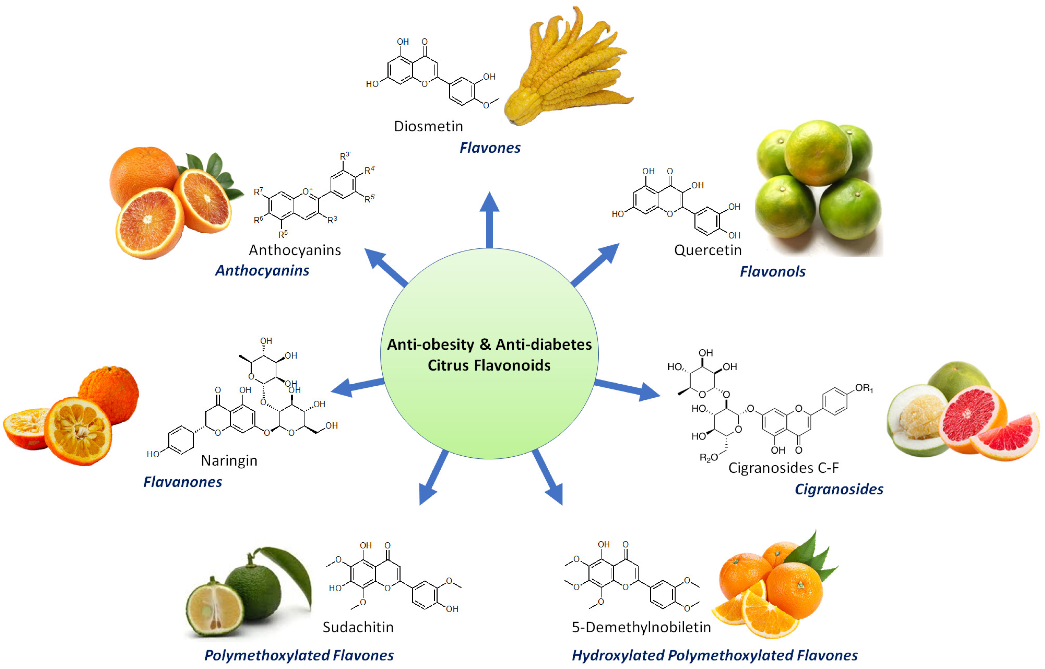
2. Bioactive Flavonoids from Citrus Fruit Peels

2.1. Flavanones

2.1.1. Didymin

To evaluate the antidiabetic potential of didymin, a bioactive flavonoid glycoside found in various citrus fruits, Ali et al., first employed a series of cell-free enzymatic inhibitory assays and they discovered that didymin was a potent inhibitor of α-glucosidase, PTP1B, rat lens and human recombinant aldose reductase, which was further supported by the molecular docking analysis [38]. Treatment of didymin in insulin-resistant HepG2 cells also led to downregulation of PTP1B and enhanced glucose uptake. This improved insulin sensitivity was mediated via activation of IRS-1, PI3K, Akt, and GSK-3 via enhanced phosphorylations while downregulation of key enzymes involved in gluconeogenesis such as PEPCK and G6Pase [38].

2.1.2. Eriocitrin (Eriomin)

Eriocitrin (Eriomin) is an abundant flavanone in the peels of various citrus fruits including C. latifolia, C. limon, C. grandis cv Hirado, and C. leiocarpa [17,39]. Using HFD-fed and HCD-fed obese rats, Miyake et al. have proved the strong lipid-lowering effects of eriocitrin [39]. Using HepG2 cells and a diet-induced obese zebrafish model, Hiramitsu et al. further demonstrated that eriocitrin could ameliorate dyslipidemia and hepatic steatosis via upregulating PPARα, NRF1, ATP5J, and COX4l1, thus activating mitochondrial biogenesis [39]. The antiobesity potential of Eriocitrin was further confirmed in HFD-fed obese mice [40,41]. Kwon et al. discovered that dietary eriocitrin reduced adipose tissue mass and hepatic steatosis via decreasing lipogenesis and increasing FA oxidation in adipose tissue and liver respectively, as well as enhancing energy expenditure in adipose tissue and skeletal muscle [40]. Furthermore, insulin sensitivity was improved by eriocitrin as a result of reduced gluconeogenesis and proinflammatory responses in the liver [40]. Ferreira et al. also found that eriocitrin supplementation at 25 mg/kg body weight for 4 weeks could significantly enhance insulin sensitivity and reduce hepatic TG accumulation which were associated with the inhibitory effect of eriocitrin on oxidative stress and systemic inflammation [41].
Of note, Cesar et al. recently showed that in a randomized crossover clinical study, a 200 mg/day eriomin administration for 12 weeks was able to reduce hyperglycemia and improve diabetes-related parameters in patients with hyperglycemia above 110 mg/dL via its anti-inflammatory effect and upregulation of GLP-1, indicating the therapeutic potential of eriomin for the glycemic control in prediabetic and diabetic populations [42].

2.1.3. Hesperidin

Hesperidin is a flavonoid commonly found in citrus fruits, with especial high amount in the peels of C. tangerine, C. succosa, C.suhuiensis, and C. kinokuni [17]. In 3T3-L1 cells, hesperidin has shown potent inhibitory effects on adipogenesis and free fatty acid (FFA) secretion derived from adipocyte lipolysis, repressing FFA-induced IR [43]. This is executed through inhibition of tumor necrosis factor-alpha (TNF- α) stimulated NF-κB and ERK pathways, as well as downregulation of antilipolytic genes including perilipin and PDE3B [43]. In HepG2 cells, hesperidin efficiently blocks pancreatic lipase (PL) activity via interacting with PL by hydrogen bonds and van der Waals forces, showing therapeutic potential in managing obesity [44]. Rajan et al. further showed in palmitate (PA)-treated HepG2 cells, besides reducing TG content, hesperidin increased glucose uptake in an insulin-independent manner [45]. Further elucidation of the molecular mechanism revealed that hesperidin activated AMPK signaling, increasing phosphorylation of acetyl-CoA carboxylase (ACC) and glycogen synthase kinase 3 beta (GSK3β) while decreasing expression of HMG-CoA reductase (HMGCR) and SREBP-2 [45]. In primary bovine aortic endothelial cells, hesperidin and its metabolite hesperetin improved endothelial function via stimulation of NO production and reduction of inflammation [31].
Interestingly, in high fat diet (HFD)-fed LDLr(-/-) mice, hesperidin also exerted protective effect against atherosclerosis via pleiotropic effects such as antioxidative and anti-inflammatory effects, improvement of insulin sensitivity, reduction of hyperlipidemia and hepatic steatosis, and blockage of macrophage foam cell formation [46]. Similarly, in obesogenic cafeteria diet (CAF)-fed obese rat, hesperidin supplementation also showed therapeutic potential against metabolic syndrome via improving insulin sensitivity and lowering cholesterol, FFA and inflammation levels [47]. Hesperidin also showed significant antidiabetic effect in nicotinamide/streptozotocin (NA/STZ)-induced diabetic rats via remarkable improvements in the insulin sensitivity and antioxidant defense system [25].
Salden et al. found that in obese individuals with a flow-mediated dilation ≥ 3%, supplementation of hesperidin 2S at 450 mg/day for 6 weeks could significantly improve endothelial function and blood pressures via reduction of soluble vascular adhesion molecule-1 (VCAM-1) and intracellular adhesion molecule-1 (ICAM-1) [32].
Yet, hesperidin has relatively lower water solubility and thus poor oral bioavailability, so the scientific community have tried to modify hesperidin to α-monoglucosyl hesperidin using cyclodextrin glucanotransferase [48]. Nishikawa et al. discovered that compared with hesperidin, α-monoglucosyl hesperidin is more potent in inducing brown-like adipocyte formation and thus suppressing white adipose tissue accumulation in mice [48]. Yoshida et al. also found that although not improving the insulin sensitivity, α-monoglucosyl hesperidin did ameliorate hyperglycemia and macrophage infiltration into the adipose tissue in HFD-fed obese mice [49]. Moreover, hesperetin, the metabolite of hesperidin and an aglycone of α-monoglucosyl hesperidin, efficiently block the monocyte chemotactic protein 1 (MCP-1) expression when 3T3-L1 adipocytes cultured alone or cocultured with RAW264 macrophages [49].

2.1.4. Naringenin

Naringenin is a well-studied bioactive flavonoid extracted from citrus fruit peels. Using 3T3-L1 and coculture of 3T3-L1 and RAW264 macrophages, Yoshida et al. and Tsuhako et al. found that naringenin could be a potent antiobesity nutritional supplement due to its efficient inhibitory effects on adipogenesis and adipose tissue inflammation [12,43,50,51]. Detailed examination revealed that naringenin, besides suppressing TNF-α-stimulated FFA secretion, blocked toll-like receptor 2 (TLR2) expression during adipocyte differentiation in 3T3-L1 which is partially executed via activation of PPARγ [43,51]. Naringenin was discovered to be an activator of PPARα and PPARγ while an inhibitor of LXRα [52]. Later it was also found that naringenin reduced adiponectin protein expression and dose-dependently inhibited insulin-stimulated glucose uptake via suppressing tyrosine phosphorylation of IRS-1 [53]. Furthermore, in 3T3-L1 and macrophages coculture, naringenin suppressed TNF-α-induced TLR2 expression via attenuating activation of NF-κB and JNK pathways in differentiated adipocytes [51]. Noticeably, MCP-1 and MCP-3 expression were also consistently downregulated in adipocytes, macrophages, and a co-culture of adipocytes and macrophages [12,50]. These effects were also observed in HFD-fed obese mice and were correlated with amelioration of hyperglycemia and adipose tissue inflammation, especially, macrophage and neutrophil infiltration into adipose tissue [12,50,51]. Using human white adipocyte cultures (hADSC), Rebello et al. found that naringenin enhanced energy expenditure, and upregulated key genes involved in thermogenesis and insulin sensitivity including UCP1, PGC1α, ChREBP, and GLUT4 [54]. In human umbilical vein endothelial cells (HUVECs), Zhang et al. also discovered naringenin ameliorated high glucose-induced endothelial dysfunction via upregulating the protein level of intracellular heat shock protein 70 (iHSP70) [55].
With HFD-fed LDLr(-/-) mice, Mulvihill et al. and Burke et al. investigated the therapeutic effect of naringenin on obesity associated metabolic dysfunctions [56,57,58,59]. Naringenin supplementation to a HFD was able to reverse obesity via increased energy expenditure and hepatic fatty acid (FA) oxidation, which was accompanied by an amelioration of dyslipidemia, a reduction of inflammation and improvements in insulin sensitivity [56,57,58,59]. Elucidation of the relevant molecular mechanism revealed that naringenin stimulated PPARα-mediated transcription while downregulated SREBP-1-mediated lipogenesis both in liver and muscles [60]. Although in these mice, atherosclerotic lesions were unchanged in size, they displayed a much less macrophage content and a more stable plaque phenotype [56]. Besides this atheroprotection effect, naringenin also displayed the ability to promote atherosclerosis regression [61]. Using HFD-fed Fgf21(-/-) mice, Assini et al. found that naringenin ameliorated hyperinsulinemia and impaired glucose tolerance, partially through induction of PGC1α [62]. Ahmed et al. further discovered that in NA/STZ-induced diabetic rats, naringenin treatment potently reversed diabetic symptoms via its insulinotropic effects and insulin improving action which is partially mediated through modulating insulin receptor, GLUT4 and adiponectin expression in adipose tissue [63].
Using HFD-fed obese ovariectomized mice, Ke et al. discovered that naringenin administration placed beneficial effects on metabolic health and tumorigenesis of obesity-related postmenopausal breast cancer via activation of AMPK signaling and induction of cell death in tumor cells [64]. Similarly, Snoke et al. also found that in a colon-26 cancer cachexia mouse model, naringenin administration improved insulin sensitivity while suppressed inflammation and adenocarcinoma growth [65].

2.1.5. Naringin

Naringin is also commonly found in the peels of various citrus fruits including C. aurantium, C. natsudaidai, C. paradise, and C. grandis cv. Hirado [17]. Although also displayed strong anti-inflammatory and antioxidant activities, naringin is less studied compared with its aglycone naringenin and its effect on obesity and associated metabolic disorders remains to be fully elucidated.
In a rat model of diabetes, Ahmed et al. found that similar to naringenin, naringin also stimulated adipose tissue expression of insulin receptor β-subunit, GLUT4 and adiponectin which partially explains its alleviation effects on the content of serum insulin, C-peptide, and liver glycogen content [63]. Using an in vitro cardiomyoblast model of diabetes, Chen et al. also demonstrated the protective potential of naringin on diabetes-associated cardiomyocyte injury [66]. In H9C2 cells, naringin attenuated high glucose-induced apoptosis by inhibiting excessive ROS production, the dissipation of mitochondrial membrane potential, and the activation of the p38 MAPK signaling induced by leptin [66].

2.1.6. Narirutin

Abundant in the peels of C. shunkokan, C. sulcata, C. nobilis var Knep, and C. leiocarpa [17], narirutin is another flavone glycoside which shows potent capacities in suppressing adipogenesis and intracellular triglyceride accumulation. Rajan et al. recently found that in insulin resistant HepG2 cells, narirutin enhanced glucose uptake in an insulin-independent manner mainly through activation of the AMPK pathway [45]. Molecular docking analysis revealed that narirutin might activate AMPK by binding to the CBS domains in the regulatory gamma-subunit of AMPK. Activation of AMPK leads to enhanced phosphorylation of metabolic enzyme ACC and GSK3β as well as decreased expression of HMGCR and SREBP-2, resulting in enhanced fatty acid oxidation and reduced lipid biosynthesis [45].

2.1.7. Neohesperidin

Neohesperidin is a flavonoid enriched in the peels of C. aurantium, C. bergamia, C.glaberrima, C. hassaku, and C. changshanensis [17,67]. Using HepG2 cells, Zhang et al. found that neohesperidin dramatically increased glucose uptake and this was associated with activation of AMPK signaling [67]. In vivo studies also showed that, similar to hesperidin, neohesperidin exhibited impressive potency in suppressing intracellular triglyceride accumulation and improving insulin sensitivity [29,68]. In HFD-fed obese mice, neohesperidin administration attenuated gain of body weight, inflammation and insulin resistance [29]. This was associated with an improved diversity of gut microbiota and the modified abundance of bacteroidetes and firmicutes, indicating the potential of flavonoid-induced gut microbiota therapy against obesity [29]. Using diabetic KK-A(y) mice, Jia et al. discovered that the antidiabetic potential of neohesperidin was associated with hepatic activation of the AMPK pathway and the subsequent regulation of its target genes including stearoyl-CoA desaturase 1 (SCD-1), fatty acid synthase (FAS), and acyl-CoA oxidase (ACOX) [68].

2.2. Flavones

2.2.1. Diosmetin

Diosmetin, a monomethoxyflavone, is naturally present abundantly in the peels of citrus fruits such as C. medica var. 2 and C. suhuiensis [69,70]. Xie et al. discovered that in 3T3-L1 cells, normal diet–fed ob/ob mice and in HFD-fed obese mice, diosmetin functioned as an agonist for estrogen receptors (ERs) which subsequently provoked energy expenditure via stimulation of thermogenesis in brown adipose tissue (BAT) and acceleration of white adipose tissue (WAT) browning [30]. Diosmetin-induced upregulation of ERs in matured adipocytes and in mice adipose tissue was indispensable for the observed metabolic benefits that diosmetin placed in obese mice, as ER blockage by the antagonist fulvestrant abolished the metabolic benefits including reduction of weight gain and fat mass as well as improved insulin sensitivity [30]. Using 3T3-L1 and RAW264 macrophages coculture, Lee et al. found that diosmetin also displayed strong anti-inflammatory and antilipolytic activities [70]. These activities were mediated via downregulation of iNOS and inhibition of MAPK phosphorylation and nucleus translocation in macrophages and adipocytes which lead to suppression of inflammatory mediators such as NO, TNF-α, and MCP [70]. As such, diosmetin-rich dietary therapy may be suitable for the management of obesity-related metabolic syndromes.
Interesting, in HFD-fed SD rats, Meephat et al. further discovered that diosmetin efficiently prevent metabolic syndrome and associated cardiac abnormalities in these insulin resistant rats as diosmetin treatment improved insulin sensitivity, restored ejection faction, and decreased left ventricular hypertrophy and fibrosis [69]. Detail investigation indicates that suppression of the Ang II/AT1 receptor/NF-κB pathway was underlying these rescue effects of diosmetin [69].

2.2.2. Diosmin

Jain et al. found that diosmin, a flavone enriched in the peels of C. montana, C. latifolia, C. lumia, and C. kinokuni, was able to suspend the progression of early diabetic neuropathy in a rat model of HFD/STZ-induced type 2 diabetes [17,71]. After successful induction of diabetes as evidenced by insulin resistance and dramatically elevated blood glucose, 4 weeks of diosmin supplementation dose-dependently improved thermal hyperalgesia, cold allodynia and walking function in vivo [71]. This was associated with normalized malondialdehyde, and nitric oxide as well as restored glutathione levels and superoxide dismutase activity of diabetic mice [71].

2.2.3. Nobiletin

Nobiletin is a well-studied polymethoxylated flavone (PMF) which is extensively found in the peels of various citrus fruits [17,72]. Nobiletin has been shown to suppress adipogenesis and obesity in various in vitro and in vivo models [45]. In 3T3-L1 cells, Miyata et al. found that besides inducing adipocyte apoptosis, nobiletin increased the secretion of adiponectin while decreased the secretion of MCP-1 and resistin, thus improving the insulin sensitivity [73]. Apart from downregulating PPARγ2, Kanda et al. found that nobiletin suppressed adipogenesis via reducing the phosphorylation of CREB while enhancing the phosphorylation of STAT5 [74]. Lone et al. further demonstrated that nobiletin exerted dual modulatory effects on 3T3-L1 preadipocytes, on one hand, nobiletin reduced intracellular stress and key transcription factors involved in lipid metabolism; on the other hand, it accelerated browning in 3T3-L1 white adipocytes via induction of multiple beige-specific genes [75]. Using HepG2 and Ampkβ1-/- primary mouse hepatocyte, Morrow et al. proved that nobiletin placed a metabolic protection effect on hepatocytes via activation of AMPK and ACC [76]. Rajan et al. also observed the similar effect induced by nobiletin in PA-treated HepG2, in addition, they found that nobiletin also upregulated GSK3β while downregulated SREBP-2 and HMGCR [45]. In addition, Tsuboi showed that in J774.1 mouse macrophages, nobiletin increased cell release of high-density lipoprotein cholesterol via activation of AMPK and subsequently the LXRα-PPARγ loop pathway which upregulated ABCA1 and ABCG1 [77].
The therapeutic potentials of nobiletin against obesity, diabetes and atherosclerosis are evidenced by multiple in vivo studies [78,79]. Lee et al. showed that in diabetic ob/ob mice and HFD-fed obese mice, nobiletin significantly improved insulin sensitivity and glucose homeostasis via decreasing inflammatory adipokines such as interleukin (IL)-6 and MCP-1, while simultaneously upregulating GLUT4 and GLUT1 in various tissues [78,80]. Zhang et al. found that in both HFD-fed obese mice and rat, gut microbiota was modified by long-term oral administration of nobiletin which led to improved biotransformation, indicating the crucial contribution of gut microbiota in the in vivo antiobesity effect of nobiletin [81,82].
By employing mice with hepatic AMPK deficiency, hepatic ACC dysfunction and mice with adipocyte-specific AMPK deficiency, Morrow et al. proved the metabolic protective effect of nobiletin against HFD/HCD-induced obesity, hepatic steatosis, and dyslipidemia, in vivo [76]. Bunbupha et al. further examined nobiletin-induced protective effect on nonalcoholic fatty liver disease (NAFLD) and they found that nobiletin-treated HFD-fed rat exhibited a much less severe NAFLD phenotype including ameliorated adiposity, hyperlipidemia, insulin resistance, hepatic lipids accumulation and fibrosis [79]. This protective effect was mediated through upregulation of hepatic adiponectin receptor 1 (AdipoR1) and plasma adiponectin levels, as well as downregulation of hepatic NADPH oxidase subunit gp91(phox) [79]. Similarly, in nonobese mice, Kim et al. also observed nobiletin-induced protective effect on nonalcoholic fatty liver disease (NAFLD) and HCD-induced hypercholesterolemia [83]. This effect was associated with a reduction of systematic inflammation and atherosclerosis-associated cardiovascular markers [83]. Consist with this, Burke et al. have also demonstrated that nobiletin exhibited a strong therapeutic potential on metabolic dysfunction and cardiovascular disease as nobiletin supplement in HFD-fed LDLr(-/-) mice led to a more stable plaque phenotype of atherosclerotic lesions, in which the macrophage content was much less [56].

2.2.4. Sinensetin

Sinensetin is a rare polymethoxylated flavone which is found in certain citrus fruits including C. sunki, C. sinensis cv Valencia, C. tachibana, and C. tangerine [17,84]. Compared with other parts of the fruit, it is more abundantly found in the peels of the citrus fruits [85,86]. Kang et al. observed a strong inhibitory effects of sinensetin on adipogenesis in 3T3-L1 cells [84]. Its antiadipogenic property was partially explained by a downregulation of SREBP1c expression as well as an increased phosphorylation of PKA and hormone-sensitive lipase, indicating the involvement of the cAMP-mediated signaling pathway [84]. In 3T3-L1 cells, sinensetin blocked insulin-stimulated glucose uptake by suppressing the IRS and Akt phosphorylation while it enhanced phosphorylation of AMPK and ACC to stimulate FA oxidation [84]. Similarly, Rajan et al. found that in PA-treated HepG2 cells, sinensetin also increased AMPK, ACC, and GSK3β phosphorylation levels and decreased SREBP-2 and HMGCR expression. Nevertheless, an increase in 2-NBDG glucose uptake was observed in PA-treated HepG2 cells, indicating the tissue-specific regulation pattern of sinensetin [84], Yet, to this date, the physiological relevance of these antiadipogenic effect induced by sinensetin still need to be further investigated in vivo.

2.2.5. Sudachitin

Sudachitin is a rare polymethoxylated flavone found in the peels of C. sudachi [33,87]. Studies have shown that in cellular and animal models, the extracts from C. sudachi peel which is enriched in sudachitin, could ameliorate hyperlipidemia and obesity, possibly due to an augmented AMPK activity and PPARα transcription [87]. Shikishima et al. further proved that in a randomized controlled trial, although not affecting glycemic control and lipid profile, consumption of sudachi peel extract capsules (including sudachitin 4.9 mg/day) for 12 weeks could considerably decreased the ratio of visceral fat to subcutaneous fat, and moderately reduced waist circumference in Japanese patients at risk for developing diabetes [33].
In HFD-fed obese mice and db/db mice, sudachitin displayed strong capacity in stimulating energy expenditure and limiting weight gain via improving glucose and lipid metabolism and promoting mitochondrial biogenesis and function [88]. Elucidation of the underlying molecular mechanism in primary myocytes revealed that sudachitin placed a favorable effect on Sirt1 and PGC1α upregulation in the skeletal muscle [88].

2.2.6. Tangeretin

Tangeretin is a major polymethoxylated flavone extensively found in a variety of citrus fruit peels. Miyata et al. discovered the promising therapeutic potential of tangeretin for the management of insulin resistance as evidenced by its strong inhibitory effect on adipocyte differentiation and its favorable modulation on the adipocytokine secretion balance [73]. In 3T3-L1 cells, without affecting cell viability, tangeretin provoked the secretion of adiponectin, an insulin-sensitizing factor while simultaneously suppressed the secretion of MCP-1, an insulin resistance factor, thus dramatically reducing the intracellular triglyceride accumulation [73]. In adipocyte and macrophage coculture, Sun et al. discovered that tangeretin reduced adipocyte inflammation by reprogramming glucose metabolism and enhancing lactate accumulation which leads to increased macrophage polarization [89].
The molecular mechanisms underlying lipid-modulating effects of tangeretin have been extensively studied in various in vitro model employing mouse, rat and human hepatocytes [45,52,90,91]. Guo et al. found that in primary mouse hepatocytes, tangeretin upregulated insulin signaling pathway, increased glycogen content while decreased glucose output. This effect was mediated via inhibiting the phosphorylation of ERK1/2 [91]. Rajan et al. further discovered that in PA-treated HepG2 cells, tangeretin attenuated intracellular lipid accumulation via activation of the AMPK pathway [45]. Through transcriptome and bioinformatics analyses, Chen et al. identified a set of 13 genes associated with lipoprotein lipase (LPL) under regulation of tangeretin, among them, angiopoietin-like 3 (ANGPTL3), an inhibitor of LPL catalytic activity, is downregulated in both HepG2 and Huh-7 cells [90]. Further elucidation revealed a tangeretin-responsive element in the promoter region of ANGPTL3. Although not directly affecting the expression of ANGPTL3 upstream regulator, LXRα, molecular docking analysis predict the strong binding capacity of tangeretin to the ligand-binding domain of LXRα [90].
In a mouse model of diabetes, intragastric administration of tangeretin improved the hepatic insulin sensitivity and glucose homeostasis via deactivating the MEK-ERK1/2 pathway [91]. This tangeretin-induced beneficial effect on insulin resistance was also found in HFD-fed obese mice [90,92]. Nery et al. discovered that although not directly reducing fat accumulation in the mouse liver, tangeretin did suppressed adipocyte hypertrophy in the mice with pre-existent obesity [92]. Chen et al. observed that long-term supplementation of HFD-fed mice with tangeretin suppressed weight gain, hepatic steatosis, and glucose intolerance via improving adipose thermogenesis, ameliorating systemic inflammation and gut microbiota dysbiosis. Interestingly, Liu et al. found in C. elegans, tangeretin supplementation could even considerably augment the stress tolerance and the lifespan in an insulin/insulin like growth factor signaling dependent manner [93].

2.2.7. 3′,4′,3,5,6,7,8Heptamethoxyflavone

3,5,6,7,8,3′,4′-heptamethoxyflavone (HMF) is a major and naturally occurring polymethoxyflavone found in the peels of various citrus fruits including C. yatsusiro, C. oto, C. nobilis var Knep, C. clementine and C. unshiu [17,94]. Sawamoto et al. found that in 3T3-L1 preadipocyte, HMF treatment for 8 days dose-dependently inhibits adipocyte differentiation mainly through downregulating PPARγ, C/EBPα, and SREBP1, as well as activating PKACα and its downstream AMPK and ACC [95]. Nery et al. treated HFD-fed obese mice with HMF supplement (100 mg/kg) daily for 4 weeks and they found that although HMF did not affect adipocyte size, HMF was potent in reducing hepatic lipid accumulation which was associated with a considerably enhanced level of anti-inflammatory cytokine IL-10 [92].

2.2.8. 5-Demethylnobiletin

5-demethylnobiletin (5-OH-Nob) is a hydroxylated polymethoxylated flavone (HPMF) which is exclusively found in citrus peels. Tung et al. have shown that in 3T3-L1 preadipocytes, 5-OH-Nob could significantly suppress adipogenesis and triglyceride accumulation [72]. Moreover, to overcome the relatively poor aqueous solubility and low oral bioavailability of 5-OH-Nob, they chemically modified 5-OH-Nob to 5-acetyloxy-6,7,8,3′,4′-pentamethoxyflavone (5-Ac-Nob) which showed improved antiadipogenic and antiobesity effects both in vitro and in vivo via activation of the AMPK pathway [72].

2.2.9. 5-Hydroxy-3,6,7,8,3′,4′-Hexamethoxyflavone

5-hydroxy-3,6,7,8,3′,4′-hexamethoxyflavone (5-OH-HxMF) is a HPMF enriched in the peels of certain citrus fruits such as C. sinensis and C. reticulate [96,97]. Lai et al. demonstrated that 5-OH-HxMF dose-dependently suppressed adipogenesis both in vitro and in vivo [97]. In 3T3-L1 preadipocytes, 5-OH-HxMF considerably suppressed the protein expression of key adipogenesis-related genes including PPARγ, SREBP1 and their downstream targets aP2, ACC and FAS while activated the AMPK pathway [97]. Furthermore, 5-OH-HxMF induced a delayed cell cycle entry into G2/M phase after adipogenesis initiation in the treated preadipocytes [97]. Besides these modulations, Wang et al. further discovered that 5-OH-HxMF decreased lipid accumulation via downregulation of C/EBPs and activation of SIRT1. Interestingly, when compared with 3,5,6,7,8,3′,4′-heptamethoxyflavone (HMF), 5-OH-HxMF displayed an around 20% higher potency in preventing lipid accumulation, indicating the crucial role played by the hydroxyl group in the antiadipogenic activity [96]. In HFD-fed obese mice, 5-OH-HxMF efficiently reduced the weight gain, adipose tissue mass and hyperlipidemia [97].

2.3. Other Flavonoids

2.3.1. Anthocyanins

Anthocyanins, a class of flavonoid compounds well known to exert numerous health-promoting actions, could also be found in the peels of various citrus fruits such as C. sinensis L. Osbeck [13,24]. Treatment of 3T3-L1 preadipocytes with 2.5, 5, 10, 25 µM of anthocyanins equivalents impressively suppressed adipocyte differentiation, intracellular triglyceride accumulation and adipokine secretion. These beneficial effects were partially attributed to the antioxidative property of anthocyanins and their modulation on the expression and function of key genes involved in lipid metabolism including PPARγ, C/EBPα, SREBP-1c, ACCα, and FAS [24].

2.3.2. Cigranoside C, D, E, F

Cigranoside C, D, E, F are recently discovered flavonoids found in the peels of certain citrus fruits including C. grandis L. Osbeck, C. grandis Shatianyu, and C. paradisi Mcfad [98]. All four cigranoside displayed strong antidiabetic potential as evidenced by their strong inhibitory effect on α-amylase, α-glucosidase, and PL in a series of in vitro cell-free assays [98].

2.3.3. Quercetin

Quercetin is a flavonol pigment present in various fruits and vegetables [13]. Dhanya et al. investigated its mechanism of action in the context of diabetic skeletal muscle [99]. Through inhibition of relevant protein kinases in L6 myotubes, quercetin was proved to stimulate the glucose uptake mainly via the AMPK pathway and its downstream p38 MAPK rather than the insulin signaling pathway. The rise of intracellular calcium after quercetin treatment also indicated the possible calcium-calmodulin mediated protein kinase (CaMKK) may also be involved [99]. These action modes align quercetin with the commercial drug metformin, highlighting its therapeutic potential against T2DM [99]. Indeed, further in vivo study also confirmed its antidiabetic potential as treatment quercetin resulted in various beneficial effect in NA/STZ-induced diabetic rats including improved insulin sensitivity and ameliorated hyperglycemia and hyperlipidemia, as well as reduced serum insulin, oxidative stress and C-peptide and reduced liver glycogen content. Nevertheless, in this in vivo model, besides adiponectin, GPx, GST and SOD, quercetin treatment still executed its antidiabetic potential through, at least partially, enhancing the mRNA expression of the insulin signaling pathway including GLUT4, and insulin receptor β-subunit [25].
Table 1 summarizes these citrus flavonoids, their major molecular targets and relevant mechanisms underlying their potential against obesity and associated diseases.

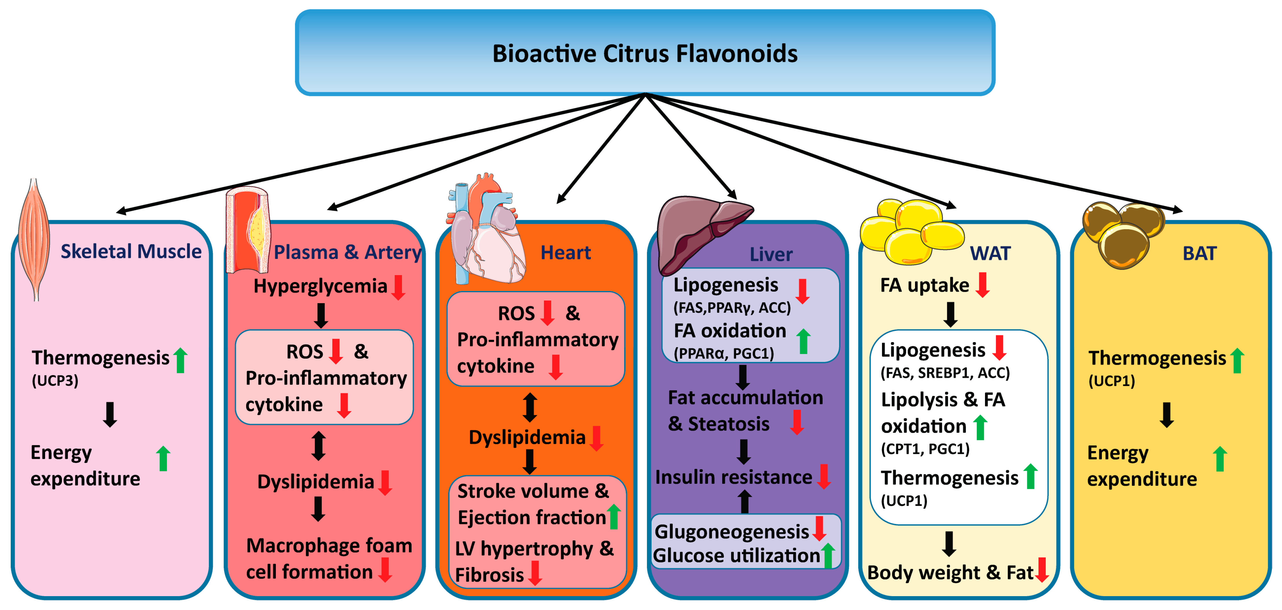
3. Discussion and Conclusions

This review aims to present the latest findings on natural flavonoids isolated from various citrus fruit peels that have shown therapeutic potential toward obesity or associated metabolic syndrome, and the underlying physiological and cellular mechanisms (Figure 2). Our investigation showed that many bioactive citrus flavonoids exerted their beneficial effects against obesity, diabetes and other associated disease through multiple targets and diverse intracellular processes. This multiple-layer regulation seems to be a favorable feature for the drug development against obesity and related metabolic diseases, since these diseases typically have a complex etiology which usually includes a network of multiple dysregulated factors and processes [2,4].
Nevertheless, several limitations need to be appropriately addressed before developing these bioactive flavonoids into clinical drugs against obesity and associated metabolism syndrome. First, optimized harvesting, processing and extraction methods for obtaining these bioactive flavonoids from citrus fruit peels still needs to be developed. For example, optimizing harvest times and thermal processing methods could help enrich, release and transform bioactive flavonoid, resulting in more efficient cellular uptake [101,102]. Second, most of the bioactive flavonoids displayed low bioavailability due to their poor water solubility, limiting their clinical use. Thus, appropriate modifications may need to be incorporated into their chemical structures to improve the bioavailability. One successful example is 5-Ac-Nob. Being chemically modified from 5-OH-Nob, 5-Ac-Nob showed higher potency in reducing intracellular TG content both in vitro and in vivo, due to its improved aqueous solubility [72]. Last but not least, although several clinical trials have shown positive results regarding the therapeutic uses of these flavonoids [31,103], the lack of adequate clinical evidence to evaluate the long-term toxicity and efficacy of these phytochemicals in humans is a major limitation for the majority of these bioactive flavonoids.
Despite all these challenges, bioactive flavonoids remain as an effective and promising alternative approach for obesity and associated diseases. Thanks to the technological advances in the fields such as metagenomics, metabolomics, and proteomics, scientists now could generate huge amount of data and examine various in vitro and in vivo effects of bioactive flavonoids in great detail [47,81,82,104].
In recent years, there has also been an explosion of interest in artificial intelligence (AI) and its potential applications across a wide range of industries. The pharmaceutical sector is no exception, with many companies now exploring how AI could be used to accelerate and improve the drug discovery process.
In theory, AI has the ability to speed up the process of discovering new drugs by orders of magnitude. This is because AI can automate many of the tasks that are currently done by human researchers, such as data collection and analysis [35]. Perhaps most importantly, AI has the potential to help researchers access and analyze huge amounts of data that would otherwise be inaccessible. By harnessing the power of big data, AI could help scientists identify patterns and trends that would otherwise be invisible information which could be critical in making key decisions during drug discovery research. In addition, AI can provide a more objective approach to analyzing data than humans alone are capable of. This is due to the fact that machines are not biased by personal beliefs or preconceptions; they simply analyze the data as they are. This objectivity can lead to faster and more accurate decision making during the drug discovery process [105].
There are numerous advantages that AI, especially machine learning, can bring to the translation of flavonoids to clinical drugs. One common application is virtual screening, where computer programs are used to identify potential small molecule ligands that could bind to and modulate the activity of a target protein, or to identify new therapeutic targets [35]. This can be a more efficient way to screen for lead compounds than traditional experimental approaches. Another area where machine learning is having an impact is in the development of predictive models for ADMET (absorption, distribution, metabolism, excretion and toxicity) properties [35,105]. These models can help assess the safety and efficacy of candidate molecules at an early stage in the drug development process, saving time and resources later on. Besides these, it is also possible that machine learning will be used to design entirely new classes of drugs. Recent advances in artificial intelligence have led to the development of programs that can generate novel compounds with specific chemical properties. If these compounds show promise in preclinical studies, they could one day be developed into new medicines [35,105]. With the assistance from AI, scientists could focus on more creative and strategic tasks, such as conceptualizing new experiments or interpreting results [105,106].
In conclusion, citrus fruit peels, although usually regarded as waste, contain numerous flavonoids that have shown therapeutic potential for obesity and diabetes mellitus. The chemical structure-based elucidation of these antiobesity and antidiabetes mechanisms provides scientific bases for using citrus fruit peels as sources of nutraceuticals and pharmaceuticals against obesity and diabetes. This review thus highlights the therapeutic and economic potential of these citrus fruit byproducts. Proper management of citrus fruit peel waste will not only considerably eliminate the relevant environmental concerns, but also expand the alternative sources of high-value bioactive flavonoids that could alleviate the global burden of obesity and related diseases.

Author Contributions

Investigation: K.L., Y.M.Y.; visualization: K.L., Y.M.Y.; conceptualization: K.L.; writing—original draft preparation: K.L., Y.M.Y.; writing—review and editing: K.L., Y.M.Y. All authors have read and agreed to the published version of the manuscript.

Funding

This research received no external funding.

Institutional Review Board Statement

Not applicable.

Informed Consent Statement

Not applicable.

Data Availability Statement

Not applicable.

Acknowledgments

We would like to thank DFG SFB1116 “Master Switches in Cardiac Ischemia”, Heinrich Heine University Duesseldorf, University Hospital Duesseldorf, and Nanyang Technological University for their support.

Conflicts of Interest

The authors declare no potential conflict of interest.

Abbreviations

2-(N-(7-Nitrobenz-2-oxa-1,3-diazol-4-yl)Amino)-2-Deoxyglucose (2-NBDG); 3-hydroxy-3-methylglutaryl-CoA reductase (HMGCR); 3-isobutyl-1-methylxanthine (IBMX); 5-acetyloxy-6,7,8,3′,4′-pentamethoxyflavone (5-Ac-Nob); 5-demethylnobiletin (5-OH-Nob); 5-hydroxy-3,6,7,8,3′,4′-hexamethoxyflavone (5-OH-HxMF); Abnormal dauer formation protein 16 (DAF-16); Acetyl CoA Carboxylase (ACC); Acid-binding protein (aP2); Acyl-CoA synthetase (ACS); Acyl-CoA oxidase (ACO); Adiponectin receptor 1 (AdipoR1); Adenosine 5′-monophosphate-activated protein kinase (AMPK); Absorption, distribution, metabolism, excretion and toxicity (ADMET); Advanced glycation end-product (AGE); Akt substrate of 160 kDa (AS160); AMP-activated protein kinase (AMPK); Angiopoietin-like 3 (ANGPTL3); Angiotensin II (Ang II); Angiotensin II type 1 (AT1); ATP binding cassette subfamily A member 1 (ABCA1); ATP binding cassette subfamily G member 1 (ABCA1); ATP-binding cassette transporters (ABC); ATP Synthase Peripheral Stalk Subunit F6 (ATP5J); Bone marrow-derived macrophages (BMDMs); Brown adipose tissue (BAT); Calcium-calmodulin mediated protein kinase (CaMKK); cAMP-response element-binding protein (CREB); Carbohydrate-responsive element-binding protein (ChREBP); Carnitine palmitoyltransferase I (CPT1); CCAAT enhancer-binding protein-alpha (C/EBPα); Chemokine (C-C motif) ligand (CCL); Citrate synthase activity (CSA); c-Jun NH2-terminal kinase (JNK); Cytochrome c oxidase subunit 4 (COX4l1); Endothelial NO synthase (eNOS); Estrogen receptor (ER); Fatty acid (FA); Fatty acid synthetase (FAS); Fatty acid translocase (FAT/CD36); Fibroblast growth factor 21 (FGF21); Free fatty acid (FFA); G-protein-coupled bile acid receptor, Gpbar1 (TGR5); Glucagon-like peptide 1 (GLP-1); Glucocorticoid receptor (GR); Glucose 6-phosphatase (G6Pase); Glucose transporter Typ 4 (GLUT4); Glutathione peroxidase (GPx); Glutathione-S-transferase (GST); Glycogen synthase (GS); Glycogen synthase kinase 3 beta (GSK3β); Heat shock protein (HSP); Heat shock transcription factor 1(HSF1); Heptamethoxyflavone (HMF); High-cholesterol diet (HCD); High fat diet (HFD); Human recombinant AR (HRAR); Human umbilical vein endothelial cell (HUVEC); Human white adipocyte cultures (hADSC); Hydroxylated polymethoxyflavones (HPMFs); Inducible nitric oxide synthase (iNOS); Insulin receptor substrate (IRS); Intercellular cell adhesion molecule-1 (ICAM-1); Intracellular heat shock protein 70 (iHSP70); Interleukin (IL); Iodothyronine Deiodinase 2 (DIO2); Liver kinase B1 (LKB1); Low-density lipoprotein (LDL); Low density lipoprotein cholesterol receptor (LDLr); Mitogen-activated protein kinase (MAPK); Monocyte chemoattractant protein-1 (MCP-1/CCL2); Nicotinamide/Streptozotocin (NA/STZ); Nitric oxide (NO); Nobiletin (Nob); Nonalcoholic fatty liver disease (NAFLD); Nuclear factor kappa-light-chain-enhancer of activated B cells (NF-κB); Nuclear liver X receptor α (LXRα); Nuclear respiratory factor 1 (NRF1); Obesogenic cafeteria diet (CAF); Ovariectomized (OVX); Palmitate (PA); Pancreatic lipase (PL); Patatin-like phospholipase domain containing 2 (pnpla2); Polymethoxyflavones (PMFs); Peroxisome proliferator-activated receptor (PPAR); Peroxisome proliferator-activated receptor gamma coactivator 1-alpha (PGC-1α); Phosphodiesterase-3B (PDE3B); Phosphoenolpyruvate carboxykinase (PEPCK); Phosphoinositide 3-kinase (PI3K); Protein kinase A (PKA); Protein kinase A catalytic subunit α (PKACα); Protein tyrosine phosphatase 1B (PTP1B); Rat lens aldose reductase (RLAR); Reactive oxygen species (ROS); References (Ref); Signal transducer and activator of transcription (STAT); Sirtuin 1 (SIRT1); Sprague-Dawley (SD); Sterol regulatory element binding transcription factor 1 (Srebf1); Sterol regulatory element-binding protein (SREBP); Stearoyl-CoA desaturase 1 (SCD-1); Superoxiddismutase (SOD); Toll-like receptor (TLR); Triacylglycerol (TG); Tumor necrosis factor-α (TNF-α); Uncoupling protein (UCP); Vascular cell adhesion molecule-1 (VCAM-1); White adipose tissue (WAT).

References

  1. Collaborators, G.B.D.O.; Afshin, A.; Forouzanfar, M.H.; Reitsma, M.B.; Sur, P.; Estep, K.; Lee, A.; Marczak, L.; Mokdad, A.H.; Moradi-Lakeh, M.; et al. Health Effects of Overweight and Obesity in 195 Countries over 25 Years. N. Engl. J. Med. 2017, 377, 13–27. [Google Scholar] [CrossRef] [PubMed]
  2. Misra, A.; Singhal, N.; Khurana, L. Obesity, the metabolic syndrome, and type 2 diabetes in developing countries: Role of dietary fats and oils. J. Am. Coll. Nutr. 2010, 29, 289S–301S. [Google Scholar] [CrossRef] [PubMed]
  3. Guilherme, A.; Virbasius, J.V.; Puri, V.; Czech, M.P. Adipocyte dysfunctions linking obesity to insulin resistance and type 2 diabetes. Nat. Rev. Mol. Cell Biol. 2008, 9, 367–377. [Google Scholar] [CrossRef] [PubMed] [Green Version]
  4. Tabak, A.G.; Herder, C.; Rathmann, W.; Brunner, E.J.; Kivimaki, M. Prediabetes: A high-risk state for diabetes development. Lancet 2012, 379, 2279–2290. [Google Scholar] [CrossRef] [PubMed] [Green Version]
  5. Rosen, E.D.; MacDougald, O.A. Adipocyte differentiation from the inside out. Nat. Rev. Mol. Cell Biol. 2006, 7, 885–896. [Google Scholar] [CrossRef]
  6. Cao, Z.; Umek, R.M.; McKnight, S.L. Regulated expression of three C/EBP isoforms during adipose conversion of 3T3-L1 cells. Genes Dev. 1991, 5, 1538–1552. [Google Scholar] [CrossRef] [Green Version]
  7. Tontonoz, P.; Hu, E.; Spiegelman, B.M. Stimulation of adipogenesis in fibroblasts by PPAR gamma 2, a lipid-activated transcription factor. Cell 1994, 79, 1147–1156. [Google Scholar] [CrossRef]
  8. Xu, J.; Liao, K. Protein kinase B/AKT 1 plays a pivotal role in insulin-like growth factor-1 receptor signaling induced 3T3-L1 adipocyte differentiation. J. Biol. Chem. 2004, 279, 35914–35922. [Google Scholar] [CrossRef] [Green Version]
  9. Giri, S.; Rattan, R.; Haq, E.; Khan, M.; Yasmin, R.; Won, J.S.; Key, L.; Singh, A.K.; Singh, I. AICAR inhibits adipocyte differentiation in 3T3L1 and restores metabolic alterations in diet-induced obesity mice model. Nutr. Metab. 2006, 3, 31. [Google Scholar] [CrossRef] [Green Version]
  10. Khan, R.M.M.; Chua, Z.J.Y.; Tan, J.C.; Yang, Y.; Liao, Z.; Zhao, Y. From Pre-Diabetes to Diabetes: Diagnosis, Treatments and Translational Research. Medicina 2019, 55, 546. [Google Scholar] [CrossRef]
  11. Han, H.Y.; Lee, S.K.; Choi, B.K.; Lee, D.R.; Lee, H.J.; Kim, T.W. Preventive Effect of Citrus aurantium Peel Extract on High-Fat Diet-Induced Non-alcoholic Fatty Liver in Mice. Biol. Pharm. Bull. 2019, 42, 255–260. [Google Scholar] [CrossRef] [Green Version]
  12. Yoshida, H.; Watanabe, H.; Ishida, A.; Watanabe, W.; Narumi, K.; Atsumi, T.; Sugita, C.; Kurokawa, M. Naringenin suppresses macrophage infiltration into adipose tissue in an early phase of high-fat diet-induced obesity. Biochem. Biophys. Res. Commun. 2014, 454, 95–101. [Google Scholar] [CrossRef]
  13. Asyifah, M.R.; Lu, K.; Ting, H.L.; Zhang, D. Hidden potential of tropical fruit waste components as a useful source of remedy for obesity. J. Agric. Food Chem. 2014, 62, 3505–3516. [Google Scholar] [CrossRef]
  14. Lu, K.; Han, M.; Ting, H.L.; Liu, Z.; Zhang, D. Scutellarin from Scutellaria baicalensis suppresses adipogenesis by upregulating PPARalpha in 3T3-L1 cells. J. Nat. Prod. 2013, 76, 672–678. [Google Scholar] [CrossRef]
  15. Li, D.; Wang, P.; Luo, Y.; Zhao, M.; Chen, F. Health benefits of anthocyanins and molecular mechanisms: Update from recent decade. Crit. Rev. Food Sci. Nutr. 2017, 57, 1729–1741. [Google Scholar] [CrossRef]
  16. Rashid, A.M.; Lu, K.; Yip, Y.M.; Zhang, D. Averrhoa carambola L. peel extract suppresses adipocyte differentiation in 3T3-L1 cells. Food Funct. 2016, 7, 881–892. [Google Scholar] [CrossRef]
  17. Nogata, Y.; Sakamoto, K.; Shiratsuchi, H.; Ishii, T.; Yano, M.; Ohta, H. Flavonoid composition of fruit tissues of citrus species. Biosci. Biotechnol. Biochem. 2006, 70, 178–192. [Google Scholar] [CrossRef] [Green Version]
  18. Roowi, S.; Crozier, A. Flavonoids in tropical citrus species. J. Agric. Food Chem. 2011, 59, 12217–12225. [Google Scholar] [CrossRef]
  19. Guo, J.; Tao, H.; Cao, Y.; Ho, C.T.; Jin, S.; Huang, Q. Prevention of Obesity and Type 2 Diabetes with Aged Citrus Peel (Chenpi) Extract. J. Agric. Food Chem. 2016, 64, 2053–2061. [Google Scholar] [CrossRef]
  20. Sharma, K.; Mahato, N.; Cho, M.H.; Lee, Y.R. Converting citrus wastes into value-added products: Economic and environmently friendly approaches. Nutrition 2017, 34, 29–46. [Google Scholar] [CrossRef]
  21. Russo, C.; Maugeri, A.; Lombardo, G.E.; Musumeci, L.; Barreca, D.; Rapisarda, A.; Cirmi, S.; Navarra, M. The Second Life of Citrus Fruit Waste: A Valuable Source of Bioactive Compounds. Molecules 2021, 26, 5991. [Google Scholar] [CrossRef] [PubMed]
  22. Kundusen, S.; Haldar, P.K.; Gupta, M.; Mazumder, U.K.; Saha, P.; Bala, A.; Bhattacharya, S.; Kar, B. Evaluation of Antihyperglycemic Activity of Citrus limetta Fruit Peel in Streptozotocin-Induced Diabetic Rats. ISRN Endocrinol. 2011, 2011, 869273. [Google Scholar] [CrossRef] [PubMed] [Green Version]
  23. Zhang, M.; Zhu, J.; Zhang, X.; Zhao, D.G.; Ma, Y.Y.; Li, D.; Ho, C.T.; Huang, Q. Aged citrus peel (chenpi) extract causes dynamic alteration of colonic microbiota in high-fat diet induced obese mice. Food Funct. 2020, 11, 2667–2678. [Google Scholar] [CrossRef] [PubMed]
  24. Tomasello, B.; Malfa, G.A.; La Mantia, A.; Miceli, N.; Sferrazzo, G.; Taviano, M.F.; Di Giacomo, C.; Renis, M.; Acquaviva, R. Anti-adipogenic and anti-oxidant effects of a standardised extract of Moro blood oranges (Citrus sinensis (L.) Osbeck) during adipocyte differentiation of 3T3-L1 preadipocytes. Nat. Prod. Res. 2021, 35, 2660–2667. [Google Scholar] [CrossRef] [PubMed]
  25. Ali, A.M.; Gabbar, M.A.; Abdel-Twab, S.M.; Fahmy, E.M.; Ebaid, H.; Alhazza, I.M.; Ahmed, O.M. Antidiabetic Potency, Antioxidant Effects, and Mode of Actions of Citrus reticulata Fruit Peel Hydroethanolic Extract, Hesperidin, and Quercetin in Nicotinamide/Streptozotocin-Induced Wistar Diabetic Rats. Oxid. Med. Cell Longev. 2020, 2020, 1730492. [Google Scholar] [CrossRef]
  26. Shen, C.Y.; Wan, L.; Wang, T.X.; Jiang, J.G. Citrus aurantium L. var. amara Engl. inhibited lipid accumulation in 3T3-L1 cells and Caenorhabditis elegans and prevented obesity in high-fat diet-fed mice. Pharmacol. Res. 2019, 147, 104347. [Google Scholar] [CrossRef]
  27. Sathiyabama, R.G.; Rajiv Gandhi, G.; Denadai, M.; Sridharan, G.; Jothi, G.; Sasikumar, P.; Siqueira Quintans, J.S.; Narain, N.; Cuevas, L.E.; Coutinho, H.D.M.; et al. Evidence of insulin-dependent signalling mechanisms produced by Citrus sinensis (L.) Osbeck fruit peel in an insulin resistant diabetic animal model. Food Chem. Toxicol. 2018, 116, 86–99. [Google Scholar] [CrossRef] [Green Version]
  28. Lin, Y.K.; Chung, Y.M.; Yang, H.T.; Lin, Y.H.; Lin, Y.H.; Hu, W.C.; Chiang, C.F. The potential of immature poken (Citrus reticulata) extract in the weight management, lipid and glucose metabolism. J. Complement. Integr. Med. 2022, 19, 279–285. [Google Scholar] [CrossRef]
  29. Lu, J.F.; Zhu, M.Q.; Zhang, H.; Liu, H.; Xia, B.; Wang, Y.L.; Shi, X.; Peng, L.; Wu, J.W. Neohesperidin attenuates obesity by altering the composition of the gut microbiota in high-fat diet-fed mice. FASEB J. 2020, 34, 12053–12071. [Google Scholar] [CrossRef]
  30. Xie, B.; Pan, D.; Liu, H.; Liu, M.; Shi, X.; Chu, X.; Lu, J.; Zhu, M.; Xia, B.; Wu, J. Diosmetin Protects Against Obesity and Metabolic Dysfunctions Through Activation of Adipose Estrogen Receptors in Mice. Mol. Nutr. Food Res. 2021, 65, e2100070. [Google Scholar] [CrossRef]
  31. Rizza, S.; Muniyappa, R.; Iantorno, M.; Kim, J.A.; Chen, H.; Pullikotil, P.; Senese, N.; Tesauro, M.; Lauro, D.; Cardillo, C.; et al. Citrus polyphenol hesperidin stimulates production of nitric oxide in endothelial cells while improving endothelial function and reducing inflammatory markers in patients with metabolic syndrome. J. Clin. Endocrinol. Metab. 2011, 96, E782–E792. [Google Scholar] [CrossRef]
  32. Salden, B.N.; Troost, F.J.; de Groot, E.; Stevens, Y.R.; Garcés-Rimón, M.; Possemiers, S.; Winkens, B.; Masclee, A.A. Randomized clinical trial on the efficacy of hesperidin 2S on validated cardiovascular biomarkers in healthy overweight individuals. Am. J. Clin. Nutr. 2016, 104, 1523–1533. [Google Scholar] [CrossRef] [Green Version]
  33. Shikishima, Y.; Tsutsumi, R.; Kawakami, A.; Miura, H.; Nii, Y.; Sakaue, H. Sudachi peel extract powder including the polymethoxylated flavone sudachitin improves visceral fat content in individuals at risk for developing diabetes. Food Sci. Nutr. 2021, 9, 4076–4084. [Google Scholar] [CrossRef]
  34. Saldivar-Gonzalez, F.I.; Aldas-Bulos, V.D.; Medina-Franco, J.L.; Plisson, F. Natural product drug discovery in the artificial intelligence era. Chem. Sci. 2022, 13, 1526–1546. [Google Scholar] [CrossRef]
  35. Schneider, G. Automating drug discovery. Nat. Rev. Drug Discov. 2018, 17, 97–113. [Google Scholar] [CrossRef]
  36. Kandemir, K.; Piskin, E.; Xiao, J.; Tomas, M.; Capanoglu, E. Fruit Juice Industry Wastes as a Source of Bioactives. J. Agric. Food Chem. 2022, 70, 6805–6832. [Google Scholar] [CrossRef]
  37. Panwar, D.; Saini, A.; Panesar, P.S.; Chopra, H.K. Unraveling the scientific perspectives of citrus by-products utilization: Progress towards circular economy. Trends Food Sci. Technol. 2021, 111, 549–562. [Google Scholar] [CrossRef]
  38. Ali, M.Y.; Zaib, S.; Rahman, M.M.; Jannat, S.; Iqbal, J.; Park, S.K.; Chang, M.S. Didymin, a dietary citrus flavonoid exhibits anti-diabetic complications and promotes glucose uptake through the activation of PI3K/Akt signaling pathway in insulin-resistant HepG2 cells. Chem. Biol. Interact. 2019, 305, 180–194. [Google Scholar] [CrossRef]
  39. Hiramitsu, M.; Shimada, Y.; Kuroyanagi, J.; Inoue, T.; Katagiri, T.; Zang, L.; Nishimura, Y.; Nishimura, N.; Tanaka, T. Eriocitrin ameliorates diet-induced hepatic steatosis with activation of mitochondrial biogenesis. Sci. Rep. 2014, 4, 3708. [Google Scholar] [CrossRef] [Green Version]
  40. Kwon, E.Y.; Choi, M.S. Eriocitrin Improves Adiposity and Related Metabolic Disorders in High-Fat Diet-Induced Obese Mice. J. Med. Food 2020, 23, 233–241. [Google Scholar] [CrossRef]
  41. Ferreira, P.S.; Manthey, J.A.; Nery, M.S.; Spolidorio, L.C.; Cesar, T.B. Low doses of eriocitrin attenuate metabolic impairment of glucose and lipids in ongoing obesogenic diet in mice. J. Nutr. Sci. 2020, 9, e59. [Google Scholar] [CrossRef] [PubMed]
  42. Cesar, T.B.; Ramos, F.M.M.; Ribeiro, C.B. Nutraceutical Eriocitrin (Eriomin) Reduces Hyperglycemia by Increasing Glucagon-Like Peptide 1 and Downregulates Systemic Inflammation: A Crossover-Randomized Clinical Trial. J. Med. Food 2022, 25, 1050–1058. [Google Scholar] [CrossRef] [PubMed]
  43. Yoshida, H.; Takamura, N.; Shuto, T.; Ogata, K.; Tokunaga, J.; Kawai, K.; Kai, H. The citrus flavonoids hesperetin and naringenin block the lipolytic actions of TNF-alpha in mouse adipocytes. Biochem. Biophys. Res. Commun. 2010, 394, 728–732. [Google Scholar] [CrossRef] [PubMed]
  44. Huang, R.; Zhang, Y.; Shen, S.; Zhi, Z.; Cheng, H.; Chen, S.; Ye, X. Antioxidant and pancreatic lipase inhibitory effects of flavonoids from different citrus peel extracts: An in vitro study. Food Chem. 2020, 326, 126785. [Google Scholar] [CrossRef] [PubMed]
  45. Rajan, P.; Natraj, P.; Ranaweera, S.S.; Dayarathne, L.A.; Lee, Y.J.; Han, C.H. Anti-adipogenic effect of the flavonoids through the activation of AMPK in palmitate (PA)-treated HepG2 cells. J. Vet. Sci. 2022, 23, e4. [Google Scholar] [CrossRef]
  46. Sun, Y.Z.; Chen, J.F.; Shen, L.M.; Zhou, J.; Wang, C.F. Anti-atherosclerotic effect of hesperidin in LDLr(-/-) mice and its possible mechanism. Eur. J. Pharmacol. 2017, 815, 109–117. [Google Scholar] [CrossRef]
  47. Guirro, M.; Gual-Grau, A.; Gibert-Ramos, A.; Alcaide-Hidalgo, J.M.; Canela, N.; Arola, L.; Mayneris-Perxachs, J. Metabolomics Elucidates Dose-Dependent Molecular Beneficial Effects of Hesperidin Supplementation in Rats Fed an Obesogenic Diet. Antioxidants 2020, 9, 79. [Google Scholar] [CrossRef] [Green Version]
  48. Nishikawa, S.; Hyodo, T.; Nagao, T.; Nakanishi, A.; Tandia, M.; Tsuda, T. α-Monoglucosyl Hesperidin but Not Hesperidin Induces Brown-Like Adipocyte Formation and Suppresses White Adipose Tissue Accumulation in Mice. J. Agric. Food Chem. 2019, 67, 1948–1954. [Google Scholar] [CrossRef]
  49. Yoshida, H.; Tsuhako, R.; Sugita, C.; Kurokawa, M. Glucosyl Hesperidin Has an Anti-diabetic Effect in High-Fat Diet-Induced Obese Mice. Biol. Pharm. Bull. 2021, 44, 422–430. [Google Scholar] [CrossRef]
  50. Tsuhako, R.; Yoshida, H.; Sugita, C.; Kurokawa, M. Naringenin suppresses neutrophil infiltration into adipose tissue in high-fat diet-induced obese mice. J. Nat. Med. 2020, 74, 229–237. [Google Scholar] [CrossRef]
  51. Yoshida, H.; Watanabe, W.; Oomagari, H.; Tsuruta, E.; Shida, M.; Kurokawa, M. Citrus flavonoid naringenin inhibits TLR2 expression in adipocytes. J. Nutr. Biochem. 2013, 24, 1276–1284. [Google Scholar] [CrossRef]
  52. Goldwasser, J.; Cohen, P.Y.; Yang, E.; Balaguer, P.; Yarmush, M.L.; Nahmias, Y. Transcriptional regulation of human and rat hepatic lipid metabolism by the grapefruit flavonoid naringenin: Role of PPARalpha, PPARgamma and LXRalpha. PLoS ONE 2010, 5, e12399. [Google Scholar] [CrossRef] [Green Version]
  53. Richard, A.J.; Amini-Vaughan, Z.; Ribnicky, D.M.; Stephens, J.M. Naringenin inhibits adipogenesis and reduces insulin sensitivity and adiponectin expression in adipocytes. Evid. Based Complement. Alternat. Med. 2013, 2013, 549750. [Google Scholar] [CrossRef] [Green Version]
  54. Rebello, C.J.; Greenway, F.L.; Lau, F.H.; Lin, Y.; Stephens, J.M.; Johnson, W.D.; Coulter, A.A. Naringenin Promotes Thermogenic Gene Expression in Human White Adipose Tissue. Obesity 2019, 27, 103–111. [Google Scholar] [CrossRef]
  55. Zhang, S.; Li, J.; Shi, X.; Tan, X.; Si, Q. Naringenin activates beige adipocyte browning in high fat diet-fed C57BL/6 mice by shaping the gut microbiota. Food Funct. 2022, 13, 9918–9930. [Google Scholar] [CrossRef]
  56. Burke, A.C.; Sutherland, B.G.; Telford, D.E.; Morrow, M.R.; Sawyez, C.G.; Edwards, J.Y.; Drangova, M.; Huff, M.W. Intervention with citrus flavonoids reverses obesity and improves metabolic syndrome and atherosclerosis in obese Ldlr(-/-) mice. J. Lipid. Res. 2018, 59, 1714–1728. [Google Scholar] [CrossRef] [Green Version]
  57. Mulvihill, E.E.; Assini, J.M.; Sutherland, B.G.; DiMattia, A.S.; Khami, M.; Koppes, J.B.; Sawyez, C.G.; Whitman, S.C.; Huff, M.W. Naringenin decreases progression of atherosclerosis by improving dyslipidemia in high-fat-fed low-density lipoprotein receptor-null mice. Arterioscler. Thromb. Vasc. Biol. 2010, 30, 742–748. [Google Scholar] [CrossRef] [Green Version]
  58. Assini, J.M.; Mulvihill, E.E.; Sutherland, B.G.; Telford, D.E.; Sawyez, C.G.; Felder, S.L.; Chhoker, S.; Edwards, J.Y.; Gros, R.; Huff, M.W. Naringenin prevents cholesterol-induced systemic inflammation, metabolic dysregulation, and atherosclerosis in Ldlr-/-; mice. J. Lipid. Res. 2013, 54, 711–724. [Google Scholar] [CrossRef] [PubMed] [Green Version]
  59. Burke, A.C.; Telford, D.E.; Edwards, J.Y.; Sutherland, B.G.; Sawyez, C.G.; Huff, M.W. Naringenin Supplementation to a Chow Diet Enhances Energy Expenditure and Fatty Acid Oxidation, and Reduces Adiposity in Lean, Pair-Fed Ldlr(-/-) Mice. Mol. Nutr. Food Res. 2019, 63, e1800833. [Google Scholar] [CrossRef]
  60. Mulvihill, E.E.; Allister, E.M.; Sutherland, B.G.; Telford, D.E.; Sawyez, C.G.; Edwards, J.Y.; Markle, J.M.; Hegele, R.A.; Huff, M.W. Naringenin prevents dyslipidemia, apolipoprotein B overproduction, and hyperinsulinemia in LDL receptor-null mice with diet-induced insulin resistance. Diabetes 2009, 58, 2198–2210. [Google Scholar] [CrossRef]
  61. Burke, A.C.; Sutherland, B.G.; Telford, D.E.; Morrow, M.R.; Sawyez, C.G.; Edwards, J.Y.; Huff, M.W. Naringenin enhances the regression of atherosclerosis induced by a chow diet in Ldlr(-/-) mice. Atherosclerosis 2019, 286, 60–70. [Google Scholar] [CrossRef] [PubMed]
  62. Assini, J.M.; Mulvihill, E.E.; Burke, A.C.; Sutherland, B.G.; Telford, D.E.; Chhoker, S.S.; Sawyez, C.G.; Drangova, M.; Adams, A.C.; Kharitonenkov, A.; et al. Naringenin prevents obesity, hepatic steatosis, and glucose intolerance in male mice independent of fibroblast growth factor 21. Endocrinology 2015, 156, 2087–2102. [Google Scholar] [CrossRef] [PubMed] [Green Version]
  63. Ahmed, O.M.; Hassan, M.A.; Abdel-Twab, S.M.; Abdel Azeem, M.N. Navel orange peel hydroethanolic extract, naringin and naringenin have anti-diabetic potentials in type 2 diabetic rats. Biomed. Pharmacother. 2017, 94, 197–205. [Google Scholar] [CrossRef] [PubMed]
  64. Ke, J.Y.; Banh, T.; Hsiao, Y.H.; Cole, R.M.; Straka, S.R.; Yee, L.D.; Belury, M.A. Citrus flavonoid naringenin reduces mammary tumor cell viability, adipose mass, and adipose inflammation in obese ovariectomized mice. Mol. Nutr. Food Res. 2017, 61. [Google Scholar] [CrossRef] [PubMed]
  65. Snoke, D.B.; Nishikawa, Y.; Cole, R.M.; Ni, A.; Angelotti, A.; Vodovotz, Y.; Belury, M.A. Dietary Naringenin Preserves Insulin Sensitivity and Grip Strength and Attenuates Inflammation but Accelerates Weight Loss in a Mouse Model of Cancer Cachexia. Mol. Nutr. Food Res. 2021, 65, e2100268. [Google Scholar] [CrossRef]
  66. Chen, J.; Mo, H.; Guo, R.; You, Q.; Huang, R.; Wu, K. Inhibition of the leptin-induced activation of the p38 MAPK pathway contributes to the protective effects of naringin against high glucose-induced injury in H9c2 cardiac cells. Int. J. Mol. Med. 2014, 33, 605–612. [Google Scholar] [CrossRef] [Green Version]
  67. Zhang, J.; Sun, C.; Yan, Y.; Chen, Q.; Luo, F.; Zhu, X.; Li, X.; Chen, K. Purification of naringin and neohesperidin from Huyou (Citrus changshanensis) fruit and their effects on glucose consumption in human HepG2 cells. Food Chem. 2012, 135, 1471–1478. [Google Scholar] [CrossRef]
  68. Jia, S.; Hu, Y.; Zhang, W.; Zhao, X.; Chen, Y.; Sun, C.; Li, X.; Chen, K. Hypoglycemic and hypolipidemic effects of neohesperidin derived from Citrus aurantium L. in diabetic KK-A(y) mice. Food Funct. 2015, 6, 878–886. [Google Scholar] [CrossRef]
  69. Meephat, S.; Prasatthong, P.; Rattanakanokchai, S.; Bunbupha, S.; Maneesai, P.; Pakdeechote, P. Diosmetin attenuates metabolic syndrome and left ventricular alterations via the suppression of angiotensin II/AT1 receptor/gp(91phox)/p-NF-κB protein expression in high-fat diet fed rats. Food Funct. 2021, 12, 1469–1481. [Google Scholar] [CrossRef]
  70. Lee, H.; Sung, J.; Kim, Y.; Jeong, H.S.; Lee, J. Inhibitory effect of diosmetin on inflammation and lipolysis in coculture of adipocytes and macrophages. J. Food Biochem. 2020, 44, e13261. [Google Scholar] [CrossRef]
  71. Jain, D.; Bansal, M.K.; Dalvi, R.; Upganlawar, A.; Somani, R. Protective effect of diosmin against diabetic neuropathy in experimental rats. J. Integr. Med. 2014, 12, 35–41. [Google Scholar] [CrossRef]
  72. Tung, Y.C.; Li, S.; Huang, Q.; Hung, W.L.; Ho, C.T.; Wei, G.J.; Pan, M.H. 5-Demethylnobiletin and 5-Acetoxy-6,7,8,3′,4′-pentamethoxyflavone Suppress Lipid Accumulation by Activating the LKB1-AMPK Pathway in 3T3-L1 Preadipocytes and High Fat Diet-Fed C57BL/6 Mice. J. Agric. Food Chem. 2016, 64, 3196–3205. [Google Scholar] [CrossRef]
  73. Miyata, Y.; Tanaka, H.; Shimada, A.; Sato, T.; Ito, A.; Yamanouchi, T.; Kosano, H. Regulation of adipocytokine secretion and adipocyte hypertrophy by polymethoxyflavonoids, nobiletin and tangeretin. Life Sci. 2011, 88, 613–618. [Google Scholar] [CrossRef]
  74. Kanda, K.; Nishi, K.; Kadota, A.; Nishimoto, S.; Liu, M.C.; Sugahara, T. Nobiletin suppresses adipocyte differentiation of 3T3-L1 cells by an insulin and IBMX mixture induction. Biochim. Biophys. Acta 2012, 1820, 461–468. [Google Scholar] [CrossRef]
  75. Lone, J.; Parray, H.A.; Yun, J.W. Nobiletin induces brown adipocyte-like phenotype and ameliorates stress in 3T3-L1 adipocytes. Biochimie 2018, 146, 97–104. [Google Scholar] [CrossRef]
  76. Morrow, N.M.; Burke, A.C.; Samsoondar, J.P.; Seigel, K.E.; Wang, A.; Telford, D.E.; Sutherland, B.G.; O’Dwyer, C.; Steinberg, G.R.; Fullerton, M.D.; et al. The citrus flavonoid nobiletin confers protection from metabolic dysregulation in high-fat-fed mice independent of AMPK. J. Lipid. Res. 2020, 61, 387–402. [Google Scholar] [CrossRef]
  77. Tsuboi, T.; Lu, R.; Yonezawa, T.; Watanabe, A.; Woo, J.T.; Abe-Dohmae, S.; Yokoyama, S. Molecular mechanism for nobiletin to enhance ABCA1/G1 expression in mouse macrophages. Atherosclerosis 2020, 297, 32–39. [Google Scholar] [CrossRef]
  78. Lee, Y.S.; Cha, B.Y.; Choi, S.S.; Choi, B.K.; Yonezawa, T.; Teruya, T.; Nagai, K.; Woo, J.T. Nobiletin improves obesity and insulin resistance in high-fat diet-induced obese mice. J. Nutr. Biochem. 2013, 24, 156–162. [Google Scholar] [CrossRef]
  79. Bunbupha, S.; Pakdeechote, P.; Maneesai, P.; Prasarttong, P. Nobiletin alleviates high-fat diet-induced nonalcoholic fatty liver disease by modulating AdipoR1 and gp91(phox) expression in rats. J. Nutr. Biochem. 2021, 87, 108526. [Google Scholar] [CrossRef]
  80. Lee, Y.S.; Cha, B.Y.; Saito, K.; Yamakawa, H.; Choi, S.S.; Yamaguchi, K.; Yonezawa, T.; Teruya, T.; Nagai, K.; Woo, J.T. Nobiletin improves hyperglycemia and insulin resistance in obese diabetic ob/ob mice. Biochem. Pharmacol. 2010, 79, 1674–1683. [Google Scholar] [CrossRef]
  81. Zhang, M.; Xin, Y.; Feng, K.; Yin, B.; Kan, Q.; Xiao, J.; Cao, Y.; Ho, C.T.; Huang, Q. Comparative Analyses of Bioavailability, Biotransformation, and Excretion of Nobiletin in Lean and Obese Rats. J. Agric. Food Chem. 2020, 68, 10709–10718. [Google Scholar] [CrossRef] [PubMed]
  82. Zhang, M.; Zhang, X.; Zhu, J.; Zhao, D.G.; Ma, Y.Y.; Li, D.; Ho, C.T.; Huang, Q. Bidirectional interaction of nobiletin and gut microbiota in mice fed with a high-fat diet. Food Funct. 2021, 12, 3516–3526. [Google Scholar] [CrossRef] [PubMed]
  83. Kim, Y.J.; Yoon, D.S.; Jung, U.J. Efficacy of nobiletin in improving hypercholesterolemia and nonalcoholic fatty liver disease in high-cholesterol diet-fed mice. Nutr. Res. Pract. 2021, 15, 431–443. [Google Scholar] [CrossRef] [PubMed]
  84. Kang, S.I.; Shin, H.S.; Ko, H.C.; Kim, S.J. Effects of sinensetin on lipid metabolism in mature 3T3-L1 adipocytes. Phytother. Res. 2013, 27, 131–134. [Google Scholar] [CrossRef] [PubMed]
  85. Youn, K.; Yu, Y.; Lee, J.; Jeong, W.S.; Ho, C.T.; Jun, M. Polymethoxyflavones: Novel beta-Secretase (BACE1) Inhibitors from Citrus Peels. Nutrients 2017, 9, 973. [Google Scholar] [CrossRef] [Green Version]
  86. Manthey, J.A.; Grohmann, K. Phenols in citrus peel byproducts. Concentrations of hydroxycinnamates and polymethoxylated flavones in citrus peel molasses. J. Agric. Food Chem. 2001, 49, 3268–3273. [Google Scholar] [CrossRef]
  87. Xu, W.; Miyamoto, L.; Aihara, H.; Yamaoka, T.; Tanaka, N.; Tsuchihashi, Y.; Ikeda, Y.; Tamaki, T.; Kashiwada, Y.; Tsuchiya, K. Methanol extraction fraction from Citrus Sudachi peel exerts lipid reducing effects in cultured cells. J. Med. Investig. 2018, 65, 225–230. [Google Scholar] [CrossRef] [Green Version]
  88. Tsutsumi, R.; Yoshida, T.; Nii, Y.; Okahisa, N.; Iwata, S.; Tsukayama, M.; Hashimoto, R.; Taniguchi, Y.; Sakaue, H.; Hosaka, T.; et al. Sudachitin, a polymethoxylated flavone, improves glucose and lipid metabolism by increasing mitochondrial biogenesis in skeletal muscle. Nutr. Metab. 2014, 11, 32. [Google Scholar] [CrossRef] [Green Version]
  89. Sun, Y.; Liu, L.; Qiu, C. Tangeretin protects mice from diet-induced metabolic inflammation via activating adipose lactate accumulation and macrophage M2 polarization. Biochem. Biophys. Res. Commun. 2022, 630, 16–23. [Google Scholar] [CrossRef]
  90. Chen, P.Y.; Chao, T.Y.; Hsu, H.J.; Wang, C.Y.; Lin, C.Y.; Gao, W.Y.; Wu, M.J.; Yen, J.H. The Lipid-Modulating Effect of Tangeretin on the Inhibition of Angiopoietin-like 3 (ANGPTL3) Gene Expression through Regulation of LXRα Activation in Hepatic Cells. Int. J. Mol. Sci. 2021, 22, 9853. [Google Scholar] [CrossRef]
  91. Guo, J.; Chen, J.; Ren, W.; Zhu, Y.; Zhao, Q.; Zhang, K.; Su, D.; Qiu, C.; Zhang, W.; Li, K. Citrus flavone tangeretin is a potential insulin sensitizer targeting hepatocytes through suppressing MEK-ERK1/2 pathway. Biochem. Biophys. Res. Commun. 2020, 529, 277–282. [Google Scholar] [CrossRef]
  92. Nery, M.; Ferreira, P.S.; Gonçalves, D.R.; Spolidorio, L.C.; Manthey, J.A.; Cesar, T.B. Physiological effects of tangeretin and heptamethoxyflavone on obese C57BL/6J mice fed a high-fat diet and analyses of the metabolites originating from these two polymethoxylated flavones. Food Sci. Nutr. 2021, 9, 1997–2009. [Google Scholar] [CrossRef]
  93. Liu, Y.; Zhou, Z.; Yin, L.; Zhu, M.; Wang, F.; Zhang, L.; Wang, H.; Zhou, Z.; Zhu, H.; Huang, C.; et al. Tangeretin promotes lifespan associated with insulin/insulin-like growth factor-1 signaling pathway and heat resistance in Caenorhabditis elegans. Biofactors 2022, 48, 442–453. [Google Scholar] [CrossRef]
  94. Kim, H.I.; Jeong, Y.U.; Kim, J.H.; Park, Y.J. 3,5,6,7,8,3′,4′-Heptamethoxyflavone, a Citrus Flavonoid, Inhibits Collagenase Activity and Induces Type I Procollagen Synthesis in HDFn Cells. Int. J. Mol. Sci. 2018, 19, 620. [Google Scholar] [CrossRef] [Green Version]
  95. Sawamoto, A.; Nakanishi, M.; Okuyama, S.; Furukawa, Y.; Nakajima, M. Heptamethoxyflavone inhibits adipogenesis via enhancing PKA signaling. Eur. J. Pharmacol. 2019, 865, 172758. [Google Scholar] [CrossRef]
  96. Wang, Y.; Lee, P.S.; Chen, Y.F.; Ho, C.T.; Pan, M.H. Suppression of Adipogenesis by 5-Hydroxy-3,6,7,8,3′,4′-Hexamethoxyflavone from Orange Peel in 3T3-L1 Cells. J. Med. Food 2016, 19, 830–835. [Google Scholar] [CrossRef]
  97. Lai, C.S.; Ho, M.H.; Tsai, M.L.; Li, S.; Badmaev, V.; Ho, C.T.; Pan, M.H. Suppression of adipogenesis and obesity in high-fat induced mouse model by hydroxylated polymethoxyflavones. J. Agric. Food Chem. 2013, 61, 10320–10328. [Google Scholar] [CrossRef]
  98. Deng, M.; Dong, L.; Jia, X.; Huang, F.; Chi, J.; Muhammad, Z.; Ma, Q.; Zhao, D.; Zhang, M.; Zhang, R. The flavonoid profiles in the pulp of different pomelo (Citrus grandis L. Osbeck) and grapefruit (Citrus paradisi Mcfad) cultivars and their in vitro bioactivity. Food Chem. X 2022, 15, 100368. [Google Scholar] [CrossRef]
  99. Dhanya, R.; Arya, A.D.; Nisha, P.; Jayamurthy, P. Quercetin, a Lead Compound against Type 2 Diabetes Ameliorates Glucose Uptake via AMPK Pathway in Skeletal Muscle Cell Line. Front. Pharmacol. 2017, 8, 336. [Google Scholar] [CrossRef] [Green Version]
  100. Chen, Q.; Wang, D.; Gu, Y.; Jiang, Z.; Zhou, Z. Tangeretin prevents obesity by modulating systemic inflammation, fat browning, and gut microbiota in high-fat diet-induced obese C57BL/6 mice. J. Nutr. Biochem. 2022, 101, 108943. [Google Scholar] [CrossRef]
  101. Sung, J.; Suh, J.H.; Wang, Y. Effects of heat treatment of mandarin peel on flavonoid profiles and lipid accumulation in 3T3-L1 adipocytes. J. Food Drug Anal. 2019, 27, 729–735. [Google Scholar] [CrossRef] [PubMed] [Green Version]
  102. Zeng, S.L.; Li, S.Z.; Lai, C.J.; Wei, M.Y.; Chen, B.Z.; Li, P.; Zheng, G.D.; Liu, E.H. Evaluation of anti-lipase activity and bioactive flavonoids in the Citri Reticulatae Pericarpium from different harvest time. Phytomedicine 2018, 43, 103–109. [Google Scholar] [CrossRef] [PubMed]
  103. Kang, S.; Song, S.; Lee, J.; Chang, H.; Lee, S. Clinical Investigations of the Effect of Citrus unshiu Peel Pellet on Obesity and Lipid Profile. Evid. Based Complement. Alternat. Med. 2018, 2018, 4341961. [Google Scholar] [CrossRef] [PubMed] [Green Version]
  104. Pla-Pagà, L.; Guirro, M.; Gual-Grau, A.; Gibert-Ramos, A.; Foguet-Romero, E.; Catalán, Ú.; Mayneris-Perxachs, J.; Canela, N.; Valls, R.M.; Arola, L.; et al. Proteomic Analysis of Heart and Kidney Tissues in Healthy and Metabolic Syndrome Rats after Hesperidin Supplementation. Mol. Nutr. Food Res. 2020, 64, e1901063. [Google Scholar] [CrossRef]
  105. Schneider, P.; Walters, W.P.; Plowright, A.T.; Sieroka, N.; Listgarten, J.; Goodnow, R.A., Jr.; Fisher, J.; Jansen, J.M.; Duca, J.S.; Rush, T.S.; et al. Rethinking drug design in the artificial intelligence era. Nat. Rev. Drug Discov. 2020, 19, 353–364. [Google Scholar] [CrossRef]
  106. King, R.D.; Rowland, J.; Oliver, S.G.; Young, M.; Aubrey, W.; Byrne, E.; Liakata, M.; Markham, M.; Pir, P.; Soldatova, L.N.; et al. The automation of science. Science 2009, 324, 85–89. [Google Scholar] [CrossRef]
Figure 1. Representative antiobesity and antidiabetes flavonoids isolated from citrus fruit peels.
Futurepharmacol 03 00002 g001
Figure 2. Major physiological and cellular mechanisms underlying the therapeutic potential of bioactive citrus flavonoids toward obesity and associated metabolic syndrome. (The figure was partially generated using images from Servier Medical Art, licensed under a Creative Commons Attribution 3.0 Unported License).
Futurepharmacol 03 00002 g002
Table 1. Flavonoids from citrus fruit peel with therapeutic potentials against obesity and diabetes.
FlavonoidsConstituentFruit SourceChemical StructureMechanismMolecular Pathways (Ref.)In VitroIn Vivo
DidyminFlavanoneC. reticulata, etc.Futurepharmacol 03 00002 i001Improvement of insulin sensitivity, Amelioration of hyperglycemiaα-glucosidase, PTP1B, RLAR, HRAR, AGE, ONOO–, ROS, IRS-1, PI3K, Akt, GSK-3, PEPCK and G6Pase [38]Insulin-resistant HepG2 [38]
Eriocitrin (Eriomin)FlavanoneC. latifolia,
C. limon,
C. leiocarpa, C. grandis cv Hirado, etc.
Futurepharmacol 03 00002 i002Suppression of adipogenesis, oxidative stress and inflammation, Amelioration of dyslipidemia and hepatic steatosis, Reduction of liver lipid accumulation, Activation of mitochondrial biogenesis, Enhancement of FA oxidation, Improvement of insulin sensitivity, Reduction of body weightPPARα, NRF1, ATP5J, COX4l1 [39], ACC, PPARα, PPARγ, UCP1, PGC1α, SREBP1, SREBP2 [40], Resistin, Leptin [41], GLP-1, TNF-α, IL-6 [42]HepG2 [39]DIO-zebrafish [39], HFD-fed and HCD-fed obese rats [39], HFD-fed obese mice [40,41]
HesperidinFlavanoneC. tangerina,
C. suhuiensis, C. kinokuni, C. succosa, etc.
Futurepharmacol 03 00002 i003Suppression of adipogenesis, TG accumulation, oxidative stress and inflammation, Reduction of body weight and liver glycogen content, Improvement of insulin sensitivity and endothelial function, Amelioration of hyperglycemia and hyperlipidemia, Suppression of serum insulin and C-peptide, Inhibition of macrophage foam cell formation and TNF-α-stimulated FFA secretion
AMPK, ACC, GSK3β, SREBP-2, HMGCR [45], ACCα, FAS, ABCG8, ABCA1, ABCG1, ROS [46], GPx, GST, SOD, GLUT-4, insulin receptor β-subunit [25], PL [44], NF-κB, TNF-α, IL-6, ERK, perilipin and PDE3B [43], Akt, AMPK, eNOS, NO, VCAM-1 [31], VCAM-1, ICAM-1, selectin [32]
PA-treated HepG2 [45], HepG2 [44], 3T3-L1 [43], Primary bovine aortic endothelial cells [31]HFD-fed LDLr(-/-) mice [46], NA/STZ-induced diabetic rats [25], CAF-fed obese rat [47]
FlavonoidsConstituentFruit SourceChemical StructureMechanismMolecular Pathways (Ref.)In VitroIn Vivo
NaringeninFlavanoneC. aurantium, etc.Futurepharmacol 03 00002 i004Suppression of adipogenesis, Amelioration of hyperglycemia, hyperinsulinemia and dyslipidemia, Reduction of adipose tissue inflammation, Reduction of body weight, Inhibition of TNF-α-stimulated FFA secretion, Inhibition of muscle strength loss, Improvement of endothelial function, Amelioration of metabolic syndrome and atherosclerosis, Enhancement of atherosclerosis regression, Induction of insulin resistance, Increase of energy expenditurePPARγ, TLR2, NF-κB, JNK, TNF-α and MCP-1 [51], AMPKα, Cyclin D1, MCP-1, IL-6, leptin [64], NF-κB, TNF-α, IL-6, ERK, perilipin and PDE3B [43], adiponectin, PGC1α, IL-6 [65], iHSP70, HSF1, Akt, AS160, NF-κB, IL-6, VCAM-1, ICAM-1 [55], Pnpla2, PGC1α, CPT1A [61], adiponectin, IRS-1 [53], TNF-α, IL1b, CCL2, CCL3 [58], PPARα, PGC1α, SREBP-1 [60], UCP1, PGC1α, PGC1β, adiponectin, ChREBP, GLUT4 [54], MCP-1, JNK [12], MCP-1, MCP-3 [50], IR, GLUT4, adiponectin [63]3T3-L1, 3T3-L1 and RAW264 macrophages coculture [12,50,51], HUVEC [55], hADSC [54]HFD-fed obese mice [50,51], HFD-fed obese ovariectomized mice [64], Colon-26 cancer cachexia mice [65], HFD-fed LDLr(-/-) mice [56,57,58,60,61], HFD-fed Fgf21(-/-) mice [62], NA/STZ-induced diabetic rats [63]
FlavonoidsConstituentFruit SourceChemical StructureMechanismMolecular Pathways (Ref.)In VitroIn Vivo
NaringinFlavanoneC. aurantium, C. natsudaidai, C. paradisi,
C. grandis cv. Hirado, etc.
Futurepharmacol 03 00002 i005Inhibition of high glucose-induced cardiomyocyte injury, Suppression of apoptosis, oxidative stress and mitochondrial damage, Improvement of insulin sensitivityp38, ROS [66], IR, GLUT4, adiponectin [63]H9c2 [66]NA/STZ-induced diabetic rats [63]
NarirutinFlavanoneC. shunkokan, C. sulcata,
C. leiocarpa,
C. nobilis var Knep, etc.
Futurepharmacol 03 00002 i006Suppression of adipogenesis, Suppression of TG accumulationAMPK, ACC, GSK3β, SREBP-2, HMGCR [45]PA-treated HepG2 [45]
NeohesperidinFlavanoneC. aurantium, C. bergamia, C. glaberrima, C. hassaku, C. changshanensis, etc.Futurepharmacol 03 00002 i007Suppression of TG accumulation, Improvement of insulin sensitivity, Amelioration of hyperglycemia and hyperlipidemia, Reduction of body weight and inflammation
AMPK [67], SCD-1, FAS, ACOX [68]HepG2 [67]Diabetic KK-A(y) mice [68], HFD-fed obese mice [29]
DiosmetinFlavoneC. suhuiensis, C. medica var. 2, etc.Futurepharmacol 03 00002 i008Amelioration of hypertension, hyperglycemia, insulin resistance, dyslipidemia, LV hypertrophy and fibrosis, Reduction of body weight and fat mass, Improvements of insulin sensitivity, Inhibition of inflammation, Inhibition of lipolysisAng II, AT1 receptor, gp91phox, p-NF-κB [69], ERs [30], iNOS, MAPKs, NF-kB, TNF-α, p50, MCP-1 [70]3T3-L1 [30], 3T3-L1 and RAW264 macrophages coculture [70]HFD-fed SD rats [69], HFD-fed obese mice [30]
FlavonoidsConstituentFruit SourceChemical StructureMechanismMolecular Pathways (Ref.)In VitroIn Vivo
DiosminFlavoneC. montana, C. latifolia,
C. lumia,
C. kinokuni, etc.
Futurepharmacol 03 00002 i009Prevention of diabetic neuropathy progressionNO, SOD [71] HFD/STZ-induced diabetic rats [71]
NobiletinPolymethoxylated flavoneC. reticulata, C. tangerina, C. suhuiensis, C. tachibana, etc. Futurepharmacol 03 00002 i010Suppression of adipogenesis, Suppression of TG accumulation, Induction of apoptosis, Modulation of gut microbiota, Suppression of FA synthesis and enhancement of FA oxidation, Reduction of obesity, hepatic steatosis, and dyslipidemia, Improvement of insulin sensitivity, Amelioration of hyperglycemia, hypercholesterolemia and NAFLD, Amelioration of metabolic syndrome and atherosclerosis, Increase of cellular cholesterol releaseadiponectin, MCP-1, resistin, and caspase 3 [73], AMPK, ACC, GSK3β, SREBP-2, HMGCR [45], AMPK, ACC [76], LDL, IL-1β, IL-6 [83], ABCA1, ABCG1, PPARγ, LXRα [77], AdipoR1, gp91 [79], adiponectin, MCP-1, IL-6, PPARγ, GLUT4, GLUT1, Akt [78,80], C/EBPβ, PPARδ, PPARα, PGC-1α, UCP1, AMPK, JNK [75], PPARγ, CREB, STAT5 [74]3T3-L1 [72,73,74,75], PA-treated HepG2 [45], HepG2, Ampkβ1-/- primary mouse hepatocyte [76], J774.1 mouse macrophages [77]HFD-fed obese mice [72,82], Ampkβ1-/-, AccDKI, and iβ1β2AKO mice [76], HFD-fed obese rats [79,81], HCD-fed nonobese mice [83], HFD-fed LDLr(-/-) mice [56], diabetic ob/ob mice [80]
FlavonoidsConstituentFruit SourceChemical StructureMechanismMolecular Pathways (Ref.)In VitroIn Vivo
SinensetinPolymethoxylated flavoneC. sunki,
C. sinensis cv Valencia,
C. tachibana, C. tangerina, etc.
Futurepharmacol 03 00002 i011Suppression of adipogenesis, Suppression of TG accumulationAMPK, ACC, GSK3β, SREBP-2, HMGCR [45], SREBP1c, PKA, IRS, Akt, AMPK, ACC [84]PA-treated HepG2 [45], 3T3-L1 [84]
SudachitinPolymethoxylated flavoneC. sudachi, etc.Futurepharmacol 03 00002 i012Suppression of adipogenesis, Improvement of insulin sensitivity, Amelioration of hyperglycemia and dyslipidemia, Reduction of body weight, Increase of energy expenditureSirt1, PGC1α [88] db/db mice [88], HFD-fed obese mice [88]
TangeretinPolymethoxylated flavoneC. reticulata, C. tangerina, C. succosa,
C. suhuiensis, etc.
Futurepharmacol 03 00002 i013Suppression of adipogenesis, Suppression of TG accumulation, Improvement of liver insulin sensitivity and glucose homeostasis, Reduction of body weight and inflammation, Suppression of gut microbiota dysbiosis, Prolongation of life spanadiponectin and MCP-1 [73], AMPK, ACC, GSK3β, SREBP-2, HMGCR [45], MEK, ERK1/2 [91], ANGPTL3, LPL, LXRα [90], IL-10 [92], DAF-16, HSP-16.2, HSP-16.49 [93], LXRα, PPARγ, PPARα, PGC1α [52]3T3-L1 [73], PA-treated HepG2 [45], Primary mouse hepatocytes [91], HepG2, primary rat hepatocytes and Huh-7 [52,90], BMDM, BMDM and adipocyte coculture [89]db/db mice [91], HFD-fed obese mice [92,100], Caenorhabditis elegans [93]
FlavonoidsConstituentFruit SourceChemical StructureMechanismMolecular Pathways (Ref.)In VitroIn Vivo
3′,4′,3,5,6,7,8-HeptamethoxyflavonePolymethoxylated flavoneC. yatsusiro, C. oto,
C. clementina,
C. nobilis var Knep, etc.
Futurepharmacol 03 00002 i014Suppression of adipogenesis, Amelioration of hyperglycemia and hyperlipidemiaPPARγ, C/EBPα, SREBP1, PKACα, AMPK, ACC [95], IL-10 [92]3T3-L1 [95]HFD-fed obese mice [92]
5-DemethylnobiletinHydroxylated polymethoxylated flavoneC. reticulata, etc.Futurepharmacol 03 00002 i015Suppression of adipogenesis, Suppression of TG accumulation 3T3-L1 [72]
5-Hydroxy-3,6,7,8,3′,4′-HexamethoxyflavoneHydroxylated polymethoxylated flavoneC. sinensis,
C. reticulate, etc.
Futurepharmacol 03 00002 i016Suppression of adipogenesis, Delay of cell cycle progression, Suppression of hepatic lipid accumulationPPARγ, SREBP1, aP2, FAS, ACC, AMPK [97], C/EBPs, SIRT1 [96]3T3-L1 [96,97]HFD-fed obese mice [97]
AnthocyaninsFlavonoidsC. sinensis L. Osbeck, etc.Futurepharmacol 03 00002 i017Suppression of adipogenesis, Suppression of TG accumulationROS, adipokine, PPARγ, C/EBPα, SREBP-1c, ACCα, FAS and CSA [24] 3T3-L1 [24]
Cigranoside C, D, E, FFlavonoids C. grandis L. Osbeck,
C. grandis Shatianyu,
C. paradisi Mcfad, etc.
Futurepharmacol 03 00002 i018Suppression of α-amylase, α-glucosidase, and PLα-amylase, α-glucosidase, PL [98]cell-free assay [98]
FlavonoidsConstituentFruit SourceChemical StructureMechanismMolecular Pathways (Ref.)In VitroIn Vivo
QuercetinFlavonolC. reticulata, etc.Futurepharmacol 03 00002 i019Improvement of insulin sensitivity, Amelioration of hyperglycemia and hyperlipidemia, Suppression of serum insulin and C-peptide, Reduction of oxidative stress, Reduction of liver glycogen contentadiponectin, GPx, GST, SOD, GLUT-4, insulin receptor β-subunit [25], AMPK, p38, CaMKK, GLUT4 [99]L6 myotubes [99]NA/STZ-induced diabetic rats [25]
Disclaimer/Publisher’s Note: The statements, opinions and data contained in all publications are solely those of the individual author(s) and contributor(s) and not of MDPI and/or the editor(s). MDPI and/or the editor(s) disclaim responsibility for any injury to people or property resulting from any ideas, methods, instructions or products referred to in the content.

