Article

Topical Application of ASPP 092, a Diarylheptanoid Isolated from Curcuma comosa Roxb, Accelerates Wound Healing

by
Waratta Hemtong
1,2,
Aporn Chuncharunee
1 and
Gopinathan Pillai Sreekanth
3,*,†
1
Department of Anatomy, Faculty of Medicine Siriraj Hospital, Mahidol University, Bangkok 10700, Thailand
2
Department of Thai Traditional Medicine, Phetchaburi Rajabhat University, Phetchaburi 76000, Thailand
3
Siriraj Center of Research Excellence for Molecular Medicine, Faculty of Medicine Siriraj Hospital, Mahidol University, Bangkok 10700, Thailand
*
Author to whom correspondence should be addressed.
Current address: Division of Applied Biology, CSIR—Indian Institute of Chemical Technology (CSIR-IICT), Hyderabad 500007, India.
Future Pharmacol. 2023, 3(1), 1-13; https://doi.org/10.3390/futurepharmacol3010001
Submission received: 30 November 2022 / Revised: 16 December 2022 / Accepted: 19 December 2022 / Published: 21 December 2022

Abstract

:
Wound healing is the restorative process of skin or tissue injury, composed of the inflammatory, proliferative, maturation, and remodeling phases. The current study aimed to examine the efficacy of ASPP 092 (a well-characterized diarylheptanoid from Curcuma comosa Roxb) in modulating wound healing. Full-thickness excision wounds were made in rats and treated with either ASPP 092 (dose: 1 mg/mL and 2 mg/mL) or mupirocin (bioequivalent formulation). A control group treated with the vehicle (gel base) was also maintained. The healing efficacy of ASPP 092 was evaluated based on gross appearance, wound closure, and histopathology on days 3, 7, and 12 post-wounding. The expression of cyclooxygenase-2 (COX-2) among the groups was also determined on day 3 post-wounding. Our results suggest that ASPP 092 treatment accelerated wound healing, as evidenced by rapid wound closure, re-epithelialization, and granulation of tissue formation with fewer inflammatory cells. More fibroblasts, collagen fibers, and blood vessels originated with reduced COX-2 expression in the wounds, demonstrating the anti-inflammatory potential of ASPP 092 in experimental wounds. In conclusion, our findings, for the first time, preliminarily identified the potential of ASPP 092 in accelerating wound healing; however, more detailed studies on its mechanism of action in wound healing are required.

1. Introduction

Wound healing is the normal body process in which the skin or tissues are repaired after an injury. It is mechanistically a highly regulated process that can be differentiated into three overlapping and interdependent phases: inflammatory, proliferative, and maturation and remodeling [1,2,3]. The key factors and processes of skin wound healing were recently reviewed [4]. After a blood vessel injury, the initial inflammatory phase forms a clot through platelet degranulation to attain homeostasis. The blood vessels then dilate, allowing the inflammatory cells to mantle the wound area at the inflammatory phase. The tissue regeneration process at the inflammatory phase initiates wound healing, where an intermediate immunomodulatory mechanism determines the recovery period [5]. The growth factors and cytokines involved in the inflammatory phase initiate the signals to activate the fibroblasts and keratinocytes at the proliferation phase, where re-epithelialization, angiogenesis, and granulation occur. Later, the collagen forms networking to tightly cross-link the cells, increasing the tensile strength of the tissue around the wound area and scar formation [6,7].
In chronic wounds, different harmful bacterial species were reported to place a microbiota environment at the wound site that causes damage to the host [8], which may lead to delayed healing. Topical antimicrobial agents were divided into disinfectants, antiseptics, and antibiotics, where only antiseptics and antibiotics were applied to the living wound [9]. The topical application of these antimicrobial agents was found to be more beneficial than systemic antimicrobial agents in the case of wounds due to their higher absorption in the skin [9]. Mupirocin is an established antibiotic used to treat chronic wounds [10]. Several well-established studies used mupirocin as a standard reference drug to evaluate the efficacy of new molecules, natural compounds, and their derived compounds in experimental wound-healing models [11,12,13]. In a recent study, other than the antibacterial action, it was reported to stimulate the growth factors and proliferation of human keratinocytes for skin regeneration [14].
Curcuma comosa Roxb. (C. comosa) is a native Asian plant commonly seen in Thailand, Indonesia, and Malaysia. This plant belongs to the Zingiberaceae family, which has been popularly used in Thai traditional medicine for treating postpartum uterine bleeding [15] with well-characterized estrogenic activity in both in vitro and in vivo models [16,17,18]. The phenolic and nonphenolic composition of C. comosa was isolated [17,19], where its diarylheptanoid, ASPP 092 exhibited antioxidant properties [20]. Recently, we identified the anti-inflammatory properties of ASPP 092 in an ear edema model in mice [21]; the current study aimed to investigate the efficacy of ASPP 092 in the inflammatory phases of wound healing.

2. Materials and Methods

2.1. Animals

Six-week-old female Wistar rats with an average weight of 180–200 g were obtained from the National Laboratory Animal Center, Mahidol University, Salaya Campus, Nakhonpathom Province, Thailand, and were housed at a controlled temperature (25 ± 2 °C), the humidity of 65%, and a 12 h light/dark cycle. The animals were allowed free access to standard rat chow and water ad libitum. The experimental protocol was approved by Siriraj Animal Care and Use Committee (SiACUC), Faculty of Medicine Siriraj Hospital, Mahidol University, Thailand (No. SI-ACUP 012/2555).

2.2. Experimental Wound Procedures, Treatments, and Sample Collection

Rhizomes of C. comosa were procured from the Nakhorn Pathom Province in Thailand (Voucher Number: SCMU No. 300). The diarylheptanoid of C. comosa, ASPP 092 [(3S)-1-(3,4-dihydroxy-phenyl)-7-phenyl-(6E)-6-hepten-3-ol] was isolated as previously described [19] and its structure is represented in the Figure 1. The purity of the compound used in the study was >98%, determined by HPLC. The isolated compounds were mixed with a gel base (5% sodium carboxy-methyl-cellulose solution, 14% glycerin as emollient and moisturizer, 0.1% w/w methyl-paraben, and 0.02% propyl-paraben for preservation) before being applied to the animals. Two different concentrations of ASPP 092, including 1 mg/mL (n = 12) and 2 mg/mL (n = 12), were tested with the reference drug (mupirocin) at 8 mg/mL (n = 12). A group of rats with no reference drug or ASPP 092 was also maintained as the control group (n = 12). Four animals from each group were sacrificed with an overdose of pentobarbital sodium on days 3, 7, and 12 post-wounding.

2.2.1. Punch Wound Procedure in Rats

On the day of the experiment (day 0), the animals were anesthetized by intraperitoneal injection of pentobarbital sodium (Nembutal®, Akorn Pharmaceuticals, Gurnee, IL, USA) at a dosage of 50 mg/kg body weight. The dorsal aspect of the upper part of the rats was shaved using an electric clipper and disinfected with 70% alcohol. A 6 mm diameter skin biopsy punch (Acuderm Inc., Ft. Lauderdale, FL, USA) was induced to create full-thickness wounds under aseptic conditions and photographed. Each animal had four wounds, with two on each side.

2.2.2. Wound Treatment in Rats

Wound sites on each animal were labeled and applied topically with the gel base (control), mupirocin (reference drug), and ASPP 092 at both 1 mg/mL and 2 mg/mL (test compound at two different dosages). After that, the wounds in each animal were covered with Urgoderm No. 1681 (URGO, Chenove, France) hypoallergenic dressing retention sheet and adhesive non-woven fabric (Fixomull, Hamburg, Germany). Then the Neotape (BEC-VET, Nonthaburi, Thailand) was used to prevent Urgoderm and non-woven fabric from slipping and sutured tightly to the skin with silk, U.S.P. 2/0. The wound-induced animals were sacrificed with an overdose of pentobarbital sodium on days 3, 7, and 12 post-wounding. The wounds were observed and photographed at different time intervals. Then the wound tissues were removed and stored in a solution containing paraformaldehyde for histological evaluation.

2.3. Quantification of Wound Contraction

Primarily, all the wound sites were labeled after the punch and photographed (day 0). Furthermore, on days 3, 7, and 12, the same wound sites were again photographed at the same position. The same digital camera (Panasonic Lumix DMC-FX78, Osaka, Japan) was used to capture the photographs in a fixed position on days 0, 3, 7, and 12 to eliminate technical errors. The changes in the size of wound areas were quantified by the Motic Image plus 2.0 program of the stereoscope and expressed as the percentage of wound contraction (or wound healing) using the formula:
%   wound   healing = wound   area   day   0 - wound   area   days   ( 3 / 7 / 12 ) wound   area   day   0

2.4. Histopathology

The experimental rats were sacrificed under anesthesia using an overdose of pentobarbital sodium (Nembutal®) on days 3, 7, and 12 post-wounding. The wound tissues on these indicated days were harvested, fixed in 4% paraformaldehyde overnight, dehydrated through the graded alcohol, embedded in paraffin, cut into 4 µm thicknesses, and mounted onto slides. The serial sections were deparaffinized and stained with hematoxylin–eosin. The histopathological changes in wound healing were then evaluated in terms of epithelial proliferation, granulation tissue formation and organization, and newly formed capillaries under a light microscope.

2.5. Analysis of Wound Re-Epithelialization

Re-epithelialization in the experimental wounds was investigated using a previously established protocol [22]. Briefly, the hematoxylin-and-eosin-stained slides were positioned in a microscope stage and underwent a computer-assisted morphometric analysis. The wound areas with restored epithelium were analyzed from the dermal to the epithelial edge. The images for each experimental animal were obtained and analyzed. The data obtained were expressed as mean ± standard error of the mean.

2.6. Immunohistochemistry

Paraffin-embedded wound sections on days 3, 7, and 12 were deparaffinized, rehydrated, and antigen retrieval was conducted by microwave heating in 10 mM sodium citrate buffer (pH 6.0) for 15 min, and then the endogenous peroxidase activity and nonspecific reaction blocked for 5 min. Tissue sections were incubated with (1:100) anti-COX-2 polyclonal antibody (ab15191, Abcam, Cambridge, UK) as primary antibody in a humidified chamber overnight at 4 °C, washed with PBS, and then incubated with the secondary antibody solution (HRP anti-rabbit antibody; Cat No. 31460, Dako Laboratories, Glostrup, Denmark) for 1 h, washed with PBS. Sections were detected of immunoreactivity with a solution containing 0.1 M 3, 3′-diaminobenzidine (DAB) in 0.05 M Tris-buffered saline (TBS) with 0.5 mL H2O2 (3%) DAB solution (Dako Laboratories, Glostrup, Denmark) for 5 min and the reaction terminated with distilled water. The sections were counterstained with hematoxylin. The analysis of immunohistochemical staining was assessed by light microscopy. COX-2 expression was stained brownish.

2.7. Statistical Analysis

The data were expressed as means ± standard error of the mean and analyzed using one-way ANOVA followed by Bonferroni’s test. p < 0.05 was considered to be statistically significant.

3. Results

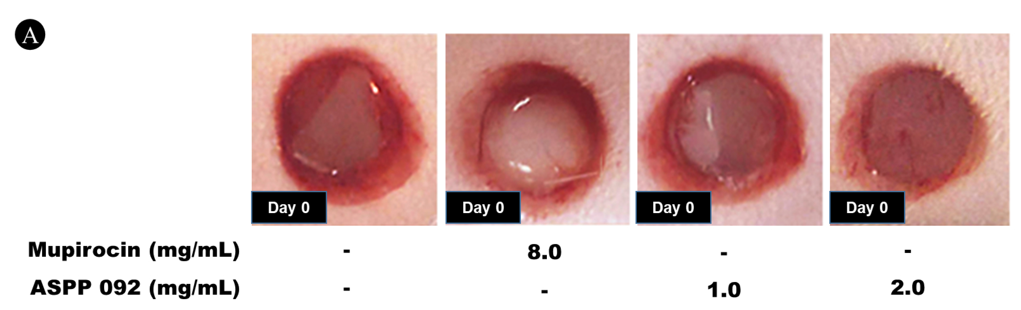
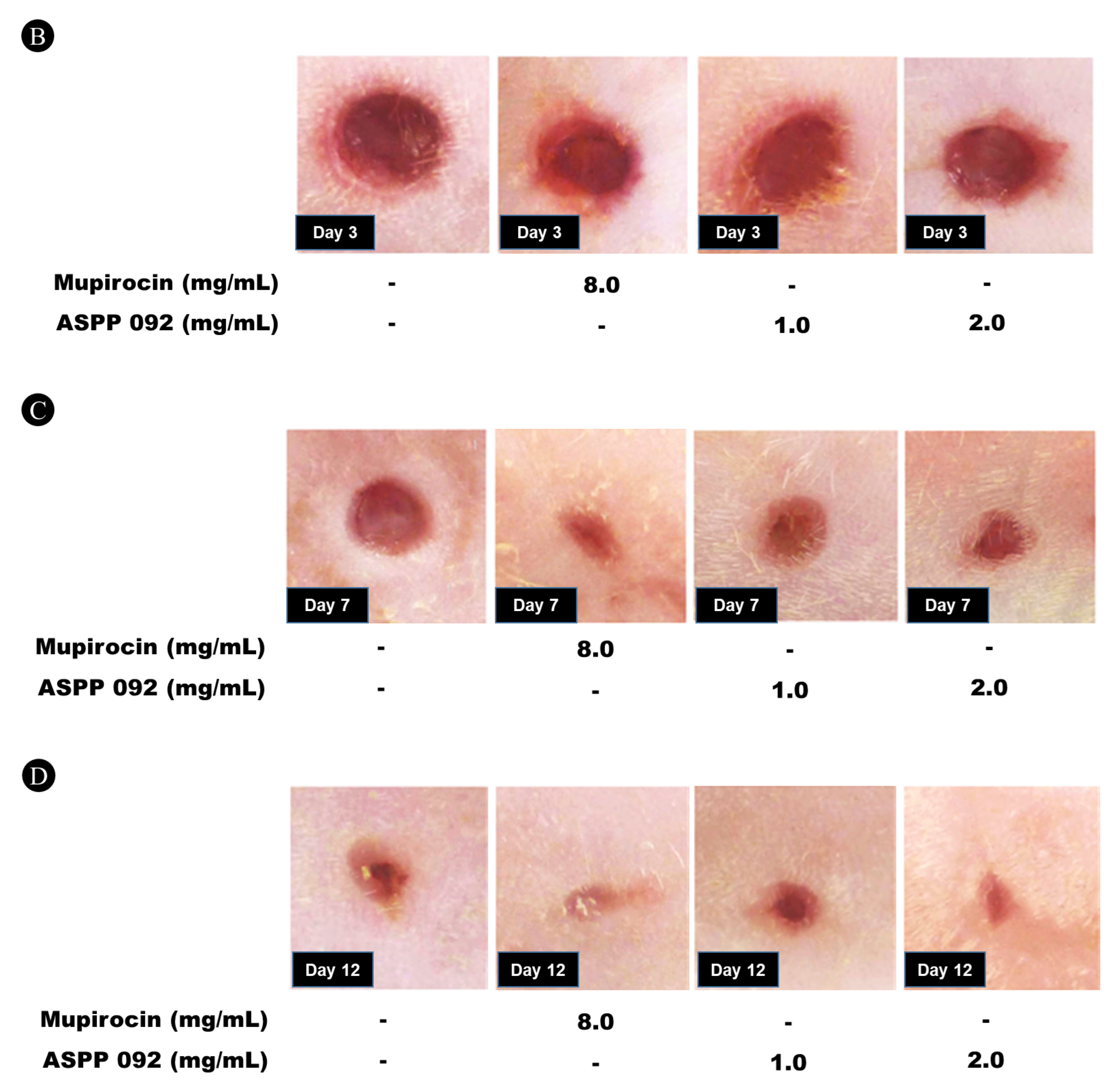
3.1. ASPP 092 Treatment Improved Wound Healing in Rats

The experimental wounds were visually examined and photographed at the indicated time intervals, including the initial day of the wound represented as day 0 (Figure 2A), day 3 (Figure 2B), day 7 (Figure 2C), and day 12 (Figure 2D) post-wounding. The experimental rats maintained good health over the study period, with no observable infection in the wound area. Significant changes in the wound sizes in rats treated with ASPP 092 and mupirocin were observed on days 3 and 7 post-wounding (Figure 2B,C). The changes in the wound sizes upon treatment with ASPP 092 at both 1 mg/mL and 2 mg/mL were dose-dependent. On day 7, neither ASPP 092- or mupirocin-treated rats exhibited any signs of inflammation in gross appearance (Figure 2D). Although the mupirocin-treated rats showed a significantly higher improvement than the ASPP 092-treated rats on both days 3 (Figure 2B) and 7 (Figure 2C), the wounds of all groups of rats were found to be closed entirely on day 12, with the entire epidermis and keratin formation (Figure 2D).

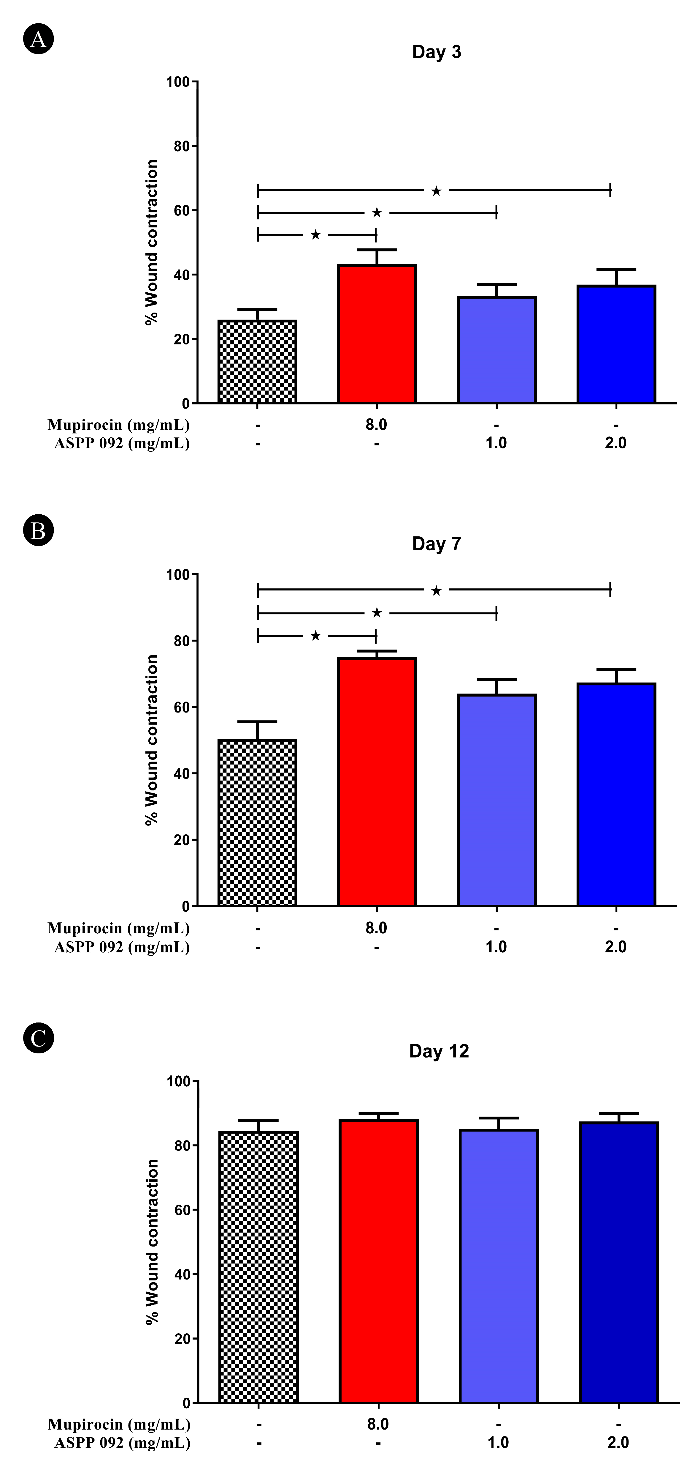
3.2. ASPP 092 Treatment Improved Wound Contraction in the Experimental Rats

The average percentages of wound contraction in the experimental rats were calculated based on the data obtained on days 3, 7, and 12 that were then compared with the observations on day 0. On day 3, the percentage wound contractions were found to be 26.03 ± 3.08, 39.29 ± 4.44, 31.41 ± 3.5, and 36.92 ± 4.69 in the control, mupirocin-treated, ASPP 092 (1 mg/mL), and ASPP 092 (2 mg/mL) groups, respectively (Figure 3A). More significant improvements in the wounds were found on day 7 (Figure 3B). Specifically, the wound area was significantly reduced in the mupirocin-treated and ASPP 092-treated groups of rats. On day 7, the wound contractions were increased to 50.33 ± 5.2, 75.05 ± 1.81, 64.04 ± 4.26, and 67.42 ± 3.88 in the control, mupirocin-treated, ASPP 092 (1 mg/mL), and ASPP 092 (2 mg/mL) groups, respectively (Figure 3B). However, on day 12, the wound-healing process progressed in all groups of rats. The wound contractions were found to be 84.57 ± 3.12, 88.2 ± 1.77, 85.15 ± 3.39, and 87.44 ± 2.5 in the control, mupirocin-treated, ASPP 092 (1 mg/mL), and ASPP 092 (2 mg/mL) groups, respectively (Figure 3C). We found significant wound contractions in the experimental rats treated with either mupirocin or ASPP 092 on days 3 and 7; however, no substantial changes in wound contraction were observed on day 12, suggesting the potential of ASPP 092 in speeding up the early phases of healing of the experimental wound in rats.

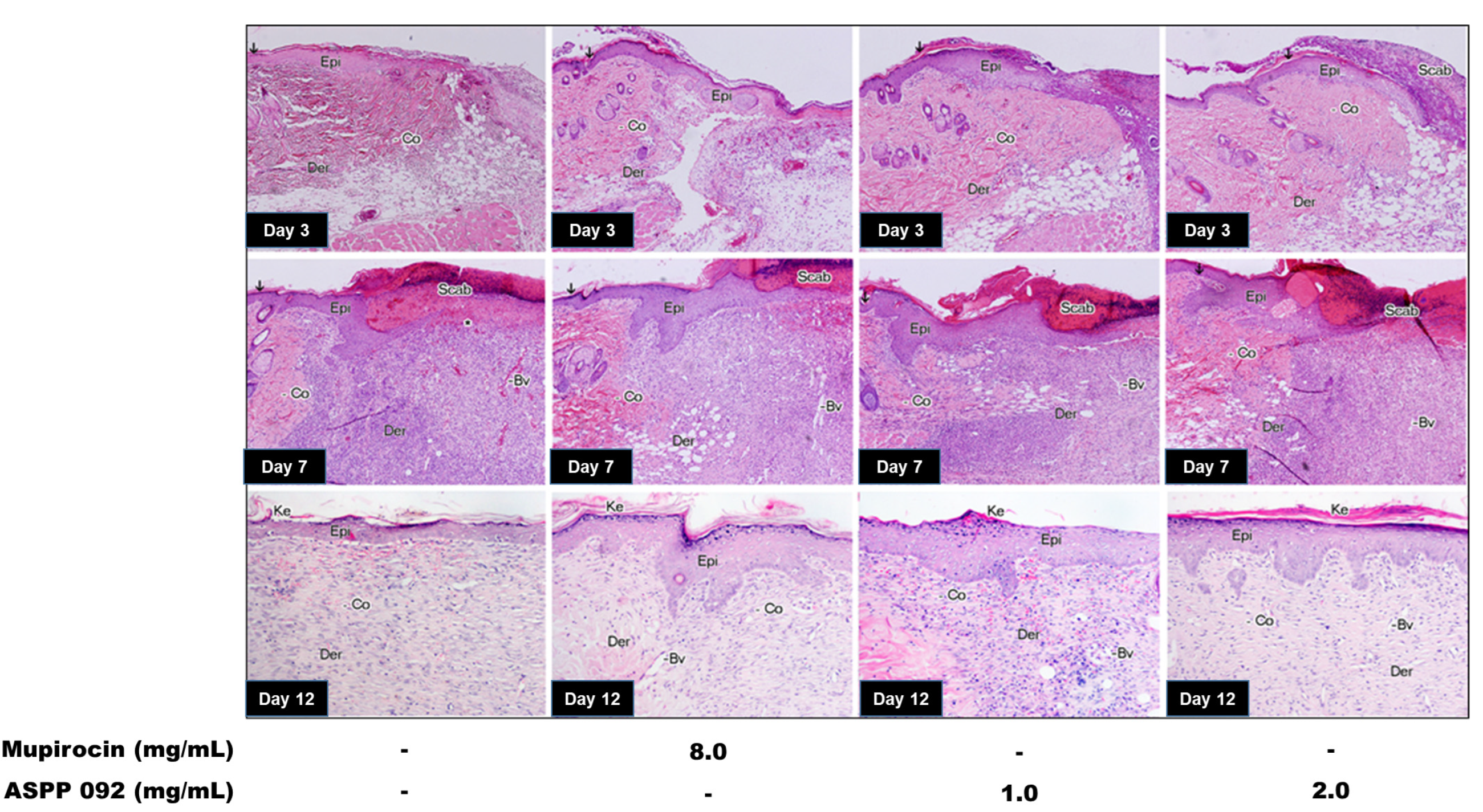
3.3. ASPP 092 Improved the Histopathology in the Wound Area

Histopathology analysis of the wound area tissues in the mupirocin- and ASPP 092-treated rats was compared with the control group. The wound-healing process for the inflammatory, proliferative, and maturation and remodeling phases was characterized at different time points (days 3, 7, and 12) in the wound (Figure 4).
On day 3 (Figure 4), the experimentally induced wounds treated with either mupirocin or ASPP 092 displayed significant retention in the inflammatory phase overlapping the initiation of the proliferative phase. The wound gaps were found to be established with an accumulation of hemorrhagic fibrin clots (scabs) with a migratory tongue of epidermal cells (epithelial cells) under the scabs. The signs of re-epithelialization in all the wounds originated; however, a well-established epidermal layer was only observed in the mupirocin- and ASPP 092-treated wounds compared to the control groups. In the dermal layer, the fewest signs of inflammatory cells, including mast cells, macrophages, and neutrophils, were seen with abundant granular tissues, including fibroblasts and collagen. Moreover, angiogenesis was also seen in the wounds treated with either Mupirocin or ASPP 092; however, the degree of angiogenesis in the control group was found to be less. This finding suggests the remarkable changes on day 3 when the experimental wounds were treated with different concentrations of ASPP 092. Interestingly, a higher grade of improvement in the wounds was observed with the highest dosage of ASPP 092 (2 mg/mL) and mupirocin.
On day 7 (Figure 4), the proliferation stage overlapped with the initiation of the maturation and remodeling phase in mupirocin and ASPP 092 at different concentrations in a dose-dependent manner. The epidermal layer exhibited higher proliferation on day seven than on day 3 in mupirocin and to varying concentrations of ASPP 092. A continuous incomplete epidermal layer was observed under the scab in the control wounds. Moreover, a thick epidermis migration toward the central area was observed in the mupirocin- and ASPP 092-treated groups compared to the control group. In the dermal region, the control wound displayed inflammatory cells; however, they were found to be comparably less in the mupirocin and to varying concentrations of ASPP 092 treatment. In addition, fewer fibroblasts, collagen fiber, and newly formed blood vessels were seen in the control group. However, the mupirocin and varying concentrations of the ASPP 092 treatment wound groups revealed the most intense granulation tissue composed of more fibroblasts, and collagen fiber from the edges of the wound migrated into the central area with newly formed blood vessels.
On day 12 (Figure 4), all the experimental wounds had shrunk and were in the maturation and remodeling phase of wound healing. All the experimental wounds exhibited a covered epidermis (neoepithelium) on the wound surface with the stratum basale, stratum spinosum, stratum granulosum, and stratum corneum. At the stratum corneum, the keratin on the superficial surface of the epidermis had already been generated. The mupirocin- and ASPP 092-treated groups presented higher thicknesses of the epidermis and more columnar appearances than the control group. In the dermal layer, all the wounds showed dense, irregularly arranged connective tissue, as in normal dermis, which had collagen bundles, hair follicles, and new blood vessels. However, the mupirocin- and ASPP 092-treated wounds were more extensively developed than the control-treated wound.
In conclusion, the wound-healing process was faster at the different stages in mupirocin- and ASPP 092-treated wounds than in control wounds. The treatment with either mupirocin or ASPP 092 at 2 mg/mL dosage effectively healed the experimental wounds with a consistent scar formation, where the fewest inflammatory cells were observed. In addition, fibroblast synthesis (fibroplasia), collagen synthesis, angiogenesis (neovascularization), and re-epithelialization were prominent in the wounds treated with either mupirocin or ASPP 092 at 2 mg/mL dosage.

3.4. ASPP 092 Improved the Re-Epithelialization in the Experimental Wounds

Re-epithelialization in the experimental wounds was analyzed using morphometric analysis (Table 1). A higher epithelial migration was found in the mupirocin- and ASPP 092 (2 mg/mL)-treated wounds. The re-epithelial regeneration was higher on day 7 compared to day 3 in the mupirocin- and ASPP 092-treated groups. In comparison with the day 7 observations of both concentrations of ASPP 092, including 1 mg/mL and 2 mg/mL, a higher grade of re-epithelialization was seen in ASPP 092 at a 2 mg/mL concentration.

3.5. ASPP 092 Reduced the Cyclooxygenase-2 (COX-2) Expressions in the Experimental Wounds

Immunohistochemical staining with COX-2 antibody was conducted to preliminarily evaluate the anti-inflammatory potential of ASPP 092 in the experimental wounds (Figure 5). Our result suggests that signs of inflammation occurred in both the epidermal and dermal compartments. A higher expression of COX-2 was found in the basal keratinocytes, inflammatory cells, and stromal cells. The COX-2-expressed keratinocytes were integrated into the newly formed epithelium near the wound margin. The COX-2 positive cells were expressed in keratinocytes on the stratum basale and suprabasal layer of the stratum spinosum of the developing epidermal tongue (neoepithelium) near the wound margin. The wounds treated with ASPP 092 displayed fewer inflammatory cells (dark brown nuclei) on the basal layer of the epidermis and dermis, whereas control wounds showed higher COX-2 positive cells. The higher COX-2 expression in the control group suggests delayed wound closure. Our results preliminarily indicate that ASPP 092 may accelerate wound repair by exerting its anti-inflammatory properties.

4. Discussion

In traditional medicine, herbal formulations were widely used to treat various disease conditions, which has been comprehensively reviewed [23,24]. In the traditional healthcare system, Curcuma comosa is one of the herbal formulations reported to show several effects [25]. Diarylheptanoids isolated from Curcuma comosa have become popular due to their various activities in treating diseases; however, their oral administration exhibits a short half-life [26]. In leukemic cell lines, the cytotoxicity and antiproliferative effects of the diarylheptanoids isolated from C. comosa were previously established [27]. Recently, one of the popular diarylheptanoids, ASPP 092, isolated from C. comosa, was shown to exhibit neuronal protection against hydrogen-peroxide-induced toxicity via nrf-2 activation [28]. In an experimental model, ASPP 092 moderated the inflammatory cytokines and improved ethyl-phenylpropionate (EPP)-induced inflammation [21]. The current study identified that ASPP 092 could accelerate wound healing, modulating the stages of wound healing in rats. Our findings were consistent with the recently identified compounds of natural origin [29,30], which can moderate wound-healing processes in rats.
The signaling molecules and regeneration patterns in wound healing have been comprehensively reviewed [4]. The different phases of skin wound healing and various aspects of skin wound care and regeneration have previously been explained [31]. The current study was designed to evaluate the therapeutic potential of ASPP 092 at different phases of experimentally induced wound care management. When the wounds were treated with ASPP 092, the granulation tissue formation was accompanied by markedly fewer inflammatory cells. In addition, more fibroblasts, collagen fibers, and newly generated blood vessels were seen. These effects may be due to the anti-inflammatory and antioxidant properties of ASPP 092, which needs validation.
The extract of C. comosa and its diarylheptanoid was reported to suppress the release of pro-inflammatory cytokines such as interferon-gamma (INF-γ), interleukin-4 (IL-4), tumor necrosis factor α (TNF-α), and interleukin- 1β (IL-1β) [32], and the expression of inducible nitric oxide synthase (iNOS) and cyclooxygenase 2 (COX-2) [33]. Inflammatory cytokines were reported to contribute to the imbalance of redox status in various diseases [34]. The ethanolic extract of C. comosa and its diarylheptanoids displayed antioxidant properties by modulating the superoxide dismutase (SOD), decreasing the production of kidney malondialdehyde (MDA), increasing glutathione (GSH) content, and restoring glutathione peroxidase (GPx) activities in mice with cisplatin-induced nephrotoxicity [20]. The diarylheptanoids (ASPP 092 and ASPP 049) of C. comosa were also reported to protect against hydrogen-peroxide (H2O2)-induced oxidative stress by modulating the lipid peroxidation that causes cell death in human retinal pigment epithelial (ARPE-19) cells [28]. In addition, these diarylheptanoids were identified as increasing the antioxidant enzymes, including glutathione peroxidase, superoxide dismutase, and catalase [35]. However, in the current study, wound healing was assessed only by wound closure analysis, histopathology, and an inflammatory marker (COX-2 expression), which requires a more comprehensive evaluation of the inflammatory cytokines and antioxidants in the experimental wounds treated with ASPP 092.
Histopathology findings suggest that ASPP 092 treatment enhanced the healing by re-epithelialization and wound contraction, which causes an increase in tensile strength. A higher concentration of ASPP 092 (2 mg/mL) accelerated wound healing, though it was not as effective as the mupirocin antibiotics. This may be due to several factors affecting the wound-healing process, including oxygenation, infection, and stress. A better understanding of these factors may lead to a better experimental therapeutics design that could improve wound healing [36].
COX-2 was reported to be a crucial marker for inflammation [37]. The structure–activity relationship (SAR) of different COX-2 inhibitors was previously reviewed [38]. The SAR among the diarylheptanoids isolated from the barks of Alnus japonica Steudel displayed that the keto-enol group in the heptane moiety or a caffeoyl group in the aromatic ring were vital for its inhibitory activities on COX-2 production [39]. However, in the case of the diarylheptanoids obtained from Curcuma xanthorrhiza, the presence of diene ketone ensured better skin-penetrating properties [40]. In the case of the diarylheptanoids isolated from the Curcuma comosa for its estrogenic activity, a keto group at C3 and an absence of hydroxyl moiety in the ring B were reported to be essential [16]. Molecular docking studies determined that the presence of β-hydroxy-β-aryl-alkanoic acid class was the most potent group exhibiting the highest inhibition of COX-2 when orally administered in animals [41]; interestingly, they were structurally similar to ibuprofen, which is in the class of nonsteroidal anti-inflammatory drugs (NSAID).
We preliminarily evaluated the COX-2 expressions in the ASPP 092-treated wounds using an immunohistochemistry analysis in the current study. The ASPP 092-treated wounds displayed fewer inflammatory and COX-2 positive cells in the epidermal and dermal layers. This result was consistent with the previous studies stating that another diarylheptanoid of C. comosa, ASPP 049, reduces the COX-2 expressions to exert the anti-inflammatory potential [33]. In another study, ASPP 092 was reported to reduce the expression of COX-2 in an experimentally induced inflammation model in mice [21]. Our results were consistent with several natural compounds, showing that COX-2 is an established marker to preliminarily identify the anti-inflammatory potential of compounds [42]

5. Conclusions

ASPP 092 accelerates the healing in experimentally induced wounds; however, its potential in acute injuries may need more experimentation. Our study identified that ASPP 092 modulates inflammation and influences the different phases of wound healing, which are limited to the preliminary histopathology observations. Further studies on the effect of ASPP 092 on the inflammatory cytokines and factors contributing to the inflammatory signals in the wounds need thorough investigation.

Author Contributions

Conceptualization, A.C. and G.P.S.; methodology, A.C. and G.P.S.; software, G.P.S.; validation, W.H., A.C. and G.P.S.; formal analysis, A.C. and G.P.S.; investigation, W.H.; resources, A.C.; data curation, A.C. and G.P.S.; writing—original draft preparation, G.P.S.; writing—review and editing, A.C. and G.P.S.; visualization, W.H., A.C. and G.P.S.; supervision, A.C. and G.P.S.; project administration, A.C.; funding acquisition, A.C. and G.P.S. All authors have read and agreed to the published version of the manuscript.

Funding

This research was supported by Mahidol University Grant (R016120002) to Gopinathan Pillai Sreekanth. The institutional funding of the Faculty of Medicine Siriraj Hospital, Mahidol University, Thailand, also supported this research to Waratta Hemthong and Aporn Chuncharunee.

Institutional Review Board Statement

The animal study protocol was approved by Siriraj Animal Care and Use Committee (SiACUC), Faculty of Medicine Siriraj Hospital, Mahidol University, Thailand (No. SI-ACUP 012/2555 dated 21 August 2012) for studies involving animals.

Informed Consent Statement

Not applicable.

Data Availability Statement

The data presented in this study are available on request from the authors.

Acknowledgments

We sincerely thank Pawinee Piyachaturawat (Department of Physiology, Faculty of Science, Mahidol University, Bangkok, Thailand) and Apichart Suksamrarn (Department of Chemistry, Faculty of Science, Ramkhamhaeng University, Bangkok, Thailand) for providing ASPP 092 and guidance throughout the study. We also express our sincere gratitude to Remya Ramesh (CSIR-IICT, Hyderabad) for the technical discussions and Poornendu Krishnaprasad (CSIR-URDIP, Pune) for the critical reading and editing of the manuscript.

Conflicts of Interest

The authors declare no conflict of interest.

References

  1. Midwood, K.S.; Williams, L.V.; Schwarzbauer, J.E. Tissue repair and the dynamics of the extracellular matrix. Int. J. Biochem. Cell Biol. 2004, 36, 1031–1037. [Google Scholar] [CrossRef] [PubMed]
  2. Collier, M. Wound-bed management: Key principles for practice. Prof. Nurse 2002, 18, 221–225. [Google Scholar] [PubMed]
  3. Nguyen, H.; Merrill, B.J.; Polak, L.; Nikolova, M.; Rendl, M.; Shaver, T.M.; Pasolli, H.A.; Fuchs, E. Tcf3 and Tcf4 are essential for long-term homeostasis of skin epithelia. Nat. Genet. 2009, 41, 1068–1075. [Google Scholar] [CrossRef] [PubMed] [Green Version]
  4. Sorg, H.; Sorg, C.G. Skin wound healing: Of players, patterns and processes. Eur. Surg. Res. 2022. ahead of print. [Google Scholar] [CrossRef]
  5. Wang, Z.; Qi, F.; Luo, H.; Xu, G.; Wang, D. Inflammatory Microenvironment of Skin Wounds. Front. Immunol. 2022, 13, 789274. [Google Scholar] [CrossRef]
  6. Gilmore, M.A. Phases of wound healing. Dimens. Oncol. Nurs. 1991, 5, 32–34. [Google Scholar]
  7. Broughton, G., 2nd; Janis, J.E.; Attinger, C.E. The basic science of wound healing. Plast. Reconstr. Surg. 2006, 117, 12S–34S. [Google Scholar] [CrossRef]
  8. Rahim, K.; Saleha, S.; Zhu, X.; Huo, L.; Basit, A.; Franco, O.L. Bacterial Contribution in Chronicity of Wounds. Microb. Ecol. 2017, 73, 710–721. [Google Scholar] [CrossRef]
  9. Punjataewakupt, A.; Napavichayanun, S.; Aramwit, P. The downside of antimicrobial agents for wound healing. Eur. J. Clin. Microbiol. Infect. Dis. 2019, 38, 39–54. [Google Scholar] [CrossRef]
  10. Sritharadol, R.; Nakpheng, T.; Wan Sia Heng, P.; Srichana, T. Development of a topical mupirocin spray for antibacterial and wound-healing applications. Drug Dev. Ind. Pharm. 2017, 43, 1715–1728. [Google Scholar] [CrossRef]
  11. Nayak, S.B.; Rodrigues, V.; Maharaj, S.; Bhogadi, V.S. Wound healing activity of the fruit skin of Punica granatum. J. Med. Food 2013, 16, 857–861. [Google Scholar] [CrossRef] [PubMed]
  12. Nayak, B.S.; Ramdeen, R.; Adogwa, A.; Ramsubhag, A.; Marshall, J.R. Wound-healing potential of an ethanol extract of Carica papaya (Caricaceae) seeds. Int. Wound J. 2012, 9, 650–655. [Google Scholar] [CrossRef] [PubMed]
  13. Chuncharunee, A.; Waikakul, S.; Wongkajornsilp, A.; Chongkolwatana, V.; Chuncharunee, L.; Sirimontaporn, A.; Rungruang, T.; Sreekanth, G.P. Invalid freeze-dried platelet gel promotes wound healing. Saudi Pharm. J. 2019, 27, 33–40. [Google Scholar] [CrossRef] [PubMed]
  14. Twilley, D.; Reva, O.; Meyer, D.; Lall, N. Mupirocin Promotes Wound Healing by Stimulating Growth Factor Production and Proliferation of Human Keratinocytes. Front. Pharm. 2022, 13, 862112. [Google Scholar] [CrossRef] [PubMed]
  15. Piyachaturawat, P.; Ercharuporn, S.; Suksamrarn, A. Uterotrophic Effect of Curcuma comosa in Rats. Int. J. Pharmacogn. 1995, 33, 334–338. [Google Scholar] [CrossRef]
  16. Winuthayanon, W.; Suksen, K.; Boonchird, C.; Chuncharunee, A.; Ponglikitmongkol, M.; Suksamrarn, A.; Piyachaturawat, P. Estrogenic activity of diarylheptanoids from Curcuma comosa Roxb. Requires metabolic activation. J. Agric. Food Chem. 2009, 57, 840–845. [Google Scholar] [CrossRef]
  17. Winuthayanon, W.; Piyachaturawat, P.; Suksamrarn, A.; Ponglikitmongkol, M.; Arao, Y.; Hewitt, S.C.; Korach, K.S. Diarylheptanoid phytoestrogens isolated from the medicinal plant Curcuma comosa: Biologic actions in vitro and in vivo indicate estrogen receptor-dependent mechanisms. Environ. Health Perspect. 2009, 117, 1155–1161. [Google Scholar] [CrossRef] [Green Version]
  18. Winuthayanon, W.; Piyachaturawat, P.; Suksamrarn, A.; Burns, K.A.; Arao, Y.; Hewitt, S.C.; Pedersen, L.C.; Korach, K.S. The natural estrogenic compound diarylheptanoid (D3): In Vitro mechanisms of action and in vivo uterine responses via estrogen receptor alpha. Environ. Health Perspect. 2013, 121, 433–439. [Google Scholar] [CrossRef] [Green Version]
  19. Suksamrarn, A.; Ponglikitmongkol, M.; Wongkrajang, K.; Chindaduang, A.; Kittidanairak, S.; Jankam, A.; Yingyongnarongkul, B.E.; Kittipanumat, N.; Chokchaisiri, R.; Khetkam, P.; et al. Diarylheptanoids, new phytoestrogens from the rhizomes of Curcuma comosa: Isolation, chemical modification and estrogenic activity evaluation. Bioorg. Med. Chem. 2008, 16, 6891–6902. [Google Scholar] [CrossRef]
  20. Jariyawat, S.; Kigpituck, P.; Suksen, K.; Chuncharunee, A.; Chaovanalikit, A.; Piyachaturawat, P. Protection against cisplatin-induced nephrotoxicity in mice by Curcuma comosa Roxb. ethanol extract. J. Nat. Med. 2009, 63, 430–436. [Google Scholar] [CrossRef]
  21. Chuncharunee, A.; Khosuk, P.; Naovarat, R.; Kaliyadan, F.; Sreekanth, G.P. ASPP 092, a phenolic diarylheptanoid from Curcuma comosa suppresses experimentally-induced inflammatory ear edema in mice. Saudi J. Biol. Sci. 2021, 28, 5937–5946. [Google Scholar] [CrossRef] [PubMed]
  22. Lai, J.J.; Lai, K.P.; Chuang, K.H.; Chang, P.; Yu, I.C.; Lin, W.J.; Chang, C. Monocyte/macrophage androgen receptor suppresses cutaneous wound healing in mice by enhancing local TNF-alpha expression. J. Clin. Investig. 2009, 119, 3739–3751. [Google Scholar] [CrossRef] [PubMed] [Green Version]
  23. Nugraha, R.V.; Ridwansyah, H.; Ghozali, M.; Khairani, A.F.; Atik, N. Traditional Herbal Medicine Candidates as Complementary Treatments for COVID-19: A Review of Their Mechanisms, Pros and Cons. Evid.-Based Complement. Altern. Med. 2020, 2020, 2560645. [Google Scholar] [CrossRef] [PubMed]
  24. Zamawe, C.; King, C.; Jennings, H.M.; Mandiwa, C.; Fottrell, E. Effectiveness and safety of herbal medicines for induction of labour: A systematic review and meta-analysis. BMJ Open 2018, 8, e022499. [Google Scholar] [CrossRef] [PubMed] [Green Version]
  25. Burapan, S.; Kim, M.; Paisooksantivatana, Y.; Eser, B.E.; Han, J. Thai Curcuma Species: Antioxidant and Bioactive Compounds. Foods 2020, 9, 1219. [Google Scholar] [CrossRef]
  26. Tuntiyasawasdikul, S.; Sripanidkulchai, B. Curcuma comosa loaded transfersomal gel for transdermal application: Formulation, In Vitro and In Vivo evaluation. Drug Dev. Ind. Pharm. 2021, 47, 1824–1834. [Google Scholar] [CrossRef]
  27. Viriyaadhammaa, N.; Saiai, A.; Neimkhum, W.; Nirachonkul, W.; Chaiyana, W.; Chiampanichayakul, S.; Tima, S.; Usuki, T.; Duangmano, S.; Anuchapreeda, S. Cytotoxic and Antiproliferative Effects of Diarylheptanoids Isolated from Curcuma comosa Rhizomes on Leukaemic Cells. Molecules 2020, 25, 5476. [Google Scholar] [CrossRef]
  28. Khin Aung, Z.M.; Jantaratnotai, N.; Piyachaturawat, P.; Sanvarinda, P. A pure compound from Curcuma comosa Roxb. protects neurons against hydrogen peroxide-induced neurotoxicity via the activation of Nrf-2. Heliyon 2022, 8, e11228. [Google Scholar] [CrossRef]
  29. Hmood, A.A.; Feki, A.; Eleroui, M.; Kammoun, I.; Kallel, R.; Boudawara, T.; Hakim, A.; Hilali, A.; Hassouni, A.O.; Suleiman, A.A.J.; et al. Biological activities and wound healing potential of a water-soluble polysaccharide isolated from Glycyrrhiza glabra in Wistar rat. Braz. J. Biol. 2022, 84, e265447. [Google Scholar] [CrossRef]
  30. Rambe, P.S.; Putra, I.B.; Yosi, A. The effect of roselle leaf (Hibiscus sabdariffa L.) extract gel on wound healing. J. Med. Life 2022, 15, 1246–1251. [Google Scholar] [CrossRef]
  31. Tottoli, E.M.; Dorati, R.; Genta, I.; Chiesa, E.; Pisani, S.; Conti, B. Skin Wound Healing Process and New Emerging Technologies for Skin Wound Care and Regeneration. Pharmaceutics 2020, 12, 735. [Google Scholar] [CrossRef] [PubMed]
  32. Sodsai, A.; Piyachaturawat, P.; Sophasan, S.; Suksamrarn, A.; Vongsakul, M. Suppression by Curcuma comosa Roxb. of pro-inflammatory cytokine secretion in phorbol-12-myristate-13-acetate stimulated human mononuclear cells. Int. Immunopharmacol. 2007, 7, 524–531. [Google Scholar] [CrossRef] [PubMed]
  33. Thampithak, A.; Jaisin, Y.; Meesarapee, B.; Chongthammakun, S.; Piyachaturawat, P.; Govitrapong, P.; Supavilai, P.; Sanvarinda, Y. Transcriptional regulation of iNOS and COX-2 by a novel compound from Curcuma comosa in lipopolysaccharide-induced microglial activation. Neurosci. Lett. 2009, 462, 171–175. [Google Scholar] [CrossRef] [PubMed]
  34. Ajeeshkumar, K.K.; Vishnu, K.V.; Navaneethan, R.; Raj, K.; Remyakumari, K.R.; Swaminathan, T.R.; Suseela, M.; Asha, K.K.; Sreekanth, G.P. Proteoglycans isolated from the bramble shark cartilage show potential anti-osteoarthritic properties. Inflammopharmacology 2019, 27, 175–187. [Google Scholar] [CrossRef] [PubMed]
  35. Jitsanong, T.; Khanobdee, K.; Piyachaturawat, P.; Wongprasert, K. Diarylheptanoid 7-(3,4 dihydroxyphenyl)-5-hydroxy-1-phenyl-(1E)-1-heptene from Curcuma comosa Roxb. protects retinal pigment epithelial cells against oxidative stress-induced cell death. Toxicol. Vitr. 2011, 25, 167–176. [Google Scholar] [CrossRef] [PubMed]
  36. Guo, S.; Dipietro, L.A. Factors affecting wound healing. J. Dent. Res. 2010, 89, 219–229. [Google Scholar] [CrossRef]
  37. Kuwano, T.; Nakao, S.; Yamamoto, H.; Tsuneyoshi, M.; Yamamoto, T.; Kuwano, M.; Ono, M. Cyclooxygenase 2 is a key enzyme for inflammatory cytokine-induced angiogenesis. FASEB J. 2004, 18, 300–310. [Google Scholar] [CrossRef]
  38. Zarghi, A.; Arfaei, S. Selective COX-2 Inhibitors: A Review of Their Structure-Activity Relationships. Iran. J. Pharm. Res. 2011, 10, 655–683. [Google Scholar]
  39. Kim, H.J.; Yeom, S.H.; Kim, M.K.; Shim, J.G.; Paek, I.N.; Lee, M.W. Nitric oxide and prostaglandin E2 synthesis inhibitory activities of diarylheptanoids from the barks of Alnus japonica steudel. Arch. Pharm. Res. 2005, 28, 177–179. [Google Scholar] [CrossRef]
  40. Claeson, P.; Pongprayoon, U.; Sematong, T.; Tuchinda, P.; Reutrakul, V.; Soontornsaratune, P.; Taylor, W.C. Non-Phenolic Linear Diarylheptanoids from Curcuma xanthorrhiza: A Novel Type of Topical Anti-Inflammatory Agents: Structure-Activity Relationship. Planta Med. 1996, 62, 236–240. [Google Scholar] [CrossRef]
  41. Dilber, S.P.; Dobric, S.; Juranic, Z.D.; Markovic, B.D.; Vladimirov, S.M.; Juranic, I.O. Docking studies and anti-inflammatory activity of beta-hydroxy-beta-arylpropanoic acids. Molecules 2008, 13, 603–615. [Google Scholar] [CrossRef] [PubMed] [Green Version]
  42. Bralley, E.E.; Greenspan, P.; Hargrove, J.L.; Wicker, L.; Hartle, D.K. Topical anti-inflammatory activity of Polygonum cuspidatum extract in the TPA model of mouse ear inflammation. J. Inflamm. 2008, 5, 1. [Google Scholar] [CrossRef] [PubMed]
Figure 1. Structure of ASPP 092 [7-(3,4-dihydroxy-phenyl)-5-hydroxy-1-phenyl-(1E)-1-hepten]; molecular weight: 298.38.
Futurepharmacol 03 00001 g001
Figure 2. The morphological changes in the wounds when the experimentally induced wounds were treated with mupirocin or ASPP 092 at concentrations of 1 mg/mL and 2 mg/mL. A vehicle control group was also maintained. The effect of these treatments at different time points, including (A) Day 0, (B) Day 3, (C) Day 7, and (D) Day 12, are represented.
Futurepharmacol 03 00001 g002aFuturepharmacol 03 00001 g002b
Figure 3. Percentage of wound healing at different time intervals. The experimentally induced wounds were treated with either mupirocin or ASPP 092 at concentrations of 1 mg/mL and 2 mg/mL. A vehicle control group was also maintained. The percentage of wound healing at different time points, including (A) Day 3, (B) Day 7, and (C) Day 12, are represented. The data obtained for each time point are represented as mean + SEM. * p < 0.05 was considered to be statistically significant.
Futurepharmacol 03 00001 g003
Figure 4. Histopathological observations in the wounds treated with ASPP 092. The experimentally induced wounds were treated with either mupirocin or ASPP 092 at concentrations of 1 mg/mL and 2 mg/mL. A vehicle control group was also maintained. Histopathology sections of the wounds stained with hematoxylin and eosin (H&E) at different time points, including days 3, 7, and 12, were conducted and represented (arrow, wound margin; Bv, blood vessel; Co, collagen fibers; Der, dermis; Epi, epidermis; Ke, keratin; *, inflammatory cells; ×100).
Futurepharmacol 03 00001 g004
Figure 5. COX-2 expressions in the experimentally induced wounds treated with ASPP 092. The experimentally induced wounds were treated with either mupirocin or ASPP 092 at concentrations of 1 mg/mL and 2 mg/mL. A vehicle control group was also maintained. Immunohistochemistry analysis was conducted to estimate the COX-2 expressions in the experimental wounds. Immunostaining for COX-2 (dark brown nuclei) in the epidermis and dermis of rat skin wounds on day 3 is represented. The density of COX-2 positive cells on stratum basale and lower part of stratum spinosum, fibroblasts, and endothelial cells of new vessels are also shown (Bv, blood vessel; Der, dermis; Epi, epidermis; *, inflammatory cell; ×200).
Futurepharmacol 03 00001 g005
Table 1. Re-epithelialization in the experimental wounds at different time intervals. The experimentally induced wounds were treated with either mupirocin or ASPP 092 at concentrations of 1 mg/mL and 2 mg/mL. A vehicle control group was also maintained. Re-epithelialization in the wounds was calculated and expressed as percentage changes. The analyzed data are represented as mean ± SEM. * p < 0.05 was considered to be statistically significant.
Groups of Experimental WoundsRe-Epithelialization (%)
Day 3Day 7Day 12
Control Group27.43 ± 3.9281.6 ± 5.32100
Mupirocin (8 mg/mL)-treated47.65 ± 1.89100 *100
ASPP 092 (1 mg/mL)-treated37.38 ± 2.69 *88.22 ± 4.52100
ASPP 092 (2 mg/mL)-treated43.14 ± 2.87 *100 *100
Disclaimer/Publisher’s Note: The statements, opinions and data contained in all publications are solely those of the individual author(s) and contributor(s) and not of MDPI and/or the editor(s). MDPI and/or the editor(s) disclaim responsibility for any injury to people or property resulting from any ideas, methods, instructions or products referred to in the content.

