Review

The Effects of SARS-CoV-2 on the Angiopoietin/Tie Axis and the Vascular Endothelium

by
Dolgormaa Janchivlamdan
1,
Maitreyi Shivkumar
2 and
Harprit Singh
1,*
1
Leicester School of Allied Health Sciences, De Montfort University, The Gateway, Leicester LE1 9BH, UK
2
Leicester School of Pharmacy, De Montfort University, The Gateway, Leicester LE1 9BH, UK
*
Author to whom correspondence should be addressed.
Encyclopedia 2024, 4(1), 544-557; https://doi.org/10.3390/encyclopedia4010035
Submission received: 31 January 2024 / Revised: 27 February 2024 / Accepted: 6 March 2024 / Published: 11 March 2024
(This article belongs to the Collection Encyclopedia of COVID-19)

Abstract

:
Severe acute respiratory syndrome coronavirus 2 (SARS-CoV-2) infection can cause potentially life-threatening coronavirus disease (COVID-19). COVID-19 is a multisystem disease and is associated with significant respiratory distress, systemic hyperinflammation, vasculitis, and multi-organ failure. SARS-CoV-2 causes the deterioration of numerous systems, with increasing evidence implying that COVID-19 affects the endothelium and vascular function. The endothelium is important for preserving vascular tone and homeostasis. The overactivation and dysfunction of endothelial cells are significant outcomes of severity in patients with COVID-19. The Angiopoietin 1/Tie 2 pathway plays an important role in endothelium quiescence and vessel stability. The disruption of Angiopoietin/Tie balance affects the vessel contact barrier and leads to vessel leakage, and this in turn causes endothelial dysfunction. Although vascular instability through SARS-CoV-2 is associated with endothelial dysfunction, it is still not understood if the virus affects the Angiopoietin/Tie axis directly or via other mechanisms such as cytokine storm and/or immune response associated with the infection. This review provides an overview of the impact SARS-CoV-2 has on endothelial function and more specifically on the Angiopoietin/Tie pathway.

1. Introduction to Coronaviruses

Coronaviruses (CoVs) are a family of single-stranded RNA viruses that are a source of respiratory and digestive diseases in humans and animals [1]. The coronavirus family is separated into four different genera, i.e., Alphacoronavirus (α-CoV), Betacoronavirus (β-CoV), Gammacoronavirus (ϒ-CoV), and Deltacoronavirus (δ-CoV), which are part of the subfamily of Orthocoronavirinae. α-CoVs and β-CoVs cause disease in humans and other mammals, whereas ϒ-CoVs and δ-CoVs cause disease in avian species [2]. Human coronavirus (HCoV) OC43 and HCoV-HKU1 belong to β-CoV, whereas HCoV-229E and HCoV-NL-63 belong to α-CoV. These four human coronavirus species result in varying severity [3]. Severe acute respiratory syndrome coronavirus (SARS-CoV-1), Middle East respiratory syndrome coronavirus (MERS-CoV), and, more recently, severe acute respiratory syndrome coronavirus 2 (SARS-CoV-2) belong to β-CoV genera and all of them result in more severe symptoms like pneumonia [4].

1.1. Virion Composition of Coronaviruses

The diameter of coronaviruses is about 80–120 nm in length, and the virus is surrounded by two layers of lipid molecules with envelope (E), spike (S), nucleocapsid (N), and membrane (M) proteins within the lipid bilayer. The S protein belongs to class I transmembrane proteins and is around 128–160 kDa prior to covalently attaching to carbohydrates and 150–200 kDa following N-linked glycosylation [5,6]. S protein cleaves into three identical units of polypeptides (S1–S3) by proteases found on target cells. S1 is involved in binding to receptors and S2 for membrane fusion. The endoplasmic reticulum (ER) stress response is stimulated through the infective process of the virus by S protein, which leads to apoptosis through unfolded protein response activation [7]. The M protein has a molecular weight of 25–30 kDa and has three functional regions which span the phospholipid bilayer [8]. M protein plays a role in viral infection processes such as RNA packaging and virion formation. The E protein is a relatively small protein with a molecular weight of 8–12 kDa and is involved in the assembly and release of virions [9]. N protein has a molecular weight of 43–50 kDa and supports the packaging of RNA into ribonucleocapsids. It also plays a role in numerous activities, including regulating virus assembly by interacting with M protein and the viral genome, increasing the replication and transcription of RNA genome, and in escaping the host immune reaction. Coronaviruses have one of the largest viral RNA genomes, roughly 27–32 kilobases (kb) [10]. The 5′-end of the genome encodes non-structural proteins, including proteases and transcription factors, and the 3′-end encodes structural proteins as well as cis-acting RNA elements crucial for RNA production [11].

1.2. SARS-CoV-2 Infection

SARS-CoV-2 infection can progress into a critical and potentially fatal respiratory disorder and pulmonary damage in patients through affected upper and lower respiratory epithelial cells, including alveolar type 2 (AT2) cells. The entry of SARS-CoV-2 into the target cell is the first step in the infection process and development of disease. Entry begins with attachment of the SARS-CoV-2 S glycoprotein to the host surface angiotensin converting enzyme 2 (ACE2), followed by S protein cleavage by cell surface transmembrane serine protease 2 (TMPRSS2) into the S1 and S2 domains. The S1 subunit contains the receptor-binding domain (RBD) that recognises and binds to the ACE2 receptor, and therefore regulates virus specificity to the host cell and the infection processes that lead to disease [12]. The RBD shifts from an upright posture to connect with the ACE2 receptor to a horizontal posture to escape the immune response. After S protein binds to ACE2 on the target cell surface via its RBD, cleavage by TMPRSS2 reveals the fusion peptide on the S2 subunit to initiate the fusion process of the virus to the host cell membrane [13]. TMPRSS2 is expressed in the plasma membrane of upper airway, pulmonary, digestive, heart, prostate, and liver epithelial cells [14]. SARS-CoV-2 has also been shown to use endosomal cysteine proteases cathepsin B (Cat B) and Cat L to enter the host cell aside from TMPRSS2 [15]. Kawase M et al. (2012) implied that concurrent suppression of surface receptor TMPRSS2, Cat L, and Cat B effectively inhibits virion entry into host cells in vitro [16].
Both non-specific and virus-specific immune responses contribute towards COVID-19, with escape from the innate immune response strongly affecting viral RNA replication and progression of infection. The protein kinase B (Akt) signalling pathway, which is important for cell survival, is abnormally regulated by SARS-CoV-2 to support its survival and replication. Nuclear factor-kB (NF-kb) transcription factor has been suggested to be highly active in severe inflammation during SARS-CoV-2 infection [16]. RNA replication of the SARS-CoV-2 genome forms double-stranded RNA (dsRNA), which upregulates the cytoplasmic innate immune pathway via stimulation of melanoma differentiation-associated protein 5 (MDA5) and retinoic acid-inducible gene I (RIG-I). This in turn triggers signalling by mitochondrial antiviral-signalling protein (MAVS), leading to release of type I and type III interferons [17], resulting in the activation of antiviral interferon-stimulated genes (ISGs) in neighbouring cells via the Janus kinase/signal transducer and activator of transcription (JAK/STAT) signalling pathway [18]. However, studies have shown that SARS-CoV-2 as well as SARS-CoV and MERS-CoV have various mechanisms to inhibit interferon-related immune responses, with the non-structural open reading frame 6 (Orf6) protein being involved in immune evasion and viral structural proteins inhibiting type I interferon-regulated antiviral immune responses [19,20].
The viral proteases 3-chymotrypsin-like protease (3CLpro) and papain-like protease (PLpro) are also crucial for viral replication, with roles in hydrolysing the viral polyprotein. PLpro also disrupts the host non-specific immune reaction by targeting various host cell proteins [21]. RNA-dependent RNA polymerase (RdRp) is important for SARS-CoV-2 virus transcription, RNA genome replication, phenotype changes, and regulation of post-transcription of host cell gene expression [22]. SARS-CoV-2 also affects the target cell through its cytocidal effect, which promotes host cell death and lysis via an immune-regulated process. The formation of syncytia by fusion of SARS-CoV-2-infected cells, via formation of replication complexes formed through the movement of vesicles and disturbance of Golgi apparatus, has also been observed [23]. Rapid and unregulated non-specific responses to viral infection lead to a rise in inflammatory cytokines and immune cell activation in the lower respiratory tract, triggering damage to cells and tissues and thus exacerbation of COVID-19 [24].

2. The Endothelium

The circulatory system contains numerous blood vessels, including arteries, veins, capillaries, and lymph vessels, which transport blood, haemocytes, oxygen, nutrients, hormones, vitamins, minerals, and lymphatic fluids, which are important to supporting the survival of the human body [25]. Endothelial cells are a thin monolayer that mainly lines the interior surface of vasculature and are commonly shielded by pericytes that promote vascular stability. These cells have various roles and are predominately surrounded by smooth muscle and connective tissue. The main function of endothelial cells is regulating haemostasis, coagulation, thrombosis, permeability, inflammatory processes, remodelling of new vessels, blood vessel tone, vasorelaxation and vasoconstriction, blood flow and pressure, and forming the barrier between vessels and tissues [26,27,28]. The endothelium has a capacity to respond to physical damage and chemical molecules. The stability of the endothelium depends on the regulation of vascular contractile activity of vascular smooth muscle cells, vasculitis, and cellular attachment to other cells [29]. Physiological activity of certain endothelium signalling mediators, including nitric oxide, prostacyclins, and angiopoietins, are important for all features of blood vessel stability and integrity. Therefore, the metabolic function of endothelial cells is crucial for the constant adaptation of blood vessel tone, which in turn regulates blood pressure. Additionally, endothelial cells have a key role in decreasing the formation of blood clots and blood thickness equilibrium in the blood stream, including the normal control of white blood cell transport from blood vessels to tissues [30].

2.1. Angiopoietin Family

Angiopoietins belong to a family of vascular growth factor glycoproteins which control the formation of new blood vessels and promote angiogenesis and foetal/adult vascular development. The four isoforms, Angiopoietin (Angpt) 1, 2, 3, 4, are structurally comparable and are ligands of the Tunica Interna endothelial cell tyrosine kinase (Tie) receptor, predominantly expressed in endothelial cells [31]. Angpt 1 and 2 are key isoforms of this glycoprotein family and have been thoroughly studied, whereas Angpt 3 and 4 are less well studied. Although Angpt 3 is expressed in mice and humans, it is not biologically active in human endothelial cells. The specific and precise equilibrium of Angpt 1 and 2 is important to controlling vascular stability [32]. The alteration of these protein level ratios is connected with multiple diseases, such as cancer, sepsis, and coronary artery diseases [33].

2.2. The Function of Angiopoietin 1

Angpt 1 has a molecular weight of 70 kDa and consists of 498 residues [34]. Angpt 1 is suggested to be a significant angiogenic growth factor and constitutive paracrine agonist of tyrosine kinase receptor (Tie2) and supports blood vessel stability and integrity. Witzenbichler et al. (1998) reported that binding of Angpt 1 to Tie 2 in vascular endothelial cells leads to the phosphorylation of several tyrosine residues on the carboxy terminus of the receptor, initiating various intracellular secondary messengers that contribute to endothelial function, including vessel sprouting and cell viability [35]. The absence of Angpt 1 was shown to lead to defects in blood vessel growth and foetal blood vessel development in Angpt 1 knockout mice [36]. The upregulation of Angpt 1 in a genetically modified mouse model increased angiogenesis and decreased vascular permeability [37]. Angpt 1 does not bind to the Tie 1 receptor; however, it indirectly activates it through the transphosphorylation of active Tie 2 receptors [38]. Angpt 1-induced PI3K/Akt leads to the addition of phosphate groups and inhibition of the forkhead transcription factor Foxo1 in vascular endothelial cells [39]. This transcription factor is involved in endothelial cell death and controls the synthesis of various mediators such as Angpt 2. Angpt 1 phosphorylation of the Tie 2 receptor also facilitates adhesion to pericytes and smooth muscle cells, which in turn leads to blood vessel protection and stability [40].

2.3. The Function of Angiopoietin 2

Angpt 1 and 2 have 60% homology in their amino acid sequence. Like Angpt 1, Angpt 2 is capable of binding to Tie 2 receptor. Angpt 2 is stored in Weibel–Palade bodies (WPBs) in endothelial cells. The release of Angpt 2 is highly controlled and exerts an autocrine action [41]. Angpt 2 has the opposite effect from Angpt 1; it facilitates vascular permeability, obstructs endothelial barrier activity, and impairs vascular integrity [42]. Although Angpt 2 obstructs the vascular protective activity of Angpt 1 after it binds to Tie 2 receptor, in some cases, this antagonist ligand can stimulate Tie 2 signalling through an unknown mechanism. In the presence of vascular endothelial growth factor (VEGF), Angpt 2 promotes angiogenesis, blood vessel sprouting, and destabilisation [43]. Also, Angpt 2 promotes pericyte detachment by decreasing the cooperation between endothelial cells and pericytes, vascular smooth muscle cells, and the extracellular matrix [44]. Also, some studies reported that upregulation of Angpt 2 in vitro activates vascular endothelial cell movement during the reorganisation stage of angiogenesis and increases cell viability via Tie 2 interaction and PI3K/Akt stimulation [45]. In physiological conditions, Angpt 2 is generally released in regions of structural changes to bloods, vessel such as the gonads, the womb, and the placenta. Its release is rapidly activated by various stimuli including thrombin, histamine, hypoxia, and VEGF. Angpt 2 also stimulates vascular endothelial cells to release inflammatory cytokines including TNF-α [46]. Angpt 2 interacts with white blood cells, including monocytes, macrophages, and neutrophils, throughout the inflammation process [47].

2.4. Tie Receptors

Tie 1 and Tie 2 are members of a specific receptor tyrosine kinase (RTK) family which are expressed predominantly in endothelial and hematopoietic cells. Tie 2 receptor interacts with Angpt 1, 2, 4, whereas Tie 1 is thought to be incapable of binding to Angpts and alternative ligands of Tie 1 have not yet been discovered [48]. Tie receptors contain three epidermal growth factor (EGF) domains amidst two immunoglobulin (Ig)-like domains and three fibronectin type 3 domains. Their Ig-like domains and EGF-like domains are important and sufficient to bind Angpt 1 and Angpt 2 [49]. Tie receptors play a role in the structural change and maturation of blood vessels during embryogenesis [50]. Tie 1 orphan receptor is important for blood vessel formation, integrity, and sprouting in the embryonic stage. Tie 1 synthesis decreases in adulthood, but its levels remain the same in endothelial cells of the renal, cardiovascular, and respiratory systems [51]. Tie 2 is the main signalling receptor of the Angpt pathway and receptor clustering is important for the stimulation of this transmembrane receptor [52]. Tie 1 is capable of forming heterodimer complexes with Tie 2, which decreases the activation of Angpt 1 and supports the activity of Angpt 2, whereas Tie2/Tie2 homodimers activate Angpt 1 signalling and vascular integrity. The process of Tie 2 receptor activation is still not well understood, apart from the binding of the carboxyl terminus domain of Angpt to the ligand-binding domain of this receptor [53].

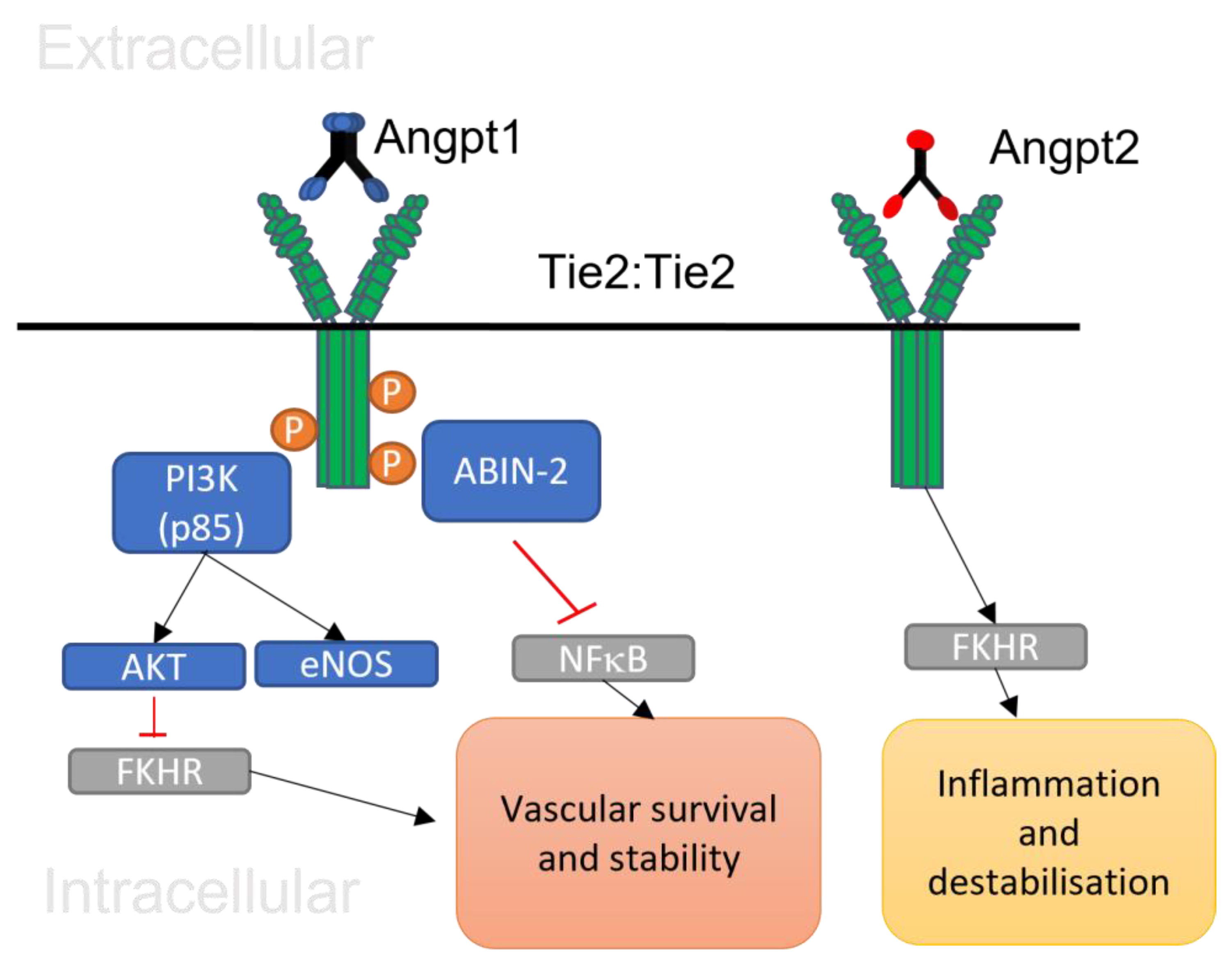
2.5. Angiopoietin 1/Tie 2 Signalling

After the binding of the five subunits of multimeric Angpt 1 to the Tie 2 receptor at the cell-to-cell junctional complex, Tie 2 transmembrane receptor is phosphorylated and starts the signalling pathway, resulting in the regulation of multiple processes, including blood vessel stability and endothelial cell survival, as illustrated in Figure 1 [53,54]. Angpt 1 activates PI3K by recruiting the regulatory p85 subunit in Tie 2, which results in the stimulation of Akt and nitric oxide synthase 3 (eNOS) to inhibit endothelial apoptosis. The stimulation of Akt by the Angpt 1/Tie 2 signalling pathway leads to the phosphorylation and obstruction of the Forkhead transcription factor (FKHR) gene to support cell survival and vessel stability [55,56,57,58,59,60]. The Angpt 1/Tie pathway is also involved in cortical actin cytoskeleton stability by regulating GTPase and decreasing inflammatory molecules through the inhibition of NFκB protein [61,62,63,64,65,66].
As discussed above, Tie 1/Tie 2 heterodimer receptors can affect Angpt 1/Tie 2 signalling. Tumour necrosis factor-α (TNF-α) is a crucial inflammatory cytokine that is able to stimulate Tie 1 ectodomain cleavage in endothelial cells. TNF-α was also shown to elevate Tie 2 expression, leading to changes in the ratio of Tie 1 to Tie 2. Similar observations have been found with VEGF, which is capable of cleaving the ectodomains of both Tie 1 and Tie 2, leading to the regulation of Angpt 1 signalling [67,68,69,70,71]. Tie 1 decreases the capacity of Angpt 1 to bind to Tie 2 and therefore decreases Tie 2 activation by its ectodomain. Cleavage of the extracellular domain of Tie 1 enhances the binding of Angpt 1 to Tie 2 and thus increases Angpt 1/Tie 2 activation [72]. Changes in Angpt 1 and Angpt 2 levels also regulate Tie 2 signalling. A decrease in Angpt 1 and increase in Angpt 2 promote vessel destabilisation under various disease states and have been associated with acute lung injury [73]. Some studies reported that the release of Angpt 1 changes after ischemic stroke and increased levels of this protein decrease the complications of stroke [74]. During atherosclerosis, Angpt 2 elevation promotes vessel inflammatory processes by stimulating NFκB and traffics white blood cells through the vessel to the affected regions [75]. Also, inflammation caused by infection leads to a decrease in Angpt 1 and suppression of the Angpt 1/Tie 2 pathway, leading to vascular instability and increased permeability. Several studies have mentioned that Angpt could be utilised as a molecular marker or treatment method to decrease the severity of lung inflammation and sepsis [76,77]. Some studies suggest that elevated Angpt 2 in various tumours stimulate neovascularisation in cancer mice models. Angpt 1/Tie 2 pathway therapies have been connected with recovery and survival rates in specific types of tumours, such as cervical and ovarian cancers [78,79,80].

3. The Impact of SARS-CoV-2 on Endothelial Cells

Post-mortem studies in COVID-19 patients have reported a procoagulant state and microvascular damage in deceased patients, demonstrating that vascular pathology caused by endothelial dysfunction is a crucial factor in the pathogenesis of SARS-CoV-2 [81,82,83]. Endothelial dysfunction during SARS-CoV-2 infection results in pro-inflammation and vascular permeability and leads to vascular destabilisation through the mechanisms shown in Figure 2 [84]. As mentioned above, the ACE2 receptor is one of the most important receptors that bind to the spike protein of SARS-CoV-2 and is expressed in various cells, including vascular endothelial cells. SARS-CoV-2 has been connected with substantial immune activation that directly influences endothelial cells via cytokine storm [85]. SARS-CoV-2 can infect host endothelial cells if high viral loads are used, and this leads to the activation of cell death. Infections of endothelial cells have been suggested to be less efficient compared to those of epithelial cells due to the lower expression of the ACE2 receptor and protease in endothelial cells [86].
Endothelial tissues cooperate with epithelial tissues in the respiratory tract airway barrier to control tissue stability and defend against infectious microorganisms. Epithelial cells communicate with endothelial cells during pathogenic infections to initiate immune cell activation. These two cells create a single layer bound by solid junctions that control vascular permeability, whereas adherent junctions control cell-to-cell and cell-to-matrix cooperation [87]. The endothelium in the lung contains macro- and micro-vessel endothelial cells, with the latter belonging to the epithelial and endothelial contact junction [88]. An increase in inflammatory cytokines causes local endothelial dysfunction and supports blood clotting and platelet activation [89]. The damage to endothelial cells caused by an intense immune reaction could affect the structure of blood vessel and airway junctions and cause vessel leakage, lung inflammation, white blood cell extravasation, and oxygen shortage [90]. Lung epithelial cells stimulate NOD-like receptor protein 3 (NLPR3) and inflammatory cytokines such as TNF-α and IL-6 during COVID-19 in order to start intense immune reactions; these processes result in apoptosis, cell injury, increased vessel permeability, and lung inflammation [91]. In addition, unregulated synthesis of neutrophil extracellular traps (NETs) and reactive oxygen species (ROS) impairs endothelial cells, activates platelet and blood clotting, and aids in acute respiratory distress syndrome and tissue injury. Nitric oxide and prostacyclin are vasodilators that help with vascular integrity. The levels of these hormones are reduced in SARS-CoV-2 and lead to endothelial dysfunction [92].
Intracellular adhesion molecules bind to fibrinogens and support fibrin aggregation to endothelial cells, which is involved in abnormal functioning of endothelial cells, blood clotting, and vessel narrowing [93]. Hyperactivation and dysfunction of endothelial cells are a significant hallmark of severity in patients with SARS-CoV-2. It is not clear if SARS-CoV-2 can directly and productively infect endothelial cells despite endothelial cells expressing the ACE2 receptor, which is necessary for viral entry [94,95,96,97]. Enrichment of SARS-CoV-2 RNA was found in lung endothelial cells in deceased COVID-19 patients [98], and infection was observed in in vitro-cultured endothelial cells [98,99]. On the other hand, infection of primary endothelial cells was not observed [100]. Despite this, it has been suggested that infection of neighbouring cells, such as epithelial and vascular pericyte cells, causes an elevated inflammatory response and cytokine storm and leads to endothelial dysfunction [86,100].
Some studies have speculated that SARS-CoV-2-infected epithelial cells detach at the contact barriers and that permits the virus to be transmitted from the base surfaces of epithelial cells to endothelial cells in the lungs. It has also been suggested that the virus might infect endothelial cells through the blood stream via the apical surface [101]. Also, others have suggested that ACE2 is released at high levels in small-vessel pericytes and that pericyte injury caused by SARS-CoV-2 might play a role in endothelial dysregulation. The loss or detachment of SARS-CoV-2-infected and injured pericytes causes endothelial dysfunction through declining endothelial barrier activity and endothelial activation [94,101].
It has also been reported that the SARS-CoV-2 spike protein stimulates damage to the vessel contact barrier in cerebral small-vessel endothelial cells [101]. Endothelial cells sense and respond to damage-associated molecular signals from surrounding virus-affected epithelial cells and endothelial cells. Furthermore, the vascular endothelial cell-regulated inflammatory process is an key part of the pathological mechanism of COVID-19 [52,102]. During infection, the inflammatory process activates endothelial cells, which synthesise tissue factors that result in blood clotting, hyperpermeability of small vessels, lung damage, and increased levels of cytokines such as TNF-α and IL-6, associated with amplified coagulation factor 1 or fibrinogen [102]. The extracellular activation of ACE2 is decreased and the renin–angiotensin system (RAS) is stimulated after SARS-CoV-2 entry into the host cell. ACE2 is important for supporting vessel relaxation molecules including angiotensins 1–7, preventing the activation of reactive oxygen species (ROS) through inhibition of angiotensin II type 1 receptor (AT1R), and regulates the equilibrium of the RAS [103]. Downregulation of nitric oxide and elevated vessel-narrowing molecules such as angiotensin 2 are important aspects of endothelial dysregulation during COVID-19. The reduction in nitric oxide by upregulation of NFκB leads to a reduction in endothelial nitric oxide synthase (eNOS) and to the synthesis of ROS, which are involved in disrupting endothelial function (Figure 2) [104]. During SARS-CoV-2 infection and cytokine storm, the increase in angiotensin type 2 receptor and reduction in angiotensin 1–7 are involved in decreasing vessel dilation, supporting white blood cell and thrombocyte adhesion, and therefore activate a pro-thrombotic, pro-coagulative state and inhibit the fibrinolysis process [105]. Moreover, angiotensin type 2 receptor upregulates nicotinamide adenine dinucleotide phosphate oxidase (NADPH oxidase 2), and increased expression of angiotensin type 2 receptor leads to an increase in oxidative stress and destabilisation of vascular activity [106]. People infected with SARS-CoV-2 displayed elevated amounts of fibrinogen, fibrin-dissolving fragments such as D-dimer, and blood clotting protein including factor 8, and decreased amounts of endothelial plasminogen activator inhibitor 1 [107].

3.1. Potential Impact of SARS-CoV-2 on Angpt/Tie Signalling

Angpt 2 plays an important role in elevated vascular inflammation and leakage through inactivating Angpt 1 and Tie signalling. Elevated levels of Angpt 2 have been detected in the blood serum of patients with vascular leakage, lung inflammation, and acute respiratory distress syndrome, and have been linked with complications like sepsis [108]. Lu et al. (2022) reported that SARS-CoV-2 infection impairs the structure of the vascular endothelial contact barrier due to damaging vascular endothelial cadherin barrier junctions and increasing inflammatory mediators including IL-6, IL-8, and Angpt 2 in vitro. Also, these cytokines were increased in SARS-CoV-2 infections compared to other coronaviruses such as NL63 [109]. The stimulation of phosphorylated mixed-lineage kinase domain-like protein (pMLKL), which supports necrotic apoptosis, occurred in small vessels with increased Angpt 2, connecting this to vessel damage in patients with COVID-19 [110]. Some studies suggested that thrombosis, vessel damage, and vascular endothelial cells apoptosis were observed in the tissue of deceased COVID-19 patients using pulmonary imaging mass cytometry. This result supports the speculation that COVID-19 is a vascular disorder [111]. SARS-CoV-2 uses the cellular machinery of infected cells, similar to other viruses, in order to produce proteins which are necessary for the transcription and assembly of the pathogen, resulting in atypical hyperstimulation of signalling mechanism of PI3/Akt/mTOR pathway [112]. Hypoxia caused by SARS-CoV-2 infection is also involved in angiogenesis. The lack of oxygen connected with continuing inflammation leads to hypoxia-inducible factor 1 alpha (HIF-1α) stabilisation, which interacts with molecular markers including VEGF, Angpt, and Tie 2 receptor. Also, angiopoietin-like 4 proteins (Angpt 4) are released in response to oxygen starvation and malnutrition by a HIF1-α-related mechanism. Angpt 4 is involved in several activities including controlling vessel damage, permeability, wound healing, vessel function, and cholesterol homeostasis [113]. Some studies reported that increased levels of Angpt 4 were related to acute respiratory distress syndrome and severity of SARS-CoV-2 infection [114]. SARS-CoV-2 infection activates HIF-1α, and upregulation of this factor is also involved in the pathophysiological mechanism of this pathogen, including a decrease in ACE2 receptor [115].
White blood cell endothelial adhesion molecules such as soluble E-selectin are activated by inflammatory cytokines. Elevated levels of soluble E-selectin and Angpt 2 have been reported in patients with SARS-CoV-2 infection [116]. Thrombocytes, except pericytes, produce large numbers of Angpt 1 protein. Thrombocytopenia is caused by apoptosis of thrombocytes, and build-up of thrombus aggregation leads to decline in serum Angpt 1 in patients with severe COVID-19 [117]. It has been speculated that after the first stage of SARS-CoV-2 infection, the induced expression of inflammatory cytokines and local hypoxia due to upregulated Angpt 2 results in the development of lung vessel injuries. Serum Angpt 2/Angpt 1 ratio is more substantially elevated in patients with severe symptoms or those who are critically ill than patients with less severe symptoms [118].

3.2. Potential Clinical Implication of Angpt/Tie Signalling in COVID-19 Patients

It is well known that endothelial dysfunction is associated with multiple organ damage [119]. Molecular imbalances of key mediators associated with ED, including decreased NO bioavailability, increase in endothelin-1 and angiotensin II, increase in intracellular adhesion molecule 1 (ICAM1) and vascular cell adhesion molecule 1 (VCAM1), and rise in plasminogen activator inhibitor 1 (PAI-1) and Angpt-2 levels, to name a few, are linked to acute kidney and liver injury, lung damage, stroke, and cardiac/peripheral vascular injury [119,120]. In vitro and early clinical evidence shows that, either by direct infection or through an indirect mechanism (cytokine storm), SARS-CoV-2 leads to ED and results in multi-organ injury [119,120,121]. So, it is important to explore therapeutic ways in which endothelial function and vascular integrity can be improved in the care of patients with long-term SARS-CoV-2 symptoms. Recent work on primary human umbilical vein endothelial cells (HUVECs) treated with plasma from patients with severe COVID-19 showed upregulated expression of several thromboinflammatory genes, including Angpt-2 and E-selectin [122]. Downregulation of anti-thrombotic genes, endothelial protein C receptor, and thrombomodulin was also observed in the treated cells. Interestingly, stimulating Tie2 receptor with its protective ligand Angpt1 or using a vascular endothelial protein tyrosine phosphatase (Tie2 kinase inhibitor) inhibitor (AKB-9778) significantly reduced the ability of severe COVID-19 plasma to induce procoagulant activity in HUVECs [122]. While the study had limitations in correlating the gene expression profile with clinical outcomes, this initial work indicates the Angpt/Tie2 axis as an attractive model to pursue.

4. Conclusions

Endothelial dysfunction plays a crucial role in the pathophysiology of SARS-CoV-2. The effect of SARS-CoV-2 on Tie receptor expression and Angpt 1 levels is not well studied; however, some studies indicate that infection induces changes in the levels of Angpts and Tie 2, which may potentially destabilise the vasculature and promote thrombosis, coagulation, inflammation, and permeability during severe COVID-19. It is also possible that SARS-CoV-2 may indirectly affect Angpt/Tie 2 signalling through inflammatory cytokine storm and stimulation of a pro-thrombotic state. Inflammatory cytokines such as TNF-α and IL-6 upregulate Angpt 2 by inhibition of nitric oxides and NFκB activation and thus suppress Angpt 1/Tie 2 signalling. Direct endothelial virus infection or infection of neighbouring cells may cause disruptions in Angpt 1/Tie 2 and endothelial dysfunction during immune activation against infection. As previously mentioned, SARS-CoV-2 is associated with long-term vascular pathology and endothelial injury. Therefore, based on the evidence, more research is required in order to understand the molecular impact SARS-CoV-2 has on endothelial dysfunction and the Angiopoietin/Tie axis to improve treatment and care.

Author Contributions

Wrote the main manuscript and completed conceptualization of the manuscript, D.J. and H.S.; reviewed the manuscript, D.J., M.S. and H.S.; prepared figures, D.J., M.S. and H.S. All authors have read and agreed to the published version of the manuscript.

Funding

This research received no external funding.

Data Availability Statement

No new data were created or analyzed in this study. Data sharing is not applicable to this article.

Conflicts of Interest

The authors declare no conflicts of interest.

References

  1. Zhu, N.; Zhang, D.; Wang, W.; Li, X.; Yang, B.; Song, J.; Zhao, X.; Huang, B.; Shi, W.; Lu, R.; et al. A Novel Coronavirus from Patients with Pneumonia in China, 2019. N. Engl. J. Med. 2020, 382, 727–733. [Google Scholar] [CrossRef] [PubMed]
  2. Banerjee, A.; Kulcsar, K.; Misra, V.; Frieman, M.; Mossman, K. Bats and Coronaviruses. Viruses 2019, 11, 41. [Google Scholar] [CrossRef]
  3. V’kovski, P.; Kratzel, A.; Steiner, S.; Stalder, H.; Thiel, V. Coronavirus biology and replication: Implications for SARS-CoV-2. Nat. Rev. Microbiol. 2020, 19, 155–170. [Google Scholar] [CrossRef] [PubMed]
  4. Li, Q.; Guan, X.; Wu, P.; Wang, X.; Zhou, L.; Tong, Y.; Ren, R.; Leung, K.S.M.; Lau, E.H.Y.; Wong, J.Y.; et al. Early Transmission Dynamics in Wuhan, China, of Novel Coronavirus-Infected Pneumonia. N. Engl. J. Med. 2020, 382, 1199–1207. [Google Scholar] [CrossRef]
  5. Gaunt, E.R.; Hardie, A.; Claas, E.C.; Simmonds, P.; Templeton, K.E. Epidemiology and clinical presentations of the four human coronaviruses 229E, HKU1, NL63, and OC43 detected over 3 years using a novel multiplex real-time PCR method. J. Clin. Microbiol. 2010, 48, 2940–2947. [Google Scholar] [CrossRef]
  6. Naskalska, A.; Dabrowska, A.; Szczepanski, A.; Milewska, A.; Jasik, K.P.; Pyrc, K. Membrane Protein of Human Coronavirus NL63 Is Responsible for Interaction with the Adhesion Receptor. J. Virol. 2019, 93, e00355-19. [Google Scholar] [CrossRef] [PubMed]
  7. Corman, V.M.; Muth, D.; Niemeyer, D.; Drosten, C. Hosts and Sources of Endemic Human Coronaviruses. Adv. Virus Res. 2018, 100, 163–188. [Google Scholar]
  8. Lalchhandama, K. The chronicles of coronaviruses: The bronchitis, the hepatitis and the common cold. Sci. Vis. 2020, 20, 43–53. [Google Scholar] [CrossRef]
  9. Gierer, S.; Bertram, S.; Kaup, F.; Wrensch, F.; Heurich, A.; Kramer-Kuhl, A.; Welsch, K.; Winkler, M.; Meyer, B.; Drosten, C.; et al. The spike protein of the emerging betacoronavirus EMC uses a novel coronavirus receptor for entry, can be activated by TMPRSS2, and is targeted by neutralizing antibodies. J. Virol. 2013, 87, 5502–5511. [Google Scholar] [CrossRef]
  10. Neuman, B.W.; Kiss, G.; Kunding, A.H.; Bhella, D.; Baksh, M.F.; Connelly, S.; Droese, B.; Klaus, J.P.; Makino, S.; Sawicki, S.G.; et al. A structural analysis of M protein in coronavirus assembly and morphology. J. Struct. Biol. 2011, 174, 11–22. [Google Scholar] [CrossRef]
  11. Lamers, M.M.; Haagmans, B.L. SARS-CoV-2 pathogenesis. Nat. Rev. Microbiol. 2022, 20, 270–284. [Google Scholar] [CrossRef]
  12. Letko, M.; Marzi, A.; Munster, V. Functional assessment of cell entry and receptor usage for SARS-CoV-2 and other lineage B betacoronaviruses. Nat. Microbiol. 2020, 5, 562–569. [Google Scholar] [CrossRef]
  13. Wang, W.; Ye, L.; Ye, L.; Li, B.; Gao, B.; Zeng, Y.; Kong, L.; Fang, X.; Zheng, H.; Wu, Z.; et al. Up-regulation of IL-6 and TNF-α induced by SARS-coronavirus spike protein in murine macrophages via NF-κB pathway. Virus Res. 2007, 128, 1–8. [Google Scholar] [CrossRef]
  14. Hoffmann, M.; Kleine-Weber, H.; Schroeder, S.; Krüger, N.; Herrler, T.; Erichsen, S.; Schiergens, T.S.; Herrler, G.; Wu, N.-H.; Nitsche, A.; et al. SARS-CoV-2 Cell Entry Depends on ACE2 and TMPRSS2 and Is Blocked by a Clinically Proven Protease Inhibitor. Cell 2020, 181, 271–280. [Google Scholar] [CrossRef] [PubMed]
  15. Ou, X.; Liu, Y.; Lei, X.; Li, P.; Mi, D.; Ren, L.; Guo, L.; Guo, R.; Chen, T.; Hu, J.; et al. Characterization of spike glycoprotein of SARS-CoV-2 on virus entry and its immune cross-reactivity with SARS-CoV. Nat. Commun. 2020, 11, 1620–1629. [Google Scholar] [CrossRef] [PubMed]
  16. Kawase, M.; Shirato, K.; van der Hoek, L.; Taguchi, F.; Matsuyama, S. Simultaneous treatment of human bronchial epithelial cells with serine and cysteine protease inhibitors prevents severe acute respiratory syndrome coronavirus entry. J. Virol. 2012, 86, 6537–6545. [Google Scholar] [CrossRef] [PubMed]
  17. Yin, X.; Riva, L.; Pu, Y.; Martin-Sancho, L.; Kanamune, J.; Yamamoto, Y.; Sakai, K.; Gotoh, S.; Miorin, L.; Paul, D.; et al. MDA5 Governs the Innate Immune Response to SARS-CoV-2 in Lung Epithelial Cells. Cell Rep. 2021, 34, 108628. [Google Scholar] [CrossRef]
  18. Lokugamage, K.G.; Hage, A.; de Vries, M.; Valero-Jimenez, A.M.; Schindewolf, C.; Dittmann, M.; Rajsbaum, R.; Menachery, V.D. Type I Interferon Susceptibility Distinguishes SARS-CoV-2 from SARS-CoV. J. Virol. 2020, 94, e01410-20. [Google Scholar] [CrossRef]
  19. Reuschl, A.-K.; Thorne, L.G.; Whelan, M.V.X.; Ragazzini, R.; Furnon, W.; Cowton, V.M.; De Lorenzo, G.; Mesner, D.; Turner, J.L.E.; Dowgier, G.; et al. Evolution of enhanced innate immune suppression by SARS-CoV-2 Omicron subvariants. Nat. Microbiol. 2024, 9, 451–463. [Google Scholar] [CrossRef] [PubMed]
  20. Kim, Y.; Shin, E. Type I and III interferon responses in SARS-CoV-2 infection. Exp. Mol. Med. 2021, 53, 750–760. [Google Scholar] [CrossRef]
  21. Shin, D.; Mukherjee, R.; Grewe, D.; Bojkova, D.; Baek, K.; Bhattacharya, A.; Schulz, L.; Widera, M.; Mehdipour, A.R.; Tascher, G.; et al. Papain-like protease regulates SARS-CoV-2 viral spread and innate immunity. Nature 2020, 587, 657–662. [Google Scholar] [CrossRef] [PubMed]
  22. Shang, J.; Ye, G.; Shi, K.; Wan, Y.; Luo, C.; Aihara, H.; Geng, Q.; Auerbach, A.; Li, F. Structural basis of receptor recognition by SARS-CoV-2. Nature 2020, 581, 221–224. [Google Scholar] [CrossRef] [PubMed]
  23. Morse, J.S.; Lalonde, T.; Xu, S.; Liu, W. Learning from the Past: Possible Urgent Prevention and Treatment Options for Severe Acute Respiratory Infections Caused by 2019-nCoV. ChemRxiv: Prepr. Serv. Chem. 2020, 21, 730–738. [Google Scholar] [CrossRef] [PubMed]
  24. Roberts, K.A.; Colley, L.; Agbaedeng, T.A.; Ellison-Hughes, G.M.; Ross, M.D. Vascular Manifestations of COVID-19—Thromboembolism and Microvascular Dysfunction. Front. Cardiovasc. Med. 2020, 7, 598400. [Google Scholar] [CrossRef]
  25. Pugsley, M.K.; Tabrizchi, R. The vascular system. An overview of structure and function. J. Pharmacol. Toxicol. Methods 2000, 44, 333–340. [Google Scholar] [CrossRef] [PubMed]
  26. Aird, W.C. Endothelium and haemostasis. Hamostaseologie 2015, 35, 11–16. [Google Scholar] [CrossRef]
  27. Jia, G.; Aroor, A.R.; Jia, C.; Sowers, J.R. Endothelial cell senescence in aging-related vascular dysfunction. Biochim. Biophys. Acta (BBA)-Mol. Basis Dis. 2019, 1865, 1802–1809. [Google Scholar] [CrossRef]
  28. Gullo, A.L.; Aragona, C.O.; Scuruchi, M.; Versace, A.G.; Saitta, A.; Imbalzano, E.; Loddo, S.; Campo, G.M.; Mandraffino, G. Endothelial progenitor cells and rheumatic disease modifying therapy. Vasc. Pharmacol. 2018, 108, 8. [Google Scholar] [CrossRef]
  29. Deanfield, J.E.; Halcox, J.P.; Rabelink, T.J. Endothelial function and dysfunction: Testing and clinical relevance. Circulation 2007, 115, 1285–1295. [Google Scholar] [CrossRef]
  30. Pearson, J.D. Normal endothelial cell function. Lupus 2000, 9, 183–188. [Google Scholar] [CrossRef] [PubMed]
  31. Buhimschi, C.S.; Bhandari, V.; Dulay, A.T.; Thung, S.; Razeq, S.S.A.; Rosenberg, V.; Han, C.S.; Ali, U.A.; Zambrano, E.; Zhao, G.; et al. Amniotic fluid angiopoietin-1, angiopoietin-2, and soluble receptor tunica interna endothelial cell kinase-2 levels and regulation in normal pregnancy and intraamniotic inflammation-induced preterm birth. J. Clin. Endocrinol. Metab. 2010, 95, 3428–3436. [Google Scholar] [CrossRef]
  32. Daly, C.; Eichten, A.; Castanaro, C.; Pasnikowski, E.; Adler, A.; Lalani, A.S.; Papadopoulos, N.; Kyle, A.H.; Minchinton, A.I.; Yancopoulos, G.D.; et al. Angiopoietin-2 functions as a Tie2 agonist in tumor models, where it limits the effects of VEGF inhibition. Cancer Res. 2013, 73, 108–118. [Google Scholar] [CrossRef]
  33. Bilimoria, J.; Singh, H. The Angiopoietin ligands and Tie receptors: Potential diagnostic biomarkers of vascular disease. J. Recept. Signal Transduct. Res. 2019, 39, 187–193. [Google Scholar] [CrossRef] [PubMed]
  34. Maisonpierre, P.C.; Suri, C.; Jones, P.F.; Bartunkova, S.; Wiegand, S.J.; Radziejewski, C.; Compton, D.; McClain, J.; Aldrich, T.H.; Papadopoulos, N.; et al. Angiopoietin-2, a natural antagonist for Tie2 that disrupts in vivo angiogenesis. Science 1997, 277, 55–60. [Google Scholar] [CrossRef] [PubMed]
  35. Witzenbichler, B.; Maisonpierre, P.C.; Jones, P.; Yancopoulos, G.D.; Isner, J.M. Chemotactic properties of angiopoietin-1 and -2, ligands for the endothelial-specific receptor tyrosine kinase Tie2. J. Biol. Chem. 1998, 273, 18514–18521. [Google Scholar] [CrossRef] [PubMed]
  36. Suri, C.; Jones, P.F.; Patan, S.; Bartunkova, S.; Maisonpierre, P.C.; Davis, S.; Sato, T.N.; Yancopoulos, G.D. Requisite role of angiopoietin-1, a ligand for the TIE2 receptor, during embryonic angiogenesis. Cell 1996, 87, 1171–1180. [Google Scholar] [CrossRef] [PubMed]
  37. Chae, J.K.; Kim, I.; Lim, S.T.; Chung, M.J.; Kim, W.H.; Kim, H.G.; Ko, J.K.; Koh, G.Y. Coadministration of angiopoietin-1 and vascular endothelial growth factor enhances collateral vascularization. Arterioscler. Thromb. Vasc. Biol. 2000, 20, 2573–2578. [Google Scholar] [CrossRef] [PubMed]
  38. Saharinen, P.; Eklund, L.; Miettinen, J.; Wirkkala, R.; Anisimov, A.; Winderlich, M.; Nottebaum, A.; Vestweber, D.; Deutsch, U.; Koh, G.Y.; et al. Angiopoietins assemble distinct Tie2 signalling complexes in endothelial cell-cell and cell-matrix contacts. Nat. Cell Biol. 2008, 10, 527–537. [Google Scholar] [CrossRef]
  39. Kim, M.; Allen, B.; Korhonen, E.; Nitschke, M.; Yang, H.; Baluk, P.; Saharinen, P.; Alitalo, K.; Daly, C.; Thurston, G.; et al. Opposing actions of angiopoietin-2 on Tie2 signalling and FOXO1 activation. J. Clin. Investig. 2016, 126, 3511–3525. [Google Scholar] [CrossRef]
  40. Cai, J.; Kehoe, O.; Smith, G.M.; Hykin, P.; Boulton, M.E. The angiopoietin/Tie-2 system regulates pericyte survival and recruitment in diabetic retinopathy. Investig. Ophthalmol. Vis. Sci. 2000, 49, 2163–2171. [Google Scholar] [CrossRef]
  41. Stratmann, A.; Risau, W.; Plate, K.H. Cell type-specific expression of angiopoietin-1 and angiopoietin-2 suggests a role in glioblastoma angiogenesis. Am. J. Pathol. 1998, 153, 1459–1466. [Google Scholar] [CrossRef]
  42. Holash, J.; Maisonpierre, P.C.; Compton, D.; Boland, P.; Alexander, C.R.; Zagzag, D.; Yancopoulos, G.D.; Wiegand, S.J. Vessel cooption, regression, and growth in tumors mediated by angiopoietins and VEGF. Science 1999, 284, 1994–1998. [Google Scholar] [CrossRef]
  43. Lobov, I.B.; Brooks, P.C.; Lang, R.A. Angiopoietin-2 displays VEGF-dependent modulation of capillary structure and endothelial cell survival in vivo. Proc. Natl. Acad. Sci. USA 2002, 99, 11205–11210. [Google Scholar] [CrossRef]
  44. Dumont, D.J.; Gradwohl, G.; Fong, G.H.; Puri, M.C.; Gertsenstein, M.; Auerbach, A.; Breitman, M.L. Dominant-negative and targeted null mutations in the endothelial receptor tyrosine kinase, tek, reveal a critical role in vasculogenesis of the embryo. Genes Dev. 1994, 8, 1897–1909. [Google Scholar] [CrossRef]
  45. Felcht, M.; Luck, R.; Schering, A.; Seidel, P.; Srivastava, K.; Hu, J.; Bartol, A.; Kienast, Y.; Vettel, C.; Loos, E.K.; et al. Angiopoietin-2 differentially regulates angiogenesis through TIE2 and integrin signaling. J. Clin. Investig. 2012, 122, 1991–2005. [Google Scholar] [CrossRef]
  46. Fiedler, U.; Scharpfenecker, M.; Koidl, S.; Hegen, A.; Grunow, V.; Schmidt, J.M.; Kriz, W.; Thurston, G.; Augustin, H.G. The Tie-2 ligand angiopoietin-2 is stored in and rapidly released upon stimulation from endothelial cell Weibel-Palade bodies. Blood 2004, 103, 4150–4156. [Google Scholar] [CrossRef] [PubMed]
  47. Scholz, A.; Plate, K.H.; Reiss, Y. Angiopoietin-2: A multifaceted cytokine that functions in both angiogenesis and inflammation. Ann. N. Y. Acad. Sci. 2015, 1347, 45–51. [Google Scholar] [CrossRef] [PubMed]
  48. Kontos, C.D.; Cha, E.H.; York, J.D.; Peters, K.G. The endothelial receptor tyrosine kinase Tie1 activates phosphatidylinositol 3-kinase and Akt to inhibit apoptosis. Mol. Cell. Biol. 2002, 22, 1704–1713. [Google Scholar] [CrossRef]
  49. Fiedler, U.; Krissl, T.; Koidl, S.; Weiss, C.; Koblizek, T.; Deutsch, U.; Martiny-Baron, G.; Marme, D.; Augustin, H.G. Angiopoietin-1 and angiopoietin-2 share the same binding domains in the Tie-2 receptor involving the first Ig-like loop and the epidermal growth factor-like repeats. J. Biol. Chem. 2003, 278, 1721–1727. [Google Scholar] [CrossRef] [PubMed]
  50. Koblizek, T.I.; Weiss, C.; Yancopoulos, G.D.; Deutsch, U.; Risau, W. Angiopoietin-1 induces sprouting angiogenesis in vitro. Curr. Biol. CB 1998, 8, 529–532. [Google Scholar] [CrossRef] [PubMed]
  51. Savant, S.; Porta, S.; Budnik, A.; Busch, K.; Hu, J.; Tisch, N.; Korn, C.; Valls, A.; Benest, A.; Terhardt, D.; et al. The orphan receptor Tie1 controls Angiogenesis and vascular remodeling by differentially regulating Tie2 in Tip and Stalk cells. Cell Rep. 2015, 12, 1761–1773. [Google Scholar] [CrossRef]
  52. Procopio, W.N.; Pelavin, P.I.; Lee, W.M.; Yeilding, N.M. Angiopoietin-1 and -2 coiled coil domains mediate distinct homo-oligomerization patterns, but fibrinogen-like domains mediate ligand activity. J. Biol. Chem. 1999, 274, 30196–30201. [Google Scholar] [CrossRef]
  53. Leppanen, V.; Saharinen, P.; Alitalo, K. Structural basis of Tie2 activation and Tie2/Tie1 heterodimerization. Proc. Natl. Acad. Sci. USA 2017, 114, 4376–4381. [Google Scholar] [CrossRef] [PubMed]
  54. Jeansson, M.; Gawlik, A.; Anderson, G.; Li, C.; Kerjaschki, D.; Henkelman, M.; Quaggin, S. Angiopoietin-1 is essential in mouse vasculature during development and in response to injury. J. Clin. Investig. 2011, 121, 2278–2289. [Google Scholar] [CrossRef]
  55. Kwak, H.J.; So, J.; Lee, S.J.; Kim, I.; Koh, G.Y. Angiopoietin-1 is an apoptosis survival factor for endothelial cells. FEBS Lett. 1999, 448, 249–253. [Google Scholar] [CrossRef]
  56. Kwak, H.J.; Lee, S.J.; Lee, Y.; Ryu, C.H.; Koh, K.N.; Choi, H.Y.; Koh, J.Y. Angiopoietin-1 inhibits irradiation and mannitol-induced apoptosis in endothelial cells. Circulation 2000, 101, 2317. [Google Scholar] [CrossRef]
  57. Kim, I.; Kim, H.G.; So, J.; Kwak, H.J.; Koh, J.Y. Angiopoietin-1 regulates endothelial cell survival through the phosphatidylinositol 3′-kinase/Akt signal transduction pathway. Circ. Res. 2000, 86, 24–29. [Google Scholar] [CrossRef]
  58. Papapetropoulos, A.; Fulton, D.; Mahboubi, K.; Kalb, R.; O’connor, D.; Li, F.; Altieri, D.; Sessa, W. Angiopoietin-1 inhibits endothelial cell apoptosis via the akt/survivin pathway. J. Biol. Chem. 2000, 275, 9102–9105. [Google Scholar] [CrossRef] [PubMed]
  59. Kontos, C.D.; Stauffer, T.P.; Yang, W.; York, J.; Huang, L.; Blanar, M.; Meyer, T.; Peters, K. Tyrosine 1101 of Tie2 is the major site of association of p85 and is required for activation of phosphatidylinositol 3-kinase and Akt. Mol. Cell Biol. 1998, 18, 4131–4140. [Google Scholar] [CrossRef] [PubMed]
  60. Jones, N.; Master, Z.; Jones, J.; Bouchard, D.; Gunji, Y.; Sasaki, H.; Daly, R.; Alitalo, K.; Dumont, D. Identification of tek/Tie2 binding partners. Binding to a multifunctional docking site mediates cell survival and migration. J. Biol. Chem. 1999, 274, 30896–30905. [Google Scholar] [CrossRef]
  61. Jones, N.; Chen, S.H.; Sturk, C.; Master, Z.; Tran, J.; Kerbel, R.; Dumont, D. A unique autophosphorylation site on Tie2/tek mediates dok-R phosphotyrosine binding domain binding and function. Mol. Cell Biol. 2003, 23, 2658–2668. [Google Scholar] [CrossRef] [PubMed]
  62. Harfouche, R.; Gratton, J.; Yancopoulos, G.D.; Noseda, M.; Karsan, A.; Hussain, S. Angiopoietin-1 activates both anti- and proapoptotic mitogen-activated protein kinases. FASEB J. 2003, 17, 1523–1525. [Google Scholar] [CrossRef] [PubMed]
  63. Kichina, J.V.; Goc, A.; Al-Husein, B.; Somanath, P.; Kandel, E. PAK1 as a therapeutic target. Expert Opin. Ther. Targets 2010, 14, 703–725. [Google Scholar] [CrossRef] [PubMed]
  64. David, S.; Ghosh, C.; Mukherjee, A.; Parikh, S. Angiopoietin-1 requires IQGAP1 to activate Rac1 and promote endothelial barrier defense. Arterioscler. Thromb. Vasc. Biol. 2011, 31, 2643–2652. [Google Scholar] [CrossRef] [PubMed]
  65. Tadros, A.; Hughes, D.P.; Dunmore, B.J. ABIN-2 protects endothelial cells from death and has a role in the antiapoptotic effect of Angiopoietin-1. Blood 2003, 102, 4407–4409. [Google Scholar] [CrossRef]
  66. Van Huffel, S.; Delaei, F.; Heyninck, K.; Valck, D.; Beyaert, R. Identification of a novel A20-binding inhibitor of nuclear factor-kappaB activation termed ABIN-2. J. Biol. Chem. 2001, 276, 30216–30223. [Google Scholar] [CrossRef]
  67. Korhonen, E.A.; Lampinen, A.; Giri, H.; Anisimov, A.; Kim, M.; Allen, B.; Fang, S.; D’Amico, G.; Sipila, T.; Lohela, M.; et al. Tie1 controls angiopoietin function in vascular remodeling and inflammation. J. Clin. Investig. 2016, 126, 3495–3510. [Google Scholar] [CrossRef]
  68. Inoue, M.; Itoh, H.; Ueda, M.; Naruko, T.; Kojima, A.; Komatsu, R.; Doi, K.; Ogawa, Y.; Tamura, N.; Takaya, K.; et al. Vascular endothelial growth factor (VEGF) expression in human coronary atherosclerotic lesions: Possible pathophysiological significance of VEGF in progression of atherosclerosis. Circulation 1998, 98, 2108–2116. [Google Scholar] [CrossRef]
  69. Singh, H.; Hansen, T.M.; Patel, N.; Brindle, N. The molecular balance between receptor tyrosine kinases Tie1 and Tie2 is dynamically controlled by VEGF and TNFa and regulates angiopoietin signalling. PLoS ONE 2012, 7, 29319. [Google Scholar]
  70. Marron, M.B.; Singh, H.; Tahir, T.A.; Kavumkal, J.; Kim, H.; Koh, J.; Brindle, N. Regulated proteolytic processing of Tie1 modulates ligand responsiveness of the receptortyrosine kinase Tie2. J. Biol. Chem. 2007, 282, 30509–30517. [Google Scholar] [CrossRef]
  71. Findley, C.M.; Cudmore, M.J.; Ahmed, A.; Kontos, C. VEGF induces Tie2 shedding via a phosphoinositide 3-kinase/Akt-dependent pathway to modulate Tie2 signaling. Arter. Thromb. Vasc. Biol. 2007, 27, 2619–2626. [Google Scholar] [CrossRef]
  72. Seegar, T.C.; Eller, B.; Tzvetkova-Robev, D.; Kolev, M.; Henderson, C.; Nikilov, D.; Barton, W. Tie1-Tie2 interactions mediate functional differences between angiopoietin ligands. Mol. Cell 2010, 37, 643–655. [Google Scholar] [CrossRef]
  73. Schlosser, K.; Taha, M.; Deng, Y.; McIntyre, L.; Mei, S.; Stewart, D. High circulating Angiopoietin 2 levels exacerbate pulmonary inflammation but not vascular leak or mortality in endotoxin-induced lung injury in mice. Thorax 2018, 73, 248–261. [Google Scholar] [CrossRef]
  74. Zhang, Z.G.; Zhang, L.; Croll, S.D.; Chopp, M. Angiopoietin-1 reduces cerebral blood vessel leakage and ischemic lesion volume after focal cerebral embolic ischemia in mice. Neuroscience 2002, 113, 683–687. [Google Scholar] [CrossRef] [PubMed]
  75. Zernecke, A.; Weber, C. Inflammatory mediators in atherosclerotic vascular disease. Basic Res. Cardiol. 2005, 100, 93–101. [Google Scholar] [CrossRef] [PubMed]
  76. Davis, J.S.; Yeo, T.W.; Piera, K.A.; Woodberry, T.; Celermajer, D.; Stephens, D.; Anstey, N. Angiopoietin-2 is increased in sepsis and inversely associated with nitric oxide-dependent microvascular reactivity. Crit. Care 2010, 14, R89. [Google Scholar] [CrossRef]
  77. Ghosh, C.C.; Thamm, K.; Berghelli, A.V.; Schrimpf, C.; Maski, M.; Abid, T.; Milam, K.; Rajakimar, A.; Santel, A.; Kielstein, J. Drug repurposing screen identifies Foxo1-dependent Angiopoietin-2 regulation in sepsis. Crit. Care Med. 2015, 43, 230–240. [Google Scholar] [CrossRef]
  78. Leow, C.C.; Coffman, K.; Inigo, I.; Breen, S.; Czapiga, M.; Soukharev, S.; Gingles, N.; Peterson, N.; Fazenbaker, C.; Woods, R.; et al. MEDI3617, a human anti angiopoietin 2 monoclonal antibody, inhibits angiogenesis and tumor growth in human tumor xenograft models. Int. J. Oncol. 2012, 40, 1321–1330. [Google Scholar] [CrossRef] [PubMed]
  79. White, R.R.; Shan, S.; Rusconi, C.P.; Shetty, G.; Dewhirst, M.; Kontos, C.; Sullenger, B. Inhibition of rat corneal angiogenesis by a nuclease-resistant RNA aptamer specific for Angiopoietin-2. Proc. Natl. Acad. Sci. USA 2003, 100, 5028–5033. [Google Scholar] [CrossRef]
  80. Yang, P.; Chen, N.; Yang, D.; Crane, J.; Yang, S.; Wang, H.; Dong, R.; Yi, X.; Xie, L.; Jing, G.; et al. The ratio of serum Angiopoietin-1 to Angiopoietin-2 in patients with cervical cancer is a valuable diagnostic and prognostic biomarker. PeerJ 2017, 5, 3387. [Google Scholar] [CrossRef]
  81. Ackermann, M.; Verleden, S.E.; Kuehnel, M.; Haverich, A.; Welte, T.; Laenger, F.; Vanstapel, A.; Werlein, C.; Stark, H.; Tzankov, A.; et al. Pulmonary vascular endothelialitis, thrombosis, and angiogenesis in Covid-19. N. Engl. J. Med. 2020, 383, 120–128. [Google Scholar] [CrossRef]
  82. Goshua, G.; Pine, A.B.; Meizlish, M.L.; Chang, C.; Zhang, H.; Bahel, P.; Baluha, A.; Bar, N.; Bona, R.; Burns, A.; et al. Endotheliopathy in COVID-19-associated coagulopathy: Evidence from a single-centre, cross-sectional study. Lancet Haematol. 2020, 7, e575–e582. [Google Scholar] [CrossRef]
  83. Smadja, D.M.; Mentzer, S.J.; Fontenay, M.; Laffan, M.; Ackermann, M.; Helms, J.; Jonigk, D.; Chocron, R.; Pier, G.; Gendron, N.; et al. COVID-19 is a systemic vascular hemopathy: Insight for mechanistic and clinical aspects. Angiogenesis 2021, 24, 755–788. [Google Scholar] [CrossRef]
  84. Teuwen, L.A.; Geldhof, V.; Pasut, A.; Carmeliet, P. COVID-19: The vasculature unleashed. Nat. Rev. Immunol. 2020, 20, 389–391. [Google Scholar] [CrossRef]
  85. Varga, Z.; Flammer, A.J.; Steiger, P.; Haberecker, M.; Andermatt, R.; Zinkernagel, A.S.; Mehra, M.R.; Schuepbach, R.A.; Ruschitzka, F.; Moch, H. Endothelial cell infection and endotheliitis in COVID-19. Lancet 2020, 395, 1417–1418. [Google Scholar] [CrossRef]
  86. Schimmel, L.; Chew, K.Y.; Stocks, C.J.; Yordanov, T.E.; Essebier, P.; Kulasinghe, A.; Monkman, J.; Dos Santos Miggiolaro, A.F.R.; Cooper, C.; de Noronha, L.; et al. Endothelial cells are not productively infected by SARS-CoV-2. Clin. Transl. Immunol. 2021, 10, e1350. [Google Scholar] [CrossRef]
  87. Garcia-Ponce, A.; Chanez Paredes, S.; Castro Ochoa, K.F.; Schnoor, M. Regulation of endothelial and epithelial barrier functions by peptide hormones of the adrenomedullin family. Tissue Barriers 2016, 4, e1228439. [Google Scholar] [CrossRef]
  88. Millar, F.R.; Summers, C.; Griffiths, M.J.; Toshner, M.R.; Proudfoot, A.G. The pulmonary endothelium in acute respiratory distress syndrome: Insights and therapeutic opportunities. Thorax 2016, 71, 462–473. [Google Scholar] [CrossRef]
  89. Noris, M.; Benigni, A.; Remuzzi, G. The case of complement activation in COVID-19 multiorgan impact. Kidney Int. 2020, 98, 314–322. [Google Scholar] [CrossRef] [PubMed]
  90. Pelaia, C.; Tinello, C.; Vatrella, A.; De Sarro, G.; Pelaia, G. Lung under attack by COVID-19-induced cytokine storm: Pathogenic mechanisms and therapeutic implications. Ther. Adv. Respir. Dis. 2020, 14, 1–9. [Google Scholar] [CrossRef] [PubMed]
  91. Freeman, T.L.; Swartz, T.H. Targeting the NLRP3 Inflammasome in Severe COVID-19. Front. Immunol. 2020, 11, 1518. [Google Scholar] [CrossRef]
  92. Bernard, I.; Limonta, D.; Mahal, L.K.; Hobman, T.C. Endothelium Infection and Dysregulation by SARS-CoV-2: Evidence and Caveats in COVID-19. Viruses 2020, 13, 29. [Google Scholar] [CrossRef]
  93. Grobler, C.; Maphumulo, S.C.; Grobbelaar, L.M.; Bredenkamp, J.C.; Laubscher, G.J.; Lourens, P.J.; Steenkamp, J.; Kell, D.B.; Pretorius, E. Covid-19: The Rollercoaster of Fibrin (Ogen), D-Dimer, Von Willebrand Factor, P-Selectin and Their Interactions with Endothelial Cells, Platelets and Erythrocytes. Int. J. Mol. Sci. 2020, 21, 5168. [Google Scholar] [CrossRef] [PubMed]
  94. Nicin, L.; Abplanalp, W.T.; Mellentin, H.; Kattih, B.; Tombor, L.; John, D.; Schmitto, J.; Heineke, J.; Emrich, F.; Arsalan, M.; et al. Cell type specific expression of the putative SARS-CoV-2 receptor ACE2 in human hearts. Eur. Heart J. 2020, 41, 1804–1806. [Google Scholar] [CrossRef] [PubMed]
  95. Sluimer, J.C.; Gasc, J.M.; Hamming, I.; Goor, H.; Michaud, A.; Van den Akker, L.H.; Jutten, B.; Cleutjens, J.; Bijnens, A.; Corvol, P.; et al. Angiotensin converting enzyme 2 (ACE2) expression and activity in human carotid atherosclerotic lesions. J. Pathol. 2008, 215, 273–279. [Google Scholar] [CrossRef] [PubMed]
  96. Lu, R.; Zhao, X.; Li, J.; Niu, P.; Yang, B.; Wu, H.; Wang, W.; Song, H.; Huang, B.; Zhu, N.; et al. Genomic characterisation and epidemiology of 2019 novel coronavirus: Implications for virus origins and receptor binding. Lancet 2020, 395, 565–574. [Google Scholar] [CrossRef] [PubMed]
  97. Hikmet, F.; Mear, L.; Edvinsson, A.; Micke, P.; Uhlen, M.; Lindskog, C. The protein expression profile of ACE2 in human tissues. Mol. Syst. Biol. 2020, 16, 9610. [Google Scholar] [CrossRef] [PubMed]
  98. Delorey, T.M.; Ziegler, C.G.; Heimberg, G.; Normand, R.; Yang, Y.; Segerstolpe, Å.; Abbondanza, D.; Stephen, J.; Ayshwarya Subramanian, F.; Montoro, D.T. COVID-19 tissue atlases reveal SARS-CoV-2 pathology and cellular targets. Nature 2021, 595, 107–113. [Google Scholar] [CrossRef]
  99. Wang, P.; Luo, R.; Zhang, M.; Wang, Y.; Song, T.; Tao, T.; Li, Z.; Jin, L.; Zheng, H.; Chen, W.; et al. A cross-talk between epithelium and endothelium mediates human alveolar-capillary injury during SARS-CoV-2 infection. Cell Death Dis. 2020, 11, 1042. [Google Scholar] [CrossRef]
  100. Monteil, V.; Kwon, H.; Prado, P.; Hagelkrüys, A.; Wimmer, R.A.; Stahl, M.; Leopoldi, A.; Garreta, E.; Del Pozo, C.H.; Prosper, F.; et al. Inhibition of SARS-CoV-2 Infections in Engineered Human Tissues Using Clinical-Grade Soluble Human ACE2. Cell 2020, 181, 905–913. [Google Scholar] [CrossRef]
  101. Andersson, M.I.; Arancibia-Carcamo, C.V.; Auckland, K.; Baillie, J.K.; Barnes, E.; Beneke, T.; Bibi, S.; Brooks, T.; Carroll, M.; Crook, D.; et al. SARS-CoV-2 RNA detected in blood products from patients with COVID-19 is not associated with infectious virus. Wellcome Open Res. 2020, 5, 181. [Google Scholar] [CrossRef] [PubMed]
  102. Ranucci, M.; Ballotta, A.; Di Dedda, U.; Baryshnikova, E.; Dei Poli, M.; Resta, M.; Falco, M.; Albano, G.; Menicanti, L. The procoagulant pattern of patients with COVID-19 acute respiratory distress syndrome. J. Thromb. Haemost. JTH 2020, 18, 1747–1751. [Google Scholar] [CrossRef] [PubMed]
  103. Fodor, A.; Tiperciuc, B.; Login, C.; Orasan, O.H.; Lazar, A.L.; Buchman, C.; Hanghicel, P.; Sitar-Taut, A.; Suharoschi, R.; Vulturar, R.; et al. Endothelial Dysfunction, Inflammation, and Oxidative Stress in COVID-19-Mechanisms and Therapeutic Targets. Oxidative Med. Cell. Longev. 2021, 8671713. [Google Scholar] [CrossRef] [PubMed]
  104. Nagele, M.; Haubner, B.; Tanner, F.; Ruschitzka, F.; Flammer, A. Endothelial dysfunction in Covid-19: Current findings and therapeutic implications. Atherosclerosis 2020, 314, 58–62. [Google Scholar] [CrossRef] [PubMed]
  105. Miesbach, W.; Makris, M. COVID-19: Coagulopathy, Risk of Thrombosis, and the Rationale for Anticoagulation. Clin. Appl. Thromb. Hemost. 2020, 26, 1076029620938149. [Google Scholar] [CrossRef] [PubMed]
  106. Nguyen Dinh Cat, A.; Montezano, A.C.; Burger, D.; Touyz, R.M. Angiotensin II, NADPH oxidase, and redox signaling in the vasculature. Antioxid. Redox Signal. 2013, 19, 1110–1120. [Google Scholar] [CrossRef]
  107. Iba, T.; Connors, J.M.; Levy, J.H. The coagulopathy, endotheliopathy, and vasculitis of COVID-19. Inflamm. Res. 2020, 69, 1181–1189. [Google Scholar] [CrossRef]
  108. Henry, B.M.; de Oliveira, M.H.S.; Cheruiyot, I.; Benoit, J.L.; Cooper, D.S.; Lippi, G.; Le Cras, T.D.; Benoit, S.W. Circulating level of Angiopoietin-2 is associated with acute kidney injury in coronavirus disease 2019 (COVID-19). Angiogenesis 2021, 24, 403–406. [Google Scholar] [CrossRef]
  109. Lu, R.X.Z.; Lai, B.F.L.; Rafatian, N.; Gustafson, D.; Campbell, S.B.; Banerjee, A.; Kozak, R.; Mossman, K.; Mubareka, S.; Howe, K.L.; et al. Vasculature-on-a-chip platform with innate immunity enables identification of angiopoietin-1 derived peptide as a therapeutic for SARS-CoV-2 induced inflammation. Lab Chip 2022, 22, 1171–1186. [Google Scholar] [CrossRef]
  110. Del Valle, D.M.; Kim-Schulze, S.; Huang, H.; Beckmann, N.D.; Nirenberg, S.; Wang, B.; Lavin, Y.; Swartz, T.H.; Madduri, D.; Stock, A.; et al. An inflammatory cytokine signature predicts COVID-19 severity and survival. Nat. Med. 2020, 26, 1636–1643. [Google Scholar] [CrossRef]
  111. Rendeiro, A.F.; Ravichandran, H.; Bram, Y.; Chandar, V.; Kim, J.; Meydan, C.; Park, J.; Foox, J.; Hether, T.; Warren, S.; et al. The spatial landscape of lung pathology during COVID-19 progression. Nature 2021, 593, 564–569. [Google Scholar] [CrossRef]
  112. Ramaiah, M.J. mTOR inhibition and p53 activation, microRNAs: The possible therapy against pandemic COVID-19. Gene Rep. 2020, 20, 100765. [Google Scholar] [CrossRef] [PubMed]
  113. Lei, X.; Shi, F.; Basu, D.; Huq, A.; Routhier, S.; Day, R.; Jin, W. Proteolytic processing of angiopoietin-like protein 4 by proprotein convertases modulates its inhibitory effects on lipoprotein lipase activity. J. Biol. Chem. 2011, 286, 15747–15756. [Google Scholar] [CrossRef]
  114. Bhatraju, P.K.; Morrell, E.D.; Stanaway, I.B.; Sathe, N.A.; Srivastava, A.; Postelnicu, R.; Green, R.; Andrews, A.; Gonzalez, M.; Kratochvil, C.J.; et al. Angiopoietin-Like4 Is a Novel Marker of COVID-19 Severity. Crit. Care Explor. 2022, 5, e0827. [Google Scholar] [CrossRef]
  115. Reyes, A.; Corrales, N.; Galvez, N.M.S.; Bueno, S.M.; Kalergis, A.M.; Gonzalez, P.A. Contribution of hypoxia inducible factor-1 during viral infections. Virulence 2020, 11, 1482–1500. [Google Scholar] [CrossRef]
  116. Hadjadj, J.; Yatim, N.; Barnabei, L.; Corneau, A.; Boussier, J.; Smith, N.; Pere, H.; Charbit, B.; Bondet, V.; Chenevier-Gobeaux, C.; et al. Impaired type I interferon activity and inflammatory responses in severe COVID-19 patients. Science 2020, 369, 718–724. [Google Scholar] [CrossRef] [PubMed]
  117. Vassiliou, A.G.; Keskinidou, C.; Jahaj, E.; Gallos, P.; Dimopoulou, I.; Kotanidou, A.; Orfanos, S.E. ICU Admission Levels of Endothelial Biomarkers as Predictors of Mortality in Critically Ill COVID-19 Patients. Cells 2021, 10, 186. [Google Scholar] [CrossRef]
  118. Arab, O.A.; Bennis, Y.; Gauthier, P.; Dupont, H.; Kamel, S.; Mahjoub, Y. Association between inflammation, angiopoietins, and disease severity in critically ill COVID-19 patients: A prospective study. Br. J. Anaesth. 2021, 126, 127–130. [Google Scholar] [CrossRef]
  119. Xu, S.; Ilyas, I.; Weng, J. Endothelial dysfunction in COVID-19: An overview of evidence, biomarkers, mechanisms and potential therapies. Acta Pharmacol. Sin. 2023, 44, 695–709. [Google Scholar] [CrossRef]
  120. Libby, P.; Lüscher, T. COVID-19 is, in the end, an endothelial disease. Eur. Heart J. 2020, 41, 3038–3044. [Google Scholar] [CrossRef] [PubMed]
  121. Martínez-Salazar, B.; Holwerda, M.; Stüdle, C.; Piragyte, I.; Mercader, N.; Engelhardt, B.; Rieben, R.; Döring, Y. COVID-19 and the Vasculature: Current Aspects and LongTerm Consequences. Front. Cell Dev. Biol. 2022, 10, 824851. [Google Scholar] [CrossRef] [PubMed]
  122. Schmaier, A.A.; Pajares Hurtado, G.M.; Manickas-Hill, Z.J.; Sack, K.D.; Chen, S.M.; Bhambhani, V.; Quadir, J.; Nath, A.K.; Collier, A.Y.; Ngo, D.; et al. Tie2 activation protects against prothrombotic endothelial dysfunction in COVID-19. JCI Insight 2021, 6, e151527. [Google Scholar] [CrossRef] [PubMed]
Figure 1. A schematic demonstrating Angiopoietin 1 and 2 molecular signalling in normal and abnormal vasculature. The binding of Angpt 1 to Tie 2 receptor at junctional complexes results in stimulation of PI3K/Akt, antiapoptotic protein, nitric oxide synthase 3 (eNOS), and that in turn promotes endothelial cell survival throughout normal vasculature. The inhibition of FKHR by Angpt1/Tie 2 receptor phosphorylation inhibits inflammatory molecules and supports vascular stability. In abnormal conditions, the binding of Angpt 2 and Tie 2 results in pericyte loss, increased inflammatory gene expression, and vascular destabilisation.
Encyclopedia 04 00035 g001
Figure 2. A schematic demonstrating the molecular mechanisms of the effect of SARS-CoV-2 infection on endothelial cells. Viral infection affects endothelial and vascular function by several pathways. The inflammation response during viral infection stimulates the renin–angiotensin system (RAS) by elevating angiotensin 1 or alternatively through decreasing the extracellular activation of angiotensin converting enzyme 2 (ACE2). SARS-CoV-2 infection elevates reactive oxygen species (ROS) and stimulates NFκB, and that in turn reduces nitric oxides and elevates inflammatory cytokines such as TNF-α and IL-6. This mechanism results in damaging the equilibrium amidst vessel relaxation and narrowing, inflammation, blood clotting in arteries and capillaries, and thrombocyte stimulation.
Encyclopedia 04 00035 g002
Disclaimer/Publisher’s Note: The statements, opinions and data contained in all publications are solely those of the individual author(s) and contributor(s) and not of MDPI and/or the editor(s). MDPI and/or the editor(s) disclaim responsibility for any injury to people or property resulting from any ideas, methods, instructions or products referred to in the content.

