Entry

Kidney Issues Associated with COVID-19 Disease

by
Periklis Dousdampanis
1,*,
Athanasia Mouzaki
2,
Konstantina Trigka
3,
Ioannis Stefanidis
4,
Konstantinos-Eugenios Galanopoulos
5,
Ioannis-Santo Siavelis
6,
Dionysia Stathopoulou
7 and
Stelios F. Assimakopoulos
8
1
Department of Nephrology, Saint Andrews General Hospital of Patras, 26222 Patras, Greece
2
Division of Hematology, Department of Internal Medicine, Medical School, University of Patras, 26504 Patras, Greece
3
Hemodialysis Unit Kyanous Stavros Patras, 26331 Patras, Greece
4
Department of Nephrology, Faculty of Medicine, School of Health Sciences, University of Thessaly, 41223 Larisa, Greece
5
Department of Nuclear Medicine, Saint Andrews General Hospital of Patras, 26222 Patras, Greece
6
Department of Internal Medicine, Medical School, University of Patras, 26504 Patras, Greece
7
Department of Cardiology, Saint Andrews General Hospital of Patras, 26222 Patras, Greece
8
Division of Infectious Diseases, Department of Internal Medicine, Medical School, University of Patras, 26504 Patras, Greece
*
Author to whom correspondence should be addressed.
Encyclopedia 2023, 3(3), 1085-1104; https://doi.org/10.3390/encyclopedia3030079
Submission received: 24 June 2023 / Revised: 18 August 2023 / Accepted: 25 August 2023 / Published: 31 August 2023
(This article belongs to the Collection Encyclopedia of COVID-19)

Definition

:
Infection with SARS-CoV-2 and the resulting COVID-19 can cause both lung and kidney damage. SARS-CoV-2 can directly infect renal cells expressing ACE2 receptors, resulting in kidney damage, and acute kidney injury (AKI) has been reported in COVID-19 hospitalized patients. The pathophysiology of COVID-19-associated AKI is multifactorial. Local and systemic inflammation, immune system dysregulation, blood coagulation disorders, and activation of the renin-angiotensin-aldosterone system (RAAS) are factors that contribute to the development of AKI in COVID 19 disease. COVID-19 patients with kidney involvement have a poor prognosis, and patients with chronic kidney disease (CKD) infected with SARS-CoV-2 have an increased mortality risk. CKD patients with COVID-19 may develop end-stage renal disease (ESRD) requiring dialysis. In particular, patients infected with SARS-CoV-2 and requiring dialysis, as well as patients who have undergone kidney transplantation, have an increased risk of mortality and require special consideration. Nephrologists and infectious disease specialists face several clinical dilemmas in the prophylaxis and treatment of CKD patients with COVID-19. This entry presents recent data showing the effects of COVID-19 on the kidneys and CKD patients and the challenges in the management of CKD patients with COVID-19, and discusses treatment strategies for these patients.

1. Introduction

COVID-19 has a broad clinical spectrum. In the context of COVID-19 disease, the organ most commonly affected is the lung. The kidney is another organ that can be affected by SARS-CoV-2 [1].
SARS-CoV-2 invades cells by binding its spike protein to the angiotensin-converting enzyme2 (ACE2) receptor on the cell surface [1]. Therefore, SARS-CoV-2 can affect any system in which ACE-2 receptors are present, including cardiovascular, nervous, immune, gastrointestinal, and renal systems [1].
In addition to the spike protein, SARS-CoV-2 encodes several accessory proteins, including ORF3a, ORF3b, ORF6, ORF7a, ORF7b, ORF8, ORF9b, and ORF9c. These accessory proteins play various roles in viral replication, immune evasion, and pathogenesis. Studies have shown that SARS-CoV-2 can directly infect renal cells expressing ACE2 receptors, resulting in kidney damage [2]. The virus can disrupt normal kidney function and cause inflammation and injury. The presence of SARS-CoV-2 RNA was detected in kidney tissue samples from COVID-19 patients, indicating active virus replication in the kidneys. In addition, some accessory proteins of SARS-CoV-2 were found to contribute to kidney injury. The ORF3a protein, for example, has been associated with the promotion of apoptosis and inflammation in kidney cells. It can disrupt ion transport mechanisms and impair renal filtration function [2,3]. Other studies suggest that dysregulation of the Renin-Angiotensin-Aldosterone System (RAAS), which plays a critical role in regulating blood pressure and fluid balance, may also be involved in kidney complications associated with COVID-19 [4]. The interaction between the spike protein and the ACE2 receptor may disrupt the RAAS and lead to kidney dysfunction.
It should be noted that research into the specific mechanisms underlying renal problems associated with COVID-19 is ongoing and our understanding is constantly evolving. Nevertheless, the available evidence suggests that accessory proteins of SARS-CoV-2, together with the interaction between the spike protein and the ACE2 receptor, contribute to kidney complications in COVID-19 patients.
SARS-CoV-2 activates the immune system, resulting in the production of cytokines known as cytokine release syndrome [5]. This dysregulated immune response can cause systemic inflammatory syndrome, acute distress syndrome, and multi-organ damage. This inflammatory response persists even when the viral load decreases [5].
Infection of endothelial cells causes endotheliitis, which leads to vasoconstriction. Furthermore, the synergistic effect of inflammation and hypercoagulability leads to hypoperfusion, organ ischemia, and tissue damage [6].
SARS-CoV-2 is detected in the renal parenchyma [7]. Proteinuria, hematuria, and elevated BUN and creatinine levels have been noted in SARS-CoV-2 patients. Tubular damage and collapsing glomerulopathy were observed in affected patients [8,9]. Acute tubular damage characterized by mild focal acute tubular necrosis is the predominant finding in renal biopsies from COVID-19 patients with AKI [9]. In addition, peritubular erythrocyte aggregation and glomerular fibrin thrombi with ischemic collapse have been reported [9]. Moreover, collapsing glomerulopathy associated with COVID-19 is characterized by podocyte damage [10]. The collapsing glomerulopathy associated with COVID-19 has been associated with high-risk APOL1 genotypes [11]. The relationship between COVID-19 and CKD appears to be bidirectional and complicated.
The risk of dying in hospital is higher in hospitalized COVID-19 patients with acute kidney injury (AKI) or CKD (46.4%) than in hospitalized patients without AKI or CKD (7.3%) [12]. Interestingly, hospitalized patients affected with AKI or CKD require renal replacement therapy after discharge [12].
According to secondary meta-analyses [13] and nationwide analyses [14], hospitalized COVID-19 patients with CKD, including CKD stages 3–5, maintenance dialysis, and kidney transplantation, had a higher mortality rate than COVID patients without CKD [14]. An increased mortality rate of over 25–30% has been reported in hemodialyzed patients with COVID-19 [15,16]. Hemodialysis (HD) patients have several comorbidities such as diabetes and cardiovascular disease, which makes them more susceptible to SARS-CoV-2 infection [17]. In addition, HD patients have a weaker immune system, and CKD is considered a secondary immunodeficiency that increases the risk of infection and leads to increased mortality [17]. Therefore, special considerations should be made in medical management and prophylaxis.
Management of COVID-19 kidney transplant patients is another challenge. The balance between medical treatment of COVID-19 and immunosuppressive drugs to prevent graft rejection is an important clinical problem [18]. In addition, renal transplant patients usually present with atypical symptoms that complicate the diagnosis of COVID-19 [18].
CKD patients or ESRD patients on dialysis and kidney transplant patients are at increased risk of infection. Vaccination against SARS-CoV-2 has been reported to reduce severe infections and deaths in both CKD patients and renal transplant patients [18,19]. However, isolated cases of glomerulopathy have been reported after vaccination against COVID-19 [20].
The medical management of COVID-19 CKD patients is more complicated. Drug treatment includes anti-inflammatory (nonsteroidal) agents, anticoagulants, monoclonal antibodies, and multiple antiviral therapies alone or in various combinations [21].
Even after the approval of various antiviral therapies and vaccines, the mortality rate is not under control because new variants of the virus with altered genome sequences continue to emerge [21].
Long-term effects of COVID-19 on renal function have been reported [22]. Further studies are needed to explore and understand the long-term effects of the COVID-19 pandemic. Although the World Health Organization declared the end of the COVID-19 global emergency on 5 May 2023, COVID-19 remains a public health threat.

2. Acute Kidney Injury (AKI) Associated with COVID-19

AKI is a clinical syndrome characterized by abrupt deterioration of renal function. According to initial reports, COVID-19 was not associated with AKI [23,24,25]. In contrast, recent data reported an increased incidence of AKI in hospitalized patients with COVID-19 [12,26,27]. Interestingly, the incidence and severity of AKI vary by region. This discrepancy is due to both the different clinical conditions and the different definitions used to determine AKI. Several studies that used the Kidney Disease Improving Global Outcomes (KDIGO) consensus definition of AKI (characterized by a decrease in GFR over 7 days or a decrease in 6-hour urine output) reported an increased incidence (over 30–50%) of AKI in hospitalized COVID-19 patients. The incidence of AKI was higher in patients who required intensive care [25,27,28,29].
Data from a large meta-analysis that included 49,048 hospitalized COVID-19 patients reported an increased incidence of AKI (a total of 5152 patients with AKI) in the United States and Europe (over 28.6%), whereas the incidence of AKI was lower in China (over 5.5%) [30]. Interestingly, 45% of ICU patients with AKI required renal replacement therapy (RRT) [12,26,27]. According to another large retrospective study from New York City, 46% of 3993 hospitalized patients developed AKI, and 420 patients with COVID-19-associated AKI had persistent CKD [31]. Of these patients, 35% had stage 1 AKI, 19% had stage 2 AKI, and 42% had stage 3 AKI, according to the KDIGO definition and classification. Continuous renal replacement therapy, prolonged intermittent hemodialysis, conventional intermittent hemodialysis, and peritoneal dialysis have been used to treat patients with COVID-19-associated AKI [32]. It is known that the need for RRT indicates a more severe form of AKI and is an independent mortality risk factor for these patients [33]. The incidence of AKI in hospitalized US veterans ranged from 10% to 56% [34].
In-hospital mortality was 50% in patients with AKI and 8% in patients without AKI [31]. Interestingly, renal function did not recover in one-third of patients with AKI [22]. In another study from China involving 333 hospitalized patients with COVID-19, the percentage of patients who fully recovered renal function was less than 50% [8]. It should be emphasized that incomplete recovery of AKI and/or inadequate resolution of AKI may lead to long COVID-19 renal disease with poor clinical outcome. An important difference of AKI in COVID-19 patients is that renal function in other types of AKI usually recovers after 10 days in 80% of patients [8], whereas renal function in patients with COVID-19-associated AKI recovers only after 3 weeks. The impact on the development of CKD after AKI, progression of prior CKD, and end-stage renal disease remains to be determined.
It should be emphasized that the incidence of AKI has decreased over time due to our better understanding of the disease process and changes in patient management [35]. In addition, increasingly lower rates of kidney replacement therapy were observed in the second and third waves of the pandemic. Increased age, male sex, and COVID-19 disease severity were independent mortality factors in hospitalized COVID-19 patients. Mortality risk was higher in patients with advanced AKI, hematuria, and proteinuria [36]. The etiology of the association between the high mortality risk and proteinuria and hematuria in COVID-19 patients with AKI is not clear. It could be hypothesized that the presence of proteinuria and/or hematuria indicates a more severe stage of AKI. In addition, more severe stages of AKI correlate with an increased mortality risk. It should be noted that proteinuria and hematuria are not specific for COVID-19-associated AKI.
Advanced age, the presence of diabetes and obesity, cardiovascular disease, persistent CKD, acute respiratory distress syndrome and mechanical ventilation, nephrotoxic drugs, and hypotension with tissue hypoxia and vasopressor recruitment are several risk factors for the development of AKI in COVID 19 patients [37].

3. Clinical Presentation of COVID-19-Associated AKI

COVID-19-associated AKI usually presents with proteinuria or/and hematuria [8,23]. According to a study that included 701 patients with COVID-19, 44% of them had proteinuria and 26% had hematuria. Of note, the severity of proteinuria and/or hematuria was associated with the risk of mortality in these patients, indicating a significant prognostic value of these two parameters [24].
Chan et al. [31] reported that 80% of 1835 patients with AKI had hematuria and proteinuria. Of note, more than half of hospitalized patients without AKI had hematuria and 70% had proteinuria. The presence of proteinuria and hematuria in COVID-19 patients who did not meet criteria for AKI suggests the presence of renal dysfunction predisposing to AKI. In accordance with the new AKI definition for stage 1S (subclinical AKI), the presence of Fanconi syndrome has been reported in COVID-19 patients before the development of AKI [38]. Interestingly, the presence of acute proximal tubular injury observed in Fanconi syndrome may predict AKI in COVID-19 patients. In addition, proteinuria, hypophosphatemia, hyperuricosuria, and glycosuria could be considered specific biomarkers of kidney injury preceding AKI in COVID-19 patients.
The severity of proteinuria correlates with tubular damage, and can be used to identify patients predisposed to the development of AKI or those with an increased risk factor for future loss of renal function. Thus, it could be used as a marker for AKI [39]. The contribution of proteinuria to the development of CKD in these patients is unknown and remains to be determined. Identification of biomarkers for AKI in COVID-19 patients is an unresolved challenge. Interestingly, functional biomarkers such as cystatin C, damage biomarkers, and stress biomarkers could be efficient for early detection of COVID-19-associated AKI. Delayed diagnosis of AKI in COVID-19 patients may increase the mortality risk. COVID-19-associated AKI biomarkers could serve as molecular phenotyping tools for risk stratification and earlier intervention, which could improve the clinical outcome of patients.
In addition, urinalysis data should be integrated and discussed with renal biopsy results to clarify the underlying mechanism. In this regard, renal biopsy results in COVID-19 patients have shown that renal injury is predominantly tubular in critically ill patients, whereas glomerulopathies have been observed in patients with mild disease [40]. To our knowledge, there are no data combining biopsy results with urinalysis to determine the underlying mechanism of COVID-19-associated AKI.
COVID-19-associated nephropathy (COVAN), which includes focal segmental glomerulosclerosis (FSGS) and collapsing glomerulopathy, frequently occurs in black patients because patients of African descent are carriers of APOL1 risk variants [10]. COVAN presents with proteinuria in the nephritic range and microscopic hematuria [10].

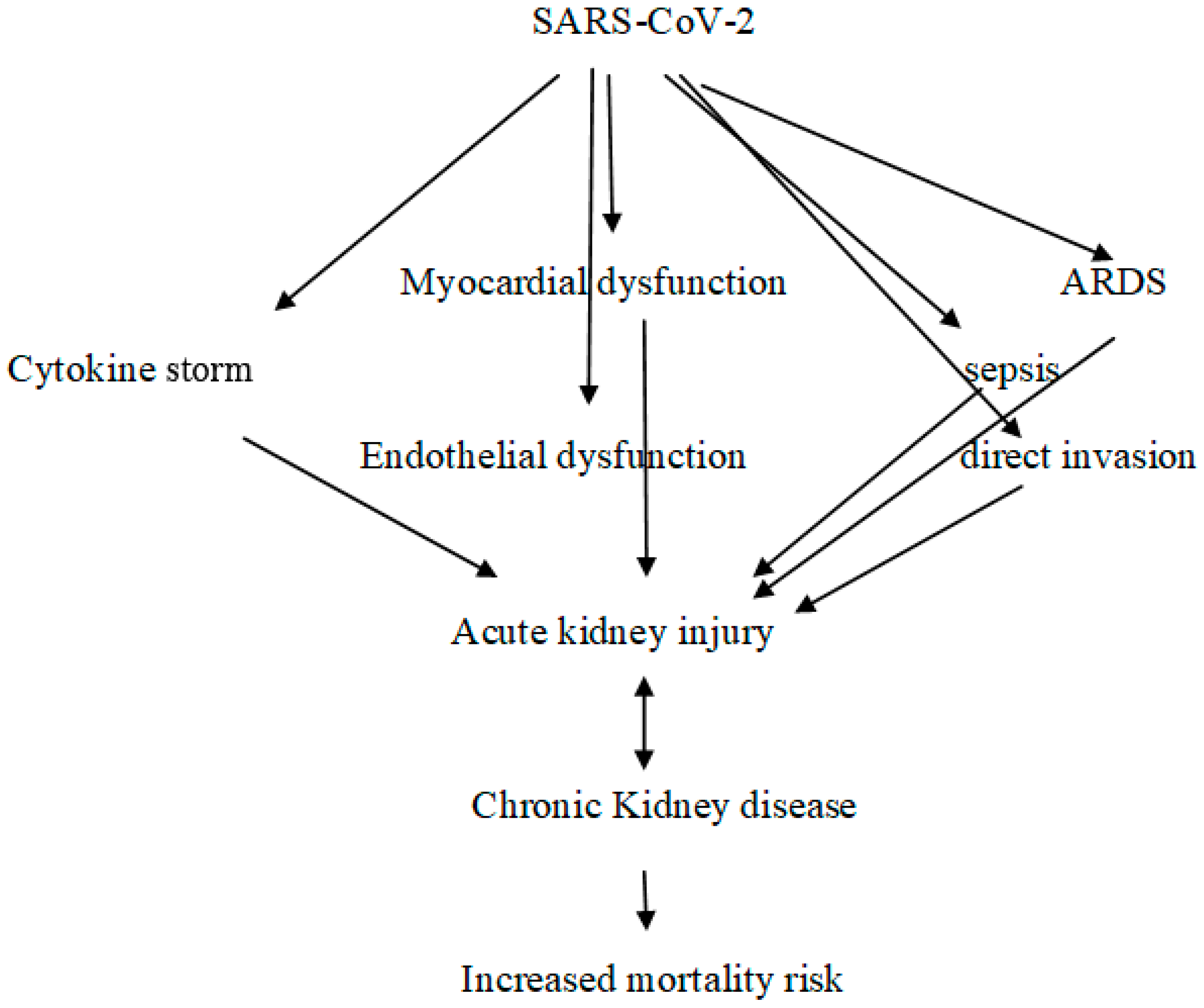
4. Pathophysiology of AKI in COVID-19 Patients

The pathophysiology of AKI in COVID-19 patients is multifactorial and complicated. Local and systemic inflammation, immune system activation, endothelial and coagulation disorders, and RAAS activation are involved in the underlying pathophysiology [41]. In addition, nonspecific factors such as mechanical ventilation, cardiac dysfunction, hemodynamic instability, and drug nephrotoxicity contribute to the development of AKI [41].
Data from autopsy studies have shown that acute tubular injury is the most common finding in COVID-19 patients with AKI [42,43,44]. Acute tubular injury with mild focal acute tubular necrosis has been observed in postmortem renal specimens from COVID-19 patients with AKI [42,43,44]. Acute tubular injury, collapsing glomerulopathy, endothelial damage, and thrombotic microangiopathy were noted in biopsies from patients with COVID-19-associated AKI [45]. In these studies, viral renal infection was not detected or was detected in only a fraction of biopsies. However, in other studies, direct viral tropism of the kidney was detected [46,47,48]. In one study [47], viral RNA was detected in the renal tissue of 23 of 32 patients with AKI compared with 3 of 7 patients without AKI. In another study [48], virus was detected in glomeruli; SARS-CoV-2 was also detected in urine samples, suggesting release of virus from infected tubular epithelial cells or increased glomerular filtration of viral fragments.

5. COVAN

Collapsing nephropathy is a variant of FSGS and is characterized by severe proteinuria, hypertension, and rapid loss of renal function. COVAN has been reported in African American patients with COVID-19, particularly those with apolipoprotein L1 allele [10,40]. COVAN has been observed in COVID-19 patients without severe respiratory disease and with isolated AKI, and in patients with proteinuria in the nephritic range [49]. Collapsing nephropathy has been described in HIV patients and in association with various viral infections caused mainly byparvovirusB19, cytomegalovirus, and Epstein-Barr virus [10,49]. In addition, collapsing nephropathy is associated with black race [10,49]. Collapsing glomerulopathy is characterized by collapse of glomerular tufts with hypertrophy and hyperplasia of podocytes. It is accompanied by acute tubular injury and inflammation of the interstitium [10,40].
The incidence, prognosis, and contribution to CKD and ESRD are unknown. In this context, the pathophysiology of COVAN is also unclear. Because collapsing nephropathy is observed in patients with HIV infection, COVAN might be thought to share the same mechanism as HIVAN nephropathy. Podocyte damage might play a crucial role in this pathophysiological mechanism.
Giannini et al. [50] reported that patients with COVAN have a poor prognosis, the majority of patients with COVAN developed advanced CKD and one third required renal replacement therapy or died after one year. A definitive treatment for COVAN remains unclear. Treatment with high-dose steroids in combination with inhibitors of the renin-angiotensin-aldosterone system may be beneficial in the treatment of COVAN, but this has not yet been proven [50].

6. Coagulation Disorders and Endothelial Dysfunction

Systemic microvascular and macrovascular thrombosis in kidneys and lungs was observed in COVID-19 patients [51]. SARS-CoV-2 affects the endothelium, and vascular endotheliitis has been reported in association with COVID-19 [51]. Several biomarkers of coagulation, fibrinolysis activation, and endothelial injury, including selectins and von Willebrand factor antigen, have been associated with poor prognosis and increased mortality risk [52]. Microvascular inflammation leads to endothelial activation and vasodilation with increased vascular permeability, resulting in prothrombotic states [53]. Furthermore, activation of the C5 complement pathway promotes inflammation and the coagulation cascade [54]. Necrotic cell-derived damage-associated molecular patterns may contribute to endothelial injury [55]. SARS-CoV-2 binds to platelets via ACE2 and induces thrombosis in this manner [56]. Interestingly, in severe COVID-19, high titers of prothrombotic autoantibodies were found to contribute to inflammation and thrombosis [57]. Thus, endothelial damage, platelet activation, and coagulation disorders may contribute to the pathophysiology of COVID-19-AKI.
Thrombotic microangiopathy has been reported in COVID-19 patients [58]. COVID-19-associated thrombotic microangiopathy is a severe complication with poor prognosis and is characterized by hemolytic anemia, thrombocytopenia, and renal failure [59].
SARS-CoV-2 can cause atypical hemolytic syndrome (aHUS) through endothelial dysfunction and direct activation of the alternative complement pathway [60].
The spike protein of SARS-CoV-2 is located in the renal tubules and activates the alternative complement pathway by competing with complement factor H [61]. In addition, complement elements were identified in the microvasculature of COVID-19 patients.
The membrane attack complex accumulates on renal tubules due to complement activation and promotes damage to microvascular epithelial cells, activation of the coagulation cascade, and deposition of fibrin, leading to tissue damage [62].
The anti-inflammatory and antithrombotic effects of nitric oxide, which are due to complement activation, are lost in aHUS associated with COVID-19 [63].
Thus, COVID-19-associated aHUS is an example of how SARS-CoV-2 can cause kidney injury by promoting inflammation, endothelial dysfunction, complement activation, and thrombotic microangiopathy. Interestingly, renal artery thrombosis with concomitant AKI has been reported in COVID-19 patients [64].

7. Involvement of the Immune System

In COVID-19, altered innate and adaptive immune responses have been reported, characterized by immunosenescence and including low-grade inflammation, contributing to organ dysfunction and ineffective T-cell response and antibody production [65,66], likely contributing to the high mortality risk observed in elderly patients [67].
Inflammation plays a critical role in the pathogenesis of AKI [41]. Increased production of inflammatory mediators such as TNF-α and FAS has been observed in COVID-19 [68]. These mediators bind to their specific receptors expressed by renal endothelial and epithelial cells, leading to direct injury [69]. Similar interactions have also been observed in animal models of sepsis with AKI [70].
SARS-CoV-2 can inhibit the release of IFN [71], a cytokine that suppresses viral replication. In the context of COVID-19-AKI, IFN has opposite effects: IFN is a mediator of glomerular injury and can lead to podocyte loss and glomerulosclerosis [72]. Activation of the IFN pathway promotes podocyte damage, leading to proteinuria [73]. In addition, APOL1 risk alleles trigger glomerular damage via the interferon signaling pathway [74]. On the other hand, administration of IFN to COVID-19 patients improves viral clearance and lowers IL-6 and C-reactive protein levels [71]. Patients with severe COVID-19 have autoantibodies against type I IFNs. In addition, patients with low INF-αlevels have a higher risk of severe COVID-19 than patients with high interferon levels, indicating a protective role of IFN against COVID-19 [75].
Uncontrolled and sustained activation of the complement cascade can lead to inflammation with accompanying tissue injury. Detection of C3c and C3d in renal arteries and glomerular capillaries, C3d in tubules, and C5b-9 in renal arterioles and tubular basement membrane has been reported [75]. According to these results, activation of the lectin pathway and the classical pathway appears to occur in peritubular capillaries and renal arteries, whereas the alternative pathway mediates tubular injury [75].
Complement activation is involved in endothelial dysfunction. More specifically, C5a binds to its endothelial receptor and promotes upregulation of tissue factor and loss of thrombomodulin with concomitant activation of coagulation and platelet adhesion and aggregation [76]. C5b-9 induces endothelial dysfunction, inflammation, and coagulation [76,77]. Interestingly, binding of C5a to its receptor C5aR contributes to cellular senescence by promoting DNA methylation of genes, and in this context, may play a role in the pathogenesis of AKI [77].
Decreased numbers of CD4+ and CD8+ T lymphocytes have been observed in COVID-19 patients [65]. In addition, the number of plasmacytoid dendritic cells, eosinophils, and natural killer cells is decreased [78].
Nuclear factor erythroid 2-related factor (NRF2), a transcription factor involved in the regulation of antioxidant activity, is suppressed in COVID-19 [79]. NRF2 binds to its inhibitor Kelch-like ECH-associated protein 1 (KEAP1) and remains inactive in the cytosol. Viral infection promotes oxidative stress, which leads to inactivation of KEAP1; consequently, NRF2 becomes active and induces the expression of genes responsible for stress-induced cell death [79]. In addition, NRF2 agonists have antiviral effects [80]. Data from experimental models of AKI showed that increased expression of NRF2 by T cells protects renal function [80]; specifically, NRF2 protects against kidney injury caused by ischemia and nephrotoxic drugs [81]. Thus, NRF2 is a promising therapeutic target for both COVID-19 and AKI.
Soluble ACE-2 or plasma ACE-2 is the ACE-2 enzyme domain derived from the cell surface. Soluble ACE-2 can bind SARS-CoV-2, and elevated levels of soluble ACE-2 were found in the plasma of critically ill COVID-19 patients [82]. The high affinity of the spike protein for ACE-2, which acts as a receptor for virus entry into cells, may promote the formation of SARS-CoV-2-soluble ACE-2 complexes and the formation of autoantibodies against ACE-2 in tissues [83,84]. Because ACE-2 has high homology (over 42%) with ACE, which is expressed on the surface of most types of leukocytes, autoantibodies against ACE-2 can cross-react with ACE and cause tissue inflammation and organ damage [84].
A cytokine storm is the result of hyperactivity of the immune system, including T cells, macrophages, and natural killer cells [85]. A high release of mediators and inflammatory cytokines, including IL-6, indicates both hyperactivation of the humoral immune system and a hyperinflammatory state [41]. In this context, a marked increase in acute-phase inflammatory markers, lymphopenia, and coagulation disorders have been reported in severe COVID-19 [41]. Of note, IL-6 mediates organ damage, including AKI [86,87]. According to a meta-analysis, elevated serum levels of IL-6 are associated with poor clinical outcome including death [88]. Interestingly, serum levels of IL-6 are lower in severe COVID-19 compared with those observed in sepsis and acute respiratory distress syndrome (ARDS) [89].

8. Other Factors Contributing to the Development of AKI

The acute hypoxemia observed in COVID-19 patients requiring mechanical ventilation may increase renal vascular resistance, which in turn contributes to renal hypoperfusion leading to acute tubular injury and AKI [90,91]. The increased concentrations of IL-6 caused by both decreased renal clearance and increased IL-6 production may contribute to respiratory failure and AKI, suggesting a lung-kidney interaction [91]. Interestingly, mechanical ventilation is a risk factor for the development of AKI in severe COVID-19 [92]. It appears that mechanical ventilation contributes to the pathophysiology of AKI via immune-mediated processes and hemodynamic factors [92]. Notably, the positive association between mechanical ventilation and AKI reflects the severity of the disease [34].
Interaction between the cardiovascular system and the kidneys contributes to the development of AKI. Cases of acute myocarditis and myocardial injury associated with COVID-19 have been reported [93,94]. The impaired cardiac function with decreased cardiac output and venous congestion seen in these pathological situations may contribute to the development of AKI by decreasing renal perfusion [95].
In the context of ARDS, cardiac output may decrease due to decreased right ventricular preload and increased right ventricular afterload caused by the use of high positive end-expiratory pressure and/or high tidal volumes [92]. Right-sided heart failure, in combination with increased venous pressure, leads to an increase in interstitial and tubular hydrostatic pressure, resulting in a decrease in glomerular filtration rate (GFR) [96].
Nephrotoxicity of drugs is another important risk factor for the development of AKI in COVID-19 patients. Vancomycin, colistin, and aminoglycosides must be used with caution because of their potential nephrotoxicity [97]. Calculation of GFR and dose adjustment according to renal function are required.
Remdesivir is a nucleotide analog with antiviral activity. It is excreted through the kidney and is potentially nephrotoxic, causing mitochondrial damage to renal tubular epithelial cells. Nephrotoxicity of remdesivir has been observed after prolonged use or at high doses [98,99].
Rhabdomyolysis is caused by skeletal muscle damage and commonly occurs after various viral infections, including COVID-19. Rhabdomyolysis is another nonimmune cause of AKI [100]. Myoglobin released into the blood after muscle injury exerts its nephrotoxic effects via renal vasoconstriction, formation of intratubular deposits, and direct action on tubular epithelial cells [100].
Patients on extracorporeal membrane oxygenation (ECMO) are at increased risk of developing AKI. Shaefi et al. [101] reported that 22% of patients with respiratory failure on ECMO required renal replacement therapy. Schmidt et al. [102] also reported that the percentage of patients on ECMO who required renal replacement therapy was higher (over 46%). Secondary infections such as cannula infections and ventilator-associated pneumonia, hemolysis, blending, venous congestion, and inflammation should be considered as possible mechanisms for AKI in these patients [102,103].
Overall, acute tubular injury occurs in association with COVID-19. Endothelial dysfunction, microvascular thrombi, local and systemic inflammation, and altered immune response contribute to AKI. Treatment and strategies to prevent progression of the disease and avoid the need for mechanical ventilation may prevent AKI. Until there is a specific treatment for AKI, the management of AKI associated with COVID-19 is no different from that in other situations; (see Figure 1.)

9. AKI and Progression to CKD

There is clinical evidence that AKI accelerates the progression to CKD [104]. Evidence from animal models of AKI suggests several potential mechanisms of progression to CKD, including fibrosis, microvascular rarefaction, tubular and interstitial damage, and glomerulosclerosis [105]. In addition, older age is a risk factor for COVID-19-associated AKI, and kidney aging is a risk factor for CKD. Thus, elderly patients with COVID-19 have a double risk (kidney aging +COVID-19-associated AKI) for progression to CKD.
AKI and CKD share several pathophysiological mechanisms, including glomerular and tubular cell death, local and systemic inflammation, and fibrosis [106]. In addition, several epidemiological studies reported that elderly patients with concomitant diseases such as diabetes, hypertension, heart failure, and obesity are at increased risk for COVID-19-associated AKI [103]. All of these parameters are also risk factors for CKD. Thus, there is a reciprocal relationship between AKI and CKD.
AKI causes both a decrease in the number of peritubular capillaries and a decrease in the quality of the remaining capillaries, leading to a decrease in renal blood flow [107]. At the same time, decreased renal blood flow leads to hypoxia, tubular epithelial cell damage, and apoptosis. Hypoxia triggers collagen production by fibroblasts, leading to fibrosis [108].
It must be emphasized that the long-term effects of AKI on renal function have not been fully elucidated, as most studies have focused on in-hospital outcomes, including the incidence of AKI and mortality. Therefore, there is a need for clinical studies focusing on the long-term effects of AKI on renal function.
However, two studies with 10 years of follow-up suggest that AKI is an independent risk factor for ESRD and all-cause mortality [109,110]. In addition, patients with a history of AKI had a 9-fold higher adjusted risk for CKD and a 3-fold higher adjusted risk for progression to ESRD [91].
As previously mentioned, hospitalized patients with COVID-19-associated AKI are at higher risk for renal replacement therapy, and the mortality rate was higher compared with patients without AKI [26]. Interestingly, only 30% of patients with COVID-19-associated AKI survived and recovered renal function [26].

10. COVID-19 and Cardiorenal Syndrome

Cardiac events may occur in patients with COVID-19-associated AKI, suggesting a possible role of cardiorenal syndrome [111]. As mentioned previously, in the clinical setting of COVID-19, there is an interaction between the affected organs. AKI leads to volume overload resulting in cardiac dysfunction, and conversely, cardiomyopathy caused by COVID-19 may lead to hypotension, renal hypoperfusion, and renal congestion, resulting in decreased GFR [111]. Cardiorenal syndrome carries a high risk of mortality and morbidity for COVID-19 patients and increases the healthcare costs associated with the disease [111].
SARS-CoV-2 can affect both the heart and kidneys through several mechanisms: first, a direct effect; second, a cytokine storm leading to multiorgan dysfunction; and third, induction of thrombotic microangiopathy [112]. In addition, the systemic coagulopathy associated with COVID-19 can lead to renal thrombosis and/or acute coronary syndrome [112].
Hypertension and atherosclerosis are common risk factors for CKD and cardiac dysfunction. According to the National Health Commission of China, 35% of patients with COVID-19 had hypertension, which is considered a negative prognostic factor [113]. COVID-19 patients with hypertension have higher levels of the inflammatory cytokines TNF-α and IL-6, which contributes to endothelial dysfunction and oxidative stress, which in turn promote the development of CKD and heart failure [111].
Inflammation leading to atherosclerosis has become a major problem for COVID-19 patients because it can cause cardiovascular events. Dysregulation of T cells, uncontrolled inflammation, and a cytokine storm likely contribute to the progression of atherosclerosis. Several factors involved in the pathophysiology of atherosclerosis, such as IL-1β, IL-6, ΙL-7, IL-8, TNF-α, IFNs, fibroblast growth factor, monocyte chemoattractant protein, macrophage inflammatory protein-1α, Von Willebrand factor antigen, and factor VIII, were significantly increased in COVID-19 patients [100,114].
In cardiorenal syndrome, angiotensin 2 promotes fibroblast proliferation, fibrosis, and hypertrophy and increases blood pressure by acting as a vasoconstrictor and causing water and salt retention. RAAS blockers have been successfully used to treat hypertension, CKD, and heart failure because of their vasodilatory, antifibrotic, and antiproteinuric effects, and they have recently been shown to have a protective effect on COVID-19 patients regardless of concomitant diseases [115].

11. COVID-19 in Dialysis Patients

The majority of patients on HD have advanced age and concomitant diseases, such as diabetes, hypertension, and cardiovascular disease. In addition, due to the immunological aging associated with uremia, these patients have a weak immune system with lower numbers of circulating CD4+ and CD8+ T cells compared with patients who are not on dialysis [116]. For these reasons, COVID-19 patients undergoing HD require more attention in their management, as they have a higher mortality risk and a worse prognosis.
The different clinical symptoms of COVID-19 in HD patients complicate their management. According to initial reports, the most common symptoms of COVID-19 patients in HD were diarrhea (80%), fever (60%), and fatigue (60%) [117]. In a study involving 65 HD centers, it was reported that 2% of HD patients tested positive for COVID-19; of these patients, 50% had fever and 20% were asymptomatic [118]. However, symptoms may vary due to infection with different SARS-CoV-2 variants. The proportion of asymptomatic SARS-CoV-2 infections in HD patients varies from 10% to 50% of patients [119]. In patients with advanced CKD infected with SARS-CoV-2, especially those on HD, viral clearance is delayed. More than two-thirds of these patients are SARS-CoV-2-positive.
The majority of COVID-19 patients on HD develop a strong antibody response. This may be due to HD patients developing more severe disease, likely associated with a stronger immune response to the virus. In addition, symptomatic HD patients had higher antibody titers to SARS-CoV-2 than asymptomatic patients [120]. Antibodies can protect against reinfection with SARS-CoV-2, but this protection is incomplete. According to a large observational study involving 2337 HD patients, serologic evidence of previous infection was associated with a greater than 45% risk of reinfection [121]. The absence of antibodies was found in 5% to 10% of HD patients. To note, these studies were conducted when the wild-type strain of the virus was prevalent. Prevention of reinfection by the delta and omicron variants of SARS-CoV-2 has not been studied in HD patients.
The mortality rate of COVID-19 patients on HD is 30% higher than in the general population with COVID-19 [118], and according to the European Dialysis and Transplantation Association Registry report, the mortality rate is over 25% [15]. The mortality of the first wave in HD patients was higher than the mortality of the subsequent viral variants, which were less pathogenic than those of the first wave in HD patients.
Another important issue is the high risk of SARS-CoV-2 infection in HD centers. According to a study in a single center in Wuhan, 20% of HD patients and 15% of staff were COVID-19-positive [122]. Hsu et al. [123] proposed guidelines to reduce the risk of SARS-CoV-2 transmission in HD centers.
Even now that the pandemic has subsided, there is still a risk of transmission. Therefore, the authors suggest that both patients and staff should continue to be educated and trained about COVID-19 prevention and management. In addition, screening strategies and education of HD patients about COVID-19 should continue. Rapid testing for SARS-CoV-2 in HD patients and personnel should continue to identify asymptomatic patients. It should be noted that patients and staff must be vaccinated against SARS-CoV-2.
According to a multicenter study from Wuhan involving 818 patients on peritoneal dialysis (PD), the incidence of symptomatic COVID-19 in PD patients was the same as in the general population [124].
PD is home dialysis, where the patient is isolated and the risk of infection is lower compared with HD patients. In addition, PD patients have been trained and educated in their method, and they follow hygiene rules and measures to prevent peritonitis. Therefore, PD patients should be more aware of the risk of infection.
Compared with HD, PD associated with COVID-19 may have some disadvantages. The introduction of intraperitoneal fluid from PD solutions may increase intraperitoneal pressure, which affects respiratory function.
Peritonitis is the most common complication of PD. It is well known that after peritonitis, the function of peritoneal membrane transport is impaired. Ultrafiltration failure due to peritonitis may lead to hypervolemia and dyspnea due to overload. Therefore, in severe COVID-19 and PD peritonitis, pulmonary function may worsen.
Gastrointestinal symptoms of COVID-19 have also been reported. Therefore, confusion of gastrointestinal symptoms of COVID-19 with those of PD peritonitis could lead to misdiagnosis of PD peritonitis.
Interestingly, Jeloka et al. [125] suggested that in the COVID-19 pandemic era, PD patients with peritonitis could be managed by video consultation to avoid hospitalization.
Data from a study of 419 hospitalized patients with ERSD, only 11 patients were on PD. PD patients had mild disease and 9 of 11 patients were discharged [126].
Data from an observational study in Turkey reported that the mortality rate was high (46.2%) in PD-hospitalized patients with COVID-19 [127]. In addition, according to the European Renal Association database, COVID-19 PD patients had a higher mortality risk than HD patients, even after accounting for patient characteristics and disease severity [15].

12. COVID-19 and Kidney Transplantation

Data from Spain and France revealed an average infection rate of 14/1000 transplants at risk [128]. Similar incidences have been reported by the Belgian Society of Nephrology and by other multicenter studies [129].
In the first wave, the hospitalization rate exceeded 80%. The mean age of transplant recipients with COVID-19 was 60 years, two-thirds were male, 80% had hypertension, and 25% had diabetes [129].
Kidney transplant patients may be at increased risk for severe disease due to immunosuppressive drugs and comorbidities. However, data are limited, and the incidence of ARDS in hospitalized kidney transplant patients varies from 12.1% to 68% [128].
The mortality risk is extremely high in hospitalized transplant patients with COVID-19. According to a meta-analysis that included 20 studies, the risk of mortality is higher in kidney transplant patients than in the general population. The risk of mortality ranges from 18% to 43% [129]. Older age, higher plasma viral load, and higher levels of proinflammatory markers were risk factors for mortality [130].
Most studies involved long-term stable recipients, and there are few data for the short-term period. However, the mortality rate in early transplant recipients with COVID-19 is high, ranging from 37% to 46%. Older age, high steroid doses, and mechanical ventilation were mortality risk factors [131].
Of note, the majority of studies were conducted in the first and second waves. Recent data from the United States report lower hospitalization rates (26% vs. 60%) and mortality rates (over 2%) during the predominance of the omicron variants. The mortality rate of transplant patients infected with the omicron variants was 4% [131]. Thus, the overall impression is that mortality is lower with omicron variants but higher than in the general population [131].
Previous recommendations for the use of immunosuppressive drugs (reduced dose and/or discontinuation) were for the first and second waves [132].
Given the prevalence of the “milder” variants, there is currently no need to change immunosuppressive therapy in asymptomatic transplant patients or in patients with mild disease. However, in severe disease with COVID-19 pneumonia, the recommendation of the DESCARTES working group of ERA-EDTA should be considered [131,132]. Current recommendations depend on disease severity and risk stratification [132].

13. Therapeutic Strategies for COVID-19 in CKD Patients

Antiviral Drugs and Monoclonal Antibodies

Remdesivir, nirmatrelvir with ritonavir and molnupiravil are currently approved for the treatment of COVID-19. Remdesivir is an intravenous antiviral agent approved for the treatment of COVID-19 in adults and paediatric patients who require hospitalization. Its nephrotoxicity and dose adjustments in CKD patients have not been well evaluated, but it is not recommended in eGFR < 30 mL/min. The excipient sulfobutylether-β-cyclodextrin accumulates in patients with reduced renal function. For eGFR ≥ 30 mL/min, no dosage adjustment is needed. Regarding the oral therapeutic options for COVID-19, for molnupiravir, there is no need for dosage adjustment in renal impairment, while nirmatrelvir with ritonavir require dosage adjustments according to eGFR level (<30, 30–60, ≥60 mL/min).
The antiviral monoclonal antibodies Sotrovimab and casirivimab + imdevimaband bamlanivimab + etesevimab are not active against Omicron variants and are not currently used (Sotrovimab has activity against Omicron BA.1 but not BA.2). No renal dose adjustments were required for these treatment options. Bebtelovimab presents activity against Omicron BA.1 and BA.2 and does not require dosage adjustment in patients with renal impairment. The tixagevimab-cilgavimab combination used for the pre-exposure prophylaxis against COVID-19 in immunosuppressed high-risk patients does not need dosage adjustments according to renal function impairment, but also possess limited activity against currently circulating SARS-CoV-2 variants.
With regard to monoclonal antibodies used for their anti-inflammatory action, tocilizumab is an IL-6 inhibitor with no need for dosage adjustments in patients with mild or moderate renal impairment, but there are only scarce data on severe renal impairment. Baricitinib and tofacitinib are JAK-inhibitors whose dose is adjusted according to eGFR. Anakinra is an anti-IL-1 factor that should not be used in patients with eGFR < 30 mL/min. Vilobelimab is an anti-complement factor with lack of adequate data in patients with renal impairment [21] (Table 1).

14. Vaccines

There is clinical evidence that the immunogenicity of the mRNA-1273 vaccine is better than that of the BNT162b2 vaccine in HD patients.
Anand et al. [133] reported that more than one in five HD patients had an attenuated immune response to the vaccines, suggesting that vaccination against COVID-19 is less effective in this subgroup. The authors concluded that poor health, uremia, and prolonged duration of end-stage kidney disease increase the risk of a suboptimal response.
Interestingly, the development of a serologic response is delayed in HD patients. A third dose of vaccine increases antibody titers in all HD patients, and patients with a poor initial response benefit from the third dose [17].
Antibody titers to SARS-CoV-2 were higher with the mRNA-1273 vaccine than with the BNT162b2 vaccine in patients who reached the threshold of 590 BAU/mL [17]. Accordingly, the cellular response was more intense with mRNA-1273 than with BNT162b2 vaccine [17].
Both mRNA vaccines appear to have near-maximal clinical efficacy, but these studies were conducted when wild-type virus was prevalent in healthy subjects [17]. Whether the better immunogenicity of mRNA-1273 translates into better protection for HD patients remains to be determined. In this regard, the protection of mRNA-1273 appears to be inadequate.
Limited data are available on the immunogenicity of the other vaccines in HD patients. Neutralizing antibodies to the alpha, beta, and delta variants were higher after vaccination with BNT162b2 vaccine than when AZD1222 was used.
Of note, in HD patients, antibody responses to Ad26.COV2.S vaccine were lower compared with mRNA vaccines [17].
Hsu et al. [134] reported that 67.5% ofAd26.COV2.S, 32.1%of BNT162b2, and 12.3% of mRNA-1273 recipients had no antibodies to SARS-CoV-2 four months after vaccination.
According to a recent large meta-analysis that included 5628 HD patients, the vaccines elicited an adequate immune response (immune efficiency 87.5%) and may be protective against SARS-CoV-2 despite lower immunogenicity rates compared with the general population [135]. Notably, data from a meta-analysis have shown that vaccines are effective against SARS-CoV-2 variants [136,137,138].
There are limited data on humoral response to vaccination in HD and PD patients. Some studies reported that antibody titers were higher in PD patients compared with HD patients, whereas in other studies, antibody titers were similar in PD and HD patients [139,140].
IgA nephropathy, minimal change disease, membranous nephropathy, and pauci-immune necrotizing glomerulonephritis have been reported after mRNA-based SARS-CoV-2 vaccination, but causality remains unproven [141]. In addition, many of the reported vaccine-associated glomerulonephritides have been associated with COVID-19 infection itself [142].
Diebolt et al. [143] reported that sporadic cases of new-onset glomerulonephritis after mRNA-based SARS-CoV-2 vaccination were due to temporal coincidence. Overall, cases of glomerulopathyhave been reported after vaccination against COVID-19, but there is no evidence for the role of the vaccines.

15. Post-COVID-19 Syndrome

At the beginning of the pandemic, there were several reports of the long-term consequences of COVID-19. Post-COVID-19 syndrome is defined as a heterogeneous clinical syndrome of new onset. Usually, it appears 3 months after COVID-19 infection and it is characterized by symptoms lasting at least 2 months, which are not explained by an alternative diagnosis. Huang et al. [144] reported the long-term consequences of COVID-19 patients after discharge. In 35% of patients with COVID-19-associated AKI, GFR had decreased after a follow-up period of 6 months [145]. A retrospective cohort study found that patients with COVID-19-associated AKI had a greater decrease in GFR than patients with other types of AKI [145]. Another large retrospective study reported that 90,000 patients had a higher incidence of AKI 30 days after the diagnosis of COVID-19 [146]. In addition, a higher incidence of ESRD was found in COVID-19. In this regard, Gu et al. [147] reported similar findings. More specifically, COVID-19 patients with stage 3 AKI had 17.8% higher deterioration of renal function compared with patients without AKI.
The long-term consequences of COVID-19 on renal function are of great concern. COVID-19 can affect the kidney through direct and indirect effects. It is likely that many of these effects will persist after COVID-19 recovery and lead to an increased risk of CKD or/and recurrent AKI. In addition, the presence of diabetes may delay recovery or exacerbate the disease. Interestingly, the decline of renal function in the post COVID-19 period is multifactorial. Chronic inflammation, tubular injury, maladaptive and delayed repair of AKI, cardiac disease, and diabetes may contribute to the pathogenesis of post-COVID-19 syndrome [148]. A nephrological follow-up at 3 months for at least 12 months after hospital discharge for AKI-associated COVID-19 patients is needed.
It should be noted, that there is no specific treatment for post-COVID-19 syndrome.

16. Conclusions

  • The relationship between COVID-19 and CKD is bidirectional.
  • COVID-19 causes AKI and may exacerbate persistent CKD, and CKD is a risk factor for COVID-19.
  • In this context, there are several common risk factors, including advanced age, diabetes, hypertension, and cardiovascular disease.
  • Therefore, it is not surprising that the coexistence of COVID-19 and CKD is associated with higher morbidity and mortality.
  • Further large-scale prospective cohort studies are needed to elucidate this association.

Author Contributions

P.D., conceptualization, data collection, writing, review and editing; K.T., data collection and editing; I.S., review and editing; K.-E.G., data collection; I.-S.S., data collection; D.S., data collection; A.M., review and editing; S.F.A., review and editing. All authors have read and agreed to the published version of the manuscript.

Funding

This research received no external funding.

Conflicts of Interest

The authors declare no conflict of interest.

References

  1. Jackson, C.B.; Farzan, M.; Chen, B.; Hyeryun, C. Mechanisms of SARS-CoV-2 entry into cells. Nat. Rev. Mol. Cell Biol. 2022, 23, 3–20. [Google Scholar] [CrossRef]
  2. Zandi, M.; Shafaati, M.; Kalantar-Neyestanaki, D.; Pourghadamyari, H.; Fani, M.; Soltani, S.; Kaleji, H.; Abbasi, S. The role of SARS-CoV-2 accessory proteins in immune evasion. Biomed Pharmacother 2022, 156, 113889. [Google Scholar] [CrossRef]
  3. Zandi, M. ORF9c and ORF10 as accessory proteins of SARS-CoV-2 in immune evasion. Nat. Rev. Immunol. 2022, 22, 331. [Google Scholar] [CrossRef]
  4. Dettlaff-Pokora, A.; Swierczynski, J. Dysregulation of the Renin-Angiotensin- Aldosterone System (RAA) in Patients Infected with SARS-CoV-2-Possible Clinical Consequences. Int. J. Mol. Sci. 2021, 22, 4503. [Google Scholar] [CrossRef]
  5. Hu, B.; Huang, S.; Yin, L. The cytokinestorm and COVID-19. J. Med. Virol. 2021, 93, 250–256. [Google Scholar] [CrossRef] [PubMed]
  6. Kaklamanos, A.; Belogiannis, K.; Skendros, P.; Gorgoulis, V.G.; Vlachoyiannopoulos, P.G.; Tzioufas, A.G. COVID-19 Immuno-biology: Lessons Learned, New Questions Arise. Front. Immunol. 2021, 12, 719023. [Google Scholar] [CrossRef] [PubMed]
  7. Hassler, L.; Batlle, D. Potential SARS-CoV-2 kidney infection and paths to injury. Nat. Rev. Nephrol. 2022, 18, 275–276. [Google Scholar] [CrossRef] [PubMed]
  8. Pei, G.; Zhang, Z.; Peng, J.; Liu, L.; Zhang, C.; Yu, C.; Ma, Z.; Huang, Y.; Liu, W.; Yao, Y.; et al. Renal Involvement and Early Prognosis in Patients with COVID-19 Pneumonia. J. Am. Soc. Nephrol. 2020, 31, 1157–1165. [Google Scholar] [CrossRef]
  9. Kudose, S.; Batal, I.; Santoriello, D.; Xu, K.; Barasch, J.; Peleg, Y.; Canetta, P.; Ratner, L.E.; Marasa, M.; Gharavi, A.G.; et al. Kidney Biopsy Findings in Patients with COVID-19. J. Am. Soc. Nephrol. 2020, 31, 1959–1968. [Google Scholar] [CrossRef]
  10. Velez, J.C.Q.; Caza, T.; Larsen, C.P. COVAN is the new HIVAN: The re-emergence of collapsing glomerulopathy with COVID-19. Nat. Rev. Nephrol. 2020, 16, 565–567. [Google Scholar] [CrossRef]
  11. Wu, H.; Larsen, C.P.; Hernandez-Arroyo, C.F.; Mohamed, M.M.B.; Caza, T.; Sharshir, M.; Chughtai, A.; Xie, L.; Gimenez, J.M.; Sandow, T.A.; et al. AKI and Col-lapsing Glomerulopathy Associated with COVID-19 and APOL1 High-Risk Genotype. J. Am. Soc. Nephrol. 2020, 3, 1688–1695. [Google Scholar] [CrossRef]
  12. Ng, J.H.; Hirsch, J.S.; Hazzan, A.; Wanchoo, R.; Shah, H.H.; Malieckal, D.A.; Ross, D.W.; Sharma, P.; Sakhiya, V.; Fishbane, S.; et al. Northwell Nephrology COVID-19 Research Consortium. Outcomes Among Patients Hospitalized With COVID-19 and Acute KidneyInjury. Am. J. Kidney Dis. 2021, 77, 204–215. [Google Scholar] [CrossRef]
  13. Chung, E.Y.; Palmer, S.C.; Natale, P.; Krishnan, A.; Cooper, T.E.; Saglimbene, V.M.; Ruospo, M.; Au, E.; Jayanti, S.; Liang, A.; et al. Incidence and Outcomes of COVID-19 in People with CKD: A Systematic Review and Meta-analysis. Am. J. Kidney Dis. 2021, 78, 804–815. [Google Scholar] [CrossRef]
  14. Ozturk, S.; Turgutalp, K.; Arici, M.; Odabas, A.R.; Altiparmak, M.R.; Aydin, Z.; Cebeci, E.; Basturk, T.; Soypacaci, Z.; Sahin, G.; et al. Mortality analysis of COVID-19 infection in chronic kidney disease, haemodialysis and renal transplant patients compared with patients without kidney disease: A nationwide analysis from Turkey. Nephrol. Dial. Transplant. 2020, 35, 2083–2095. [Google Scholar] [CrossRef] [PubMed]
  15. Jager, K.J.; Kramer, A.; Chesnaye, N.C.; Couchoud, C.; Sánchez-Álvarez, J.E.; Garneata, L.; Collart, F.; Hemmelder, M.H.; Ambühl, P.; Kerschbaum, J.; et al. Results from the ERA-EDTA Registry indicate a high mortality due to COVID-19 in dialysis patients and kidney transplant recipients across Europe. Kidney Int. 2020, 98, 1540–1548. [Google Scholar] [CrossRef] [PubMed]
  16. Valeri, A.M.; Robbins-Juarez, S.Y.; Stevens, J.S.; Ahn, W.; Rao, M.K.; Radhakrishnan, J.; Gharavi, A.G.; Mohan, S.; Husain, S.A. Presentation and Outcomes of Patients with ESKD and COVID-19. J. Am. Soc. Nephrol. 2020, 31, 1409–1415. [Google Scholar] [CrossRef]
  17. El Karoui, K.; De Vriese, A.S. COVID-19 in dialysis: Clinical impact, immune response, prevention, and treatment. Kidney Int. 2022, 101, 883–894. [Google Scholar] [CrossRef] [PubMed]
  18. Jawdeh, B.G. COVID-19 in Kidney Transplantation: Outcomes, Immunosuppression Management, and Operational Challenges. Adv. Chronic Kidney Dis. 2020, 27, 383–389. [Google Scholar] [CrossRef]
  19. Tylicki, L.; Biedunkiewicz, B.; Puchalska-Reglińska, E.; Gellert, R.; Burnier, M.; Wolf, J.; Dȩbska-Ślizień, A. COVID-19 vac-cination reduces mortality in patients on maintenance hemodialysis. Front. Med. (Lausanne) 2022, 9, 937167. [Google Scholar] [CrossRef]
  20. Klomjit, N.; Alexander, M.P.; Fervenza, F.C.; Zoghby, Z.; Garg, A.; Hogan, M.C.; Nasr, S.H.; Abu Minshar, M.; Zand, L. COVID-19 Vaccination and Glomerulonephritis. Kidney Int. Rep. 2021, 6, 2969–2978. [Google Scholar] [CrossRef]
  21. Kale, A.; Shelke, V.; Dagar, N.; Anders, H.J.; Gaikwad, A.B. How to use COVID-19 antiviral drugs in patients with chronic kidney disease. Front. Pharmacol. 2023, 14, 1053814. [Google Scholar] [CrossRef]
  22. Copur, S.; Berkkan, M.; Basile, C.; Tuttle, K.; Kanbay, M. Post-acute COVID-19 syndrome and kidney diseases: What do we know? J. Nephrol. 2022, 35, 795–805. [Google Scholar] [CrossRef] [PubMed]
  23. Guan, W.J.; Zhong, N.S. Clinical Characteristics of COVID-19 in China. Reply. N. Engl. J. Med. 2020, 382, 1861–1862. [Google Scholar] [PubMed]
  24. Batlle, D.; Soler, M.; Sparks, M.A.; Hiremath, S.; South, A.M.; Welling, P.A.; Swaminathan, S. COVID-19 and ACE2 in Cardi-ovascular, Lung, and Kidney Working Group. Acute Kidney Injury in COVID-19: Emerging Evidence of a Distinct Patho-physiology. J. Am. Soc. Nephrol. 2020, 31, 1380–1383. [Google Scholar] [CrossRef]
  25. Cheng, Y.; Luo, R.; Wang, K.; Zhang, M.; Wang, Z.; Dong, L.; Li, J.; Yao, Y.; Ge, S.; Xu, G. Kidney disease is associated with in-hospital death of patients with COVID-19. Kidney Int. 2020, 97, 829–838. [Google Scholar] [CrossRef] [PubMed]
  26. Hirsch, J.S.; Ng, J.H.; Ross, D.W.; Sharma, P.; Shah, H.H.; Barnett, R.L.; Hazzan, A.D.; Fishbane, S.; Jhaveri, K.D. Northwell COVID-19 Research Consortium; Northwell Nephrology COVID-19 Research Consortium. Acute kidney injury in patients hospitalized with COVID-19. Kidney Int. 2020, 98, 209–218. [Google Scholar] [CrossRef]
  27. Cummings, M.J.; Baldwin, M.R.; Abrams, D.; Jacobson, S.D.; Meyer, B.J.; Balough, E.M.; Aaron, J.G.; Claassen, J.; Rabbani, L.E.; Hastie, J.; et al. Epidemiology, clinical course, and outcomes of critically ill adults with COVID-19 in New York City: A prospective cohort study. Lancet 2020, 395, 1763–1770. [Google Scholar] [CrossRef]
  28. Gupta, S.; Coca, S.G.; Chan, L.; Melamed, M.L.; Brenner, S.K.; Hayek, S.S.; Sutherland, A.; Puri, S.; Srivastava, A.; Leonberg-Yoo, A.; et al. AKI Treated with Renal Replacement Therapy in Critically Ill Patients with COVID-19. J. Am. Soc. Nephrol. 2020, 32, 161–176. [Google Scholar] [CrossRef]
  29. Peng, S.; Wang, H.Y.; Sun, X.; Li, P.; Ye, Z.; Li, Q.; Wang, J.; Shi, X.; Liu, L.; Yao, Y.; et al. Early versus late acute kidney injury among patients with COVID-19-a multicenter study from Wuhan, China. Nephrol. Dial. Transplant. 2020, 35, 2095–2102. [Google Scholar] [CrossRef]
  30. Fu, E.L.; Janse, R.J.; de Jong, Y.; van der Endt, V.H.; Milders, J.; van der Willik, E.M.; de Rooij, E.N.; Dekkers, O.M.; Rotmans, J.I.; van Diepen, M. Acute kidney injury and kidney replacement therapy in COVID-19: A systematic review and metaanalysis. Clin. Kidney J. 2020, 13, 550–563. [Google Scholar] [CrossRef]
  31. Chan, L.; Chaudhary, K.; Saha, A.; Chauhan, K.; Vaid, A.; Zhao, S.; Paranjpe, I.; Somani, S.; Richter, F.; Miotto, R.; et al. AKI in Hospitalized Patients with COVID-19. J. Am. Soc. Nephrol. 2021, 32, 151–160. [Google Scholar] [CrossRef] [PubMed]
  32. Shemies, R.S.; Nagy, E.; Younis, D.; Sheashaa, H. Renal replacement therapy for critically ill patients with COVID-19-associated acute kidney injury: A review of current knowledge. Ther. Apher. Dial. 2021, 26, 15–23. [Google Scholar] [CrossRef] [PubMed]
  33. Pecly, I.M.D.; Azevedo, R.B.; Muxfeldt, E.S.; Botelho, B.G.; Albuquerque, G.G.; Diniz, P.H.P.; Silva, R.; Rodrigues, C.I.S. COVID-19 and chronic kidney disease: A comprehensive review. Braz. J. Nephrol. 2021, 43, 383–399. [Google Scholar]
  34. Bowe, B.; Cai, M.; Xie, Y.; Gibson, A.K.; Maddukuri, G.; Al-Aly, Z. Acute Kidney Injury in a National Cohort of Hospitalized US Veterans th COVID-19. Clin. J. Am. Soc. Nephrol. 2020, 16, 14–25. [Google Scholar] [CrossRef]
  35. Charytan, D.M.; Parnia, S.; Khatri, M.; Petrilli, C.M.; Jones, S.; Benstein, J.; Horwitz, L.I. Decreasing Incidence of Acute Kid-ney Injury in Patients with COVID-19 Critical Illness in New York City. Kidney Int. Rep. 2021, 6, 916–927. [Google Scholar] [CrossRef]
  36. Chaudhri, I.; Moffitt, R.; Taub, E.; Annadi, R.R.; Hoai, M.; Bolotova, O.; Yoo, J.; Dhaliwal, S.; Sahib, H.; Daccueil, F.; et al. Association of Proteinuria and Hematuria with Acute Kidney Injury and Mortality in Hospitalized Patients with COVID-19. Kidney Blood Press. 2020, 45, 1018–1032. [Google Scholar] [CrossRef]
  37. Hidayat, A.A.; Gunawan, V.A.; Iragama, F.R.; Alfiansyah, R.; Hertanto, D.M.; Tjempakasari, A.; Thaha, M. Risk Factors and Clinical Characteristics of Acute Kidney Injury in Patients with COVID-19: A Systematic Review and Meta-Analysis. Pathophysiology 2023, 30, 233–247. [Google Scholar] [CrossRef]
  38. Kormann, R.; Jacquot, A.; Alla, A.; Corbel, A.; Koszutski, M.; Voirin, P.; Parrilla, M.G.; Bevilacqua, S.; Schvoerer, E.; Gueant, J.-L.; et al. Coronavirus disease 2019: Acute Fanconi syndrome precedes acute kidney injury. Clin. Kidney J. 2020, 13, 362–370. [Google Scholar] [CrossRef]
  39. Werion, A.; Belkhir, L.; Perrot, M.; Schmit, G.; Aydin, S.; Chen, Z. Cliniquesuniversitaires Saint-Luc (CUSL) COVID-19 Research Group. SARS-CoV-2 causes a specific dysfunction of the kid-ney proximal tubule. Kidney Int. 2020, 98, 1296–1307. [Google Scholar] [CrossRef]
  40. Ferlicot, S.; Jamme, M.; Gaillard, F.; Oniszczuk, J.; Couturier, A.; May, O.; Grünenwald, A.; Sannier, A.; Moktefi, A.; Le Mon-nier, O.; et al. AP-HP/Universities/Inserm COVID-19 research collaboration. The spectrum of kidney bi-opsies in hospitalized patients with COVID-19, acute kidney injury, and/or proteinuria. Nephrol. Dial. Transplant. 2021, 12, gfab042. [Google Scholar]
  41. Legrand, M.; Bell, S.; Forni, L.; Joannidis, M.; Koyner, J.L.; Liu, K.; Cantaluppi, V. Pathophysiology of COVID-19-associated acute kidney injury. Nat. Rev. Nephrol. 2021, 17, 751–764. [Google Scholar] [CrossRef]
  42. Santoriello, D.; Khairallah, P.; Bomback, A.S.; Xu, K.; Kudose, S.; Bata, I.; Barasch, J.; Radhakrishnan, J.; D’Agati, V.; Markowitz. Postmortem Kidney Pathology Findings in Patients with COVID-19G. J. Am. Soc. Nephrol. 2020, 31, 2158–2167. [Google Scholar] [CrossRef]
  43. Miller, S.E.; Brealey, J.K. Visualization of putative coronavirus in kidney. Kidney Int. 2020, 98, 231–232. [Google Scholar] [CrossRef]
  44. Hanley, B.; Naresh, K.N.; Roufosse, C.; Nicholson, A.G.; Weir, J.; Cooke, G.S.; Thursz, M.; Manousou, P.; Corbett, R.; Goldin, R.; et al. Histopathological findings and viral tropism in UK patients with severe fatal COVID-19: A post-mortem study. Lancet Microbe 2020, 1, e245–e253. [Google Scholar] [CrossRef] [PubMed]
  45. Akilesh, S.; Nast, C.C.; Yamashita, M.; Henriksen, K.; Charu, V.; Troxell, M.L.; Kambham, N.; Bracamonte, E.; Houghton, D.; Ahmed, N.I.; et al. Multicenter Clinicopathologic Correlation of Kidney Biopsies Performed in COVID-19 Patients Presenting With Acute Kidney Injury or Proteinuria. Am. J. Kidney Dis. 2021, 77, 82–93.e1. [Google Scholar] [CrossRef]
  46. Braun, F.; Lütgehetmann, M.; Pfefferle, S.; Wong, M.N.; Carsten, A.; Lindenmeyer, M.T.; Nörz, D.; Heinrich, F.; Meißner, K.; Wichmann, D.; et al. SARS-CoV-2 renal tropism associates with acute kidney injury. Lancet 2020, 396, 597–598. [Google Scholar] [CrossRef]
  47. Sun, J.; Zhu, A.; Li, H.; Zheng, K.; Zhuang, Z.; Chen, Z.; Shi, Y.; Zhang, Z.; Chen, S.B.; Liu, X.; et al. Isolation of infectious SARS-CoV-2 from urine of a COVID-19 patient. Emerg. Microbes Infect. 2020, 9, 991–993. [Google Scholar] [CrossRef]
  48. Puelles, V.G.; Lütgehetmann, M.; Lindenmeyer, M.T.; Sperhake, J.P.; Wong, M.N.; Allweiss, L.; Chilla, S.; Heinemann, A.; Wanner, N.; Liu, S.; et al. Multiorgan and Renal Tropism of SARS-CoV-2. N. Engl. J. Med. 2020, 6, 590–592. [Google Scholar] [CrossRef]
  49. Schwimmer, J.A.; Markowitz, G.S.; Valeri, A.; Appel, G.B. Collapsing glomerulopathy. Semin. Nephrol. 2003, 23, 209–218. [Google Scholar] [CrossRef]
  50. Giannini, G.; Velez, J.C.; May, R.M.; Sharma, S.G.; Mohamed, M.M.; Cassol, C.A.; Larsen, C.P.; Caza, T.N. Renal Prognosis of COVID-19 Associated Nephropathy. Kidney Int. Rep. 2022, 7, 2722–2725. [Google Scholar] [CrossRef]
  51. Ackermann, M.; Verleden, S.E.; Kuehnel, M.; Haverich, A.; Welte, T.; Laenger, F.; Vanstapel, A.; Werlein, C.; Stark, H.; Tzankov, A.; et al. Pulmonary Vascular Endothelialitis, Thrombosis, and Angiogene-sis in COVID-19. N. Engl. J. Med. 2020, 9, 120–128. [Google Scholar] [CrossRef] [PubMed]
  52. Vassiliou, A.G.; Keskinidou, C.; Jahaj, E.; Gallos, P.; Dimopoulou, I.; Kotanidou, A.; Orfanos, S.E. ICU Admission Levels of Endothelial Biomarkers as Predictors of Mortality in Critically Ill COVID-19 Patients. Cells 2021, 10, 186. [Google Scholar] [CrossRef] [PubMed]
  53. Iba, T.; Connors, J.M.; Levy, J.H. The coagulopathy, endotheliopathy, and vasculitis of COVID-19. Inflamm. Res. 2020, 69, 1181–1189. [Google Scholar] [CrossRef] [PubMed]
  54. Macor, P.; Durigutto, P.; Mangogna, A.; Bussani, R.; De Maso, L.; D’errico, S.; Zanon, M.; Pozzi, N.; Meroni, P.L.; Tedesco, F. Multiple-Organ Complement Deposition on Vascular Endothelium in COVID-19 Patients. Biomedicines 2021, 9, 1003. [Google Scholar] [CrossRef] [PubMed]
  55. Ince, C. The central role of renal microcirculatory dysfunction in the pathogenesis of acute kidney injury. Nephron Clin. Pract. 2014, 127, 124–128. [Google Scholar] [CrossRef]
  56. Taha, M.; Sano, D.; Hanoudi, S.; Esber Elahi, M.; Gabali, A.; Chopra, T.; Draghici, S.; Samavat, I.L. Platelets and renal failure in the SARS-CoV-2 syndrome. Platelets 2021, 32, 130–137. [Google Scholar] [CrossRef]
  57. Zuo, Y.; Estes, S.K.; Ali, R.A.; Gandhi, A.A.; Yalavarthi, S.; Shi, H.; Sule, G.; Gockman, K.; Madison, J.A.; Zuo, M.; et al. Prothrombotic autoan-tibodies in serum from patients hospitalized with COVID-19. Sci. Med. Transl. 2020, 12, eabd3876. [Google Scholar] [CrossRef] [PubMed]
  58. Fox, S.E.; Lameira, F.S.; Rinke, E.B.; Vander Heide, R.S. Cardiac Endotheliitis and Multisystem Inflammatory Syndrome Af-ter COVID-19. Ann. Intern. Med. 2020, 173, 1025–1027. [Google Scholar] [CrossRef]
  59. Plášek, J.; Gumulec, J.; Máca, J.; Škarda, J.; Procházka, V.; Grézl, T.; Václavík, J. COVID-19 associated coagulopathy: Mecha-nisms and host-directed treatment. Am. J. Med. Sci. 2022, 363, 465–475. [Google Scholar] [CrossRef]
  60. Ville, S.; Le Bot, S.; Chapelet-Debout, A.; Blancho, G.; Fremeaux-Bacchi, V.; Deltombe, C.; Fakhouri, F. Atypical HUS relapse triggered by COVID-19. Kidney Int. 2020, 99, 267–268. [Google Scholar] [CrossRef]
  61. Yu, J.; Yuan, X.; Chen, H.; Chaturvedi, S.; Braunstein, E.M.; Brodsky, R.A. Direct activation of the alternative complement pathway by SARS-CoV-2 spike proteins is blocked by factor D inhibition. Blood 2020, 136, 2080–2089. [Google Scholar] [CrossRef] [PubMed]
  62. Perico, L.; Benigni, A.; Casiraghi, F.; Ng, L.F.P.; Renia, L.; Remuzzi, G. Immunity, endothelial injury and comple-ment-induced coagulopathy in COVID-19. Nat. Rev. Nephrol. 2021, 17, 46–64. [Google Scholar] [CrossRef] [PubMed]
  63. Fang, W.; Jiang, J.; Su, L.; Shu, T.; Liu, H.; Lai, S.; Ghiladi, R.A.; Wang, J. The role of NO in COVID-19 and potential therapeutic strategies. Free Radic. BiolMed. 2021, 163, 153–162. [Google Scholar] [CrossRef] [PubMed]
  64. El Shamy, O.; Munoz-Casablanca, N.; Coca, S.; Sharma, S.; Lookstein, R.; Uribarri, J. Bilateral Renal Artery Thrombosis in a Patient With COVID-19. Kidney Med. 2020, 3, 116–119. [Google Scholar] [CrossRef]
  65. Moderbacher, C.R.; Ramirez, S.I.; Dan, J.M.; Grifoni, A.; Hastie, K.M.; Weiskopf, D.; Belanger, S.; Abbott, R.K.; Kim, C.; Choi, J.; et al. Antigen-Specific Adaptive Immunity to SARS-CoV-2 in Acute COVID-19 and Associations with Age and Disease Severity. Cell 2020, 183, 996–1012.e19. [Google Scholar] [CrossRef]
  66. Pawelec, G. Age and immunity: What is “immunosenescence”? Exp. Gerontol. 2018, 105, 4–9. [Google Scholar] [CrossRef]
  67. Fuentes, E.; Fuentes, M.; Alarcón, M.; Palomo, I. Immune System Dysfunction in the Elderly. An. Da Acad. Bras. De Ciências 2017, 89, 285–299. [Google Scholar] [CrossRef] [PubMed]
  68. Cantaluppi, V.; Quercia, A.D.; Dellepiane, S.; Ferrario, S.; Camussi, G.; Biancone, L. Interaction between systemic inflamma-tion and renal tubular epithelial cells. Nephrol. Dial. Transplant. 2014, 29, 2004–2011. [Google Scholar] [CrossRef] [PubMed]
  69. Cutuli, S.L.; Artigas, A.; Fumagalli, R.; Monti, G.; Ranieri, V.M.; Ronco, C.; Antonelli, M. The EUPHAS 2 Collaborative Group Polymyxin-B hemoperfusion in septic patients: Analysis of a multicenter registry. Ann. Intensiv. Care 2016, 6, 1–9. [Google Scholar] [CrossRef]
  70. Zarbock, A.; Gomez, H.; Kellum, J.A. Sepsis-induced acute kidney injury revisited: Pathophysiology, prevention and future therapies. CurrOpinCritCare 2014, 20, 588–595. [Google Scholar] [CrossRef]
  71. Buszko, M.; Nita-Lazar, A.; Park, J.-H.; Schwartzberg, P.L.; Verthelyi, D.; Young, H.A.; Rosenberg, A.S. Lessons learned: New insights on the role of cytokines in COVID-19. Nat. Immunol. 2021, 22, 404–411. [Google Scholar] [CrossRef]
  72. Koning, R.; Bastard, P.; Casanova, J.-L.; Brouwer, M.C.; van de Beek, D.; van Agtmael, M.; Algera, A.G.; Appelman, B.; van Baarle, F.; Bax, D.; et al. Autoantibodies against type I interferons are associated with multi-organ failure in COVID-19 patients. Intensiv. Care Med. 2021, 47, 704–706. [Google Scholar] [CrossRef]
  73. Migliorini, A.; Angelotti, M.L.; Mulay, S.R.; Kulkarni, O.O.; Demleitner, J.; Dietrich, A.; Sagrinati, C.; Ballerini, L.; Peired, A.; Shankland, S.J.; et al. The antiviral cytokines IFN-α and IFN-β modulate parietal epithelial cells and promote podocyte loss: Implications for IFN toxicity, viral glomerulonephritis, and glomerular regeneration. Am. J. Pathol. 2013, 183, 431–440. [Google Scholar] [CrossRef]
  74. Beckerman, P.; Bi-Karchin, J.; Park, A.S.; Qiu, C.; Dummer, P.D.; Soomro, I.; Boustany-Kari, C.M.; Pullen, S.S.; Miner, J.H.; Hu, C.A. Transgenic expression of human APOL1 risk variants in podocytes induces kidney disease in mice. Nat. Med. 2017, 23, 429–438. [Google Scholar] [CrossRef]
  75. Pfister, F.; Vonbrunn, E.; Ries, T.; Jäck, H.-M.; Überla, K.; Lochnit, G.; Sheriff, A.; Herrmann, M.; Büttner-Herold, M.; Amann, K.; et al. Complement Activation in Kidneys of Patients With COVID-19. Front. Immunol. 2020, 11, 594849. [Google Scholar] [CrossRef]
  76. Vinayagam, S.; Sattu, K. SARS-CoV-2 and coagulation disorders in different organs. Life Sci. 2020, 260, 118431. [Google Scholar] [CrossRef]
  77. Castellano, G.; Franzin, R.; Sallustio, F.; Stasi, A.; Banelli, B.; Romani, M.; De Palma, G.; Lucarelli, G.; Divella, C.; Battaglia, M.; et al. Complement component C5a induces aberrant epigenetic modifications in renal tubular epithelial cells accelerating se-nescence by Wnt4/βcatenin signaling after ischemia/reperfusion injury. Aging 2019, 11, 4382–4406. [Google Scholar] [CrossRef]
  78. Laing, A.G.; Lorenc, A.; Del Molino Del Barrio, I.; Das, A.; Fish, M.; Monin, L.; Muñoz-Ruiz, M.; McKenzie, D.R.; Hayday, T.S.; Francos-Quijorna, I.; et al. A dynamic COVID-19 immune signature includes associations with poor prognosis. Nat. Med. 2020, 26, 1623–1635. [Google Scholar] [CrossRef]
  79. Olagnier, D.; Farahani, E.; Thyrsted, J.; Blay-Cadanet, J.; Herengt, A.; Idorn, M.; Hait, A.; Hernaez, B.; Knudsen, A.; Iversen, M.B.; et al. SARS-CoV2-mediated suppression of NRF2-signaling reveals potent antiviral and anti-inflammatory activity of 4-octyl-itaconate and dimethyl fumarate. Nat Commun. 2020, 2, 4938. [Google Scholar] [CrossRef]
  80. Noel, S.; Martina, M.N.; Bandapalle, S.; Racusen, L.C.; Potteti, H.R.; Hamad, A.R.; Reddy, S.P.; Rabb, H.T. Lymphocyte–Specific Activation of Nrf2 Protects from AKI. J. Am. Soc. Nephrol. 2015, 26, 2989–3000. [Google Scholar] [CrossRef]
  81. Liu, M.; Grigoryev, D.N.; Crow, M.T.; Haas, M.; Yamamoto, M.; Reddy, S.P.; Rabb, H. Transcription factor Nrf2 is protective during ischemic and nephrotoxic acute kidney injury in mice. Kidney Int. 2009, 76, 277–285. [Google Scholar] [CrossRef]
  82. Camargo, R.L.; Bombassaro, B.; Monfort-Pires, M.; Mansour, E.; Palma, A.C.; Ribeiro, L.C.; Ulaf, R.G.; Bernardes, A.F.; Nunes, T.A.; Agrela, M.V.; et al. Plasma Angiotensin II Is Increased in Critical Coronavirus Disease 2019. Front. Cardiovasc. Med. 2022, 9, 847809. [Google Scholar] [CrossRef]
  83. Beyerstedt, S.; Casaro, E.B.; Rangel, É.B. COVID-19: Angiotensin-converting enzyme 2 (ACE2) expression and tissue suscep-tibility to SARS-CoV-2 infection. Eur. J. Clin. Microbiol. Infect. Dis. 2021, 40, 905–919. [Google Scholar] [CrossRef]
  84. McMillan, P.; Dexhiemer, T.; Neubig, R.R.; Uhal, B.D. COVID-19—A Theory of Autoimmunity Against ACE-2 Explained. Front. Immunol. 2021, 12, 582166. [Google Scholar] [CrossRef]
  85. Fajgenbaum, D.C.; June, C.H. Cytokine Storm. N. Engl. J. Med. 2020, 3, 2255–2273. [Google Scholar] [CrossRef]
  86. Xia, P.; Wen, Y.; Duan, Y.; Su, H.; Cao, W.; Xiao, M.; Ma, J.; Zhou, Y.; Chen, G.; Jiang, W.; et al. Clinico-pathological Features and Outcomes of Acute Kidney Injury in Critically Ill COVID-19 with Prolonged Disease Course: A Retrospective Cohort. J. Am. Soc. Nephrol. 2020, 31, 2205–2221. [Google Scholar]
  87. Coomes, E.A.; Haghbayan, H. Interleukin-6 in COVID-19: A systematic review and meta-analysis. Rev. Med. Virol. 2020, 30, e2141. [Google Scholar] [CrossRef]
  88. Leisman, D.E.; Ronner, L.; Pinotti, R.; Taylor, M.D.; Sinha, P.; Calfee, C.S.; Hirayama, A.V.; Mastroiani, F.; Turtle, C.J.; Harhay, M.O. Cytokine elevation in severe and critical COVID-19: A rapid systematic review, meta-analysis, and comparison with other inflammatory syndromes. Lancet Respir. Med. 2020, 8, 1233–1244. [Google Scholar] [CrossRef]
  89. Kox, M.; Waalders, N.J.B.; Kooistra, E.J.; Gerretsen, J.; Pickkers, P. Cytokine Levels in Critically Ill Patients With COVID-19 and Other Conditions. JAMA 2020, 324, 1565–1567. [Google Scholar] [CrossRef]
  90. Legrand, M.; Mik, E.G.; Johannes, T.; Payen, D.; Ince, C. Renal Hypoxia and Dysoxia After Reperfusion of the Ischemic Kidney. Mol. Med. 2008, 14, 502–516. [Google Scholar]
  91. Hall, A.; Busse, L.W.; Ostermann, M. Angiotensin in Critical Care. Crit. Care 2018, 22, 1–6. [Google Scholar] [CrossRef] [PubMed]
  92. Barthélémy, R.; Beaucoté, V.; Bordier, R.; Collet, M.; Le Gall, A.; Hong, A.; de Roquetaillade, C.; Gayat, E.; Mebazaa, A.; Chousterman, B.G. Haemodynamic impact of positive end-expiratory pressure in SARS-CoV-2 acute respiratory distress syndrome: Oxygenation versus oxygen delivery. Br. J. Anaesth. 2021, 126, e70–e72. [Google Scholar] [CrossRef] [PubMed]
  93. Bajaj, R.; Sinclair, H.C.; Patel, K.; Low, B.; Pericao, A.; Manisty, C.; Guttmann, O.; Zemrak, F.; Miller, O.; Longhi, P.; et al. Delayed-onset myocarditis following COVID-19. Lancet Respir. Med. 2021, 9, e32–e34. [Google Scholar] [CrossRef]
  94. Kotecha, T.; Knight, D.S.; Razvi, Y.; Kumar, K.; Vimalesvaran, K.; Thornton, G.; Patel, R.; Chacko, L.; Brown, J.T.; Coyle, C.; et al. Faculty Opinions recommendation of Patterns of myocardial injury in recovered troponin-positive COVID-19 patients assessed by cardiovascular magnetic resonance. Eur. Heart J. 2021, 42, 1866–1878. [Google Scholar] [CrossRef] [PubMed]
  95. Legrand, M.; Dupuis, C.; Simon, C.; Gayat, E.; Mateo, J.; Lukaszewicz, A.C.; Payen, D. Association between systemic hemo-dynamics and septic acute kidney injury in critically ill patients: A retrospective observational study. Crit Care 2013, 29, R278. [Google Scholar] [CrossRef]
  96. Cruces, P.; Lillo, P.; Salas, C.; Salomon, T.; Lillo, F.; González, C.; Pacheco, A.; Hurtado, D.E. RenalDecapsulation Prevents Intrinsic Renal Compartment Syndrome in Ischemia-Reperfusion-Induced Acute Kidney Injury: A Physiologic Approach. CritCareMed 2018, 46, 216–222. [Google Scholar]
  97. Sang, L.; Chen, S.; Zheng, X.; Guan, W.; Zhang, Z.; Liang, W.; Zhong, M.; Jiang, L.; Pan, C.; Zhang, W.; et al. The incidence, risk factors and prognosis of acute kidney injury in severe and critically ill patients with COVID-19 in mainland China: A retrospective study. BMC Pulm. Med. 2020, 20, 1–10. [Google Scholar] [CrossRef]
  98. Gillenwater, S.; Rahaghi, F.; Hadeh, A. Remdesivir for the Treatment of COVID-19—Final Report. N. Engl. J. Med. 2020, 5, 1813–1826. [Google Scholar]
  99. Binois, Y.; Hachad, H.; Salem, J.-E.; Charpentier, J.; Lebrun-Vignes, B.; Pène, F.; Cariou, A.; Chiche, J.-D.; Mira, J.-P.; Nguyen, L.S. Acute Kidney Injury Associated With Lopinavir/Ritonavir Combined Therapy in Patients With COVID-19. Kidney Int. Rep. 2020, 5, 1787–1790. [Google Scholar] [CrossRef]
  100. Geng, Y.; Ma, Q.; Du, Y.-S.; Peng, N.; Yang, T.; Zhang, S.-Y.; Wu, F.-F.; Lin, H.-L.; Su, L. Rhabdomyolysis is Associated with In-Hospital Mortality in Patients With COVID-19. Shock 2021, 56, 360–367. [Google Scholar] [CrossRef]
  101. Shaefi, S.; Brenner, S.K.; Gupta, S.; O’Gara, B.P.; Krajewski, M.L.; Charytan, D.M.; Chaudhry, S. Extracorporeal mem-brane oxygenation in patients with severe respiratory failure from COVID-19. Intensive Care Med. 2021, 47, 208–221. [Google Scholar] [CrossRef] [PubMed]
  102. Schmidt, M.; Hajage, D.; Lebreton, G.; Monsel, A.; Voiriot, G.; Levy, D.; Baron, E. Extracorporeal membrane oxygena-tion for severe acute respiratory distress syndrome associated with COVID-19: A retrospective cohort study. Lancet Respir. Med. 2020, 8, 1121–1131. [Google Scholar] [CrossRef] [PubMed]
  103. Cai, X.; Wu, G.; Zhang, J.; Yang, L. Risk Factors for Acute Kidney Injury in Adult Patients With COVID-19: A Systematic Re-view and Meta-Analysis. Front. Med. 2021, 8, 719472. [Google Scholar] [CrossRef] [PubMed]
  104. Chawla, L.S.; Eggers, P.W.; Star, R.A.; Kimmel, P.L. Acute kidney injury and chronic kidney disease as interconnected syn-dromes. N. Engl. J. Med. 2014, 371, 58–66. [Google Scholar] [CrossRef] [PubMed]
  105. Ferenbach, D.A.; Bonventre, J.V. Mechanisms of maladaptive repair after AKI leading to accelerated kidney ageing and CKD. Nat. Rev. Nephrol. 2015, 11, 264–276. [Google Scholar]
  106. Coca, S.G.; Singanamala, S.; Parikh, C.R. Chronic kidney disease after acute kidney injury: A systematic review and me-ta-analysis. Kidney Int. 2012, 81, 442–448. [Google Scholar] [CrossRef]
  107. Karimi, Z.; Masjedi, F.; Malekmakan, L.; Doostkam, A.; Roozbeh, J.; Ghahramani, N. Potential Mechanisms of the SARS-CoV-2-induced AKI Progression to CKD: A Forward-Looking Perspective. Iran. J. Kidney Dis. 2021, 15, 243–255. [Google Scholar]
  108. Tanaka, S.; Tanaka, T.; Nangaku, M. Hypoxia as a key player in the AKI-to-CKD transition. Am. J. Physiol. Physiol. 2014, 307, F1187–F1195. [Google Scholar]
  109. Newsome, B.B.; Warnock, D.G.; McClellan, W.M.; Herzog, C.A.; Kiefe, C.I.; Eggers, P.W.; Allison, J.J. Long-term risk of mortality and end-stage renal disease among the elderly after small increases in serum creatinine level during hospitaliza-tion for acute myocardial infarction. Arch Intern. Med. 2008, 168, 609–616. [Google Scholar] [CrossRef]
  110. Coca, S.G.; Yusuf, B.; Shlipak, M.G.; Garg, A.X.; Parikh, C.R. Long-term Risk of Mortality and Other Adverse Outcomes After Acute Kidney Injury: A Systematic Review and Meta-analysis. Am. J. Kidney Dis. 2009, 53, 961–973. [Google Scholar] [CrossRef]
  111. Sifaat, M.; Patel, P.; Sheikh, R.; Ghaffar, D.; Vaishnav, H.; Nahar, L.; Rupani, S.; Quadri, S.J. Cardiorenal Disease in COVID-19 Patients. Renin Angiotensin Aldosterone Systtem 2022, 18, 4640788. [Google Scholar] [CrossRef] [PubMed]
  112. Aleksova, A.; Fluca, A.L.; Gagno, G.; Pierri, A.; Padoan, L.; Derin, A.; Moretti, R.; Noveska, E.A.; Azzalini, E.; D’Errico, S.; et al. Long-term effect of SARS-CoV-2 infection on cardiovascu-lar outcomes and all-cause mortality. Life Sci. 2022, 1, 121018. [Google Scholar] [CrossRef]
  113. Zheng, Y.-Y.; Ma, Y.-T.; Zhang, J.-Y.; Xie, X. COVID-19 and the cardiovascular system. Nat. Rev. Cardiol. 2020, 17, 259–260. [Google Scholar] [CrossRef]
  114. Huang, C.; Wang, Y.; Li, X.; Ren, L.; Zhao, J.; Hu, Y.; Zhang, L.; Fan, G.; Xu, J.; Gu, X.; et al. Clinical features of patients infected with 2019 Novel coronavirus in Wuhan, China. Lancet 2020, 395, 497–506. [Google Scholar] [CrossRef] [PubMed]
  115. Baral, R.; Tsampasian, V.; Debski, M.; Moran, B.; Garg, P.; Clark, A.; Vassiliou, V.S. Association between renin-angiotensin-aldosterone system inhibitors and clini-cal outcomes in patients with COVID-19: A systematic review and meta-analysis. JAMA Netw. Open 2021, 4, e213594. [Google Scholar] [CrossRef] [PubMed]
  116. Xiong, J.; Qiao, Y.; Yu, Z.; Huang, Y.; Yang, K.; He, T.; Zhao, J. T-Lymphocyte Subsets Alteration, Infection and Renal Outcome in Advanced Chronic Kidney Disease. Front Med. (Lausanne) 2021, 9, 742419. [Google Scholar] [CrossRef]
  117. Wang, R.; Liao, C.; He, H.; Hu, C.; Wei, Z.; Hong, Z.; Zhang, C.; Liao, M.; Shui, H. COVID-19 in Hemodialysis Patients: A Report of 5 Cases. Am. J. Kidney Dis. 2020, 76, 141–143. [Google Scholar] [CrossRef]
  118. Xiong, F.; Tang, H.; Liu, L.; Tu, C.; Tian, J.B.; Lei, C.T.; Liu, J.; Dong, J.W.; Chen, W.L.; Wang, X.H. Clinical characteristics of and medical interventions for COVID-19 in hemodialysis pa-tients in wuhan, China. J. Am. Soc. Nephrol. 2020, 31, 1387. [Google Scholar] [CrossRef]
  119. Clarke, C.; Prendecki, M.; Dhutia, A.; Ali, M.A.; Sajjad, H.; Shivakumar, O.; Lightstone, L.; Kelleher, P.; Pickering, M.C.; Thomas, D.; et al. High Prevalence of Asymptomatic COVID-19 Infection in Hemodialysis Patients Detected Using Serologic Screening. J. Am. Soc. Nephrol. 2020, 31, 1969–1975. [Google Scholar] [CrossRef]
  120. Boulanger, H.; Saksi, S.A.; Achiche, J.; Batusanski, F.; Stawiarski, N.; Diddaoui, A.; Fromentin, L.; Chawki, M. SARS-CoV-2 Antibodies in Hemodialysis Patients Six Months after Infection Compared to Healthcare Workers. Int. J. Nephrol. 2021, 2021, 4747221. [Google Scholar] [CrossRef]
  121. Cohen, D.E.; Sibbel, S.; Marlowe, G.; Bludorn, K.; Miller, D.; Kelley, T.; Connaire, J.; Young, A.; Tentori, F.; Brunelli, S.M. Antibody status, disease history, and incidence of SARS-CoV-2 infection among patients on chronic dialysis. J. Am. Soc. Nephrol. 2021, 32, 1880–1886. [Google Scholar] [CrossRef]
  122. Ma, Y.; Diao, B.; Lv, X.; Zhu, J.; Liang, W.; Liu, L.; Bu, W.; Cheng, H.; Zhang, S.; Yang, L.; et al. 2019 novel coronavirus disease in hemodialysis (HD) patients: Report from one HD center in Wuhan, China. medRxiv 2020. [Google Scholar] [CrossRef]
  123. Hsu, C.M.; Weiner, D.E. COVID-19 in dialysis patients: Outlasting and outsmarting a pandemic. Kidney Int. 2020, 98, 1402–1404. [Google Scholar] [CrossRef]
  124. Jiang, H.J.; Tang, H.; Xiong, F.; Chen, W.L.; Tian, J.B.; Sun, J.; Dong, J.W.; Wang, X.H.; Jin, X.F.; Ding, Y.Q.; et al. COVID-19 in peritoneal dialysis patients. Clin. J. Am. Soc. Nephrol. 2020, 31, 121–123. [Google Scholar] [CrossRef] [PubMed]
  125. Jeloka, T.; Gupta, A.; Prasad, N.; Varughese, S.; Mahajan, S.; Nayak, K.S.; Agarwal, S.K.; Abraham, G. COVID-19 Working Group of Indian Society of Nephrology. Indian J. Nephrol. 2020, 30, 171–173. [Google Scholar]
  126. Sachdeva, M.; Uppal, N.N.; Hirsch, J.S.; Ng, J.H.; Malieckal, D.; Fishbane, S.; Jhaveri, K.D. Northwell COVID-19 Research Consortium and the Northwell Nephrology COVID-19 Research Consortium. COVID-19 in Hospitalized Patients on Chronic Peritoneal Dialysis: A Case Series. Am. J. Nephrol. 2020, 51, 669–674. [Google Scholar] [CrossRef] [PubMed]
  127. Ozturk, S.; Gursu, M.; Arici, M.; Sahin, I.; Eren, N.; Yilmaz, M.; Koyuncu, S.; Karahisar Sirali, S. Evaluation of Out-comes of Peritoneal Dialysis Patients in the Post-COVID-19 Period: A National Multicenter Case-Control Study from Turkey. Nephron 2023, 147, 272–280. [Google Scholar] [CrossRef] [PubMed]
  128. Toapanta, N.; Torres, I.B.; Sellarés, J.; Chamoun, B.; Serón, D.; Moreso, F. Kidney transplantation and COVID-19 renal and patient prognosis. Clin. Kidney J. 2021, 14 (Suppl. S1), i21–i29. [Google Scholar] [CrossRef]
  129. Mahalingasivam, V.; Craik, A.; Tomlinson, L.A.; Ge, L.; Hou, L.; Wang, Q.; Yang, K.; Fogarty, D.G.; Keenan, C. A systematic review of COVID-19 and kidney transplantation. Kidney Int. Rep. 2021, 6, 24–45. [Google Scholar] [CrossRef]
  130. Hilbrands, L.B.; Duivenvoorden, R.; Vart, P.; Franssen, C.F.M.; Hemmelder, M.H.; Jager, K.J.; Kieneker, L.M. COVID-19-related mortality in kidney transplant and dialysis patients: Results of the ERACODA collaboration. Nephrol. Dial. Transplant 2020, 35, 1973–1983. [Google Scholar] [CrossRef]
  131. Gandolfini, I.; Crespo, M.; Hellemans, R.; Maggiore, U.; Mariat, C.; Mjoen, G.; Oniscu, G.C.; Peruzzi, L.; Sever, M.S.; Watschinger, B.; et al. Issues regarding COVID-19 in kidney transplantation in the ERA of the Omicron variant: A commentary by the ERA Descartes Working Group. Nephrol. Dial. Transplant 2022, 22, 1824–1829. [Google Scholar] [CrossRef] [PubMed]
  132. Maggiore, U.; Abramowicz, D.; Crespo, M.; Mariat, C.; Mjoen, G.; Peruzzi, L.; Sever, M.S.; Oniscu, G.C.; Hilbrands, L.; Watschinger, B. How should I manage immunosuppression in a kidney transplant patient with COVID-19? An ERA-EDTA DESCARTES expert opinion. Nephrol. Dial. Transplant. 2020, 35, 899–904. [Google Scholar] [CrossRef] [PubMed]
  133. Anand, S.; Montez-Rath, M.E.; Han, J.; Garcia, P.; Cadden, L.; Hunsader, P.; Kerschmann, R.; Beyer, P.; Dittrich, M.; Block, G.A.; et al. Antibody Response to COVID-19 Vaccination in Patients Receiving Dialysis. J. Am. Soc. Nephrol. 2021, 32, 2435–2438. [Google Scholar] [CrossRef]
  134. Hsu, C.M.; Weiner, D.E.; Manley, H.J.; Aweh, G.N.; Ladik, V.; Frament, J.; Miskulin, D.; Argyropoulos, C.; Abreo, K.; Chin, A.; et al. Seroresponse to SARS-CoV-2 Vaccines among Maintenance Dialysis Patients over 6 Months. Clin. J. Am. Soc. Nephrol. 2022, 17, 403–413. [Google Scholar] [CrossRef] [PubMed]
  135. Peiyao, R.; Mengjie, Y.; Xiaogang, S.; Wenfang, H.; Danna, Z.; Yuqun, Z.; Juan, J.; Qiang, H. Immunogenicity and safety of SARS-CoV-2 vaccine in hemodialysis patients: A systematic review and meta-analysis. Front. Public Health 2022, 10, 951096. [Google Scholar] [CrossRef]
  136. Fiolet, T.; Kherabi, Y.; MacDonald, C.J.; Ghosn, J.; Peiffer-Smadja, N. Comparing COVID-19 vaccines for their characteristics, efficacy and effectiveness against SARS-CoV-2 and variants of concern: A narrative review. Clin. Microbiol. Infect. 2022, 28, 202–221. [Google Scholar] [CrossRef]
  137. Chenchula, S.; Karunakaran, P.; Sharma, S.; Chavan, M. Current evidence on efficacy of COVID-19 booster dose vaccination against the Omicron variant: A systematic review. J. Med. Virol. 2022, 94, 2969–2976. [Google Scholar] [CrossRef]
  138. Zhou, Z.; Zhu, Y.; Chu, M. Role of COVID-19 Vaccines in SARS-CoV-2 Variants. Front. Immunol. 2022, 13, 898192. [Google Scholar] [CrossRef]
  139. Zheng, Q.; Wang, M.; Cheng, Y.; Liu, J.; Feng, Z.; Ye, L. Antibody response and safety of COVID-19 vaccine in peritoneal dialysis patients. J. Infect. 2022, 85, e167–e171. [Google Scholar] [CrossRef]
  140. Panizo, N.; Albert, E.; Giménez-Civera, E.; Puchades, M.J.; D’Marco, L.; Gandía-Salmerón, L.; Giménez, E.; Torre, I.; Sancho, A.; Gavela, E.; et al. Dynamics of SARS-CoV-2-Spike-reactive antibody and T-cell responses in chronic kidney disease patients within 3 months after COVID-19 full vaccination. Clin. Kidney J. 2022, 15, 1562–1573. [Google Scholar] [CrossRef]
  141. ElSawi, H.A.; Elborollosy, A. Immune-mediated adverse events post-COVID vaccination and types of vaccines: A systematic review and meta-analysis. Egypt. J. Intern. Med. 2022, 34, 1–11. [Google Scholar] [CrossRef]
  142. Klomjit, N.; Zand, L.; Cornell, L.D.; Alexander, M.P. COVID-19 and Glomerular Diseases. Kidney Int. Rep. 2023, 8, 1137–1150. [Google Scholar] [CrossRef]
  143. Diebold, M.; Locher, E.; Boide, P.; Enzler-Tschudy, A.; Faivre, A.; Fischer, I.; Helmchen, B.; Hopfer, H.; Kim, M.J.; Moll, S.; et al. Incidence of new onset glomerulonephritis after SARS-CoV-2 mRNA vaccination is not increased. Kidney Int. 2022, 102, 1409–1419. [Google Scholar] [CrossRef]
  144. Huang, C.; Huang, L.; Wang, Y.; Li, X.; Ren, L.; Gu, X.; Kang, L.; Guo, L.; Liu, M.; Zhou, X.; et al. 6-month conse-quences of COVID-19 in patients discharged from hospital: A cohort study. Lancet. 2023, 401, e21–e33. [Google Scholar] [CrossRef] [PubMed]
  145. Nugent, J.; Aklilu, A.; Yamamoto, Y.; Simonov, M.; Li, F.; Biswas, A.; Ghazi, L.; Greenberg, H.; Mansour, G.; Moledina, G.; et al. Assessment of Acute Kidney Injury and Longitudinal Kidney Function After Hospital Discharge Among Patients with and Without COVID-19. JAMA Netw. Open 2021, 4, e211095. [Google Scholar] [CrossRef] [PubMed]
  146. Bowe, B.; Xie, Y.; Xu, E.; Al-Aly, Z. Kidney Outcomes in Long COVID. J. Am. Soc. Nephrol. 2021, 32, 2851–2862. [Google Scholar] [CrossRef] [PubMed]
  147. Gu, X.; Huang, L.; Cui, D.; Wang, Y.; Wang, Y.; Xu, J.; Shang, L.; Fan, G.; Cao, B. Association of acute kidney injury with 1-year outcome of kidney function in hospital survivors with COVID-19: A cohort study. Ebiomedicine 2022, 76, 103817. [Google Scholar] [CrossRef]
  148. Schiffl, H.; Lang, S.M. Long-term interplay between COVID-19 and chronic kidney disease. Int. Urol. Nephrol. 2023, 55, 1977–1984. [Google Scholar] [CrossRef]
Figure 1. The interplay of SARS-CoV-2 and the kidney.
Encyclopedia 03 00079 g001
Table 1. Antiviral drugs and monoclonal antibodies used in COVID-19 patients.
Antiviral Drugs
  (1) Intravenous
Remdesevir: no renal dosage adjustment is needed but not recommended in e-GFR ≤ 30 mL/min
  (2) Oral
Molnupiravir: no renal dosage adjustement
Nirmatrelvir + Ritonavir : renal dosage adjustement
Monoclonal Antibodies
Sotrovimab: no renal dosage adjustement
Casirivimab + imdevimaband: no renal dosage adjustement
Bamlanivimab + etesevimab: no renal dosage adjustement
Bebtelovimab: no renal dosage adjustement
Tixagevimab-cilgavimab: no renal dosage adjustement
Baricitinib: renal dosage adjustement
Tofacitinib: renal dosage adjustement
Anakinra: not recommended in e-GFR ≤ 30 mL/min
Vilobelimab: no data in CKD patients
Disclaimer/Publisher’s Note: The statements, opinions and data contained in all publications are solely those of the individual author(s) and contributor(s) and not of MDPI and/or the editor(s). MDPI and/or the editor(s) disclaim responsibility for any injury to people or property resulting from any ideas, methods, instructions or products referred to in the content.

