Article

PluMu—A Mu-like Bacteriophage Infecting Actinobacillus pleuropneumoniae

1
Section of Paediatric Infectious Disease, Imperial College London, St Mary’s Campus, London W2 1PG, UK
2
Section of Virology, Imperial College London, St Mary’s Campus, London W2 1PG, UK
3
Pathobiology and Population Sciences, Royal Veterinary College, Hawkshead Campus, Hatfield AL9 7TA, UK
4
Department of Pathology, Animal and Plant Health Agency, Addlestone KT15 3NB, UK
*
Author to whom correspondence should be addressed.
These authors contributed equally to this work.
Appl. Microbiol. 2024, 4(1), 520-535; https://doi.org/10.3390/applmicrobiol4010037
Submission received: 9 February 2024 / Revised: 5 March 2024 / Accepted: 9 March 2024 / Published: 17 March 2024

Abstract

:
Actinobacillus pleuropneumoniae is the causative agent of pleuropneumonia, an economically important lung disease in pigs. In draft genomes of two Cypriot clinical A. pleuropneumoniae isolates (MIDG3457 and MIDG3459), we previously identified single genomic regions with homology to Mu-like bacteriophage and presented preliminary evidence of active phage. Here, updated Phastest genomic analysis identified two loci in both MIDG3457 and MIDG3459 that were predicted to encode proteins with high homology to, and whose organisation was characteristic of, Mu-like phages. Phylogenetically, the closest matches were with Mannheimia Vb and Glaesserella SuMu phages. Phastest scored the loci as “complete”, indicating they produced active phage. PCR amplification of the Mu-like phage c and tail genes from DNase-treated polyethylene glycol 8000 (PEG)-precipitated supernatants of MIDG3457 and MIDG3459 (grown in either Brain Heart Infusion-NAD or Grace’s Insect Medium-NAD broth) indicated the presence of intact virions. The phages from MIDG3457 and MIDG3459 were named PluMu 3457-1, 3457-2, and PluMu 3459-1 and PluMu 3459-2, respectively. Transmission electron microscopy (TEM) of the PEG-precipitated supernatants of broth-grown MIDG3459 identified virions with icosahedral heads and tails, consistent with other Mu-like phages. We conclude that MIDG3459 produces an active Mu-like phage.

1. Introduction

Actinobacillus pleuropneumoniae is a Gram-negative encapsulated facultative anaerobic bacterial pathogen that causes pleuropneumonia in pigs and is responsible for substantial economic losses in the worldwide pig industry [1]. A brief introduction to the characteristics of A. pleuropneumoniae and its importance to the pig industry is given below since comprehensive reviews covering aspects such as microbial characterisation, the impact of the bacterium on swine farming, including its role in the porcine respiratory disease complex, epidemiology, virulence factors, role of biofilms, bacterial and host factors underlying susceptibility to disease, control, and vaccines, have been published [1,2,3,4,5,6,7,8,9,10,11,12,13,14]. In brief, based on surface carbohydrates, principally capsule, there are 19 known serovars [15], and the bacterium is further classified as biovar 1 or biovar 2 if they are NAD-dependent or NAD-independent, respectively. Some serovars are considered more virulent than others. For example, serovars 1, 5, 9/11 and 16 are considered of high virulence, as they express ApxI and ApxII toxins; those expressing ApxII and ApxIII toxins, such as serovar 8, are considered of lesser virulence; and those expressing only one Apx toxin are considered the least virulent [4]. The control of A. pleuropneumoniae is through good husbandry practices, e.g., reduction/elimination of stressors (such as poor ventilation and/or temperature), vaccination, and/or antibiotic use [1]. First-generation killed whole-cell (bacterin) vaccines are the most widely used, but do not prevent colonisation or transmission; second-generation Apx-toxin recombinant or toxoid vaccines have been associated with safety concerns; and third-generation live attenuated vaccines are in development [8]. Epidemiologically, knowing the serovar is important as it is used to determine the most relevant bacterin vaccine for a geographic location and also to identify the emergence of high virulence isolates [5]. Mobile genetic elements facilitate the spread of antimicrobial resistance (AMR), and small plasmids and integrative conjugative elements carrying resistance genes have been reported in isolates of A. pleuropneumoniae [9,16]. However, to our knowledge, no active phage has been described in A. pleuropneumoniae; although, recent Phaster [17] and ProPhage Hunter [18] analyses identified possible phage in some isolates [19,20]. However, no data were presented, demonstrating the production of viral particles. In a whole genome sequencing (WGS) study of A. pleuropneumoniae isolates from throughout the world, preliminary manual genome annotation and experimental analysis identified single Mu-like phage loci in two Cypriot isolates, MIDG3457 and MIDG3459—hereafter referred to as 3457 and 3459 [21]. Mu phage was originally described in enterics, such as Escherichia coli [22] and Shigella [23], but Mu-like phages have also been reported in Glaesserella (Haemophilus) parasuis [24] and Mannheimia haemolytica [25], which, like A. pleuropneumoniae, are members of the Pasteurellaceae family. Mu-phages are double-stranded DNA phages of the Caudovirales (tailed bacteriophage) order, and the Myoviridae family. The aim of this study was to carry out an updated genomic analysis of the Mu-like phage in 3457 and 3459 (named PluMu 3457 and PluMu 3459, respectively), and determine whether the phages were active, with a view to possible use in bacteriophage therapy. Here, we have reannotated the Mu-like phage loci in MIDG3457 and MIDG3459 and show the production of a Mu-like phage as adjudged by DNA-sequence and molecular analyses, and transmission electron microscopy (TEM).

2. Materials and Methods

2.1. Bacterial Strains and Growth Conditions

Three porcine lung sample isolates of A. pleuropneumoniae were used: MIDG2331 (hereafter referred to as 2331), and 3457 and 3459 were isolated in Cyprus (2011) and are serovar 11 and 1, respectively, as determined by PCR [15], while 2331 was isolated in the UK (1995) and is serovar 8 [26]. Genome analyses indicated the potential presence (in 3457 and 3459) [21] or absence (in 2331) [26] of a Mu-like prophage. All bacterial strains were cultivated either on 1.5% agar plates containing Brain Heart Infusion (BHI) (BD® Bacto, Fisher Scientific, Loughborough, UK, Cat: 211662) supplemented with 0.01% NAD (MP Biomedicals, Cambridge, UK, Cat: 100319), or in liquid broth. BHI-NAD agar plates were placed in a static incubator at 37 °C and 5% CO2 overnight. Liquid culture was in either BHI-NAD broth or Grace’s Insect Medium [27] (Fisher Scientific, Loughborough, UK, Cat: 11595030), supplemented with 0.01% NAD. For the growth curves, overnight A. pleuropneumoniae agar plate cultures were resuspended in 10 mL of broth, and triplicate suspensions in 50 mL Falcon™ centrifuge tubes were placed in a shaking incubator (37 °C, 180 rpm) for up to 4 h with OD600 measurements taken at hourly intervals. Mean OD600 values across triplicates for each isolate were calculated.

2.2. Genomic Comparisons

Originally, single Mu-like phages were identified in 3457 and 3459 by manual whole genome annotation [21]. Here, an up-to-date annotation was performed using the phage prediction program Phastest [28]. The genomes of 3457 (ENA accession number: ERS155414) and 3459 (ENA accession number: ERS155416) were submitted to Phastest and, in each case, two intact phages were predicted (see Section 3.1). These DNA sequences were extracted from the Phastest outputs and used in subsequent analyses. The genome sequence of 2331 (Genbank accession number: LN908249.1), which does not contain a Mu-like phage, has been previously published [26]. Confirmation of Phastest predicted prophages in 3457 and 3459 was verified against a nucleotide database of Mu-like viruses (taxid: 10677) and using tblastx [29]. Further comparative analyses on nucleotide and protein sequence levels were performed using blastn, tblastx, and EasyFig [30]. Comparison of PluMu was also made against the following genomes: Burkholderia phage BcepMu (AY539836.1), Escherichia phage Mu (AF083977.1), Escherichia phage D108 (NC_013594.1), Glaesserella phage SuMu (JF832915.1), Mannheimia phage vB (KP137440.1), Pseudomonas phage D3112 (AY394005.1), Rhodobacter phage RcapMu (NC_016165.1), Shigella phage SfMu (NC_027382.1), and Vibrio phage Martha 12B12 (NC_021070.1). Phylogenetic trees were constructed by uploading fasta files of phage genomes to the NGphylogeny server at www.ngphylogeny.fr (accessed on 4 March 2024) [31] and choosing the PhyML analysis suite. Newick output files were visualised using the Phylogenetic tReE viSualisaTion (PRESTO) [32].

2.3. Precipitation of Viral Supernatants

Liquid (10 mL) bacterial cultures were grown in a shaking incubator, followed by centrifugation at 4000× g for 10 min, and the supernatants were passed through 0.22 μm Millex filters (Merck, Gillingham, UK, Cat: SLGRV04). Filtered supernatants were precipitated through the addition of sterile 20% w/v polyethylene glycol-8000 (PEG) (Sigma-Aldrich, Gillingham, UK, Cat: 89510) in 2.5 M NaCl (Sigma-Aldrich, Gillingham, UK, Cat: S5886) in a 1:5 ratio. Precipitated samples were stored at 4 °C overnight, centrifuged at 4500× g for 40 min at 4 °C, and pellets resuspended in 150 μL of 1× phosphate-buffered saline (pH 7.4, Fisher Scientific, Loughborough, UK, Cat: 10010015). Precipitated samples were either kept at 4 °C for up to a week or stored at −20 °C before further analyses.

2.4. PluMu Production

The presence of PluMu in bacterial cultures was shown by the specific PCR amplification of phage DNA regions using bacterial or PluMu DNA extracted from liquid cultures or derived-filtered or filtered PEG-precipitated supernatants. The QIAamp DNA Mini (Cat: 61304) and Fast Cycling kits (Cat: 203745) (QIAGEN, Manchester, UK) were used for DNA extraction and PCR amplification, respectively, according to the manufacturer’s instructions. Three primer pairs were synthesised by Eurofins (Ebersberg, Germany) and are listed in Table 1. Primers c_For and c_Rev target a fragment of the c gene that encodes the late transcription activator protein C that controls the four “late” promoters, Pmom, Plys, PI and PP [33]. Tail-_For and Tail_Rev primers amplify a fragment of a Mu tail protein gene. SodC_For and SodC_Rev amplify a fragment of the A. pleuropneumoniae sodC gene (encoding Cu, Zn superoxide dismutase) and the adjacent asd (encoding aspartate semialdehyde dehydrogenase) gene [34], which served as a bacterial control. Initial optimisation experiments established that a multiplex PCR with all primers was not possible. Specifically, the addition of sodC-targeting primers interfered with the amplification of the c and tail genes. Therefore, duplicate samples were subjected to a multiplex PCR targeting c and tail genes and a separate sodC-targeting PCR. The PCR cycling conditions were as follows: an initial denaturation step at 95 °C for 15 min; 30 cycles of 94 °C for 30 s, 57 °C for 90 s, and 72 °C for 90 s, followed by a final step of 10 min at 72 °C. All primers were at a final concentration of 0.2 μM as per the manufacturer’s recommendations. All PCRs were performed using an Applied Biosystems Veriti Thermal Cycler (Thermo Fisher Scientific, Loughborough, UK, Model: 4375786).
Amplicons were separated on 1% w/v agarose (Sigma-Aldrich, UK, Cat: A9539) gels and stained using 1 µg/mL ethidium bromide (Fluka, Gillingham, UK, Cat: 46067). The GeneRuler 100 bp Plus DNA Ladder (Thermo Fisher Scientific, Loughborough, UK, Cat: SM0321) was used as the marker. When required, gDNA derived from bacterial cultures, filtered supernatant, and filtered PEG-precipitated supernatants was removed using the TURBO DNA-free™ Kit (Invitrogen™, Thermo Fisher Scientific, Loughborough, UK, Cat: AM1907), according to the manufacturer’s instructions.

2.5. Transmission Electron Microscopy (TEM)

Filtered (0.22 μm, Anachem, Leicester, UK, Cat. ALF422A) PEG-precipitated supernatants of 2331 and 3459 were analysed. Samples (5 μL) were added to carbon-coated copper grids (300 mesh, Fisher Scientific, Loughborough, UK, Cat: AGS 162-3), stained with 2% phosphotungstic acid (Sigma-Aldrich, Gillingham, UK, Cat: P4006) for 5 min, dried using filter paper (no. 40 ashless, Whatman, Little Chalfont, UK, Cat: 144-090), and examined using a Tecnai bioTWIN transmission electron microscope (Thermo Fisher Scientific, Loughborough, UK).

3. Results

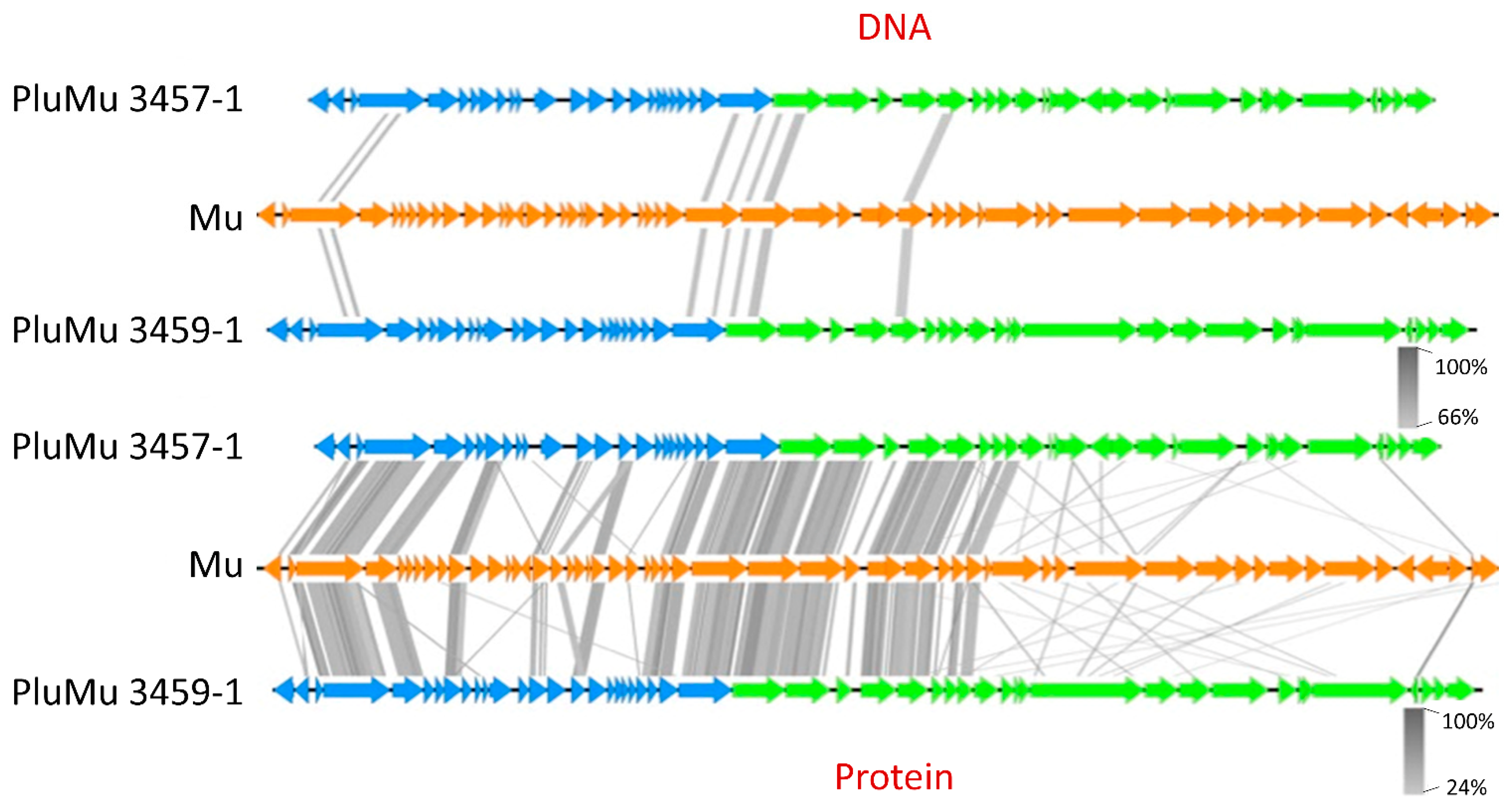
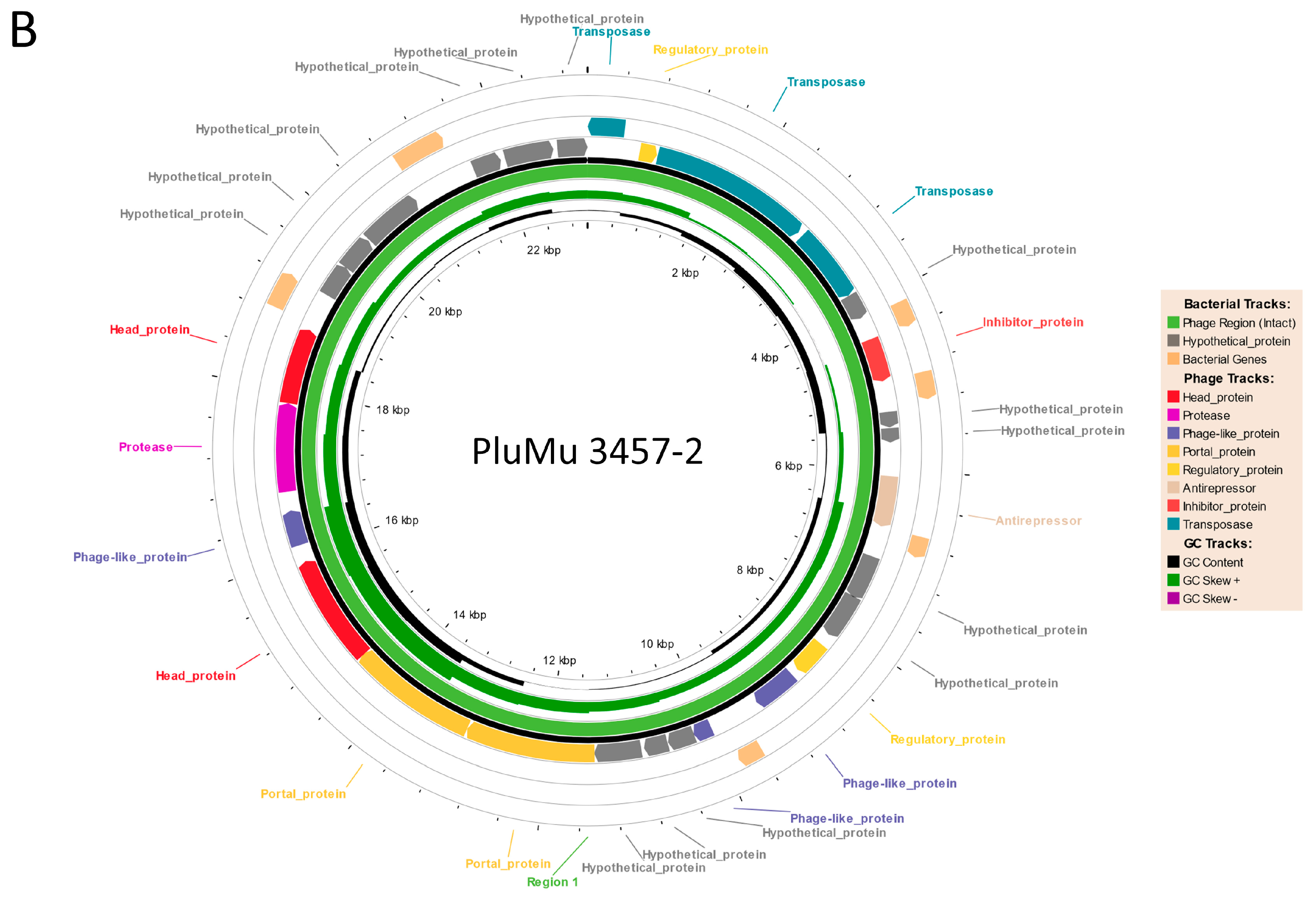
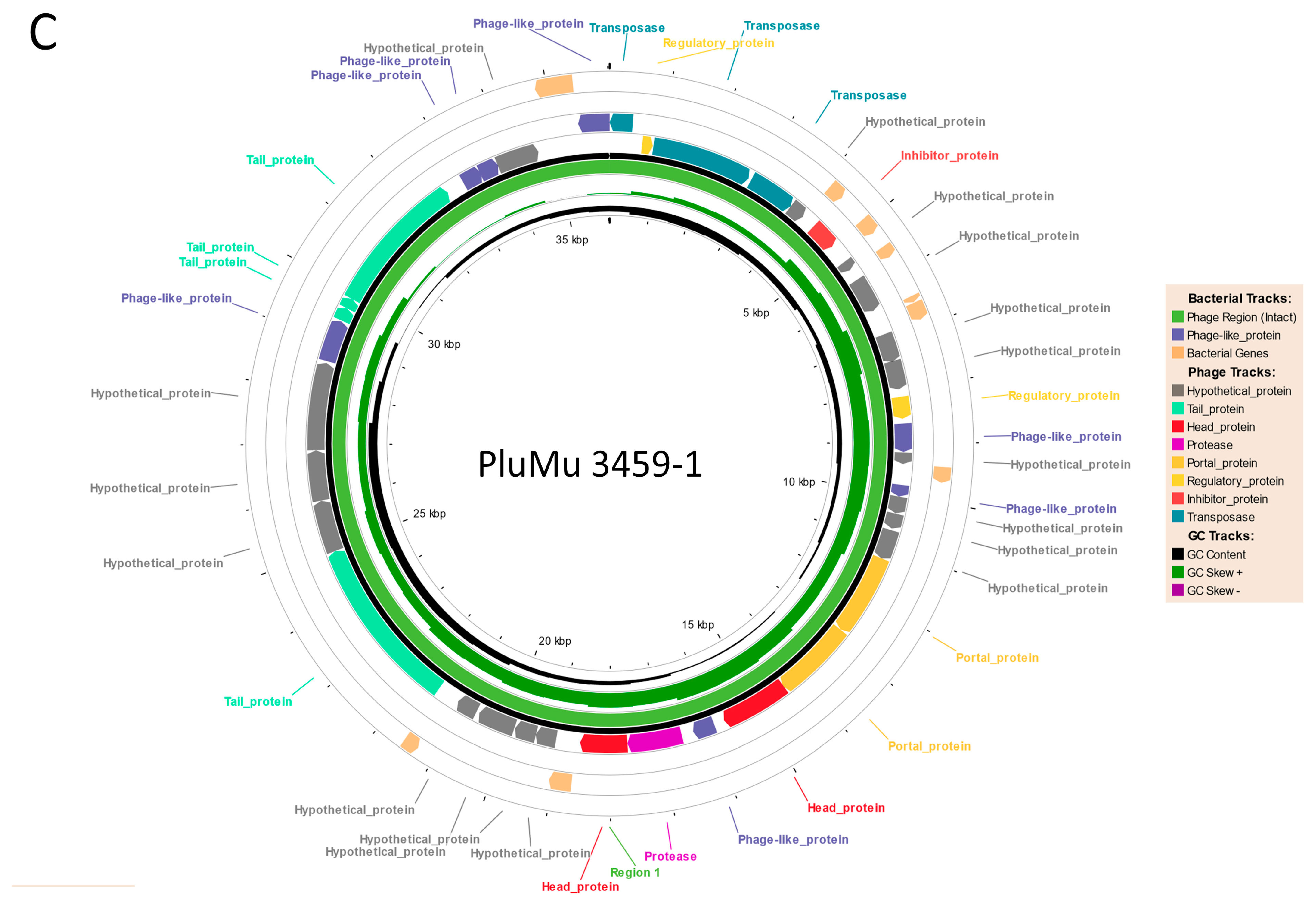
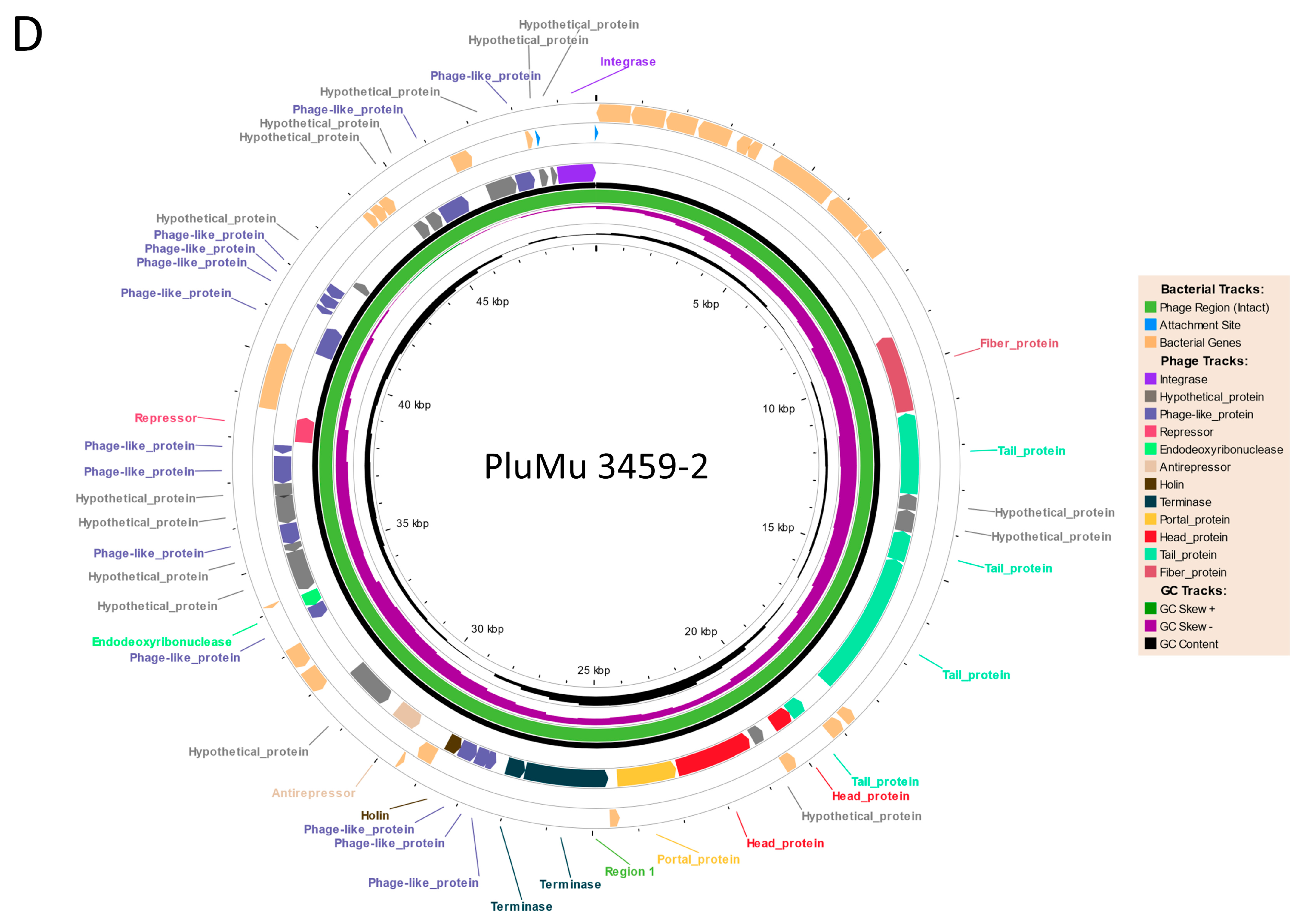
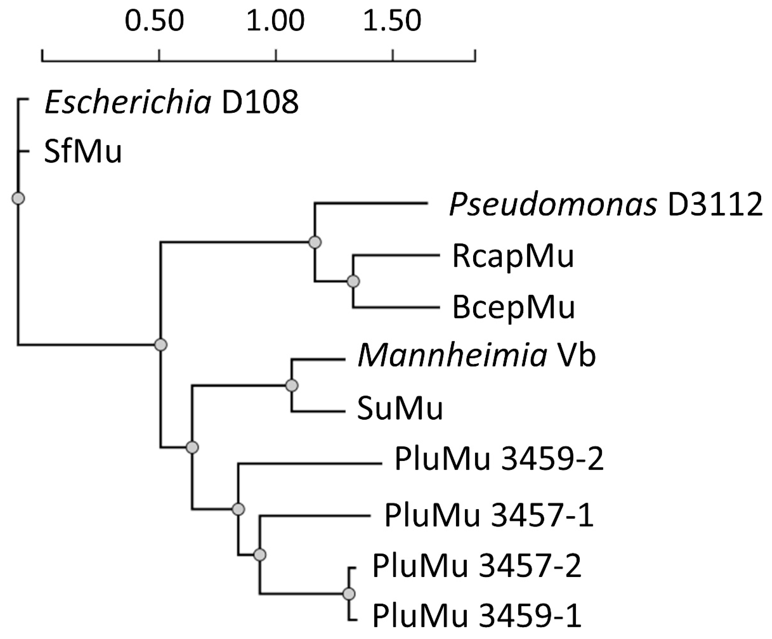
3.1. Genomic Comparisons

A summary of the results of Phastest analyses on the previously identified manually annotated phage genome sequences from 3457 and 3459 is shown in Table 2. Phastest identified two DNA regions in the 3457 genome that encode proteins with high similarity to other Mu-like phages; Region 1 (PluMu 3457-1) of 40.8 kb and Region 2 (PluMu 3457-2) of 23 kb are predicted to encode 56 and 35 proteins, respectively. In the 3459 genome, Phastest also predicted two regions encoding proteins with high similarity to other Mu-like phages; Region 1 (PluMu 3459-1) of 36 kb and Region 2 (PluMu 3459-2) of 50 kb are predicted to encode 48 and 68 proteins, respectively. Hypotheticals are the subset of phage hit proteins with significant hits with proteins in other phage genomes but are of unknown function.
All four predicted phages were ranked as “intact” (Table 2). An intact score is based on the homology to known intact phage, the identity of the proteins (e.g., head, integrase, fibre, portal, terminase, protease or lysin), the size and number of proteins in the region, and the percentage of the phage-related and hypothetical proteins in the region. The maximum score is 150, and >90 is rated as intact. All but PluMu 3457-2 (score = 100), which has the smallest locus, have scores of 140 or 150. The various PluMu phage sequences were inserted in different locations in the 3457 and 3459 genomes. For Mu-phage sequence orientations with X at the 5′ and Y at the 3′ end, the following insertion sites were found: PluMu 3457-1 is between recO, encoding a DNA repair protein and qseB; PluMu 3457-2 is between genes encoding the 50S ribosomal protein L25 and an uncharacterised protein; PluMu 3459-1 is between rseA, encoding a Sigma E negative regulatory protein [35], and an uncharacterised gene; PluMu 3459-2 interrupts the thiH gene, encoding an iminoacetate-synthetase, which is upstream of the gene encoding the 50S ribosomal protein L25. In each case, the most common blastp hits are against proteins encoded by Mu-like phages. The highest number of hits for PluMu 3457-1 (n = 9) and PluMu 3459-2 (n = 9) were against Mannheimia Vb (NC_028956), and PluMu 3457-2 (n = 18) and PluMu 3459-1 (n = 18) were against Escherichia D108 (NC_013594) Mu phages (Tables S1–S4). Whilst there is comparatively little identity at the DNA level (Figure 1 top), there is high similarity at the protein level, especially at the 5′ end (Figure 1 bottom), between the PluMu 3457-1 and PluMu 3459-1 (phages with the highest completeness score in 3457 and 3459, respectively), and reference Escherichia (AF083977) Mu phage.
Phastest circular outputs of the organisation of PluMu “intact” phage in the 3457 and 3459 genomes are shown in Figure 2A–D, with details of the predicted CDSs, top blastp hits, and significant E-values in Tables S1–S4. All have a typical Mu-like phage organisation, i.e., latency, early, and middle (region finishing downstream of the c gene), followed by the late lysis-head-tail region, with key predicted genes and proteins as detailed by Harshey [36] and the ViralZone (https://viralzone.expasy.org/4356, accessed on 9 February 2024), available through the ViralZone server [37]. These include genes encoding integrases, repressors, anti-repressors, transposases, portal proteins, terminases, and those required for tail, and head formation.
A phylogenetic tree also demonstrated their relatedness to known Mu-phages (Figure 3). All four PluMu phages were closely related to each other, especially PluMu 3457-2 and PluMu 3459-1, and then to a subbranch including Mannheimia Vb and Glaesserella SuMu, the parent bacteria of which, like A. pleuropneumoniae, are members of the Pasteurellaceae. Thus, the Phastest genomic analyses indicated the presence of two phages each in 3457 and 3459, with characteristic Mu-like phage organisation and intact completeness scores indicating their ability to produce active virions.

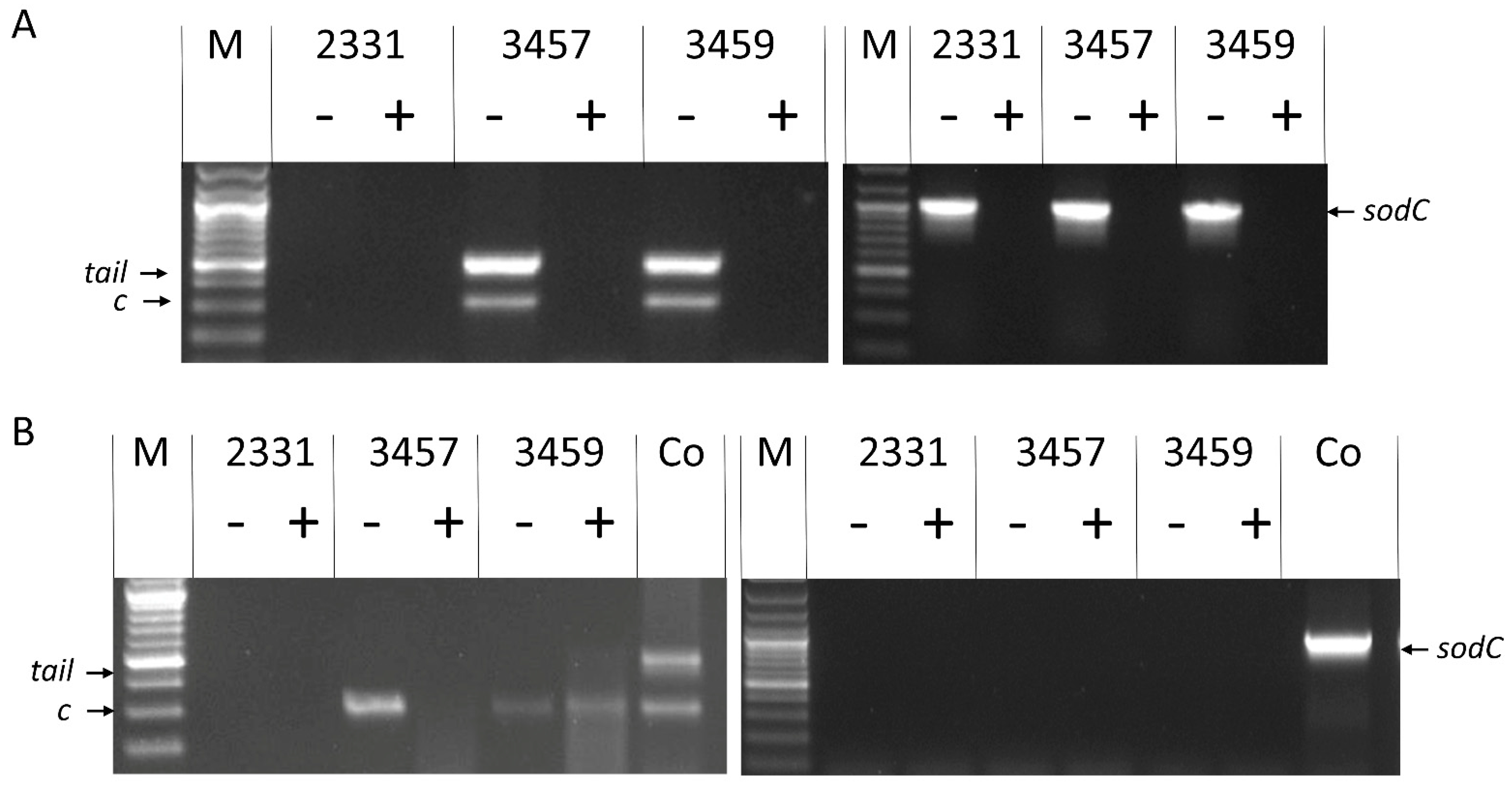
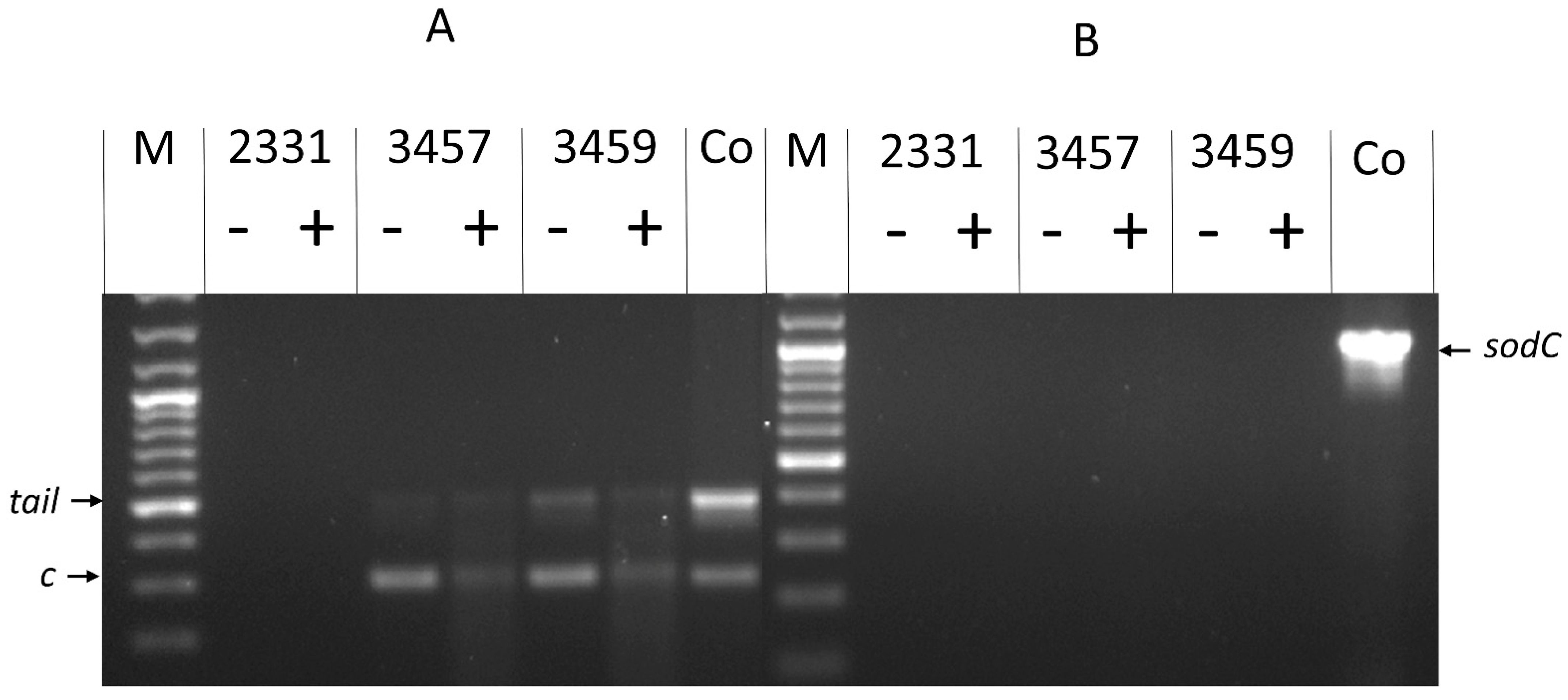
3.2. Molecular Analyses Indicate That 3457 and 3459 Produce Active Phage

Genome analysis established the presence of Mu-like prophages in the genomes of 3457 and 3459. Initially, we used a molecular biology approach to screen for the production of PluMu. Three pairs of PCR primers were designed: two targeting PluMu genes and one a non-PluMu gene in the 3457 and 3459 genomes. Specifically, the PluMu genes targeted were those encoding the late transcriptional activator protein C which is important for the expression of morphogenic structures [38] and encoding a tail protein. An A. pleuropneumoniae sodC-asd (hereafter referred to as sodC) amplicon [34] was used as a control to indicate the presence of bacterial gDNA. Since no amplification of sodC occurred in the presence of the tail and c gene primers, sodC amplifications were performed separately on the same samples. The rationale was that the DNA within intact, active virions would be protected against DNase treatment, whereas any contaminating bacterial genomic DNA (detectable by sodC amplification) would be digested, so amplification of the c and tail genes, but not sodC, from DNase-treated PEG-precipitated samples would indicate the presence of active virions. PCR was performed on sample preparations of 3457, 3459, and 2331; on isolated gDNA (Figure 4A) and on the PEG-precipitated filtered supernatants from growth in BHI-NAD (Figure 4B). Initial PCRs established that the PluMu c (264 bp) and tail (448 bp) genes were amplified when gDNA from 3457 and 3459, but not 2331 (Figure 4A left panel), was used as a template, whereas sodC was amplified from gDNA of all three isolates (Figure 4A right panel). In all cases, no amplicons were found after DNase treatment.
To varying degrees, there is an amplification of the c and tail genes from the PEG-precipitated samples from 3457 and 3459, both before and after DNase treatment (Figure 4B left panel), indicating the presence of intact virions protecting the DNA, but no sodC amplification (Figure 4B right panel) either before or after DNase treatment, indicating no contaminating gDNA was present in the samples, even before DNase treatment.
The presence of c and tail, and the absence of sodC, amplicons in DNase-treated PEG-precipitated supernatants suggested the presence of phage particles. We also evaluated the growth of all three isolates in Grace’s Insect Medium-NAD, the rationale being that it was both defined and clear, characteristics useful for reproducibility and observing phage by TEM, respectively. All three strains grew in NAD-supplemented Grace’s Insect Medium, although not to the same extent as in BHI-NAD broth (Figure 5). The presence of active phage particles in 3457 and 3459 was, again, indicated by the same c, tail (Figure 6 left panel), and sodC (Figure 6 right panel) PCR amplification results as obtained with BHI-NAD broth-grown bacteria.

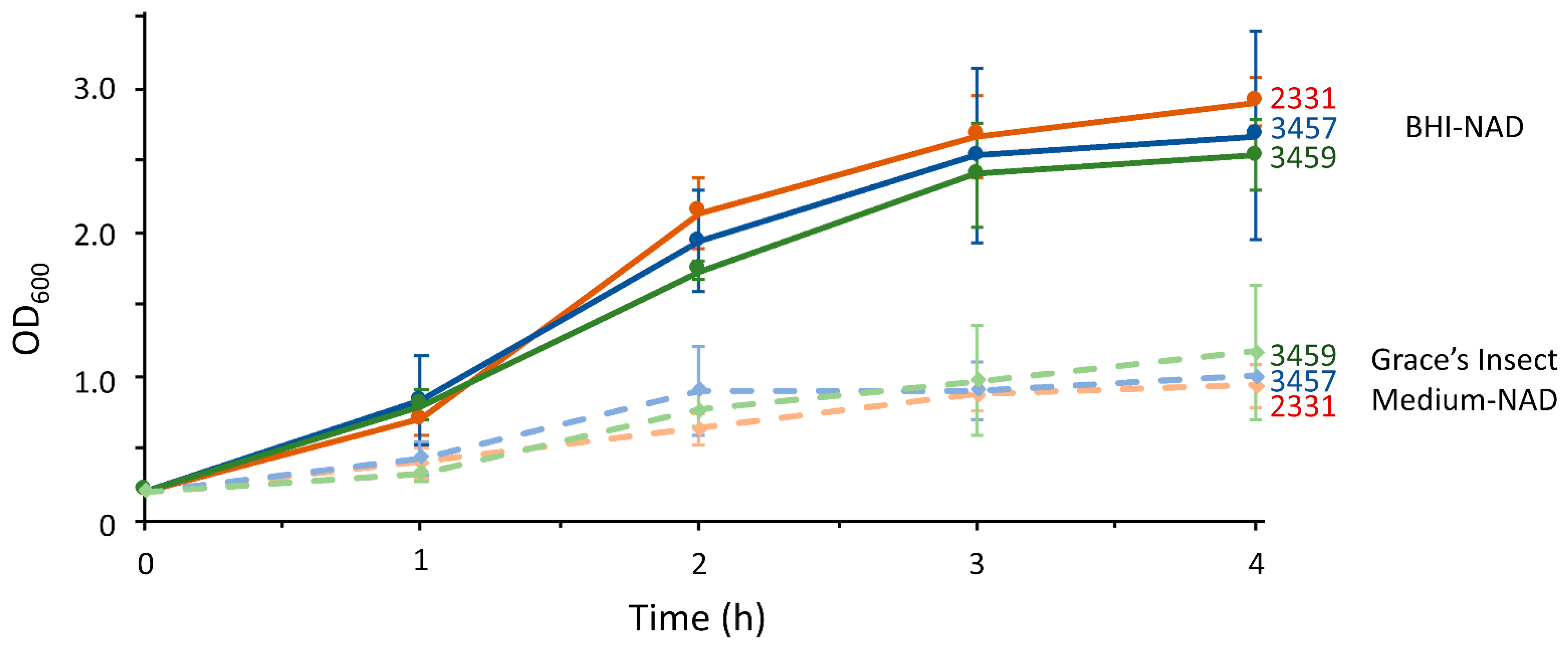
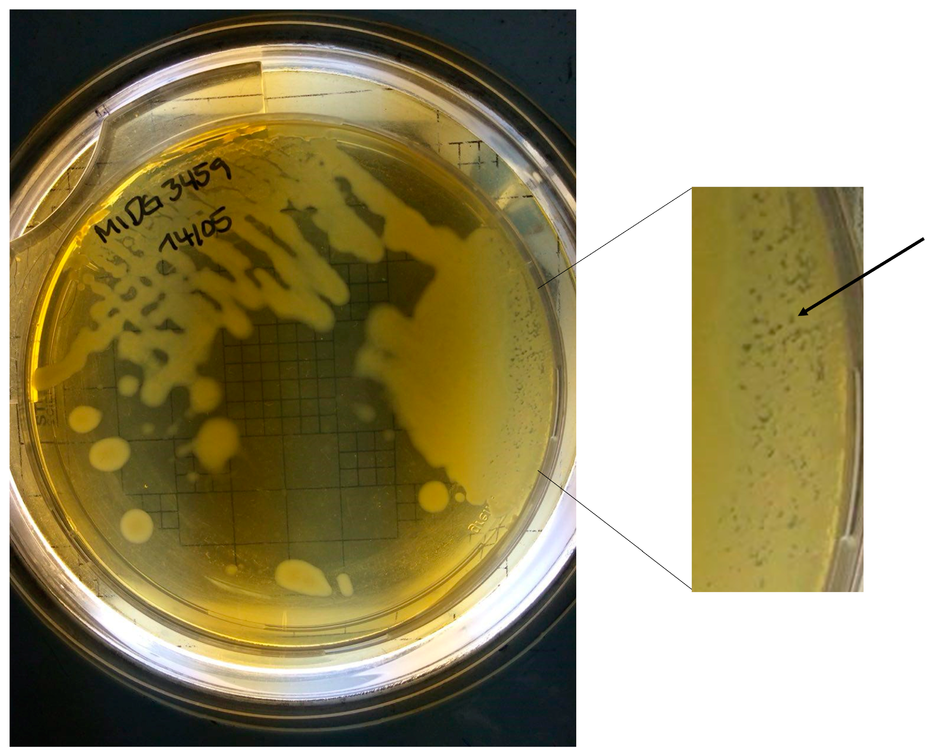
3.3. Plaque Formation in 3459 Grown on a BHI-NAD Plate

We also noticed plaque formation of 3459, but not 3457, when cultures were streaked out on BHI-NAD agar plates (Figure 7) and incubated overnight, indicating the presence of active phage. No such plaque formation was found with 2331. Indeed, we have not observed plaque formation of any A. pleuropneumoniae isolate in our collection (n = c. 600) on BHI-NAD or any other agar plates.

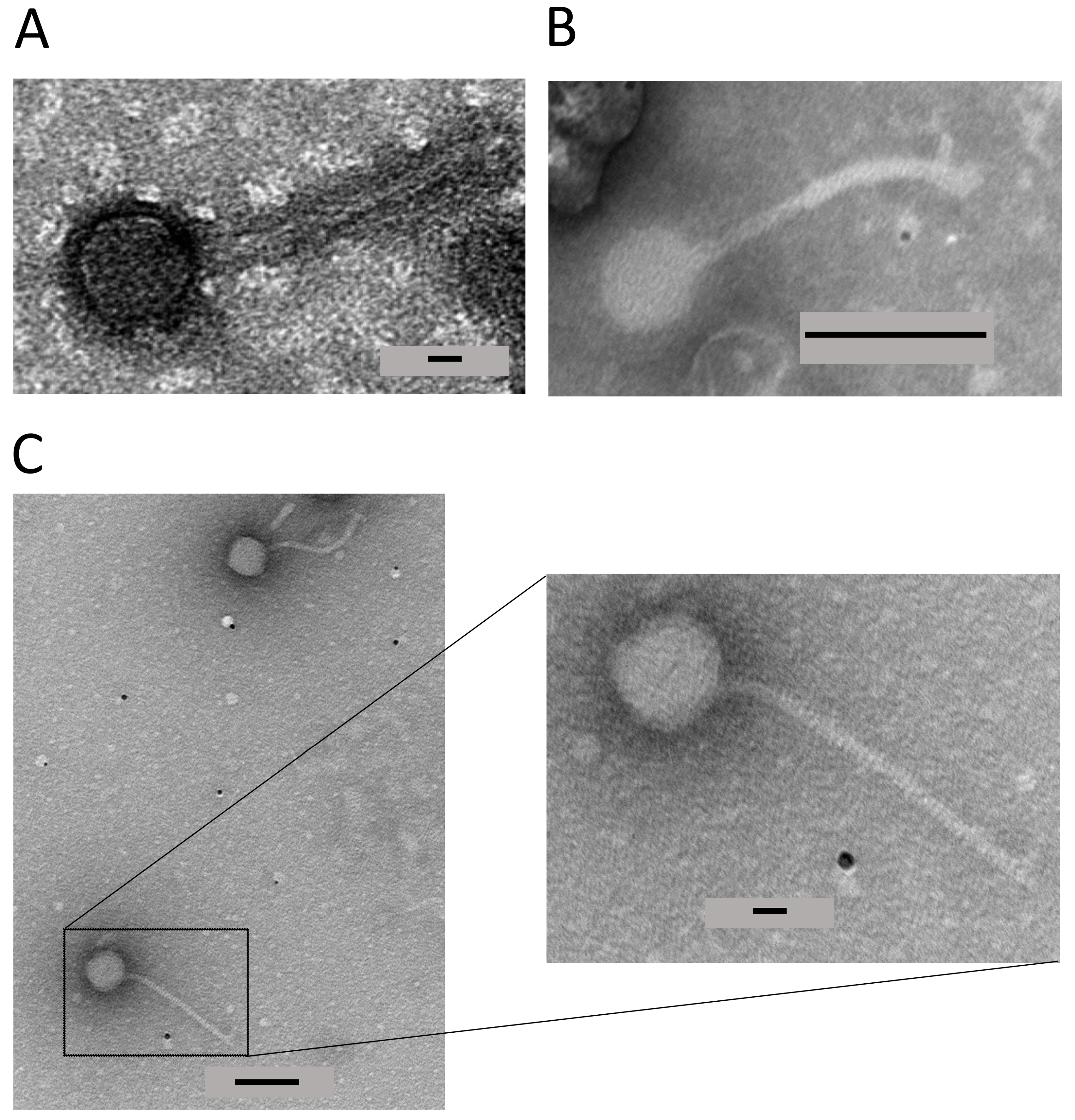
3.4. TEM Identifies a Mu-like Phage in 3459

Because of time and resource constraints and the observation of visible plaques on BHI + NAD plates, the TEM analysis focused on 3459. Initial experiments analysed samples from 3459 grown in BHI-NAD broth as the molecular analyses indicated the presence of phage (Section 3.2). Intact particles were observed but there was a high background (Figure 8A). Thus, 3459 was subsequently grown in Grace’s Insect Medium-NAD, as molecular analyses had also indicated the presence of intact phage (Section 3.2). Mu-like phage particles were found visible in PEG-precipitated supernatants (Figure 8A–C). The virion enlarged in Figure 8C insert had a head diameter of 62.7 nm, a tail length of 202 nm, and a tail diameter of 9.4 nm The phage particles had classic Mu-like phage tail-like structures attached to icosahedral-like heads. No phage particles were found with similarly derived samples from 2331 grown in either medium.

4. Discussion

The starting point for this study was preliminary manual annotation indicating the presence of Mu-like phage genomes in two Cypriot isolates of A. pleuropneumoniae, 3457 and 3459 [21]. Here, we have updated that molecular analysis and provided further evidence that active Mu-like phages, which we have named PluMu, are present in these isolates. In the original manual annotation [21], only one Mu-like phage was identified in each of 3457 and 3459. Updated Phastest analyses, however, identified two Mu-like phages in each isolate. Each phage identified was ranked as “intact”, indicative of producing active phage. While their identity at the nucleotide level was poor, high levels of identity were seen at the protein level, especially at the 5′ ends, as illustrated by the analysis of PluMu 3457-1 and 3457-2 (Figure 1). Variation in the 3′ region in Mu-like phages is a common feature, as the encoded proteins are involved in phage-specific functions, including tail formation and attachment. One of the genes that showed similarity to known Mu-phage (by both blastn and tblastn comparisons) was that encoding the portal protein gp29 homolog (PluMu 3457-1 = CDS 31, PluMu 3457-2 = CDS 19; PluMu 3459-1 = CDS 19; PluMu 3459-2 = CDS 11). The presence of this gene has been reported as a potential marker of virulent isolates and reference strains in G. parasuis [39], the bacterium where SuMu was described [24]. Further analysis will be required to determine if the PluMu gp29 homolog (or any other PluMu gene) is similarly a marker of virulent isolates. Phastest did not predict an attachment site for PluMu 3457 or PluMu 3459, unsurprising given the lack of knowledge of A. pleuropneumoniae phage biology. Others have used Phaster [20], or another phage identification program Prophage Hunter [19], to search A. pleuropneumoniae genomes for complete and/or active phage. Whole genome analysis identified 13 “complete” [20], and 21 “active” phages, with 18 being most closely related to Mannheimia Vb [19]. In the latter study, isolate origins included Australia, Asia, Europe, and North and South America, and comprised a range of serovars, i.e., 1–2, 4–8, and 10–11. All the serovar 11 (like 3457) isolates predicted to have active phage were from Europe, although the serovar 1 (like 3459) isolate was from China [19]. Whilst neither of these studies has shown active phage, our results suggest that the presence of Mu-like phage, especially ones related to Mannheimia Vb, is comparatively common in A. pleuropneumoniae genomes. Serovar 11 and 1 isolates are considered of high virulence by virtue of expression of ApxI and ApxII [4] and 3457 and 3459 were clinical isolates derived from the lungs of acutely infected pigs. Whether harbouring active PluMu makes an additional contribution to virulence remains to be determined.
To determine whether 3457 or 3459 harboured active phage, as the original manual annotation and Phastest and other genomic analyses had indicated, we initially used a molecular biology approach. This involved the PCR amplification of two PluMu genes (c and tail) and A. pleuropneumoniae sodC-asd sequences as markers of PluMu or bacterial gDNA, respectively. Despite the high specificity in PCR amplification, multiplexing all three primer pairs was unsuccessful, despite balancing melting temperatures, and checking for potential cross-dimers and sufficient size differences in amplicon size. There was some preferential amplification of the c gene in samples grown in Grace’s Insect Medium-NAD compared to BHI-NAD, but overall PCR analyses were reproducible across a range of different sample preparations. There was the amplification of c and tail genes in 3457 and 3459 PEG-precipitated supernatants derived from BHI-NAD and Grace’s Insect Medium-NAD media even after DNase treatment. There was no amplification of sodC from these samples, indicating they were free of bacterial gDNA. The results strongly suggested the presence of active PluMu, with their virion-enclosed genomes being protected from DNase treatment. To our knowledge, this is the first description of the use of defined Grace’s Insect Medium-NAD to grow A. pleuropneumoniae. Many groups have used Herriott’s defined medium [40] to grow A. pleuropneumoniae or derivatives, e.g., where the amino acid stock solution adapted from the Neisseria defined medium [41,42,43] was substituted for Herriott’s amino acid stock solution [44]. These media are comparatively more complex to make than Grace’s Insect Medium-NAD. Whilst the growth of A. pleuropneumoniae was not as good as in conventional BHI-NAD broth (Figure 5), further work to increase the growth rate and/or yield of cultures grown in Grace’s Insect Medium may be valuable, given the advantages of defined media in identifying gene and protein responses to changing environments.
In this study, the use of Grace’s Insect Medium was invaluable in the TEM analysis. PluMu 3459 virions were observed in PEG-precipitated supernatants derived from BHI-NAD-grown bacteria, but the background was high (Figure 8). PluMu particles were visible in PEG-precipitated supernatants derived from 3459 grown in Grace’s Insect Medium (Figure 8B,C). The isohedral heads and long tails are classic for Mu-like phage. There is some variation in the head diameter and tail length, but those identified for PluMu 3459 are comparable to those reported for other Mu-like phages. For example, the head diameter measurement of ~62 nm is similar to bacteriophage Mu (54 nm). However, its tail length of ~200 nm is significantly longer than that of Mu (100 nm), but comparable to BcepMu (220 nm) [45] and RcapMu (>200 nm) [46]. We are unable to categorically say whether both (or just one of) PluMu 3459-1 and PluMu 3459-2 are active phages. This question could be answered by mutation of key genes, e.g., those involved in head and/or tail formation. However, 3459, a particularly mucoid isolate, was refractory to mutant construction via conventional electroporation or natural transformation, methods we have used widely or developed [34,35,47,48]. In summary, the combination of genome and molecular analyses is consistent with the PluMu phage in 3457 and 3459 being active, confirmed by TEM for 3459.
There has been a resurgence of interest in the use of phage therapy as a strategy to treat bacterial infections, especially those caused by AMR bacteria [49]. A. pleuropneumoniae AMR is an increasing problem [9]. Additionally, eliminating A. pleuropneumoniae from herds is notoriously difficult, where complete depopulation and repopulation of a herd and deep cleansing may be required [1]. It is particularly difficult to eliminate A. pleuropneumoniae which are colonising porcine tonsillar crypts, and carrier animals can be the source of acute infections in naive herds. The recognition of phage in A. pleuropneumoniae raises the possibility of their use to treat infection caused by AMR isolates and for eliminating tonsillar carriage, and for cleaning farms, e.g., in depopulation/repopulation approaches, where inefficient cleaning is known to have considerable economic significance [50]. The biofilm mode of growth is considered as important for the growth of the bacterium in the lungs and the environment [6], and a potential role for phage in reducing or eliminating A. pleuropneumoniae in these niches remains to be determined. However, Mu-like phages, like PluMu, are temperate; lytic phages are used for therapy, and none have been described in A. pleuropneumoniae. With 19 serovars of A. pleuropneumoniae, it is also likely that a cocktail of phages would be required for therapy and or environmental decontamination. Nonetheless, the identification of PluMu in different serovars and countries, as in this and other studies [19,20], indicates that active A. pleuropneumoniae lytic phage may be found in future studies. Alternative approaches, such as the use of constitutively lytic phage-plasmids for therapeutic use, may also be possible in the future [51].

Supplementary Materials

The following supporting information can be downloaded at: https://www.mdpi.com/article/10.3390/applmicrobiol4010037/s1, Phastest outputs showing CDS position, top blastp hits, and E-values for PluMu phage: Table S1: PluMu 3457-1; Table S2: PluMu 3457-2; Table S3: PluMu 3459-1; Table S4: PluMu 3459-2.

Author Contributions

Conceptualisation, M.A.S., A.N.R., J.T.B., Y.L. and P.R.L.; methodology, L.J.B., R.F.C., Y.W., M.A.S., W.C., D.J.E., Y.L., J.T.B. and P.R.L.; validation, L.J.B., R.F.C., Y.W. and J.T.B.; formal analysis, L.J.B., R.F.C., Y.W., Y.L. and J.T.B.; investigation, L.J.B., R.F.C., Y.W., W.C., D.J.E., Y.L. and J.T.B.; resources, M.A.S., A.N.R. and P.R.L.; data curation, L.J.B., R.F.C., Y.W., J.T.B. and P.R.L.; writing—original draft preparation, P.R.L.; writing—review and editing, all; visualisation, all; supervision, M.A.S., A.N.R., J.T.B., Y.L. and P.R.L.; project administration, P.R.L.; funding acquisition, M.A.S., A.N.R. and P.R.L. All authors have read and agreed to the published version of the manuscript.

Funding

This research was funded by the Biotechnology and Biological Sciences Research Council, grant numbers BB/S002103/1, BB/S005897/1, and, in part, BB/G018553/1, and BB/G020744/1.

Data Availability Statement

The data supporting the results and the MSc thesis of R.F.C. is freely available from the corresponding author. The Phastest outputs can also be obtained from the Phastest web server (last accessed 4 March 2024): PluMu 3457-1; https://phastest.ca/submissions/ZZ_6bde62ff01: PluMu 3457-2; https://phastest.ca/submissions/ZZ_48e9dfb4fc: PluMu 3459-1; https://phastest.ca/submissions/ZZ_7a9eb7ffd9: PluMu 3459-2; https://phastest.ca/submissions/ZZ_debae37127.

Conflicts of Interest

The authors declare no conflicts of interest. The funders had no role in the design of the study; in the collection, analyses, or interpretation of data; in the writing of the manuscript; or in the decision to publish the results.

References

  1. Sassu, E.L.; Bossé, J.T.; Tobias, T.J.; Gottschalk, M.; Langford, P.R.; Hennig-Pauka, I. Update on Actinobacillus pleuropneumoniae-knowledge, gaps and challenges. Transbound. Emerg. Dis. 2018, 65 (Suppl. 1), 72–90. [Google Scholar] [CrossRef] [PubMed]
  2. Bossé, J.T.; Janson, H.; Sheehan, B.J.; Beddek, A.J.; Rycroft, A.N.; Kroll, J.S.; Langford, P.R. Actinobacillus pleuropneumoniae: Pathobiology and pathogenesis of infection. Microbes Infect. 2002, 4, 225–235. [Google Scholar] [CrossRef] [PubMed]
  3. Chiers, K.; De Waele, T.; Pasmans, F.; Ducatelle, R.; Haesebrouck, F. Virulence factors of Actinobacillus pleuropneumoniae involved in colonization, persistence and induction of lesions in its porcine host. Vet. Res. 2010, 41, 65. [Google Scholar] [CrossRef]
  4. Frey, J. Virulence in Actinobacillus pleuropneumoniae and RTX toxins. Trends Microbiol. 1995, 3, 257–261. [Google Scholar] [CrossRef] [PubMed]
  5. Gottschalk, M. The challenge of detecting herds sub-clinically infected with Actinobacillus pleuropneumoniae. Vet. J. 2015, 206, 30–38. [Google Scholar] [CrossRef]
  6. Hathroubi, S.; Loera-Muro, A.; Guerrero-Barrera, A.L.; Tremblay, Y.D.N.; Jacques, M. Actinobacillus pleuropneumoniae biofilms: Role in pathogenicity and potential impact for vaccination development. Anim. Health Res. Rev. 2018, 19, 17–30. [Google Scholar] [CrossRef]
  7. Langford, P.R.; Stringer, O.W.; Li, Y.; Bossé, J.T. Application of the MISTEACHINGS disease susceptibility framework to Actinobacillus pleuropneumoniae to identify research gaps: An exemplar of a veterinary pathogen. Anim. Health Res. Rev. 2021, 22, 120–135. [Google Scholar] [CrossRef]
  8. Loera-Muro, A.; Angulo, C. New trends in innovative vaccine development against Actinobacillus pleuropneumoniae. Vet. Microbiol. 2018, 217, 66–75. [Google Scholar] [CrossRef]
  9. Michael, G.B.; Bossé, J.T.; Schwarz, S. Antimicrobial resistance in Pasteurellaceae of veterinary origin. Microbiol. Spectr. 2018, 6. [Google Scholar] [CrossRef]
  10. Nahar, N.; Turni, C.; Tram, G.; Blackall, P.J.; Atack, J.M. Actinobacillus pleuropneumoniae: The molecular determinants of virulence and pathogenesis. Adv. Microb. Physiol. 2021, 78, 179–216. [Google Scholar] [CrossRef]
  11. Soto Perezchica, M.M.; Guerrero Barrera, A.L.; Avelar Gonzalez, F.J.; Quezada Tristan, T.; Macias Marin, O. Actinobacillus pleuropneumoniae, surface proteins and virulence: A review. Front. Vet. Sci. 2023, 10, 1276712. [Google Scholar] [CrossRef] [PubMed]
  12. Stringer, O.W.; Li, Y.; Bossé, J.T.; Langford, P.R. JMM Profile: Actinobacillus pleuropneumoniae: A major cause of lung disease in pigs but difficult to control and eradicate. J. Med. Microbiol. 2022, 71, 001483. [Google Scholar] [CrossRef] [PubMed]
  13. Boeters, M.; Garcia-Morante, B.; van Schaik, G.; Segales, J.; Rushton, J.; Steeneveld, W. The economic impact of endemic respiratory disease in pigs and related interventions—A systematic review. Porcine Health Manag. 2023, 9, 45. [Google Scholar] [CrossRef] [PubMed]
  14. Saade, G.; Deblanc, C.; Bougon, J.; Marois-Crehan, C.; Fablet, C.; Auray, G.; Belloc, C.; Leblanc-Maridor, M.; Gagnon, C.A.; Zhu, J.; et al. Coinfections and their molecular consequences in the porcine respiratory tract. Vet. Res. 2020, 51, 80. [Google Scholar] [CrossRef]
  15. Stringer, O.W.; Bossé, J.T.; Lacouture, S.; Gottschalk, M.; Fodor, L.; Angen, Ø.; Velazquez, E.; Penny, P.; Lei, L.; Langford, P.R.; et al. Proposal of Actinobacillus pleuropneumoniae serovar 19, and reformulation of previous multiplex PCRs for capsule-specific typing of all known serovars. Vet. Microbiol. 2021, 255, 109021. [Google Scholar] [CrossRef]
  16. da Silva, G.C.; Gonçalves, O.S.; Rosa, J.N.; França, K.C.; Bossé, J.T.; Santana, M.F.; Langford, P.R.; Bazzolli, D.M.S. Mobile genetic elements drive antimicrobial resistance gene spread in Pasteurellaceae species. Front. Microbiol. 2021, 12, 773284. [Google Scholar] [CrossRef]
  17. Arndt, D.; Grant, J.R.; Marcu, A.; Sajed, T.; Pon, A.; Liang, Y.; Wishart, D.S. PHASTER: A better, faster version of the PHAST phage search tool. Nucleic Acids Res. 2016, 44, W16–W21. [Google Scholar] [CrossRef]
  18. Song, W.; Sun, H.X.; Zhang, C.; Cheng, L.; Peng, Y.; Deng, Z.; Wang, D.; Wang, Y.; Hu, M.; Liu, W.; et al. Prophage Hunter: An integrative hunting tool for active prophages. Nucleic Acids Res. 2019, 47, W74–W80. [Google Scholar] [CrossRef] [PubMed]
  19. Guitart-Matas, J.; Gonzalez-Escalona, N.; Maguire, M.; Vilaro, A.; Martinez-Urtaza, J.; Fraile, L.; Migura-Garcia, L. Revealing genomic insights of the unexplored porcine pathogen Actinobacillus pleuropneumoniae using whole genome sequencing. Microbiol. Spectr. 2022, 10, e0118522. [Google Scholar] [CrossRef] [PubMed]
  20. Prado, I.G.O.; da Silva, G.C.; Crispim, J.S.; Vidigal, P.M.P.; Nascimento, M.; Santana, M.F.; Bazzolli, D.M.S. Comparative genomics of Actinobacillus pleuropneumoniae serotype 8 reveals the importance of prophages in the genetic variability of the species. Int. J. Genom. 2020, 2020, 9354204. [Google Scholar] [CrossRef]
  21. Crespo Fernandez, R. PluMu: A Mu-like Bacteriophage Infecting the Major Pig Pathogen Actinobacillus pleuropneumoniae. MSc Thesis, Imperial College, London, UK, 2015. [Google Scholar]
  22. Taylor, A.L. Bacteriophage-induced mutation in Escherichia coli. Proc. Natl. Acad. Sci. USA 1963, 50, 1043–1051. [Google Scholar] [CrossRef]
  23. Jakhetia, R.; Verma, N.K. Identification and molecular characterisation of a novel Mu-Like bacteriophage, SfMu, of Shigella flexneri. PLoS ONE 2015, 10, e0124053. [Google Scholar] [CrossRef]
  24. Zehr, E.S.; Tabatabai, L.B.; Bayles, D.O. Genomic and proteomic characterization of SuMu, a Mu-like bacteriophage infecting Haemophilus parasuis. BMC Genom. 2012, 13, 331. [Google Scholar] [CrossRef] [PubMed]
  25. Hsu, Y.H.; Cook, S.R.; Alexander, T.W.; Klima, C.L.; Niu, Y.D.; Selinger, L.B.; McAllister, T.A. Investigation of Mannheimia haemolytica bacteriophages relative to host diversity. J. Appl. Microbiol. 2013, 114, 1592–1603. [Google Scholar] [CrossRef] [PubMed]
  26. Bossé, J.T.; Chaudhuri, R.R.; Li, Y.; Leanse, L.G.; Fernandez Crespo, R.; Coupland, P.; Holden, M.T.; Bazzolli, D.M.; Maskell, D.J.; Tucker, A.W.; et al. Complete genome sequence of MIDG2331, a genetically tractable serovar 8 clinical isolate of Actinobacillus pleuropneumoniae. Genome Announc. 2016, 4, e01667-15. [Google Scholar] [CrossRef]
  27. Grace, T.D. Establishment of four strains of cells from insect tissues grown in vitro. Nature 1962, 195, 788–789. [Google Scholar] [CrossRef] [PubMed]
  28. Wishart, D.S.; Han, S.; Saha, S.; Oler, E.; Peters, H.; Grant, J.R.; Stothard, P.; Gautam, V. PHASTEST: Faster than PHASTER, better than PHAST. Nucleic Acids Res. 2023, 51, W443–W450. [Google Scholar] [CrossRef] [PubMed]
  29. Altschul, S.F.; Lipman, D.J. Protein database searches for multiple alignments. Proc. Natl. Acad. Sci. USA 1990, 87, 5509–5513. [Google Scholar] [CrossRef] [PubMed]
  30. Sullivan, M.J.; Petty, N.K.; Beatson, S.A. Easyfig: A genome comparison visualizer. Bioinformatics 2011, 27, 1009–1010. [Google Scholar] [CrossRef]
  31. Lemoine, F.; Correia, D.; Lefort, V.; Doppelt-Azeroual, O.; Mareuil, F.; Cohen-Boulakia, S.; Gascuel, O. NGPhylogeny.fr: New generation phylogenetic services for non-specialists. Nucleic Acids Res. 2019, 47, W260–W265. [Google Scholar] [CrossRef]
  32. Guindon, S.; Gascuel, O. A simple, fast, and accurate algorithm to estimate large phylogenies by maximum likelihood. Syst. Biol. 2003, 52, 696–704. [Google Scholar] [CrossRef]
  33. Sun, W.; Hattman, S.; Kool, E. Interaction of the bacteriophage Mu transcriptional activator protein, C, with its target site in the mom promoter. J. Mol. Biol. 1997, 273, 765–774. [Google Scholar] [CrossRef]
  34. Langford, P.R.; Loynds, B.M.; Kroll, J.S. Cloning and molecular characterization of Cu,Zn superoxide dismutase from Actinobacillus pleuropneumoniae. Infect. Immun. 1996, 64, 5035–5041. [Google Scholar] [CrossRef] [PubMed]
  35. Bossé, J.T.; Sinha, S.; Li, M.S.; O’Dwyer, C.A.; Nash, J.H.; Rycroft, A.N.; Kroll, J.S.; Langford, P.R. Regulation of pga operon expression and biofilm formation in Actinobacillus pleuropneumoniae by sigmaE and H-NS. J. Bacteriol. 2010, 192, 2414–2423. [Google Scholar] [CrossRef] [PubMed]
  36. Harshey, R.M. Transposable phage Mu. Microbiol. Spectr. 2014, 2. [Google Scholar] [CrossRef]
  37. De Castro, E.; Hulo, C.; Masson, P.; Auchincloss, A.; Bridge, A.; Le Mercier, P. ViralZone 2024 provides higher-resolution images and advanced virus-specific resources. Nucleic Acids Res. 2024, 52, D817–D821. [Google Scholar] [CrossRef] [PubMed]
  38. Margolin, W.; Rao, G.; Howe, M.M. Bacteriophage Mu late promoters: Four late transcripts initiate near a conserved sequence. J. Bacteriol. 1989, 171, 2003–2018. [Google Scholar] [CrossRef] [PubMed]
  39. Zehr, E.S.; Tabatabai, L.B. Detection of a bacteriophage gene encoding a Mu-like portal protein in Haemophilus parasuis reference strains and field isolates by nested polymerase chain reaction. J. Vet. Diagn. Investig. 2011, 23, 538–542. [Google Scholar] [CrossRef]
  40. Herriott, R.M.; Meyer, E.Y.; Vogt, M.; Modan, M. Defined medium for growth of Haemophilus influenzae. J. Bacteriol. 1970, 101, 513–516. [Google Scholar] [CrossRef] [PubMed]
  41. Hodgetts, A.; Bossé, J.T.; Kroll, J.S.; Langford, P.R. Analysis of differential protein expression in Actinobacillus pleuropneumoniae by Surface Enhanced Laser Desorption Ionisation--ProteinChip (SELDI) technology. Vet. Microbiol. 2004, 99, 215–225. [Google Scholar] [CrossRef]
  42. Ingham, A.; Zhang, Y.; Prideaux, C. Attenuation of Actinobacillus pleuropneumoniae by inactivation of aroQ. Vet. Microbiol. 2002, 84, 263–273. [Google Scholar] [CrossRef] [PubMed]
  43. Niven, D.F.; Levesque, M. V-Factor-dependent growth of Actinobacillus pleuropneumoniae biotype 2 (Bertschinger 2008/76). Int. J. Syst. Bacteriol. 1988, 38, 319–320. [Google Scholar] [CrossRef]
  44. Wagner, T.K.; Mulks, M.H. Identification of the Actinobacillus pleuropneumoniae leucine-responsive regulatory protein and its involvement in the regulation of in vivo-induced genes. Infect. Immun. 2007, 75, 91–103. [Google Scholar] [CrossRef]
  45. Summer, E.J.; Gonzalez, C.F.; Carlisle, T.; Mebane, L.M.; Cass, A.M.; Savva, C.G.; LiPuma, J.; Young, R. Burkholderia cenocepacia phage BcepMu and a family of Mu-like phages encoding potential pathogenesis factors. J. Mol. Biol. 2004, 340, 49–65. [Google Scholar] [CrossRef]
  46. Fogg, P.C.; Hynes, A.P.; Digby, E.; Lang, A.S.; Beatty, J.T. Characterization of a newly discovered Mu-like bacteriophage, RcapMu, in Rhodobacter capsulatus strain SB1003. Virology 2011, 421, 211–221. [Google Scholar] [CrossRef] [PubMed]
  47. Bossé, J.T.; Nash, J.H.; Kroll, J.S.; Langford, P.R. Harnessing natural transformation in Actinobacillus pleuropneumoniae: A simple method for allelic replacements. FEMS Microbiol. Lett. 2004, 233, 277–281. [Google Scholar] [CrossRef]
  48. Bossé, J.T.; Soares-Bazzolli, D.M.; Li, Y.; Wren, B.W.; Tucker, A.W.; Maskell, D.J.; Rycroft, A.N.; Langford, P.R.; BRaDP1T Consortium. The generation of successive unmarked mutations and chromosomal insertion of heterologous genes in Actinobacillus pleuropneumoniae using natural transformation. PLoS ONE 2014, 9, e111252. [Google Scholar] [CrossRef]
  49. Zalewska-Piatek, B. Phage therapy-challenges, opportunities and future prospects. Pharmaceuticals 2023, 16, 1638. [Google Scholar] [CrossRef]
  50. Stygar, A.H.; Niemi, J.K.; Oliviero, C.; Laurila, T.; Heinonen, M. Economic value of mitigating Actinobacillus pleuropneumoniae infections in pig fattening herds. Agric. Syst. 2016, 144, 113–121. [Google Scholar] [CrossRef]
  51. Shan, X.; Szabo, R.E.; Cordero, O.X. Mutation-induced infections of phage-plasmids. Nat. Commun. 2023, 14, 2049. [Google Scholar] [CrossRef]
Figure 1. Sequence comparison of the reference Escherichia Mu bacteriophage (AF083977 and PluMu 3457-1 and 3459-1. Comparison is made at the nucleotide (top) and protein (bottom) levels. Genes in dark blue are those in the regulatory segment and those in green are related to viral particle synthesis. The grey scale on the bottom right represents the level of homology.
Applmicrobiol 04 00037 g001
Figure 2. Phastest genome outputs showing organisation and predicted functions of proteins of (A) PluMu 3457-1; (B) PluMu 3457-2; (C) PluMu 3459-1; and (D) PluMu 3459-2. See Tables S1–S4 for a list of predicted CDS, top blastp hits, and E-values, upon which the Figures are based.
Applmicrobiol 04 00037 g002aApplmicrobiol 04 00037 g002bApplmicrobiol 04 00037 g002cApplmicrobiol 04 00037 g002d
Figure 3. Phylogenetic tree with distances to determine the evolutionary distances between 11 Mu-like phages. All PluMu phages (especially, PluMu 3457-2 and PluMu 3459-1) are closely related. All PluMus are also closely related to phages identified in other Pasteurellaceae, e.g., Mannheimia Vb and Glaesserella SuMu.
Applmicrobiol 04 00037 g003
Figure 4. Molecular identification of phage in 3457 and 3459 in PEG-precipitated supernatants after growth in BHI-NAD. (A) Amplification of the c and tail genes present in PluMu 3457 and PluMu 3459 genomes (left panel) and bacterial sodC (right panel), using gDNA as a template without (-) and with (+) prior DNase treatment. (B) Amplification of c and/or tail genes (left panel) and sodC (right panel). DNA templates were PEG-precipitated supernatants without (-) and with (+) prior DNase treatment. Co = 3459 gDNA. M = GeneRuler 100 bp Plus DNA Ladder. Size of amplicons: c = 264 bp, tail = 448 bp, sodC = 1035 bp).
Applmicrobiol 04 00037 g004
Figure 5. As shown, 2331, 3457 and 3459 grow faster in BHI-NAD and Grace’s Insect Medium-NAD as measured by OD600. Error bars are mean + SD of triplicate cultures.
Applmicrobiol 04 00037 g005
Figure 6. Molecular identification of active phage in PEG-precipitated supernatants of 3457 and 3459 grown in Grace’s Insect Medium-NAD. (A) Amplification of c and tail genes when PEG-precipitated supernatants were used as the template without (-) and with (+) prior DNase treatment. (B) Amplification of A. pleuropneumoniae sodC when PEG-precipitated supernatants were used templates without (-) and with (+) prior DNase treatment. Co = control = 3459 gDNA. M = GeneRuler 100 bp Plus DNA Ladder. Size of amplicons: c = 264 bp, tail = 448 bp, sodC = 1035 bp).
Applmicrobiol 04 00037 g006
Figure 7. 3459 grown on a BHI-NAD agar plate showing plaque formation as indicated by the arrow.
Applmicrobiol 04 00037 g007
Figure 8. Presence of phage virions in PEG-precipitated supernatants of 3459 grown in (A) BHI-NAD or (B,C) Grace’s Insect Medium-NAD. Magnification bars: (A) 10 nm, (B,C) = 100 nm, and ((C) insert) = 20 nm. The magnification bar in each micrograph is denoted by the varying-sized solid black bars inside the grey boxes.
Applmicrobiol 04 00037 g008
Table 1. Sequences of primer pairs that were used in this study.
Target GenePrimers5′–3′Amplicon Size (bp)
Mu cc_ForCGCAGTTGTTAGTTGAAGTG264
c_RevATTTGATGTCGCTTCCGTTG
Mu tailTail_ForTTGGCACGATTATTGAAGCAAAACG448
Tail_RevAAACACTCGTGACCCGTCATAAGG
sodCSodC_ForGATTAAGCTACCTACAAGCGGTGCAC1035
SodC_RevCCGTGTCGACATCCTTGTTTCCTTTTACAG
Table 2. Summary features of PluMu phages identified in the genomes of 3457 and 3459 by Phastest [28]. See text and Tables S1–S4 for further details. A completeness score > 90 = intact. The maximum score is 150.
PluMu 3457-1PluMu 3457-2PluMu 3459-1PluMu 3459-2
Region Length (bp)41,86223,04136,02849,839
Completeness (score)Intact (140)Intact (100)Intact (150)Intact (140)
tRNA0000
Total proteins56354868
Phage hit proteins41293942
Hypothetical proteins12141713
Bacterial proteins1561126
Attachment site
prediction
NoNoNoNo
GC%39.640.341.139.9
Disclaimer/Publisher’s Note: The statements, opinions and data contained in all publications are solely those of the individual author(s) and contributor(s) and not of MDPI and/or the editor(s). MDPI and/or the editor(s) disclaim responsibility for any injury to people or property resulting from any ideas, methods, instructions or products referred to in the content.

