Article

Selection of Listeria monocytogenes InlA-Binding Peptides Using Phage Display—Novel Compounds for Diagnostic Applications?

by
Julia Kenzel
1,
Dagmar Adeline Brüggemann
2 and
Susanne Aileen Funke
1,*
1
Institute for Bioanalysis, Coburg University of Applied Sciences and Arts, D-96450 Coburg, Germany
2
Department of Safety and Quality of Meat, Max Rubner-Institut, Federal Research Institute of Nutrition and Food, D-95326 Kulmbach, Germany
*
Author to whom correspondence should be addressed.
Appl. Microbiol. 2022, 2(4), 921-933; https://doi.org/10.3390/applmicrobiol2040070
Submission received: 7 October 2022 / Revised: 3 November 2022 / Accepted: 5 November 2022 / Published: 10 November 2022

Abstract

:
Listeria monocytogenes is a pathogenic, gram-positive bacterium causing foodborne infections and listeriosis, an infection responsible for serious medical conditions, especially for pregnant women, newborns, or people with a weak immune system. Even after antibiotic treatment, 30% of clinical infections result in death. L. monocytogenes is able to enter and multiply in mammalian cells. Invasion into epithelial cells in the human intestine is mediated by the interaction of the bacterial surface protein internalin A (InlA) with the host cell receptor E-cadherin (E-cad). We have used phage display to select InlA-specific peptides consisting of 12 amino acids using a randomized, recombinant peptide library. We could demonstrate that the selected peptides bound to recombinant InlA protein as well as to L. monocytogenes cells. In vitro, some of the peptides inhibited the interaction between recombinant InlA and human E-cad. As far as we know, this is the first publication on the development of InlA-specific peptide ligands. In the future, our peptides might be used for the development of innovative diagnostic tools or even therapeutic approaches.

1. Introduction

The gram-positive bacterium Listeria monocytogenes which can proliferate in food products, especially in meat and convenience foodstuffs, is the agent of the foodborne infection denominated listeriosis [1]. Listeria is tolerant to a wide range of food-preserving conditions, including low temperatures, low pH, and high salt concentrations [2]. The bacterium crosses the intestinal barrier, leading to systemic infection. In addition, the pathogen can colonize the host gut and penetrate, for example, the placental or the blood-brain barrier, progressing in complications such as meningitis and encephalitis as well as fetal infections up to abortion [3,4]. Foodborne listeriosis is a comparably rare disease (European commission notification rate of 0.42 cases per 100,000 population in 2017, increasing tendency), but stands out with high mortality rates primarily in immunosuppressed patients despite antibiotic treatment [5].
In recent years, the biological process of L. monocytogenes cell infection has been studied intensely in cell culture. The entry of L. monocytogenes into (non-phagocytic) cells is mediated to a large extent by the action of two of its surface proteins called internalin A (InlA) and internalin B (InlB). These proteins are leucine-rich repeat proteins, which are expressed on the surface of all L. monocytogenes serovars, but not on cells of other, more harmless Listeria species [6,7]. They mediate specific invasion into host cells via vacuolization after their direct interaction with human cell surface proteins. A pore-forming protein listeriolysin O and a phospholipase provide an escape from the vacuole. As soon as L. monocytogenes has entered the cytosol, it can move, multiply and enter neighboring cells [8,9].
Adherence to epithelial cells and cellular uptake is mediated by InlA via binding to its surface receptor E-cadherin (E-cad) [10,11,12]. E-cad is a calcium-dependent cell-cell adhesion molecule involved in tissue formation and suppression of cancer. The protein is composed of extracellular immunoglobulin-like domains (EC1 to EC5), a transmembrane α-helix, and an intracellular domain that binds β-catenin [13]. The extracellular domain EC1 is responsible for the interplay between homogenous cadherins on neighboring cells and was described as the target for InlA [14]. The structure of the InlA-E-cad complex was described in detail in 2002. The human EC1 is specifically recognized by the leucine-rich repeat domain of InlA [15].
Phage display technology was described first in 1985 [16] and has been successfully used for the selection of peptide ligands or antibody fragments for therapeutic and diagnostic applications [17]. Peptides and antibody fragments can be easily immobilized, for example, on biosensor surfaces, and applications have been reported for the detection of different pathogenic bacterial species, like Escherichia coli, Salmonella typhimurium, Bacillus anthracis, and Staphylococcus aureus [18,19,20,21,22,23].
For phage display, polypeptide libraries can be fused to bacteriophage coat proteins. The selection of target-binding phages, also called biopanning, consists of the incubation of the phage display peptide library with the target molecules, commonly coated on a plate, elimination of the unbound phages by washing and the elution of the target-bound phages, followed by amplification of eluted phages.
In 2017, we have reviewed recent publications on phage display-based identification of peptides or antibody fragments binding selectively to L. monocytogenes. We found a variety of articles reporting on phage display selection using intact Listeria cells, dead or alive, as targets. In some cases, counter-selective steps were applied to ensure species specificity. In nearly all of the studies, the binding epitope on the cell surface was not known [24]. Recently, Hust and coworkers published monoclonal antibodies, selected by phage display, binding Internalins A and B, leading to a comparably specific detection of L. monocytogenes [25].
Here, we report on the selection of small peptide ligands using recombinant InlA protein as a target for a phage display procedure. The selected peptides could be interesting for the development of novel diagnostic or therapeutic approaches. The selected peptides interacted with recombinant InlA protein as well as with Listeria monocytogenes cells. In vitro, the interaction between InlA and E-cad could be inhibited successfully.

2. Materials and Methods

2.1. Growth of Bacterial Strains

Listeria monocytogenes strains (ATCC19111 (1/2a) and ATCC13932 (4b)) were grown in brain heart infusion (BHI) medium (0.75% (w/v) pig brain infusion, 1% (w/v) pig heart infusion, 1% (w/v) peptone, 2% (w/v) glucose, 0.5% (w/v) sodium chloride, 2.5% (w/v) disodium phosphate, pH 7.4) (Carl Roth GmbH, Karlsruhe, Germany) Hor on brain-heart infusion agar plates (BHI substituted with 1.5% (w/v) agar) at 30 °C.
E. coli ER2738 (New England Biolabs, Frankfurt a. M., Germany) and E. coli BL21 (DE3) (Life Technologies GmbH, Darmstadt, Germany) were cultured in Lennox’s Luria-Bertani (LB) broth (1% (w/v) tryptone, 0.5 (w/v) yeast extract, 0.5% (w/v) sodium chloride, pH 7.0) (Carl Roth GmbH + Co. KG, Karlsruhe, Germany) at 37 °C.
Stock cultures of all bacteria were stored in 15% glycerol at −80 °C.

2.2. Protein Purification

A pGEX-6P1 vector encoding the residues 36 to 496 of InlA from L. monocytogenes was used for protein purification as published in Schubert et al., 2002 [15] with some modifications. InlA (36–496) was expressed as a glutathione-S-transferase (GST) fusion protein in E. coli BL21 cells. Affinity chromatography was performed with Glutathione Sepharose 4B (GE Healthcare, Freiburg, Germany). Elution included Precision Protease cleavage of the N-terminal GST-tag or incubation with glutathione for the elution of the fusion protein GST-InlA. A second purification step was done using MonoQ Sepharose Fast Flow (GE Healthcare, Freiburg, Germany).

2.3. Selection of Phages with Affinity to Internalin A

Four rounds of surface biopanning were performed employing a phage library displaying 12-mer random peptides (Ph.D.-12, New England Biolabs, Frankfurt a. M., Germany) as described in the manufacturer’s instructions with minor modifications. Then, 10 µg purified recombinant InlA (36–496) diluted in 0.1 M NaHCO3 (pH 8.6) was immobilized on 96-well plates and incubated with 1 × 10¹¹ pfu (100 µL/well) phage display library Ph.D-12. Nonbinding phages were removed by six washing steps with TBST (50 mM Tris-HCl, pH 7.5, 150 mM NaCl, 0.1% (v/v) Tween 20), the bound phages were eluted with 100 µL 0.2 M Glycin-HCl (pH 2.2) containing 1 mg/mL (w/v) BSA. The eluate was neutralized with 1 M Tris-HCl (pH 9.1) and was used for titering and amplification in E. coli ER2738. In each round of biopanning, the titer of phages was determined, and the enrichment efficiency was evaluated. For the following selection rounds the enriched eluate from the previous biopanning round (1 × 10¹¹ pfu in TBST) was incubated with InlA.

2.4. Phage-Enzyme-Linked Immunosorbent Assay (ELISA) Binding Assay

A phage-binding assay was performed using ELISA with the eluates from selection rounds three and four. Following plaque amplification in 10 mL ER2738 and titering, unique phage clones were screened for their ability to bind to InlA by ELISA. Then, 1 µg InlA (36–496) in 0.1 M NaHCO3 (pH 8.6) was coated on microtiter plates overnight at 4 °C. Wells with NaHCO3 only served as the negative control. The wells were blocked with 200 µL 5% (w/v) BSA in PBS (137 mM NaCl, 2.7 mM KCl, 1.4 mM KH2PO4, 10 mM Na2HPO4 × 2 H2O, pH 7.5) for 1 h at room temperature and washed four times with 100 µL TBST (0.1% (v/v) Tween 20). Then, 3 × 10¹⁰ pfu (the volume was calculated for each phage clone on the basis of the titering) diluted in 5% (w/v) BSA in PBS were added and incubated for 1 h at room temperature with agitation. The plate was washed as described above. Following incubation with 200 µL HRP conjugated anti-M13 monoclonal antibody (1:5000 dilution in 5% (w/v) BSA in PBS; GE Healthcare, Freiburg, Germany) at room temperature for 1 h, the plate was washed as described before. 100 µL 3,3′,5,5′-tetramethylbenzidine (TMB) substrate (Bio-Rad Laboratories GmbH, Munich, Germany) was incubated for 20 min at 23 °C before the reaction was stopped with 100 µL 20% (v/v) H2SO4. The plate was read at OD 450 nm using a plate reader Multiscan Go (Thermo Fisher Scientific, Darmstadt, Germany).

2.5. Isolation of Phage DNA and DNA Sequencing

Positive phages from the Phage ELISA Binding Assay were selected for DNA isolation and DNA sequencing by GATC Biotech AG (Köln, Germany). The DNA sequences were deciphered and translated into 12-mer amino acids using FinchTV software and the ExPASy translate tool (https://web.expasy.org/translate/, accessed on 23 November 2015). The sequences were aligned using the CLUSTAL Omega program (http://www.ebi.ac.uk/Tools/msa/clustalo/, accessed on 23 November 2015) and analyzed using the SAROTUP tool (http://immunet.cn/sarotup/, accessed on 23 November 2015. Net charge, hydrophobicity, and molecular weight were calculated using the Antimicrobial Peptide Database server (http://aps.unmc.edu/AP/main.php, accessed on 02 December 2015).

2.6. Peptide Synthesis

Based on the phage ELISA results, the peptide sequences expressed by phage clones showing the greatest affinity to InlA were chemically synthesized with a FAM-label (5(6)-carboxyfluorescein) by JPT Peptide Technologies GmbH (Berlin, Germany) at >95% purity (Table 1).

2.7. Detection of Peptide Binding to Internalin A

A 96-well microtiter plate (Greiner Bio-One International GmbH, Frickenhausen, Germany) was coated with 1 µg InlA (36–496) in 50 mM carbonate buffer (pH 9.8) with an overnight incubation at 4 °C with agitation. After three washing steps with 300 µL PBS, the plate was blocked with 300 µL of 5% (w/v) BSA in PBS for 1 h at 23 °C. After three additional washing steps as described above, 100 µL FAM-labeled peptides were added at a final concentration of 5 µg/mL–50 µg/mL in PBS with incubation at 23 °C for 15 min. The plate was washed three times with 300 µL PBST (0.01% (v/v) Tween 20) before 100 µL sheep anti-FITC horseradish peroxidase conjugate (1:5000 dilution in PBST) (AbD Serotec, Puchheim, Germany) was added and incubated for 1 h at 23 °C. After washing steps as described above, 100 µL 3,3′,5,5′-tetramethylbenzidine (TMB) substrate (Bio-Rad Laboratories GmbH, Munich, Germany) was added and incubated for 20 min at 23 °C before the reaction was stopped with 100 µL 20% (v/v) H2SO4. Absorbance was measured at OD 450 nm using a plate reader Multiscan Go (Thermo Fisher Scientific, Darmstadt, Germany).

2.8. Detection of Peptide Binding to Listeria Monocytogenes Using ELISA

An overnight culture of L. monocytogenes was set to OD600 of one. Then, 3 mL of the culture were harvested by centrifugation at 3000× g and 4 °C for 10 min. The cell pellet was resuspended in 3 mL PBST (0.01% (v/v) Tween 20), centrifuged at 3000× g, and resuspended in 3 mL PBST. After this, 100 µL of the washed bacteria, 100 µL of the FAM-peptides diluted in PBST at a final concentration of 20 µg/mL or 50 µg/mL and 100 µL of HRP conjugated anti-FITC antibody (1:5000 dilution) (AbD Serotec, Puchheim, Germany) were brought to a volume of 500 µL with PBST. Following incubation at 4 °C for 2 h with agitation, the cells were centrifuged at 5000× g and 4 °C for 5 min. The cell pellet was washed twice by resuspension in 250 µL PBST and centrifugation as described above. Then, 100 µL of the resuspended cell pellet was incubated with 100 µL of 3,3′,5,5′-tetramethylbenzidine (TMB) substrate (Bio-Rad Laboratories GmbH, Munich, Germany) in a microtiter plate for 20 min at 23 °C before the reaction was stopped with 20% (v/v) H2SO4. The plate was read at 450 nm using a plate reader Multiscan Go (Thermo Fisher Scientific, Darmstadt, Germany).

2.9. Detection of Peptide Binding to Listeria Monocytogenes Using Fluorescence Microscopy

An overnight culture of L. monocytogenes was diluted and grown until an OD of 0.1. The cell suspension was diluted and centrifuged at 3000× g for 10 min at 4 °C. The cell pellet was resuspended in 100 μL PBS and added to 1.8 mL 2.5% (v/v) glutaraldehyde in PBS. After incubation for 20 min and centrifugation as described above, the cell pellet was resuspended in 1 mL PBS and washed by centrifugation. Then, 30 μL of the cell suspension were added to microscope slides and dried at 90 °C for about 45 min. After washing with PBS, 30 µL of peptide solution in a final concentration of 100 µg/mL was added and incubated for 1 h at room temperature. The cells were counterstained with DAPI for 30 min. After washing with PBS, 5 μL of mounting media (Carl Roth GmbH + Co. KG, Karlsruhe, Germany) were added. The results were imaged using a Zeiss AXIO Observer. Z1 fluorescence microscope (Carl Zeiss AG, Oberkochen, Germany ) with a 63×/1.40 Oil objective, an AxioCam 506 mono camera, and ZEN pro 2012 software.

2.10. Detection of E-cad GST-InlA Interaction

Microtiter plates (Greiner Bio-One International GmbH, Frickenhausen, Germany) were coated overnight at 4 °C with 1 µg of E-cad (1–621) (Abcam, Cambridge, UK) diluted in 100 mM NaHCO3 (pH 8.6) in a final concentration of 10 µg/mL. After blocking with 200 µL of 1% (w/v) BSA in TBS (20 mM Tris-HCl, pH 7.5, 150 mM NaCl) for 1 h at 4 °C, the plate was washed four times with 100 µL TBS. Then, 100 µL of GST as control or GST-InlA (36–496) of various concentrations diluted in TBS containing 0.1% (w/v) BSA was added and incubated at 21 °C for 1 h. The plate was washed four times with 100 µL TBST (0.1% (v/v) Tween 20) before incubation with 100 µL rabbit anti-GST antibody (1 µg/mL diluted in TBS containing 1% (w/v) BSA; Carl Roth GmbH + Co. KG, Karlsruhe, Germany) for 1 h at 21 °C. Following washing as described above, 100 µL anti-rabbit HRP conjugate (Carl Roth GmbH + Co. KG, Karlsruhe, Germany) was incubated for 1 h at 21 °C. Antibodies were detected using 3,3′,5,5′-tetramethylbenzidine (TMB) substrate (Bio-Rad Laboratories GmbH, Munich, Germany) and 20% (v/v) H2SO4 to stop the color development. The absorbance was determined at 450 nm in a plate reader Multiscan Go (Thermo Fisher Scientific, Darmstadt).

2.11. Inhibition of the E-cad GST-InlA Interaction Using InlA-Binding Peptides

For the next step, 100 µL of GST-InlA was incubated with 100 µL of various concentrations of FAM-labeled peptides (5–100 µg/mL) for 2.5 h at 4 °C with agitation before the compounds were added to a microtiter plate coated with E-cad as mentioned above. After incubation at 21 °C for 1 h, the ELISA was performed as described above.

3. Results

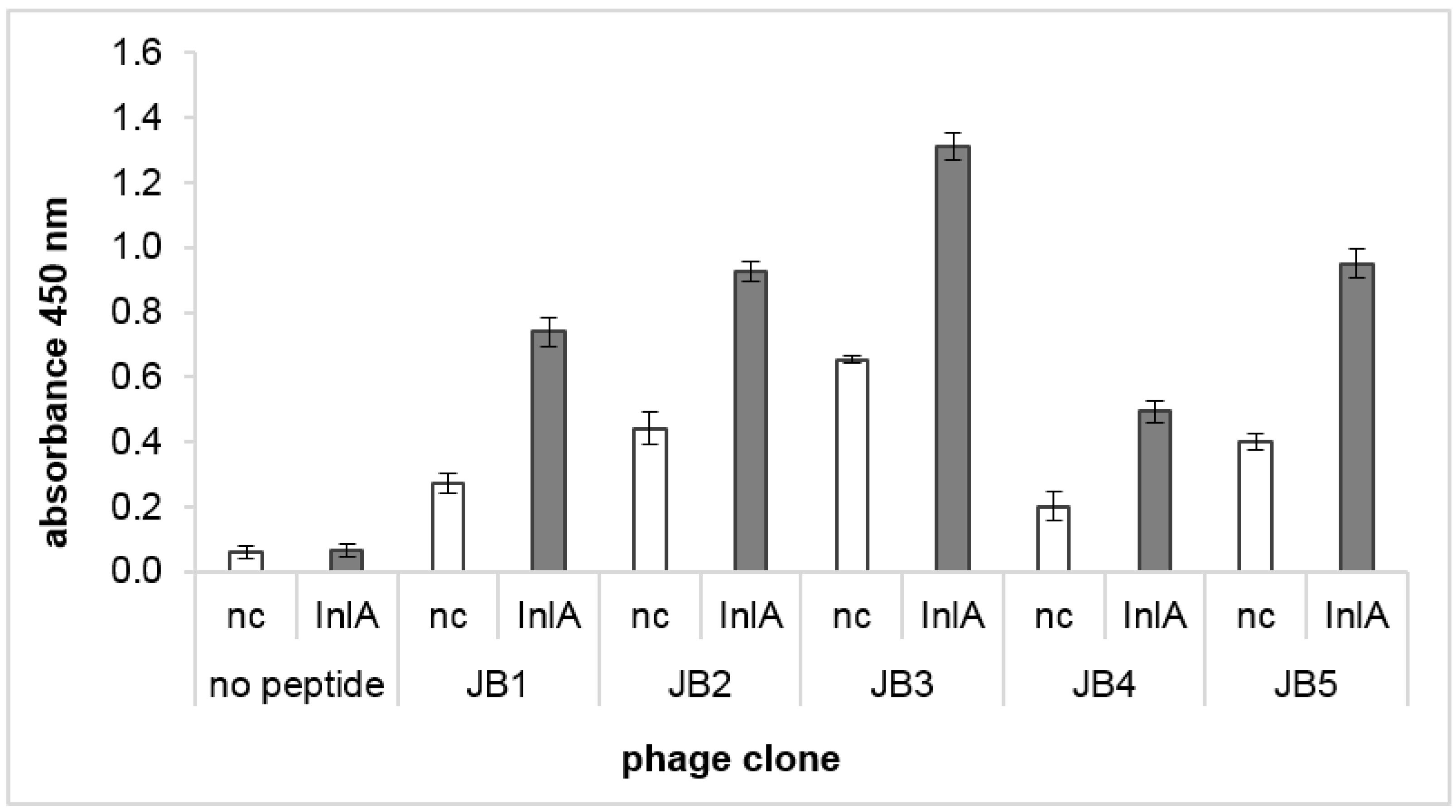
3.1. Biopanning of a Phage Display Library against InlA of L. monocytogenes Resulted in Selection of Five Peptides

To identify novel peptides selectively binding InlA, a phage display peptide library was used in a biopanning approach against InlA. The selection was performed against recombinant InlA (36–496) representing amino acids that are required for the interaction between InlA and E-cad (LRR region) [11,15]. Enrichment level was monitored after each round of selection indicating an increase in InlA affinity and effective enrichment of the phage clones in selection round three and four (Supplementary Figure S1). The negative controls without InlA showed significantly lower signals indicating a positive selection against InlA.
Four rounds of biopanning were performed. The eluted phages from selection rounds three and four were further analyzed by single colony analysis. These plaques were randomly picked, amplified, and separately tested for their binding affinity to InlA (Supplementary Figure S2). Genomic DNA was extracted and sequenced from phage clones showing comparably high binding affinity to InlA. The translated amino acid sequences encoded by the inserts identified 11 peptides with three phage clones having the same amino acid sequence, GLHTSATNLYLH. The sequences, the frequency of occurrence, and the properties of the peptides are summarized in Table 2.
Based on database research sequences showing characteristics of target-unrelated peptides such as antibody binders and phages with propagation advantage were excluded for further analysis (VVSPDMNLLLTN, SLDGAGAALRTS, and GHYTNSEWGFQE). Phage clones presenting peptide sequences JB1, JB2, JB3, JB4, and JB5 were tested by phage ELISA binding assay (Figure 1). For that, the titer of phages was determined and the same amount of pfu was used for ELISA. The phage clone representing peptide JB3 showed the highest ELISA signal followed by phages representing peptides JB2 and JB5. The binding of the phages representing peptide JB4 was very low but present.

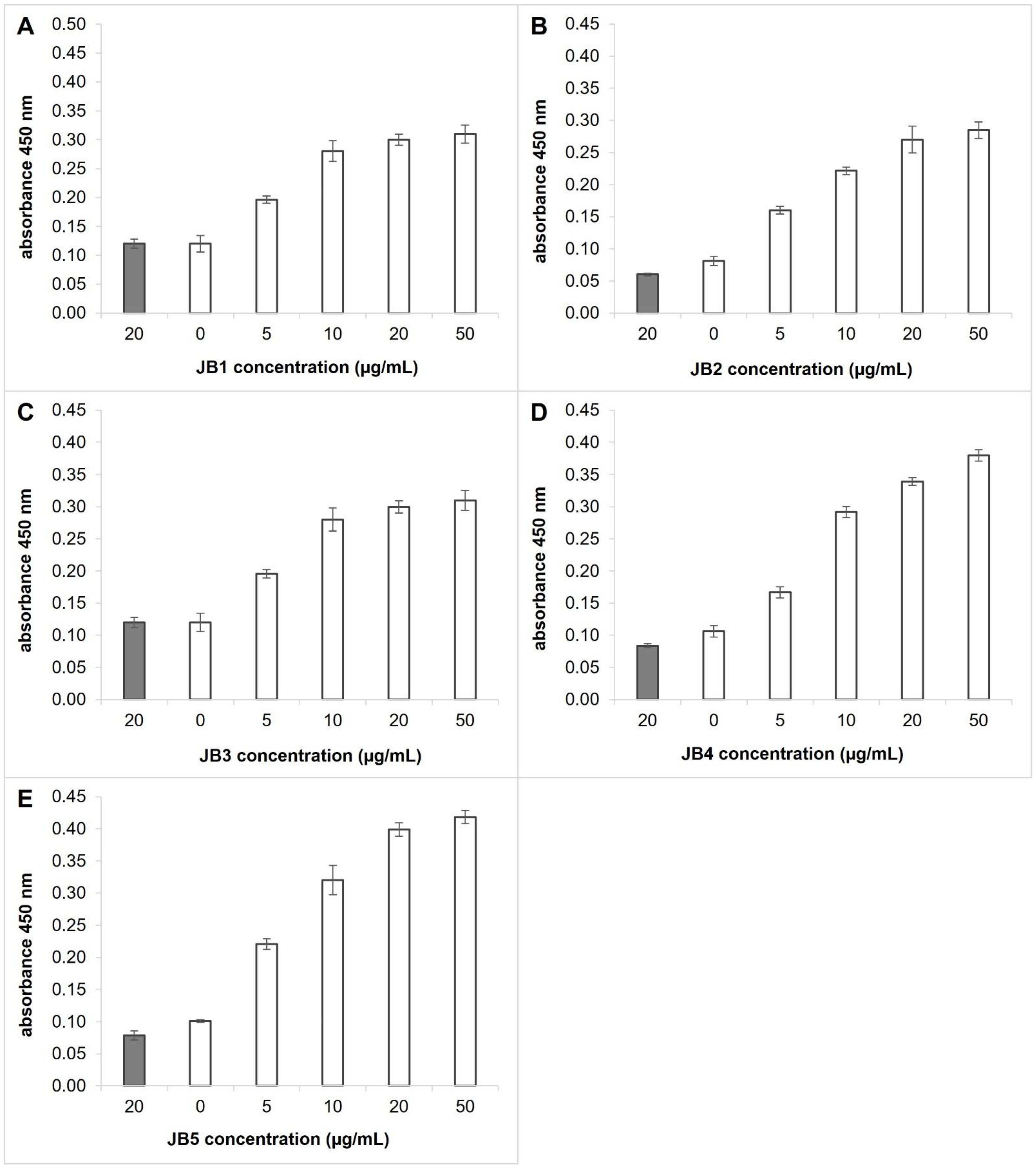
3.2. Selected Peptides Interact with InlA of L. monocytogenes

To characterize the ability of the selected peptides to interact with the target InlA, the five peptides JB1, JB2, JB3, JB4, and JB5 were synthesized with a lysin-linker and a FAM tag.
The binding of these peptides to InlA was evaluated by ELISA on recombinant InlA. The results showed that all five peptides are able to bind to InlA (Figure 2). All peptides gave comparable signals with InlA. It was observed that with an increased concentration of the peptides, there was an increment in the value of the interaction signal indicating an interaction in a concentration-dependent manner.

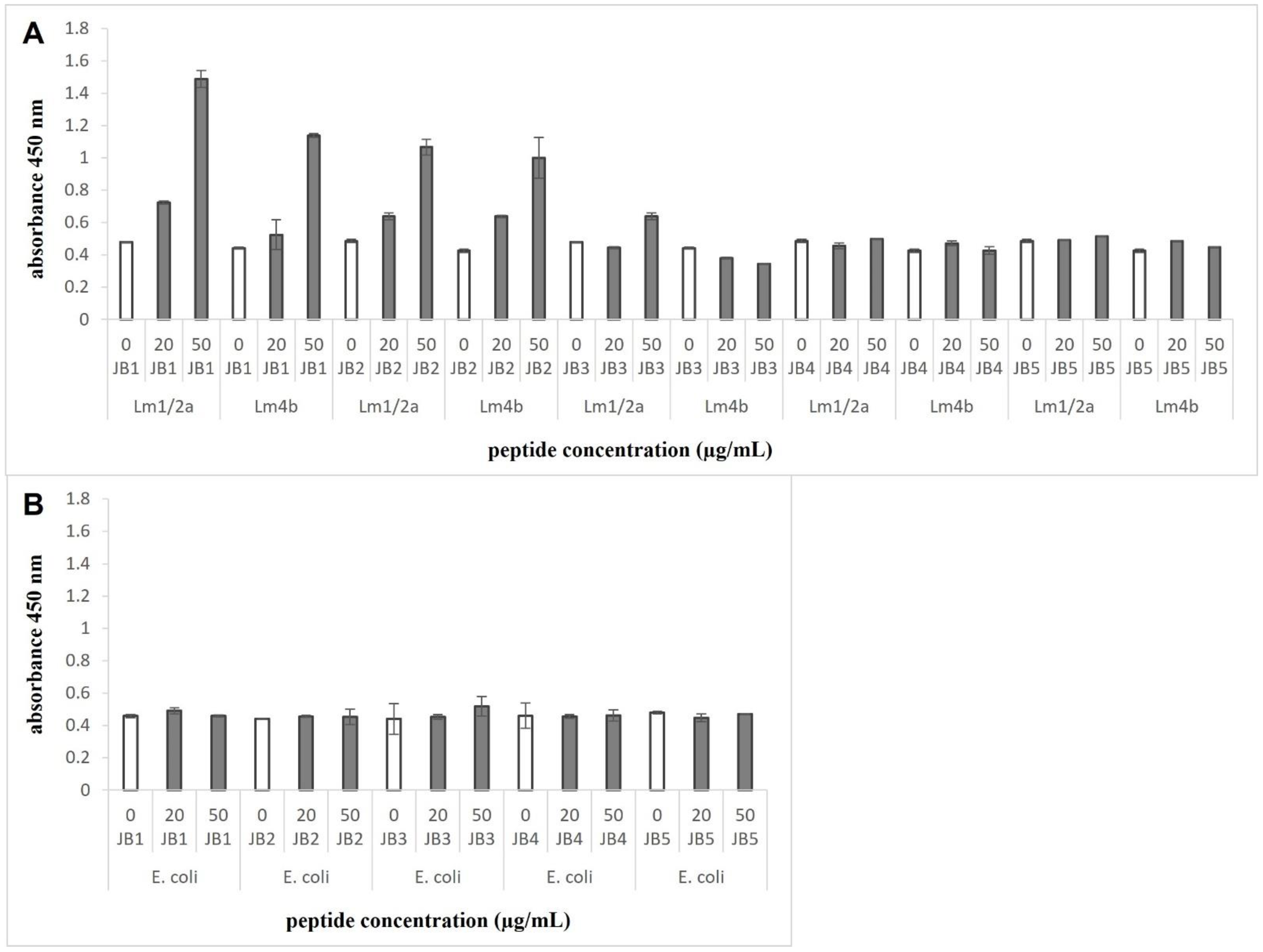
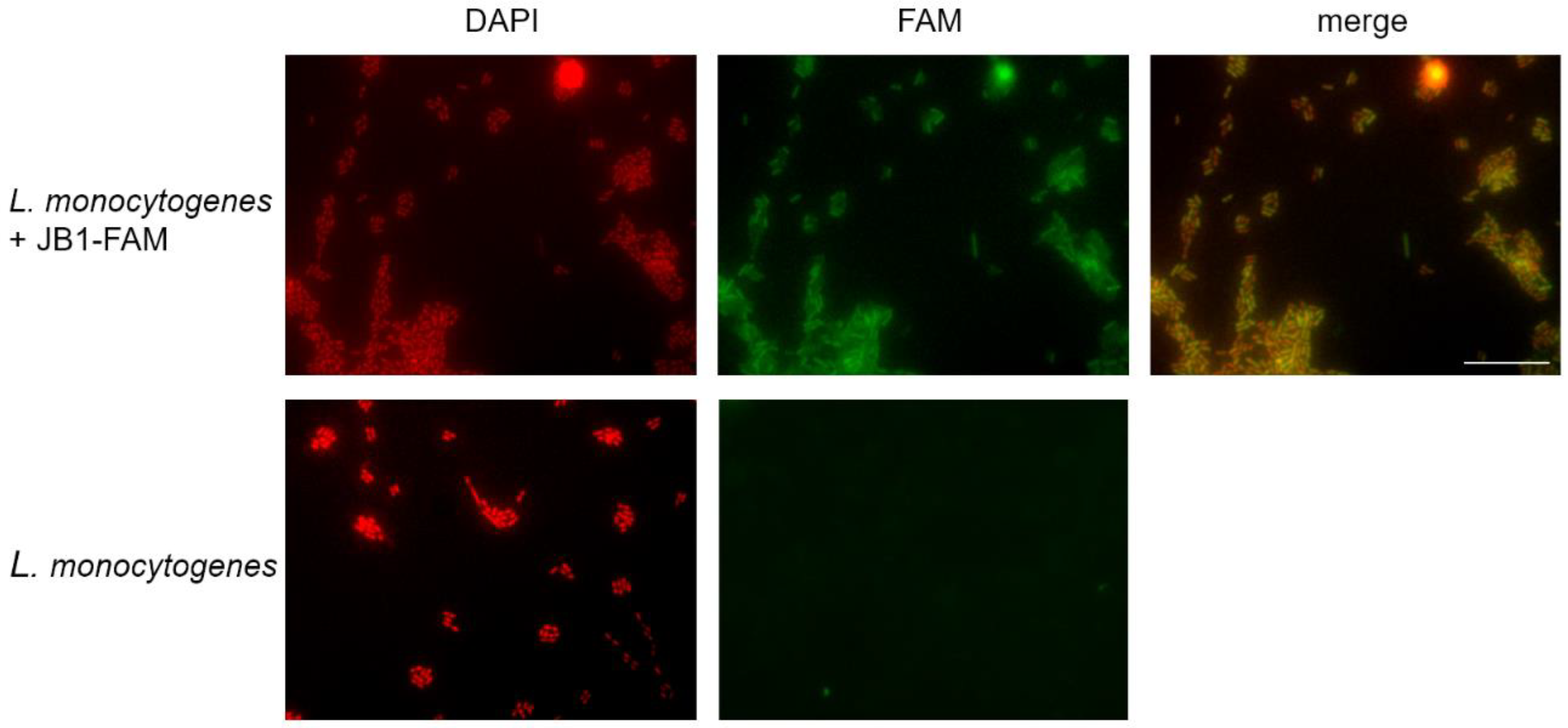
3.3. JB1 and JB2 Demonstrate Binding to L. monocytogenes

The peptides were tested by whole-cell ELISA to determine their ability to bind L. monocytogenes and non-target bacteria E. coli. Two different serotypes of L. monocytogenes were compared using the pathogenic serotypes 4b and 1/2a expressing InlA. All five peptides were incubated individually with the bacteria. Interaction of the peptides was detected by absorbance measurements. The results in Figure 3 demonstrate that peptides JB3, JB4, and JB5 exhibited low or no interaction with L. monocytogenes as well as with E. coli, while peptides JB1 and JB2 bound to L. monocytogenes but did not bind to E. coli. For JB1, higher absorbance values were measured, demonstrating that JB1 was more sensitive to L. monocytogenes than JB2. The signals of serotype 4b and 1/2a showed almost no difference in the values, indicating that these peptides were able to bind to both serotypes.
The binding of the labeled peptide JB1 to L. monocytogenes was also confirmed by fluorescence microscopy. After incubation of JB1-FAM with the cells, FAM signals were enriched by the bacteria (Figure 4). Hence, peptides JB1 and JB2 were chosen for further experiments.

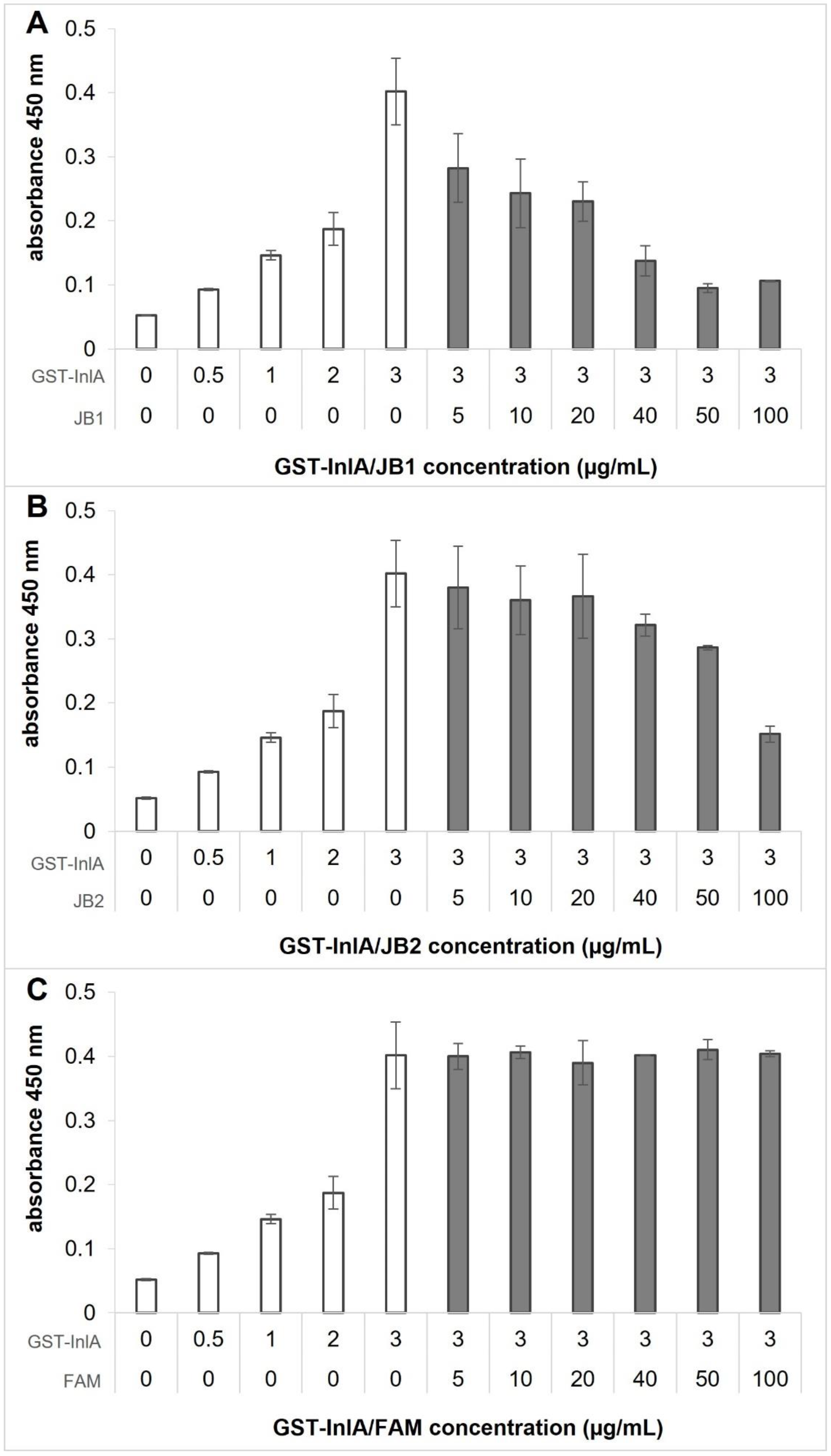
3.4. JB1 and JB2 Compete with E-cad for Binding to InlA

The effect of InlA-binding peptides on the interaction capability between InlA and E-cad was assessed by ELISA. Signals of bound InlA were measured with and without prior incubation of InlA with the peptides JB1 and JB2. The binding of InlA to E-cad was significantly reduced by the peptides JB1 and JB2 (Figure 5). Especially peptide JB1 inhibited the interaction between InlA and E-cad with increasing concentrations. At 50 µg/mL JB1 no interaction signals were detectable. Inhibitory effects as a result of the presence of the FAM tag were excluded by incubation with FAM instead of the peptides (Figure 5C). The results suggest that JB1, JB2, and E-cad interact with the same sites, or sites located close by in InlA.

4. Discussion

Listeriosis, which is terminal in approximately 30% of the reported cases despite antibiotic therapy, represents a major public health problem [26]. Increased incidence of the disease and multidrug-resistant strains of L. monocytogenes [27,28,29,30] require rapid and species-specific monitoring methods to prevent foodborne infections effectively [31]. Currently, detection methods for L. monocytogenes are culture-based techniques that are labor and time intensive. More advanced methods include real-time polymerase chain reaction (RT-PCR) or immunological techniques, for example, enzyme-linked immunosorbent assay (ELISA) or biosensor methods, but those methods require trained personnel and specialized infrastructure [32,33,34].
The genus Listeria can widely be found in the environment. Only the species L. monocytogenes is pathogenic to humans. Listeria species are closely related making it difficult to specifically detect L. monocytogenes and distinguish them from other Listeria species [35,36]. Antibodies selective and sensitive enough to unequivocally detect L. monocytogenes could not be developed yet [37,38,39,40,41]. Antibody fragments or specific peptides, selected, for example, by phage display procedures, might be an interesting alternative for antibodies offering high stability, long lifetime, and standardized production at low expense [42,43,44]. The phage display method was previously used for the selection of specific ligands in neurodegenerative diseases research, cancer research, and research of infectious diseases [17,17,45,46].
Specific peptides binding bacterial surfaces have shown high potential in the development of new methods for the detection and identification of different bacteria [47]. A variety of peptides were already selected to bind to the surface of L. monocytogenes (as reviewed by Kenzel et al., 2018) [24]. In all the articles published up to date, whole cells were used as targets in the phage display process. Cell surfaces are composed of lipids, carbohydrates, and proteins resulting in the selection of peptides with unknown targets, binding different epitopes present on the bacterial surface [48,49].
Here, we planned to identify peptide binders that specifically bind to the surface protein InlA of L. monocytogenes. InlA is only expressed on the surface of the pathogenic species L. monocytogenes [50] and offers an interesting target for the development of peptides with sufficient specificity for the pathogen, which could have the potential to be interesting for diagnostic and therapeutic approaches.
The selection was performed against recombinant InlA (36–496) representing amino acids that are required for the interaction between InlA and E-cad [11,15]. After four rounds of biopanning, five peptide sequences GLHTSATNLYLH (JB1), DSQFNKYSIATV (JB2), SGVYKVAYDWQH (JB3), QFDYMRPANDTH (JB4) and SNSIDKVNRPIN (JB5) (Table 2) were identified to bind the target. Three in addition identified sequences showing characteristics of target unrelated peptides were excluded for further experiments. Binding to InlA of peptide-presenting phage clones was investigated using an ELISA with the same amount of phages (pfu) (Figure 1) to ensure comparability of the measured binding of the phage clones. Peptides often have a KD within the micromolar range and are less affine than antibodies. The peptides in the NEB Ph.D. libraries are fused to the phage coat protein III and are therefore represented five-fold. The binding of single synthetic peptides to InlA was successfully demonstrated by ELISA (Figure 2). All five peptides bound to recombinant InlA with comparable strength and in a dose-dependent manner (Figure 2). Exact binding affinities will have to be compared using, for example, surface plasmon resonance or related methods in the future.
The binding efficiency of the peptides to whole cells of L. monocytogenes tested by ELISA revealed that peptides JB1 and JB2 bind to L. monocytogenes serotypes 1/2a and 4b, which are often found in infected patients, and do not cross-react with non-target bacteria E. coli (Figure 3). An interesting experiment for the future would be to investigate the specific binding of the peptides to InlA using InlA deficient L. monocytogenes strains as well as other L. monocytogenes serotypes.
Although the peptides JB3, JB4, and JB5 were able to show binding to recombinant InlA, binding could not be demonstrated to L. monocytogenes cells (Figure 3). Since the conformation of recombinant protein might differ from its native structure on intact cells, and the surrounding is different, the binding capacity of peptides binding recombinant protein can get lost or can be significantly reduced using whole cells. Interaction studies with further Listeria species and other bacteria are required to ensure the high specificity of the selected peptides and will be investigated in an upcoming study.
Flachbartova et al. isolated peptides interacting with the surface of an L. monocytogenes clinical isolate via combinatorial phage display. Surprisingly, two of the three selected peptides showed antimicrobial activity [51]. The toxicity of our selected peptides to L. monocytogenes cells was excluded by measurements of bacterial growth with peptide incubation for a period of 18 h (data not shown).
Listerial binding and penetration of epithelial cells are mediated through specific interaction of the bacterial protein InlA with its host cell receptor E-cad [10,11,12]. Since peptides JB1 and JB2 demonstrated binding to InlA and L. monocytogenes, we further investigated the ability of these peptides to inhibit the interaction between InlA and E-cad. Results are demonstrated in Figure 5 indicating that both peptides are able to inhibit the interaction in a concentration-dependent manner. Especially JB1 showed a complete inhibition of InlA binding to E-cad at peptide concentrations of 50 µg/mL, indicated by undetectable significant signals in ELISA (see Figure 5). The binding of InlA to E-cad was also significantly inhibited by JB2 at 100 µg/mL. These results suggest that the selected InlA binding peptides and E-cad interact with the same sites in InlA. Since InlA is involved in the adherence of L. monocytogenes to epithelial cells, blocking of InlA interaction site for E-cad by peptide binding could result in a prohibited invasion of the bacteria into host cells. This conclusion has to be investigated by further experiments in vivo.

5. Conclusions

In summary, we have used phage display for the first time to identify small peptides binding to a species-specific surface protein of L. monocytogenes. The identification of these peptides can be a significant contribution to use in diagnostic or therapeutic applications. In later applications, the peptides could easily be labeled with fluorophores, facilitating rapid detection of Listeria in food samples or even human cells.

Supplementary Materials

The following supporting information can be downloaded at: https://www.mdpi.com/article/10.3390/applmicrobiol2040070/s1, Figure S1: Screening of recovered phages for binding efficiency after each round of biopannings against InlA (enrichment ELISA); Figure S2: Screening of single phage clones for binding affinity to InlA.

Author Contributions

Conceptualization, S.A.F. and J.K.; methodology, S.A.F. and J.K.; validation, resources, S.A.F. and D.A.B.; data curation, J.K.; writing—original draft preparation, S.A.F. and J.K.; writing—review and editing, S.A.F., J.K. and D.A.B.; supervision, S.A.F. and D.A.B.; project administration, J.K.; funding acquisition, S.A.F. and D.A.B. All authors have read and agreed to the published version of the manuscript.

Funding

This work was supported by Technology Allianz Oberfranken (TAO).

Institutional Review Board Statement

Not applicable.

Data Availability Statement

Data are contained within the article or Supplementary Materials.

Acknowledgments

The vector for the purification of recombinant InlA protein was kindly provided by Joop van den Heuvel (Helmholtz Centre for Infectious Research, Braunschweig). We thank Matthias Noll (Coburg University of applied sciences and arts, Coburg) for providing the L. monocytogenes strain.

Conflicts of Interest

All authors declare that they have no conflict of interest.

References

  1. Zhu, Q.; Gooneratne, R.; Hussain, M.A. Listeria monocytogenes in Fresh Produce: Outbreaks, Prevalence and Contamination Levels. Foods 2017, 6, 21. [Google Scholar] [CrossRef] [PubMed] [Green Version]
  2. Carpentier, B.; Cerf, O. Review—Persistence of Listeria monocytogenes in food industry equipment and premises. Int. J. Food Microbiol. 2011, 145, 1–8. [Google Scholar] [CrossRef] [PubMed]
  3. Doran, K.S.; Banerjee, A.; Disson, O.; Lecuit, M. Concepts and mechanisms: Crossing host barriers. Cold Spring Harb. Perspect. Med. 2013, 3, a013433. [Google Scholar] [CrossRef] [PubMed] [Green Version]
  4. Lecuit, M. Human listeriosis and animal models. Microbes Infect. 2007, 9, 1216–1225. [Google Scholar] [CrossRef] [PubMed]
  5. Hof, H. History and epidemiology of listeriosis. FEMS Immunol. Med. Microbiol. 2003, 35, 199–202. [Google Scholar] [CrossRef]
  6. Cabanes, D.; Dehoux, P.; Dussurget, O.; Frangeul, L.; Cossart, P. Surface proteins and pathogenis potential of Listeria monocytogenes. Trends Microbiol. 2002, 10, 238–245. [Google Scholar] [CrossRef]
  7. Glaser, P.; Frangeul, L.; Buchrieser, C.; Rusniok, C.; Amend, A.; Baquero, F.; Berche, P.; Bloecker, H.; Brandt, P.; Chakraborty, T.; et al. Comparative genomics of Listeria species. Science 2001, 294, 849–852. [Google Scholar] [CrossRef] [Green Version]
  8. Disson, O.; Lecuit, M. In vitro and in vivo models to study human listeriosis: Mind the gap. Microbes Infect. 2013, 15, 971–980. [Google Scholar] [CrossRef]
  9. Tilney, L.G.; Portnoy, D.A. Actin filaments and the growth, movement, and spread of the intracellular bacterial parasite, Listeria monocytogenes. J. Cell Biol. 1989, 109, 1597–1608. [Google Scholar] [CrossRef]
  10. Gaillard, J.L.; Berche, P.; Frehel, C.; Gouin, E.; Cossart, P. Entry of L. monocytogenes into cells is mediated y internalin, a repeat protein reminiscent of surface antigens from gram-positive cocci. Cell 1991, 65, 1127–1141. [Google Scholar] [CrossRef]
  11. Lecuit, M.; Ohayon, H.; Braun, L.; Mengaud, J.; Cossart, P. Internalin of Listeria monocytogenes with an intact leucine-rich repeat region is sufficient to promote internalization. Infect. Immun. 1997, 65, 5309–5319. [Google Scholar] [CrossRef] [PubMed] [Green Version]
  12. Mengaud, J.; Ohayon, H.; Gounon, P.; Mege, R.M.; Cossart, P. E-cadherin is the receptor for internalin, a surfece protein required for entry of L. monocytogenes into epithelial cells. Cell 1996, 84, 923–932. [Google Scholar] [CrossRef] [Green Version]
  13. van Roy, F.; Berx, G. The cell-cell adhesion molecule E-cadherin. Cell Mol. Life Sci. 2008, 65, 3756–3788. [Google Scholar] [CrossRef] [PubMed]
  14. Lecuit, M.; Dramsi, S.; Gottardi, C.; Fedor-Chaiken, M.; Gumbiner, B.; Cossart, P. A single amino acid in E-cadherin responsible for host specificity towords the hman pathogen Listeria monocytogenes. EMBO J. 1999, 18, 3956–3963. [Google Scholar] [CrossRef] [PubMed] [Green Version]
  15. Schubert, W.D.; Urbanke, C.; Ziehm, T.; Beier, V.; Machner, M.P.; Domann, E.; Wehland, J.; Chakraborty, T.; Heinz, D.W. Structure of internalin, a major invasion protein of Listeria monocytogenes, in complex with its human receptor E-cadherin. Cell 2002, 111, 825–836. [Google Scholar] [CrossRef] [Green Version]
  16. Smith, G.P. Filamentous fusion phage: Novel expression vectors that display cloned antigens on the virion surface. Science 1985, 228, 1315–1317. [Google Scholar] [CrossRef]
  17. Krumpe, L.R.; Mori, T. Potential of phage-displayed peptide library technology to identify functional targeting peptides. Expert Opin. Drug Discov. 2007, 2, 525–537. [Google Scholar] [CrossRef] [Green Version]
  18. Balasubramanian, S.; Sorokulova, I.B.; Vodyanoy, V.J.; Simonian, A.L. Lytic phage as a specific and selective probe for detection of Staphylococcus aureus—A surface plasmon resonance spectroscopic study. Biosens. Bioelectron. 2007, 22, 948–955. [Google Scholar] [CrossRef]
  19. Edgar, R.; McKinstry, M.; Hwang, J.; Oppenheim, A.B.; Fekete, R.A.; Giulian, G.; Merril, C.; Nagashima, K.; Adhya, S. High-sensitivity bacterial detection using biotin-tagged phage and quantum-dot nanocomplexes. Proc. Natl. Acad. Sci. USA 2006, 103, 4841–4845. [Google Scholar] [CrossRef] [Green Version]
  20. Goldman, E.R.; Anderson, G.P.; Liu, J.L.; Delehanty, J.B.; Sherwood, L.J.; Osborn, L.E.; Cummins, L.B.; Hayhurst, A. Facile generation of heat-stable antiviral and antitoxin single domain antibodies from a semisynthetic llama library. Anal. Chem. 2006, 78, 8245–8255. [Google Scholar] [CrossRef]
  21. Olsen, E.V.; Pathirana, S.T.; Samoylov, A.M.; Barbaree, J.M.; Chin, B.A.; Neely, W.C.; Vodyanoy, V. Specific and selective biosensor for Salmonella and its detection in the environment. J. Microbiol. Methods 2003, 53, 273–285. [Google Scholar] [CrossRef]
  22. Rao, S.S.; Mohan, K.V.K.; Gao, Y.; Atreya, C.D. Identification and evaluation of a novel peptide binding to the cell surface of Staphylococcus aureus. Microbiol. Res. 2013, 168, 106–112. [Google Scholar] [CrossRef] [PubMed]
  23. Sorokulova, I.B.; Olsen, E.V.; Chen, I.-H.; Fiebor, B.; Barbaree, J.M.; Vodyanoy, V.J.; Chin, B.A.; Petrenko, V.A. Landscape phage probes for Salmonella typhimurium. J. Microbiol. Methods 2005, 63, 55–72. [Google Scholar] [CrossRef]
  24. Kenzel, J.; Schlegelmilch, L.; Funke, S.A. Phage display selection of specific ligands for Listeria monocytogenes—novel tools for diagnostic of therapeutic purposes. J. Microbiol. Biotechnol. 2018, 7, 31–38. [Google Scholar]
  25. Moreira, G.M.S.G.; Gronow, S.; Dübel, S.; Mendonça, M.; Moreira, Â.N.; Conceição, F.R.; Hust, M. Phage Display-Derived Monoclonal Antibodies Against Internalins A and B Allow Specific Detection of Listeria monocytogenes. Front. Public Health 2022, 10, 712657. [Google Scholar] [CrossRef] [PubMed]
  26. Behravesh, C.B.; Jones, T.F.; Vugia, D.J.; Long, C.; Marcus, R.; Smith, K.; Thomas, S.; Zansky, S.; Fullerton, K.E.; Henao, O.L.; et al. Deaths Associated with Bacterial Pathogens Transmitted Commonly Through Food: Foodborne Diseases Active Surveillance Network (FoodNet), 1996–2005. J. Infect. Dis. 2011, 204, 263–267. [Google Scholar] [CrossRef] [Green Version]
  27. Aureli, P.; Ferrini, A.M.; Mannoni, V.; Hodzic, S.; Wedell-Weergaard, C.; Oliva, B. Susceptibility of Listeria monocytogenes isolated from food in Italy to antibiotics. Int. J. Food Microbiol. 2003, 83, 325–330. [Google Scholar] [CrossRef]
  28. Li, L.; Olsen, R.H.; Ye, L.; Wang, W.; Shi, L.; Yan, H.; Meng, H. Characterization of Antimicrobial Resistance of Listeria monocytogenes Strains Isolated from a Pork Processing Plant and Its Respective Meat Markets in Southern China. Foodborne Pathog. Dis. 2016, 13, 262–268. [Google Scholar] [CrossRef]
  29. Li, Q.; Sherwood, J.S.; Logue, C.M. Antimicrobial resistance of Listeria spp. recovered from processed bison. Lett. Appl. Microbiol. 2007, 44, 86–91. [Google Scholar] [CrossRef]
  30. Poyart-Salmeron, C.; Carlier, C.; Trieu-Cuot, P.; Courtieu, A.L.; Courvalin, P. Transferable plasmid-mediated antibiotic resistance in Listeria monocytogenes. Lancet 1990, 335, 1422–1426. [Google Scholar] [CrossRef]
  31. Zhao, X.; Lin, C.-W.; Wang, J.; Oh, D.H. Advances in rapid detection methods for foodborne pathogens. J. Microbiol. Biotechnol. 2014, 24, 297–312. [Google Scholar] [CrossRef] [PubMed]
  32. Ohk, S.-H.; Bhunia, A.K. Multiplex fiber optic biosensor for detection of Listeria monocytogenes, Escherichia coli O157:H7 and Salmonella enterica from ready-to-eat meat samples. Food Microbiol. 2013, 33, 166–171. [Google Scholar] [CrossRef]
  33. Pochop, J.; Kačániová, M.; Hleba, L.; Lopasovský, L.; Bobková, A.; Zeleňáková, L.; Stričík, M. Detection of Listeria monocytogenes in ready-to-eat food by Step One real-time polymerase chain reaction. J. Environ. Sci. Health B 2012, 47, 212–216. [Google Scholar] [CrossRef] [PubMed]
  34. Portanti, O.; Di Febo, T.; Luciani, M.; Pompilii, C.; Lelli, R.; Semprini, P. Development and validation of an antigen capture ELISA based on monoclonal antibodies specific for Listeria monocytogenes in food. Vet. Ital. 2011, 47, 281–290. [Google Scholar]
  35. Hof, H.; Hefner, P. Pathogenicity of Listeria monocytogenes in comparison to other Listeria species. Infection 1988, 16, S141–S1444. [Google Scholar] [CrossRef] [PubMed]
  36. Paoli, G.C.; Kleina, L.G.; Brewster, J.D. Development of Listeria monocytogenes-specific immunomagnetic beads using a single-chain antibody fragment. Foodborne Pathog. Dis. 2007, 4, 74–83. [Google Scholar] [CrossRef] [PubMed]
  37. Bhunia, A.K.; Ball, P.H.; Fuad, A.T.; Kurz, B.W.; Emerson, J.W.; Johnson, M.G. Development and characterization of a monoclonal antibody specific for Listeria monocytogenes and Listeria innocua. Infect. Immun. 1991, 59, 3176–3184. [Google Scholar] [CrossRef] [Green Version]
  38. Charlier, C.; Perrodeau, É.; Leclercq, A.; Cazenave, B.; Pilmis, B.; Henry, B.; Lopes, A.; Maury, M.M.; Moura, A.; Goffinet, F.; et al. Clinical features and prognostic factors of listeriosis: The MONALISA national prospective cohort study. Lancet Infect. Dis. 2017, 17, 510–519. [Google Scholar] [CrossRef]
  39. Lathrop, A.A.; Jaradat, Z.W.; Haley, T.; Bhunia, A.K. Characterization and application of a Listeria monocytogenes reactive monoclonal antibody C11E9 in a resonant mirror biosensor. J. Immunol. Methods 2003, 281, 119–128. [Google Scholar] [CrossRef]
  40. Ojima-Kato, T.; Hashimura, D.; Kojima, T.; Minabe, S.; Nakano, H. In vitro generation of rabbit anti-Listeria monocytogenes monoclonal antibody using single cell based RT-PCR linked cell-free expression systems. J. Immunol. Methods 2015, 427, 58–65. [Google Scholar] [CrossRef]
  41. Sølve, M.; Boel, J.; Nørrung, B. Evaluation of a monoclonal antibody able to detect live Listeria monocytogenes and Listeria innocua. Int. J. Food Microbiol. 2000, 57, 219–224. [Google Scholar] [CrossRef]
  42. Brigati, J.R.; Samoylova, T.I.; Jayanna, P.K.; Petrenko, V.A. Phage display for generating peptide reagents. Curr. Protoc. Protein Sci. 2008, 51, 18.9.1–18.9.27. [Google Scholar] [CrossRef] [PubMed]
  43. Romanov, V.I.; Durand, D.B.; Petrenko, V.A. Phage display selection of peptides that affect prostate carcinoma cells attachment and invasion. Prostate 2001, 47, 239–251. [Google Scholar] [CrossRef] [PubMed]
  44. Smith, G.P.; Petrenko, V.A. Phage Display. Chem. Rev. 1997, 97, 391–410. [Google Scholar] [CrossRef]
  45. Funke, S.A.; Willbold, D. Mirror image phage display—A method to generate D-peptide ligands for use in diagnostic or therapeutical applications. Mol. Biosyst. 2009, 5, 783–786. [Google Scholar] [CrossRef] [PubMed]
  46. Huang, J.X.; Bishop-Hurley, S.L.; Cooper, M.A. Development of anti-infectives using phage display: Biological agents against bacteria, viruses, and parasites. Antimicrob. Agents Chemother. 2012, 56, 4569–4582. [Google Scholar] [CrossRef] [Green Version]
  47. Morton, J.; Karoonuthaisiri, N.; Charlermroj, R.; Stewart, L.D.; Elliott, C.T.; Grant, I.R. Phage display-derived binders able to distinguish Listeria monocytogenes from other Listeria species. PLoS ONE 2013, 8, e74312. [Google Scholar] [CrossRef] [Green Version]
  48. Yarbrough, D.K.; Hagerman, E.; Eckert, R.; He, J.; Choi, H.; Cao, N.; Le, K.; Hedger, J.; Qi, F.; Anderson, M.; et al. Specific binding and mineralization of calcified surfaces by small peptides. Calcif. Tissue Int. 2010, 86, 58–66. [Google Scholar] [CrossRef] [Green Version]
  49. Yarbrough, D.K.; Eckert, R.; He, J.; Hagerman, E.; Qi, F.; Lux, R.; Wu, B.; Anderson, M.H.; Shi, W. Rapid probing of biological surfaces with a sparse-matrix peptide library. PLoS ONE 2011, 6, e23551. [Google Scholar] [CrossRef]
  50. Gouin, E.; Mengaud, J.; Cossart, P. The virulence gene cluster of Listeria monocytogenes is also present in Listeria ivanovii, an animal pathogen, and Listeria seeligeri, a nonpathogenic species. Infect. Immun. 1994, 62, 3550–3553. [Google Scholar] [CrossRef] [Green Version]
  51. Flachbartova, Z.; Pulzova, L.; Bencurova, E.; Potocnakova, L.; Comor, L.; Bednarikova, Z.; Bhide, M. Inhibition of multidrug resistant Listeria monocytogenes by peptides isolated from combinatorial phage display libraries. Microbiol. Res. 2016, 188–189, 34–41. [Google Scholar] [CrossRef] [PubMed]
Figure 1. Phage-display-derived phage clones show binding to InlA. 1 µg InlA was coated on microtiter plates and incubated with 3 × 10¹⁰ pfu peptide-presenting phage clones. Bound phages were detected by ELISA as described in Materials and Methods. Averages and standard deviations of three independent measurements are shown. nc: wells without InlA coating. JB1: GLHTSATNLYLH, JB2: DSQFNKYSIATV, JB3: SGVYKVAYDWQH, JB4: QFDYMRPANDTH, JB5: SNSIDKVNRPIN.
Applmicrobiol 02 00070 g001
Figure 2. Synthesized peptides JB1, JB2, JB3, JB4, and JB5 bind to InlA. Wells of a microtiter plate were coated with 1 µg InlA blocking and incubated with increasing concentrations of FAM-labeled peptides. Grey bars represent the negative control with no InlA and incubation with 20 µg/mL peptide. Binding peptides were detected by ELISA using anti-FITC:HRP conjugate and measurement of absorbance at 450 nm. Means of three independent measurements and their standard deviations are shown. JB1: GLHTSATNLYLH (A), JB2: DSQFNKYSIATV (B), JB3: SGVYKVAYDWQH (C), JB4: QFDYMRPANDTH (D), JB5: SNSIDKVNRPIN (E).
Applmicrobiol 02 00070 g002
Figure 3. InlA-binding peptides JB1 and JB2 interact with L. monocytogenes. Bacteria, either L. monocytogenes serotype 1/2a, L. monocytogenes serotype 4b (A) or E. coli (B) was incubated with FAM-labeled peptides JB1 (GLHTSATNLYLH), JB2 (DSQFNKYSIATV), JB3 (SGVYKVAYDWQH), JB4 (QFDYMRPANDTH) und JB5 (SNSIDKVNRPIN) in a final concentration of 0 µg/mL, 20 µg/mL and 50 µg/mL. Binding peptides were detected by ELISA using anti-FITC:HRP conjugate and measurement of absorbance at 450 nm. Averages and standard deviations of three independent measurements are shown.
Applmicrobiol 02 00070 g003
Figure 4. Microscopical detection of the interaction between L. monocytogenes and JB1-FAM. 109 cells of L. monocytogenes were harvested by centrifugation and fixed with 2.5 (v/v) glutaraldehyde. The samples were washed and 30 µL suspension was placed onto glass slides following incubation with FAM-labeled JB1 (green) and DAPI (red). The lower row shows L. monocytogenes cells incubated with buffer without JB1. The scale bar represents 10 μm.
Applmicrobiol 02 00070 g004
Figure 5. InlA-binding peptides JB1 and JB2 inhibit the interaction between GST-InlA and E-cad. Different concentrations of GST-InlA were incubated with increasing concentrations of either JB1 (A), JB2 (B), or FAM (C) (grey bars) or without peptide (white bars) before the mixture was added to 1 µg E-cad coated on a microtiter plate. Protein binding was analyzed by ELISA using antibodies recognizing GST-InlA. The means of three independent measurements and their standard deviations are shown.
Applmicrobiol 02 00070 g005
Table 1. Synthesized FAM-labeled peptides identified by phage display against InlA (36–496).
PeptideSequence
JB1H-GLHTSATNLYLH-K(FAM)-NH2
JB2H-DSQFNKYSIATV-K(FAM)-NH2
JB3H-SGVYKVAYDWQH-K(FAM)-NH2
JB4H-QFDYMRPANDTH-K(FAM)-NH2
JB5H-SNSIDKVNRPIN-K(FAM)-NH2
Table 2. Characteristics of peptides identified by phage display against InlA (36-496).
CloneSequenceFrequencyNet ChargeHydrophobicityMW (Da)
JB1GLHTSATNLYLH3/11033%1326.47
JB2DSQFNKYSIATV1/11033%1372.49
JB3SGVYKVAYDWQH1/11033%1452.58
JB4QFDYMRPANDTH1/11−125%1488.61
JB5SNSIDKVNRPIN1/11+125%1350.50
VVSPDMNLLLTN2/11−150%1309.557
SLDGAGAALRTS1/11041%1118.214
GHYTNSEWGFQE1/11−216%1454.476
Publisher’s Note: MDPI stays neutral with regard to jurisdictional claims in published maps and institutional affiliations.

