Review

Catastrophic Antiphospholipid Syndrome: A Review

by
Carmine Siniscalchi
1,*,
Manuela Basaglia
1,
Michele Riva
1,
Michele Meschi
2,
Tiziana Meschi
3,
Giampiero Castaldo
3 and
Pierpaolo Di Micco
4
1
Department of Internal Medicine, Parma University Hospital, 43125 Parma, Italy
2
UOC Internal Medicine, Fidenza Hospital, 43036 Parma, Italy
3
Department of Medicine and Surgery, Parma University Hospital, 43125 Parma, Italy
4
AFO Medicina PO Santa Maria delle Grazie, Pozzuoli Naples Hospital 2 Nord, 80078 Naples, Italy
*
Author to whom correspondence should be addressed.
Immuno 2024, 4(1), 1-13; https://doi.org/10.3390/immuno4010001
Submission received: 25 October 2023 / Revised: 19 December 2023 / Accepted: 21 December 2023 / Published: 25 December 2023
(This article belongs to the Special Issue Recent Advances in Antiphospholipid Syndrome)

Abstract

:
Antiphospholipid syndrome (APS) is a systemic autoimmune disease characterized by thrombotic or obstetric events occurring in individuals who have persistent antiphospholipid antibodies. Catastrophic antiphospholipid syndrome (CAPS) is a rare and potentially fatal form of APS characterized by severe thrombotic complications occurring in multiple organs over a short period of time or simultaneously. CAPS is associated with a high (50%) death rate. Infections, multi-organ failure, and cerebral and heart thrombosis represent the main complications of this syndrome. Generally, anticoagulants, glucocorticoids, therapeutic plasmapheresis (TPE), and intravenous immunoglobulin (IVIG) are used in combination for treatment. Multidisciplinary care involving different specialists from hematology, rheumatology, nephrology, infectious disease, critical care, and obstetrics is often required due to the complexity of the disease. Recent data emphasize the effectiveness of biologics such as anti-TNF-a monoclonal antibodies (adalimumab, certolizumab), anti-CD38 monoclonal antibody (daratumumab), BAFF/Blys inhibitor (belimumab), and BTK inhibitor (zanubrutinib) against CAPS. In order to understand the underlying causes of CAPS, one future possibility involves investigating and characterizing the hereditary and acquired risk factors associated with CAPS.

1. Introduction

Catastrophic antiphospholipid syndrome (CAPS) is an extremely rare and potentially fatal variant of antiphospholipid syndrome (APS).
Antiphospholipid syndrome (APS) is a systemic autoimmune disease characterized by thrombotic or obstetric events in patients who have persistent antiphospholipid antibodies (APLA), which include lupus anticoagulant (LA), anti-2-glycoprotein I (anti-2GPI), and/or anti-cardiolipin antibodies (aCL) (Figure 1) [1]. It can cause venous, arterial, or microvascular thrombosis in the deep veins of the lower limbs, as well as in cerebral arterial circulation. Although uncommon, thrombosis can occur in hepatic veins, visceral veins, or the cerebral venous circulation.
Primary APS can develop alone or together with other autoimmune diseases, especially systemic lupus erythematosus (SLE). The APS categorization criteria were established for the first time in Sapporo in 1999 [2] and were modified during the Eleventh International Congress on APS in Sydney in 2006 [1,3,4].
However, the above-mentioned classifications are lacking in evidence-based definitions. Therefore, the American College of Rheumatology (ACR) and EULAR collaborated to produce the 2023 ACR/EULAR APS classification criteria [5].
According to the 2023 ACR/EULAR APS classification criteria, patients should be classified as having APS if they meet the entry criteria (at least one clinical and one laboratory criterion within three years of each other) and accumulate at least three points from clinical domains and three points from laboratory domains. We present a summary table of the 2023 ACR/EULAR APS classification criteria (Table 1).
Only a small percentage of patients with APS develop CAPS [1,6], which is generally induced by a precipitating event such as an infection [7,8]. CAPS is an aggressive variant of APS characterized by multiorgan thrombotic events that develop rapidly or simultaneously [9]. CAPS has a high death rate (50%), mostly due to cerebral and heart thrombosis, infections, and multiorgan failure [6,10].
The aim of this review is to assist general practitioners and experts in diagnosing and treating CAPS. Our recommendations are evidence-based wherever feasible, but given the scarcity of well-designed, randomized, controlled studies, they frequently represent expert opinion.

2. The Complexity of CAPS

Catastrophic APS (CAPS) is a life-threatening variant of APS characterized by the rapid onset of thrombotic events involving multiple organ systems, including large vessels and microvascular involvement. Approximately 1% of patients with APS develop the severe clinical picture of CAPS. In up to 50% of patients diagnosed with CAPS, it was the presenting symptom of APS [11,12,13].
CAPS is distinguished from APS by the intensity and extent of the thrombotic process. CAPS can involve the venous and arterial vascular system, targeting multiple organs simultaneously through micro- or combined micro- and macrovascular processes. Patients with CAPS or APS may have venous thromboembolic and arterial vascular disease, but CAPS patients also have macrovascular involvement leading to thrombosis affecting other organs, which, unlike APS, can progress acutely even when treated with appropriate anticoagulants. The mechanism by which a “thrombotic storm” occurs is not well understood. It is not known whether individuals with CAPS have aPL with different avidity, specificity, titer, or other antigen characteristics that differ from the aPL of APS. Based on our experience, most patients with CAPS have aPL triple positivity with anticardiolipin and anti-beta2GPI antibodies at high immunoglobulin G (IgG) titer.
CAPS is rare, affecting approximately 1% of individuals with APS or 5 per million in the general population. In the Euro-Phospholipid project, which included 1000 patients with APS followed for 10 years, only nine (0.9 percent) developed CAPS [10]. In the APS Alliance for Clinical Trials and International Networking (APS ACTION) Registry, based on 804 international patients, only nine (1%) developed CAPS [12]. In two smaller series with nearly 300 patients total, rates of CAPS were 2 and 5 percent, respectively [14,15].
The rarity of CAPS is also dangerously associated with the fact that not all individuals with CAPS have a previous diagnosis of APS, making the syndrome more difficult to diagnose and, therefore, treat quickly. In a study by Cervera et al., approximately one-half of patients carried a prior APS diagnosis and one-half did not [11].
Furthermore, CAPS often occurs in the course of other clinical conditions such as infection, surgical procedure, malignancy, or pregnancy. The diagnosis is so delayed and may be known at the time of presentation or may be identified during the evaluation of the acute presentation. In a series of 547 patients with 571 CAPS events, 68 percent had an underlying clinical condition [16]: infection (29%), surgery (9%), cancer (9%), pregnancy or postpartum (3%), or active systemic lupus erythematosus (SLE) (2%); one-half of these individuals presented with CAPS as their first manifestation of APS and 28% were known to have had a prior diagnosis of SLE [16]. Aside from the “masking” clinical conditions, it must be said that inadequate anticoagulation in a patient with known APS may also increase the risk of CAPS [17]. In the population of patients known to have aPL or APS, identifying and addressing additional thrombosis risk factors is important in preventing further thrombosis and possible progression to CAPS.
CAPS is characterized by a spectrum of clinical disorders, so is often faded in the initial stages. Indeed, it may be unclear whether an antiphospholipid antibody (aPL)-positive individual with or without a diagnosis of APS is progressing to CAPS. So, CAPS can present in an advanced stage of disease, when multiple thromboses and thrombocytopenia are already present. Consequently, vigilance is required to identify disease progression. Individuals who ultimately develop CAPS will typically manifest multiple thromboses and, generally, thrombocytopenia. Most patients have multiorgan involvement (and often multiorgan failure) on presentation [18]. Almost every organ system may be affected. In a report of 571 episodes of CAPS, the following organ systems were most likely to be affected [16]: kidneys (74%); brain (56%); lungs (55%); heart (53%); skin (45%); liver (34%); peripheral vessels (37%); and gastrointestinal tract (12%).
CAPS is characterized by multiple thromboses (microvascular and/or large vessels) occurring in several vascular beds over a relatively short period of time (days). This contrasts with APS, in which a single large vessel thrombotic event typically occurs. In CAPS, large vessel thrombosis includes deep vein thrombosis (DVT), pulmonary embolism (PE), or arterial thrombosis (e.g., ischemic stroke, myocardial infarction). Lung involvement may progress to acute respiratory distress syndrome (ARDS) and may be complicated by pulmonary hemorrhage. Pulmonary microvascular involvement generally presents as alveolar hemorrhage, which may be asymptomatic or may present with macroscopic hemoptysis. Cardiac involvement may include valvular disease (mitral, aortic) or myocardial infarction. Cutaneous manifestations may include purpura, livedo reticularis/racemosa, subungual hemorrhage, and skin necrosis [19]. Patients with CAPS may also experience abdominal pain due to thrombotic complications affecting the vasculature of the kidneys, liver, adrenal glands, spleen, intestine, mesentery, or pancreas [20]. Other less common organ involvement includes esophageal rupture, testicular/ovarian infarction, bone marrow infarction, and gastric and colonic ulcerations [20]. Central nervous system changes include acute ischemic encephalopathy with confusion, seizures, and/or focal findings. Hematological findings include thrombocytopenia and schistocytes. In a series of 280 patients with CAPS, 42 (15%) had disseminated intravascular coagulation (DIC) [11].

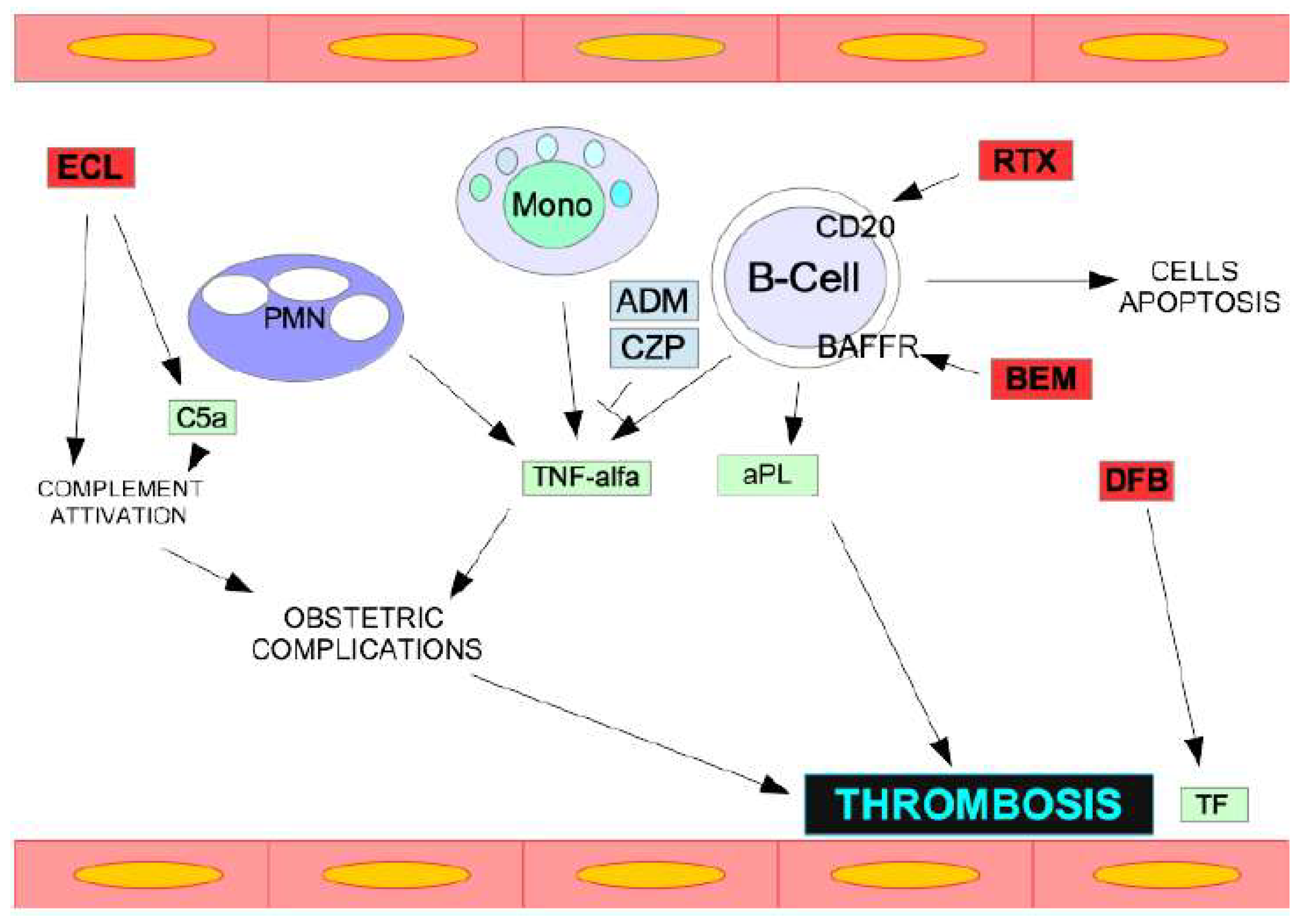
3. Pathophysiology and Mechanisms of CAPS

CAPS’s key pathogenic pathways include immune cell activation, coagulation factors, and complement activation. When complement is activated, it creates C5a, a powerful inflammatory mediator, and the C5b-9 complex [21], which promotes hemolysis and the release of free "heme" with prothrombotic activity. Complement damages vascular endothelial cells through subendothelial collagen and tissue factors, platelet activation, and the coagulation cascade. Complement can also disrupt neutrophils by generating DNA extrusion, which has anti-inflammatory and prothrombotic characteristics. In complement-mediated thrombotic microangiopathy, complement dysregulation also plays a pathophysiological role in microvascular thrombosis and acute kidney damage [21,22,23,24].
To explain the role of complement in the pathogenesis of CAPS, two sequential mechanisms have been proposed: first, aPL or complement gene abnormality predisposes one to complement dysregulation; second, an infection, inflammation, pregnancy, surgery, or other stimulus triggers complement-mediated cellular damage, with clinical manifestations of hemolysis and thrombosis [21]. The function of complement in CAPS is also responsible for the efficacy of complement-blocking medications (such as eculizumab), which are commonly utilized in refractory CAPS.

4. Not a Simple Diagnosis

A high level of suspicion for CAPS should be maintained in any patient with venous and/or arterial thrombosis with progressive multiorgan involvement, rapid clinical deterioration, and the presence of thrombocytopenia and schistocytes in the peripheral blood smear. A previous diagnosis of APS or systemic lupus erythematosus (SLE) should further increase clinical suspicion. Important indicators that suggest a patient may have CAPS rather than APS or another thrombotic disorder include rapidly progressive disease, multiorgan involvement, and a combination of large vessel and microvascular thrombosis. During pregnancy, HELLP syndrome (hemolysis, elevated liver function tests, and low platelets) and CAPS may be difficult to distinguish and may coexist [25]. Patients presenting with clinical manifestations of either of these diagnoses should be cared for by experts with experience in both conditions, given the high likelihood of maternal and neonatal complications that may progress quickly.
The classification criteria for CAPS have been proposed by the International Congress on aPL and validated for research purposes [9]. These are research criteria and should not replace the clinical judgment of a clinician with expertise in CAPS or an individual with expertise in diagnosing thrombotic disorders. However, many clinicians refer to aspects of these criteria to guide them in making a diagnosis of CAPS.

4.1. Definite CAPS

CAPS can be confidently diagnosed if all four requirements are met:
  • Involvement of three organs, systems, or tissues.
  • Manifestations appear at the same time or over the course of one week.
  • Small vessel occlusion is confirmed histologically in at least one organ or tissue.
  • The presence of aPL (anticardiolipin antibodies, anti-beta2-glycoprotein I antibodies, and/or lupus anticoagulant) is documented twice, at least 12 weeks apart.

4.2. Probable CAPS

The probable diagnosis of CAPS can occur when the following criteria are met:
  • Only two organs or tissue are involved.
  • Despite anticoagulation, a third event manifests between one week and one month after presentation.
  • No histologic evidence.
  • No laboratory confirmation of aPL.

5. Differential Diagnosis

The differential diagnosis of CAPS includes other thrombotic syndromes, particularly those with venous and arterial thrombosis at multiple or unusual sites and those with microvascular thrombosis. However, it is important to note that there is no clear distinction between these clinical conditions and CAPS that can overlap with any of these conditions. The following conditions must be considered as a differential diagnosis: disseminated intravascular coagulation (DIC); heparin-induced thrombocytopenia (HIT); thrombotic microangiopathies (TMA); small vessel vasculitis (particularly antineutrophil cytoplastic antibody (ANCA)-associated vasculitis); preeclampsia and HELLP syndrome (hemolysis, elevated liver enzymes, and low platelets); and sepsis.

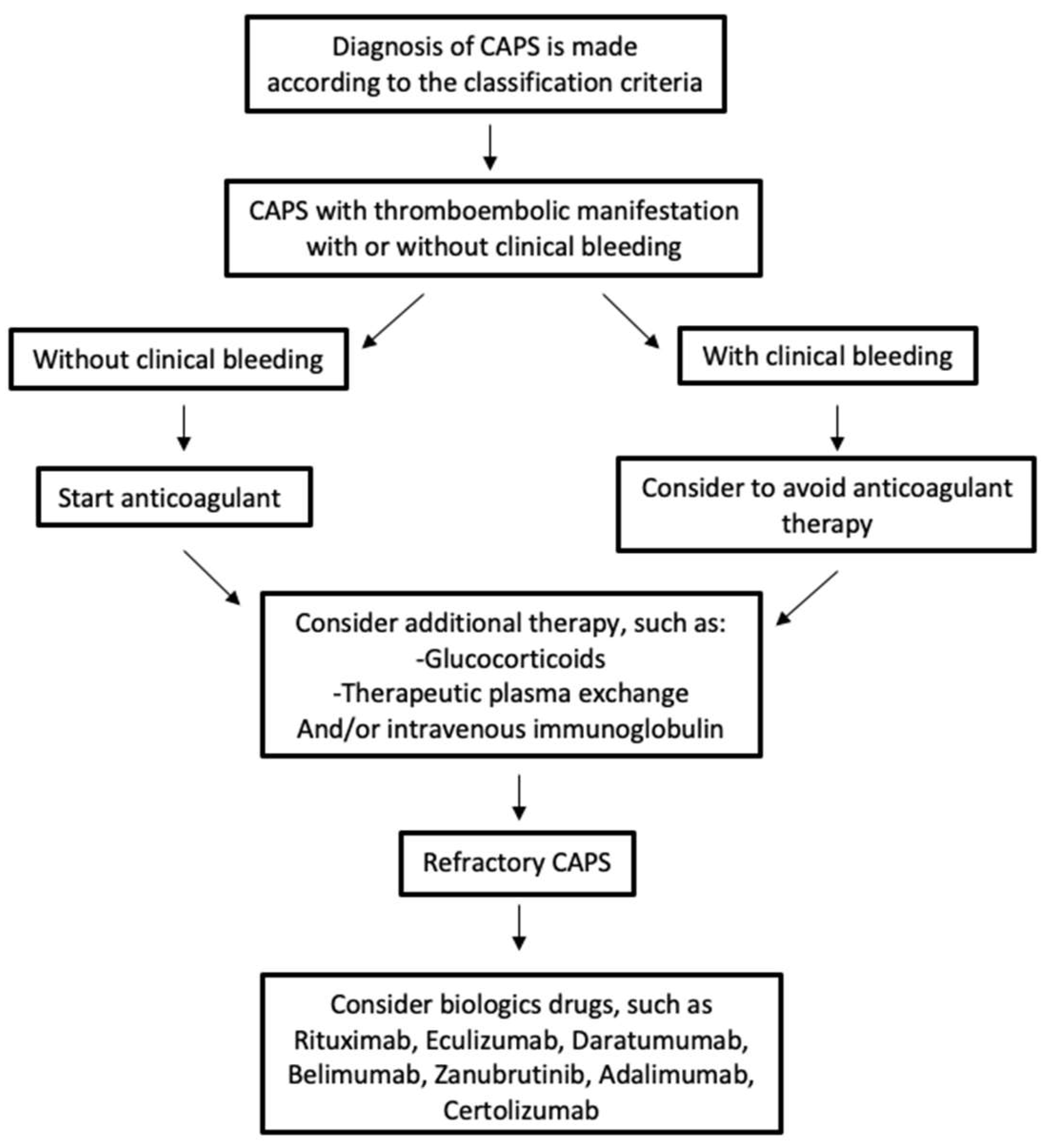
6. Management and Therapy of CAPS

The clinical and therapeutic management of patients with CAPS is not simple [21]. Very often, therapeutic decisions are made with incomplete clinical information and frequently revised as the patient’s clinical status changes or new laboratory results are available (Table 2). The optimal treatment for CAPS is unknown, and no prospective studies have been conducted on the treatment of CAPS. CAPS is generally treated with a combination of anticoagulants, glucocorticoids, and therapeutic plasmapheresis (TPE) or intravenous immunoglobulin (IVIG), sometimes referred to as triple therapy [26]. Multidisciplinary care is often required, including the involvement of hematology, rheumatology, nephrology, infectious disease, intensive care, and obstetrics teams. There are no randomized studies comparing individual therapeutic options; therefore, the proposed therapeutic approach is based on observational evidence, expert opinions, and the 2018 McMaster guidelines [27]. A 2018 report that included 471 patients noted that triple therapy (anticoagulation, glucocorticoids, and plasmapheresis or IVIG) was associated with a higher survival rate [28]; specifically, triple therapy was associated with a 71% survival rate, single therapy or a combination of two therapies showed a survival rate of 59%, while no therapy showed a survival rate of 25%. Another report documented a higher recovery rate associated with triple therapy (69%) compared to the use of fewer than three therapies (54%) [11].
Anticoagulant therapy at therapeutic doses is necessary [26,27,29]. The initial choice of anticoagulant is intravenous unfractionated heparin. Heparin is avoided (and an anticoagulant other than heparin is used) if heparin-induced thrombocytopenia (HIT) is considered likely. After recovery, hemodynamically stable patients are switched to warfarin and managed with a target international normalized ratio (INR) between 2 and 3. Direct oral anticoagulants (DOACs) are generally avoided in CAPS (and APS).
For most patients with CAPS, low-dose aspirin (100 mg/day) is also started immediately in addition to anticoagulation. Aspirin can be discontinued in patients at high risk of bleeding [27]. Anticoagulation and low-dose aspirin are generally continued indefinitely, similar to the treatment of APS, unless the risks of bleeding are judged to outweigh the benefits. No randomized trials of anticoagulant or antiplatelet therapy in CAPS have been reported. The McMaster CAPS guidelines included a meta-analysis of observational data from 325 patients that found an association between anticoagulant therapy and reduced mortality (odds ratio (OR) 0.18, 95% CI 0.09–0.38) [27].
Patients with CAPS are also treated with high-dose glucocorticoids. A typical glucocorticoid used is methylprednisolone, with a dose of 0.5 to 1 g administered intravenously once daily for three or more days, followed by oral or parenteral therapy with the equivalent of 1 mg/kg of prednisone per day with tapering initiated once the patient improves clinically [29]. It should be noted that no randomized trials of glucocorticoids in CAPS have been reported. The use of a glucocorticoid is supported by observational evidence of benefit when combined with other therapies [28].
For most patients with CAPS, therapeutic plasmapheresis (TPE) or intravenous immunoglobulin (IVIG), in addition to anticoagulants and glucocorticoids, are also suggested [27]. The mechanism by which TPE acts in CAPS is unclear; it seems likely to be due to the removal of antiphospholipid antibodies (aPL) or complement factors. Exchanges are typically performed once daily for five days (longer in patients with refractory disease); the optimal number of exchanges has not been determined. One study demonstrated that five consecutive treatments produced a 95% reduction in anticardiolipin antibodies [30]. However, the evidence supporting the use of plasmapheresis comes from observational studies and not randomized trials. These data suggest that plasmapheresis performed as a component of triple therapy is associated with improved survival [31,32]. The McMaster CAPS guidelines included a meta-analysis that found a trend toward lower mortality with plasmapheresis that did not reach statistical significance (OR 0.68, 95% CI 0.41–1.12) [30]. No specific harmful effects have been reported among CAPS patients. Despite the lack of randomized trials, CAPS is considered a first-line indication for therapeutic plasmapheresis.
The mechanism by which IVIG works in CAPS is unclear. A typical dose of IVIG is between 400 mg per kg per day for five days. Observational studies suggest that, when administered as a component of triple therapy, it is associated with improved outcomes. The McMaster CAPS guidelines included a meta-analysis of six studies that found a trend toward lower mortality with IVIG that did not reach statistical significance (OR 0.86, 95% CI 0.50–1.48) [27]. Another study involving more than 500 patients compared the relative benefits of plasmapheresis and IVIG as the third component of triple therapy and found no statistical benefit of one over the other [28].

7. Rituximab and Eculizumab in Refractory CAPS

Refractory disease includes CAPS that does not improve or worsens despite adequate anticoagulant therapy, glucocorticoids, and TPE or IVIG. For these patients, the use of rituximab (a monoclonal antibody against the B-cell antigen CD20) [33] or eculizumab (a monoclonal antibody against the C5 component of complement) may be considered (Table 3).
Some studies suggest the use of rituximab at a dose of 375 mg/m2 once a week for four weeks or 500 to 1000 mg administered twice at intervals of 7 or 14 days [35]. However, the dose has not been validated in clinical trials, and some experts use lower doses of rituximab. A 2013 review identified 20 patients treated with rituximab (8 for initial therapy and 12 for refractory CAPS) [35]. Of these 20 individuals treated with rituximab, 15 (75%) recovered, four died, and one remained in the intensive care unit at the time of publication. Further case reports describe successful treatment with rituximab, as part of initial therapy or for refractory disease [33,35,36,37].
Studies suggest the use of eculizumab at a dose of 900 mg per week for four weeks, followed by 1200 mg once every two weeks [38]. The duration of therapy is unclear; however, given the enormous costs and risk of severe sepsis, eculizumab should be stopped as soon as possible based on the patient’s clinical condition. The supporting data include small series, case reports, and a possible biologic rationale. A 2022 report identified 39 patients treated with eculizumab (6 for initial treatment and 30 for refractory disease) [38]. Of these, 29 (74%) recovered and 9 (23%) worsened; five died. These 39 individuals represented 6.7% of the 584 individuals in the registry at the time of data reporting. Small series (9 and 11 patients) described mixed responses with eculizumab, with some individuals improving (78% in one series and 45% in the other) and others not [39,40]. Individual case reports have described successful treatment with eculizumab [41,42,43,44,45,46,47].

8. CAPS during Pregnancy

Approximately 4–6% of this form of CAPS occurring during the third trimester of pregnancy or during the puerperium, and approximately fifty percent of patients, have no history of previous APS, making the diagnosis challenging and late. When compared to the general population, CAPS in pregnancy or during puerperium is related with an earlier age of onset. CAPS during pregnancy or the puerperium remains a diagnostic challenge due to a wide range of clinical signs, symptoms, and laboratory findings that frequently coincide with other obstetric problems. Because it is an uncommon condition, recommendations are based on case reports and expert opinions, and there are no randomized clinical trials on CAPS treatment [48,49,50]. Clinical signs of pregnancy can include dyspnea, elevated blood pressure, abdominal discomfort, convulsions, chest pain, proteinuria, and pulmonary embolism. Fetal morbidities, such as premature birth, growth restriction, or mortality, are associated with placental insufficiency [51,52]. CAPS symptoms and laboratory findings can overlap with other obstetric disorders including acute fatty liver of pregnancy (AFLP), thrombotic thrombocytopenic purpura (TTP), or HELLP syndrome, delaying diagnosis and treatment [53,54,55]. Catastrophic antiphospholipid syndrome remains a diagnostic challenge due to its low frequency and non-specific clinical presentation. Triple therapy, which consists of anticoagulants, corticosteroids, plasmapheresis and intravenous immunoglobulins, is typically administered to pregnant patients with CAPS [52,53,54]. Pathological antibodies and other pro-inflammatory and prothrombotic mediators can be eliminated with plasmapheresis. The standard approach is to remove 2–3 L of plasma per day for 3–5 days. Other immunosuppressive medications should be considered in case of refractory CAPS. It is reasonable to consider hydroxychloroquine for its immunosuppressive properties and relative safety. Rituximab treatment appears to be beneficial in improving CAPS in 75% of patients, according to Silver et al. [49]. Eculizumab is also suggested during pregnancy [54]. According to Mineo et al., the “1N11”, a human antibody that interferes with b2-GPI’s aPL recognition, may offer opportunities for future development of an anti-b2-GPI monoclonal antibody for new therapeutic strategies [56]. In CAPS, rapid deterioration can occur at any time, resulting in negative effects for both the mother and the fetus. Proper management is therefore critical in these individuals, since early diagnosis and prompt treatment remain critical variables for the mother’s and fetus’s favorable outcome and survival.

9. CAPS in Children

As previously mentioned, CAPS occurs in less than 1% of APS patients, and CAPS in young subjects is extremely rare, accounting for only a small percentage of all APS patients [18]. The international CAPS registry provides much of the information that we have on pediatric CAPS. There are 60 cases of CAPS with catastrophic events under the age of 18 in this registry [7]. The majority (67%) were females with no underlying autoimmune disorders (59%).
Infections were the most common trigger, especially in children (54%). Even in young adults, CAPS is frequently the first symptom of APS, which causes diagnostic delay. Multiorgan damage is present. The pulmonary, cardiac, renal, and central nervous systems are the most affected. Venous thrombosis was the most common form of peripheral vascular involvement. From 1992 to 2014, Defreitas et al. [47,57] analyzed and summarized 21 published cases of pediatric CAPS. The age ranged from 10.5 to 4.8 years on average. Fourteen (66%) of the participants were female, and 16 (76%) had no history of rheumatic diseases. Thirteen (62% of cases) had an infectious trigger, two occurred following surgery, three had systemic lupus erythematosus without infection, one had CAPS as the predominant manifestation of a malignancy, and two had no known trigger.
The US Food and Drug Administration has not approved any pharmacologic medication for the treatment of CAPS in children. When CAPS is suspected, early detection and treatment improves patient survival. As a first treatment, the CAPS Task Force suggests anticoagulants and corticosteroids [31]. Anticoagulation using heparins is the cornerstone of therapy due to its inhibitory effect on complement activation [31] as well as its thrombolytic and fibrinolytic capabilities. Anticoagulant therapy is challenging in kids. This is due to the fact that children’s hemostasis, pharmacodynamic, and pharmacokinetic responses to antithrombotics differ from those of adults [57,58]. As a result, dosing, monitoring, and drug interactions in children are more difficult. Newborns have an higher baseline aPTT, and therapeutic monitoring of unfractionated heparin is more complicated in these patients [59,60]. They have higher drug clearance, lower antithrombin levels, and a lesser ability to produce thrombin, resulting in heparin resistance [47]. Weight-based dosing may not result in the same anticoagulant effect in infants and children of different ages; thus, careful monitoring and dose adjustment are required [34]. Given its rapid onset, heparin is the anticoagulant treatment of choice in acute scenarios.
Oral warfarin or low-molecular-weight heparin (LMWH) for long-term anticoagulation depends on pragmatic considerations including compliance and ease of administration. Since warfarin can be taken as a pill rather than injecting, children are more likely to accept it. However, the challenges of maintaining a stable drug level associated with dietary modifications, drug–drug interactions, and the effects of genetic polymorphisms (CYP2C9 and VKORC1) make the use of warfarin difficult [61]. In comparison to unfractionated heparin, subcutaneous LMWH has fewer medication interactions, less monitoring requirements, and a decreased risk of heparin-induced thrombocytopenia.
Because CAPS is promoted by inflammation, it is critical to reduce the inflammation in order to arrest the disease’s progression. Glucocorticoids are utilized for this purpose. They inhibit platelet aggregation, endothelium and leukocyte adhesion, and the synthesis of plasma-derived inflammatory mediators, in addition to their general anti-inflammatory impact (reduction in C3, C5a, bradykinin, thrombin, cytokines, and nitric oxide). Steroids also lower pro-inflammatory gene transcription [62] while increasing anti-inflammatory mechanisms including cell phagocytosis, chemokinesis, and antioxidant processes. It should be noted, however, that the use of glucocorticoids has been linked to an increased risk of thromboembolic events. This should not discourage its use when necessary but suggests caution when assessing the dose and duration of glucocorticoid treatment.

10. Recurrence Risk

Although it is a rare disorder, people who recover from a first episode of CAPS may experience a recurrence. Considering the rarity of CAPS, there are not many studies on this topic. In an observational study of 58 individuals, approximately a third had a recurrence of APS-related events, which is common for APS patients; however, no patient had recurrent CAPS during the 67 months of follow-up [63]. We specified that the recurrence of thrombotic events is separated from recurrent CAPS [64].
We propose a flowchart for the management of CAPS in Figure 2.

11. Discussion

Antiphospholipid syndrome is an autoimune thrombo-inflammatory disease that presents a spectrum of clinical manifestations. The diagnosis requires positive tests for antiphospholipid antibodies in the presence of a typical clinical manifestation, but very often it is not so simple to made the diagnosis. Therefore, both misdiagnosis due to failure to recognize signs or symptoms and overdiagnosis due to overinterpretation of antiphospholipid antibody tests are common. Vitamin K antagonists remain the most appropriate treatment. Mortality, however, is >30% despite timely initiation of adequate treatment. Most deaths are due to multiple organ failure or cerebrovascular, cardiac, or infectious complications. Relapses occur in approximately one-third of patients. According to these data, CAPS remains a dangerous clinical condition, which requires surveillance, multidisciplinary competences, and the rapid institution of the correct therapy.
Recent data suggest the potential role of biologics in CAPS, such as anti-CD38 monoclonal antibody (daratumumab) [65], BAFF/Blys inhibitor (belimumab), BTK inhibitor (zanubrutinib), and anti-TNF-a monoclonal antibodies (adalimumab, certolizumab) [66]. The primary treatment for APS is still anticoagulation, although biologics’ seems to be a promising option (Table 3 and Figure 1). In order to understand the underlying causes of CAPS, one future possibility involves investigating and characterizing the hereditary and acquired risk factors associated with CAPS.

Author Contributions

Conceptualization: C.S. and P.D.M. Literature searches: C.S., M.B., M.R., M.M., G.C., T.M. and P.D.M. Writing—original draft preparation: C.S. and P.D.M. Visualization: C.S. and P.D.M. Revision of the original draft: C.S., M.B., M.R., M.M., G.C., T.M. and P.D.M. All authors have read and agreed to the published version of the manuscript.

Funding

This research received no external funding.

Conflicts of Interest

The authors declare no conflicts of interest.

References

  1. Miyakis, S.; Lockshin, M.D.; Atsumi, T.; Branch, D.W.; Brey, R.L.; Cervera, R.; Derksen, R.H.W.M.; De Groot, P.G.; Koike, T.; Meroni, P.L.; et al. International consensus statement on an update of the classification criteria for definite antiphospholipid syndrome (APS). J. Thromb. Haemost. 2006, 4, 295–306. [Google Scholar] [CrossRef] [PubMed]
  2. Wilson, W.A.; Gharavi, A.E.; Koike, T.; Lockshin, M.D.; Branch, D.W.; Piette, J.-C.; Brey, R.; Derksen, R.; Harris, E.N.; Hughes, G.R.V.; et al. International consensus statement on preliminary classification criteria for definite antiphospholipid syndrome: Report of an international workshop. Arthritis Rheum. 1999, 42, 1309–1311. [Google Scholar] [CrossRef] [PubMed]
  3. Brandt, J.T.; Triplett, D.A.; Alving, B.; Scharrer, I. Criteria for the diagnosis of lupus anticoagulants: An update. On behalf of the subcommittee on lupus anticoagulant/antiphospholipid antibody of the scientific and standardisation committee of the ISTH. Thromb. Haemost. 1995, 74, 1185–1190. [Google Scholar] [CrossRef] [PubMed]
  4. Pengo, V.; Tripodi, A.; Reber, G.; Rand, J.H.; Ortel, T.L.; Galli, M.; De Groot, P.G. Update of the guidelines for lupus anticoagulant detection. Subcommittee on lupus anticoagulant/antiphospholipid antibody of the Scientific and Standardisation committee of the International Society on Thrombosis and Haemostasis. J. Thromb. Haemost. 2009, 7, 1737–1740. [Google Scholar] [CrossRef] [PubMed]
  5. Barbhaiya, M.; Zuily, S.; Naden, R.; Hendry, A.; Manneville, F.; Amigo, M.-C.; Amoura, Z.; Andrade, D.; Andreoli, L.; Artim-Esen, B.; et al. ACR/EULAR APS Classification Criteria Collaborators. 2023 ACR/EULAR antiphospholipid syndrome classification criteria. Ann. Rheum. Dis. 2023, 82, 1258–1270. [Google Scholar] [CrossRef] [PubMed]
  6. Cervera, R.; Espinosa, G. Update on the catastrophic antiphospholipid syndrome and the “CAPS Registry”. Semin. Thromb. Hemost. 2012, 38, 333–338. [Google Scholar] [CrossRef] [PubMed]
  7. Rodríguez-Pintó, I.; Moitinho, M.; Santacreu, I.; Shoenfeld, Y.; Erkan, D.; Espinosa, G.; Cervera, R. Catastrophic antiphospholipid syndrome (CAPS): Descriptive analysis of 500 patients from the International CAPS Registry. Autoimmun. Rev. 2016, 15, 1120–1124. [Google Scholar] [CrossRef]
  8. Asherson, R.A. The catastrophic antiphospholipid syndrome, 1998. A review of the clinical features, possible pathogenesis and treatment. Lupus 1998, 7, S55–S62. [Google Scholar] [CrossRef]
  9. Asherson, R.A.; Cervera, R.; De Groot, P.G.; Erkan, D.; Boffa, M.-C.; Piette, J.-C.; Khamashta, M.A.; Shoenfeld, Y.; Catastrophic Antiphospholipid Syndrome Registry Project Group. Catastrophic Antiphospholipid Syndrome Registry Project Group Catastrophic antiphospholipid syndrome: International consensus statement on classification criteria and treatment guidelines. Lupus 2003, 12, 530–534. [Google Scholar] [CrossRef]
  10. Cervera, R.; Serrano, R.; Pons-Estel, G.J.; Ceberio-Hualde, L.; Shoenfeld, Y.; De Ramón, E.; Buonaiuto, V.; Jacobsen, S.; Zeher, M.M.; Tarr, T.; et al. Morbidity and mortality in the antiphospholipid syndrome during a 10-year period: A multicentre prospective study of 1000 patients. Ann. Rheum. Dis. 2015, 74, 1011–1018. [Google Scholar] [CrossRef]
  11. Cervera, R.; Bucciarelli, S.; Plasín, M.A.; Gómez-Puerta, J.A.; Plaza, J.; Pons-Estel, G.; Shoenfeld, Y.; Ingelmo, M.; Espinos, G. Catastrophic antiphospholipid syndrome (CAPS): Descriptive analysis of a series of 280 patients from the “CAPS Registry”. J. Autoimmun. 2009, 32, 240–245. [Google Scholar] [CrossRef] [PubMed]
  12. Sevim, E.; Zisa, D.; Andrade, D.; Sciascia, S.; Pengo, V.; Tektonidou, M.G.; Ugarte, A.; Gerosa, M.; Belmont, H.M.; Zamorano, M.A.A.; et al. Characteristics of Patients with Antiphospholipid Antibody Positivity in the APS ACTION International Clinical Database and Repository. Arthritis Care Res. 2022, 74, 324–335. [Google Scholar] [CrossRef] [PubMed]
  13. Kitchens, C.S.; Erkan, D.; Brandão, L.R.; Hahn, S.; James, A.H.; Kulkarni, R.; Pericak-Vance, M.; Vance, J.; Ortel, T.L. Thrombotic Storm Revisited: Preliminary Diagnostic Criteria Suggested by the Thrombotic Storm Study Group. Am. J. Med. 2011, 124, 290–296. [Google Scholar] [CrossRef] [PubMed]
  14. Serrano, R.; Pons-Estel, G.J.; Espinosa, G.; Quintana, R.M.; Reverter, J.C.; Tassies, D.; Monteagudo, J.; Cervera, R. Long-term follow-up of antiphospholipid syndrome: Real-life experience from a single center. Lupus 2020, 29, 1050–1059. [Google Scholar] [CrossRef] [PubMed]
  15. Taraborelli, M.; Reggia, R.; Dall’ara, F.; Fredi, M.; Andreoli, L.; Gerosa, M.; Hoxha, A.; Massaro, L.; Tonello, M.; Costedoat-Chalumeau, N.; et al. Longterm Outcome of Patients with Primary Antiphospholipid Syndrome: A Retrospective Multicenter Study. J. Rheumatol. 2017, 44, 1165–1172. [Google Scholar] [CrossRef] [PubMed]
  16. Cervera, R.; Rodríguez-Pintó, I.; Legault, K.; Erkan, D. 16th International Congress on Antiphospholipid Antibodies Task Force Report on Catastrophic Antiphospholipid Syndrome. Lupus 2020, 29, 1594–1600. [Google Scholar] [CrossRef] [PubMed]
  17. Stammler, R.; Nguyen, Y.; Yelnik, C.; Le Guern, V.; Lambert, M.; Paule, R.; Hachulla, E.; Mouthon, L.; Dupré, A.; Ackermann, F.; et al. Precipitating factors of catastrophic antiphospholipid syndrome: The role of anticoagulant treatment in a series of 112 patients. J. Thromb. Haemost. 2023, 21, 1258–1265. [Google Scholar] [CrossRef] [PubMed]
  18. Cervera, R.; Piette, J.; Font, J.; Khamashta, M.A.; Shoenfeld, Y.; Camps, M.T.; Jacobsen, S.; Lakos, G.; Tincani, A.; Kontopoulou-Griva, I.; et al. Antiphospholipid syndrome: Clinical and immunologic manifestations and patterns of disease expression in a cohort of 1000 patients. Arthritis Rheum. 2002, 46, 1019–1027. [Google Scholar] [CrossRef]
  19. Dupré, A.; Morel, N.; Yelnik, C.; Moguelet, P.; Le Guern, V.; Stammler, R.; Nguyen, Y.; Paule, R.; Dufrost, V.; Ackermann, F.; et al. Cutaneous Involvement in Catastrophic Antiphospholipid Syndrome in a Multicenter Cohort of 65 Patients. JAMA Dermatol. 2023, 159, 62–67. [Google Scholar] [CrossRef]
  20. Cervera, R.; Rodríguez-Pintó, I.; Espinosa, G. The diagnosis and clinical management of the catastrophic antiphospholipid syndrome: A comprehensive review. J. Autoimmun. 2018, 92, 1–11. [Google Scholar] [CrossRef]
  21. Chaturvedi, S.; Braunstein, E.M.; Brodsky, R.A. Antiphospholipid syndrome: Complement activation, complement gene mutations, and therapeutic implications. J. Thromb. Haemost. 2020, 19, 607–616. [Google Scholar] [CrossRef] [PubMed]
  22. Ortega-Hernandez, O.-D.; Agmon-Levin, N.; Blank, M.; Asherson, R.A.; Shoenfeld, Y. The physiopathology of the catastrophic antiphospholipid (Asherson’s) syndrome: Compelling evidence. J. Autoimmun. 2009, 32, 1–6. [Google Scholar] [CrossRef] [PubMed]
  23. Agmon-Levin, N.; Rosário, C.; Katz, B.-S.P.; Zandman-Goddard, G.; Meroni, P.; Cervera, R.; Stojanovich, L.; Blank, M.; Pierangeli, S.; Praprotnik, S.; et al. Ferritin in the antiphospholipid syndrome and its catastrophic variant (cAPS). Lupus 2013, 22, 1327–1335. [Google Scholar] [CrossRef] [PubMed]
  24. Bontadi, A.; Falcinelli, E.; Giannini, S.; Marturano, A.; Tonello, M.; Hoxha, A.; Pengo, V.; Punzi, L.; Momi, S.; Gresele, P.; et al. Platelet and endothelial activation in catastrophic and quiescent antiphospholipid syndrome. Thromb. Haemost. 2013, 109, 901–908. [Google Scholar] [CrossRef] [PubMed]
  25. George, D.; Vasanth, L.; Erkan, D.; Bass, A.; Salmon, J.; Lockshin, M.D. Primary antiphospholipid syndrome presenting as HELLP syndrome: A clinical pathology conference held by the Division of Rheumatology at Hospital for Special Surgery. HSS J. 2007, 3, 216–221. [Google Scholar] [CrossRef] [PubMed]
  26. Erkan, D. Expert Perspective: Management of Microvascular and Catastrophic Antiphospholipid Syndrome. Arthritis Rheumatol. 2021, 73, 1780–1790. [Google Scholar] [CrossRef]
  27. Legault, K.; Schunemann, H.; Hillis, C.; Yeung, C.; Akl, E.A.; Carrier, M.; Cervera, R.; Crowther, M.; Dentali, F.; Erkan, D.; et al. McMaster RARE-Bestpractices clinical practice guideline on diagnosis and management of the catastrophic antiphospholipid syndrome. J. Thromb. Haemost. 2018, 16, 1656–1664. [Google Scholar] [CrossRef]
  28. Rodríguez-Pintó, I.; Espinosa, G.; Erkan, D.; Shoenfeld, Y.; Cervera, R. The effect of triple therapy on the mortality of catastrophic anti-phospholipid syndrome patients. Rheumatology 2018, 57, 1264–1270. [Google Scholar] [CrossRef]
  29. Cervera, R.; Rodríguez-Pintó, I.; Colafrancesco, S.; Conti, F.; Valesini, G.; Rosário, C.; Agmon-Levin, N.; Shoenfeld, Y.; Ferrão, C.; Faria, R.; et al. 14th International Congress on Antiphospholipid Antibodies Task Force Report on Catastrophic Antiphospholipid Syndrome. Autoimmun. Rev. 2014, 13, 699–707. [Google Scholar] [CrossRef]
  30. Zar, T.; Kaplan, A. Predictable removal of anticardiolipin antibody by therapeutic plasma exchange (TPE) in catastrophic antiphospholipid antibody syndrome (CAPS). Clin. Nephrol. 2008, 70, 77–81. [Google Scholar] [CrossRef]
  31. Bucciarelli, S.; Espinosa, G.; Cervera, R.; Erkan, D.; Gómez-Puerta, J.A.; Ramos-Casals, M.; Font, J.; Asherson, R.A. Mortality in the catastrophic antiphospholipid syndrome: Causes of death and prognostic factors in a series of 250 patients. Arthritis Rheum. 2006, 54, 2568–2576. [Google Scholar] [CrossRef] [PubMed]
  32. Asherson, R.A.; Cervera, R.; Piette, J.-C.; Font, J.; Lie, J.T.; Burcoglu, A.; Lim, K.; Muñoz-Rodríguez, F.J.; Levy, R.A.; Boué, F.; et al. Catastrophic Antiphospholipid Syndrome: Clinical and Laboratory Features of 50 Patients. Medicine 1998, 77, 195–207. [Google Scholar] [CrossRef] [PubMed]
  33. Stanescu, C.; Andronesi, A.G.; Jurcut, C.; Gherghiceanu, M.; Vornicu, A.; Burcea, F.A.; Andronesi, T.D.; Lupusoru, G.E.; Iliuta, L.; Sorohan, B.M.; et al. Successful Treatment of Catastrophic Antiphospholipid Syndrome Using Rituximab: Case Report and Review of the Literature. Medicina 2021, 57, 912. [Google Scholar] [CrossRef] [PubMed]
  34. Andrew, M.; Schmidt, B. Use of Heparin in Newborn Infants. Semin. Thromb. Hemost. 1988, 14, 28–32. [Google Scholar] [CrossRef] [PubMed]
  35. Berman, H.; Rodríguez-Pintó, I.; Cervera, R.; Morel, N.; Costedoat-Chalumeau, N.; Erkan, D.; Shoenfeld, Y.; Espinosa, G. Rituximab use in the catastrophic antiphospholipid syndrome: Descriptive analysis of the CAPS registry patients receiving rituximab. Autoimmun. Rev. 2013, 12, 1085–1090. [Google Scholar] [CrossRef] [PubMed]
  36. Rubenstein, E.; Arkfeld, D.G.; Metyas, S.; Shinada, S.; Ehresmann, S.; A Liebman, H. Rituximab treatment for resistant antiphospholipid syndrome. J. Rheumatol. 2006, 33, 355–357. [Google Scholar] [PubMed]
  37. Shiber, S.; Yair, M. Catastrophic antiphospholipid syndrome: A case series. Isr. Med. Assoc. J. 2013, 15, 481–484. [Google Scholar]
  38. López-Benjume, B.; Rodríguez-Pintó, I.; Amigo, M.C.; Erkan, D.; Shoenfeld, Y.; Cervera, R.; Espinosa, G. Eculizumab use in catastrophic antiphospholipid syndrome (CAPS): Descriptive analysis from the “CAPS Registry”. Autoimmun. Rev. 2022, 21, 103055. [Google Scholar] [CrossRef]
  39. Yelnik, C.M.; Miranda, S.; Mekinian, A.; Lazaro, E.; Quéméneur, T.; Provot, F.; Frimat, M.; Morell-Dubois, S.; Le Guern, V.; Hachulla, E.; et al. Refractory Catastrophic Antiphospholipid Syndrome patients respond inconsistently to Eculizumab. Blood 2020, 136, 2473–2477. [Google Scholar] [CrossRef]
  40. Kello, N.; El Khoury, L.; Marder, G.; Furie, R.; Zapantis, E.; Horowitz, D.L. Secondary thrombotic microangiopathy in systemic lupus erythematosus and antiphospholipid syndrome, the role of complement and use of eculizumab: Case series and review of literature. Semin. Arthritis Rheum. 2018, 49, 74–83. [Google Scholar] [CrossRef]
  41. Shapira, I.; Andrade, D.; Allen, S.L.; Salmon, J.E. Brief Report: Induction of sustained remission in recurrent catastrophic antiphospholipid syndrome via inhibition of terminal complement with eculizumab. Arthritis Rheum. 2012, 64, 2719–2723. [Google Scholar] [CrossRef] [PubMed]
  42. Strakhan, M.; Hurtado-Sbordoni, M.; Galeas, N.; Bakirhan, K.; Alexis, K.; Elrafei, T. 36-Year-Old Female with Catastrophic Antiphospholipid Syndrome Treated with Eculizumab: A Case Report and Review of Literature. Case Rep. Hematol. 2014, 2014, 704371. [Google Scholar] [CrossRef] [PubMed]
  43. Kronbichler, A.; Frank, R.; Kirschfink, M.; Szilágyi, Á.; Csuka, D.; Prohászka, Z.; Schratzberger, P.; Lhotta, K.; Mayer, G. Efficacy of eculizumab in a patient with immunoadsorption-dependent catastrophic antiphospholipid syndrome: A case report. Medicine 2014, 93, e143. [Google Scholar] [CrossRef] [PubMed]
  44. Lonze, B.E.; Zachary, A.A.; Magro, C.M.; Desai, N.M.; Orandi, B.J.; Dagher, N.N.; Singer, A.L.; Carter-Monroe, N.; Nazarian, S.M.; Segev, D.L.; et al. Eculizumab Prevents Recurrent Antiphospholipid Antibody Syndrome and Enables Successful Renal Transplantation. Am. J. Transp. 2014, 14, 459–465. [Google Scholar] [CrossRef] [PubMed]
  45. Chidharla, A.; Syed, S.B.; Chatterjee, T.; Tarantino, M.D. A Case Report of COVID-Associated Catastrophic Antiphospholipid Syndrome Successfully Treated with Eculizumab. J. Blood Med. 2021, 12, 929–933. [Google Scholar] [CrossRef] [PubMed]
  46. Nauseef, J.T.; Lim, H.I.; DeSancho, M.T. Successful outcome with eculizumab treatment in a patient with antiphospholipid syndrome presenting with an unusual thrombotic storm. J. Thromb. Thrombolysis 2020, 52, 597–600. [Google Scholar] [CrossRef] [PubMed]
  47. Gustavsen, A.; Skattum, L.; Bergseth, G.; Lorentzen, B.; Floisand, Y.; Bosnes, V.; Mollnes, T.E.; Barratt-Due, A. Effect on mother and child of eculizumab given before caesarean section in a patient with severe antiphospholipid syndrome: A case report. Medicine 2017, 96, e6338. [Google Scholar] [CrossRef]
  48. Girish, B.; Gainder, S.; Saha, S.C.; Krishnappa, D. Rare Presentation of Catastrophic Antiphospholipid Syndrome with Myocarditis in Post-Partum Period: Case Report and Review of Literature. J. Obstet. Gynecol. India 2017, 68, 70–72. [Google Scholar] [CrossRef]
  49. Silver, R.M. Catastrophic antiphospholipid syndrome and pregnancy. Semin. Perinatol. 2018, 42, 26–32. [Google Scholar] [CrossRef]
  50. Khizroeva, J.; Bitsadze, V.; Makatsariya, A. Catastrophic antiphospholipid syndrome and pregnancy. Clinical report. J. Matern. Neonatal Med. 2018, 32, 2091–2094. [Google Scholar] [CrossRef]
  51. Hoayek, J.G.; Moussa, H.N.; Rehman, H.A.; Nasab, S.H.; Blackwell, S.C.; Sibai, B.M. Catastrophic antiphospholipid syndrome in pregnancy, a diagnosis that should not be missed. J. Matern. Neonatal Med. 2016, 29, 3950–3955. [Google Scholar] [CrossRef] [PubMed]
  52. Makatsariyaa, A.D.; Khizroevaa, J.; Bitsadzea, V.O. Catastrophic antiphospholipid syndrome (Ronald Asherson syndrome) and obstetric pathology. J. Perinat. Med. 2018, 46, 387–400. [Google Scholar] [CrossRef] [PubMed]
  53. Collict, M.; Buhagiar, W.S.; Mercieca, C.; Thake, J. Catastrophic antiphospholipid syndrome in pregnancy: A life-threatening condition. BMJ Case Rep. 2019, 12, e23086. [Google Scholar] [CrossRef] [PubMed]
  54. Hakman, E.; Mikhael, S. Clinical Report of Probable Catastrophic Antiphospholipid Syndrome in Pregnancy. Case Rep. Obstet. Gynecol. 2018, 2018, 4176456. [Google Scholar]
  55. Sadick, V.; Lane, S.; Fischer, E.; Seppelt, I.; Shetty, A.; McLean, A. Post-partum catastrophic antiphospholipid syndrome presenting with shock and digital ischaemia—A diagnostic and management challenge. J. Intensive Care Soc. 2018, 19, 357–364. [Google Scholar] [CrossRef] [PubMed]
  56. Mineo, C.; Lanier, L.; Jung, E.; Sengupta, S.; Ulrich, V.; Sacharidou, A.; Tarango, C.; Osunbunmi, O.; Shen, Y.-M.; Salmon, J.E.; et al. Identification of a monoclonal antibody that attenuates antiphospholipid syndrome-related pregnancy complications and thrombosis. PLoS ONE 2016, 11, e0158757. [Google Scholar] [CrossRef]
  57. Defreitas, M.; EdwardsRichards, A.; Raj, V.; Katsoufis, C.; Jeyapalan, A.; McLaughlin, G.; Abitbol, C. Pediatric Catastrophic Antiphospholipid Syndrome: Case Study and Literature Review. Ann. Paediatr. Rheumatol. 2014, 3, 77–87. [Google Scholar] [CrossRef]
  58. Vieira, A.; Berry, L.; Ofosu, F.; Andrew, M. Heparin sensitivity and resistance in the neonate: An explanation. Thromb. Res. 1991, 63, 85–98. [Google Scholar] [CrossRef]
  59. Andrew, M.; Vegh, P.; Johnston, M.; Bowker, J.; Ofosu, F.; Mitchell, L. Maturation of the hemostatic system during childhood. Blood 1992, 80, 1998–2005. [Google Scholar] [CrossRef]
  60. Kuhle, S.; Eulmesekian, P.; Kavanagh, B.; Massicotte, P.; Vegh, P.; Mitchell, L.G. A clinically significant incidence of bleeding in critically ill children receiving therapeutic doses of unfractionated heparin: A prospective cohort study. Haematologica 2007, 92, 244–247. [Google Scholar] [CrossRef]
  61. Newall, F.; Ignjatovic, V.; Summerhayes, R.; Gan, A.; Butt, W.; Johnston, L.; Monagle, P. In vivo age dependency of unfractionated heparin in infants and children. Thromb. Res. 2009, 123, 710–714. [Google Scholar] [CrossRef] [PubMed]
  62. Biss, T.T.; Avery, P.J.; Williams, M.D.; Brandão, L.R.; Grainger, J.D.; Kamali, F. The VKORC1 and CYP2C9 genotypes are associated with over-anticoagulation during initiation of warfarin therapy in children. J. Thromb. Haemost. 2012, 11, 373–375. [Google Scholar] [CrossRef] [PubMed]
  63. Christman, J.W.; Lancaster, L.H.; Blackwell, T.S. Nuclear factor kappa B: A pivotal role in the systemic inflammatory response syndrome and new target for therapy. Intensive Care Med. 1998, 24, 1131–1138. [Google Scholar] [CrossRef] [PubMed]
  64. Erkan, D.; Asherson, R.A.; Espinosa, G.; Cervera, R.; Font, J.; Piette, J.C.; Lockshin, M.D. Catastrophic Antiphospholipid Syndrome Registry Project Group. Long term outcome of catastrophic antiphospholipid syndrome survivors. Ann. Rheum. Dis. 2003, 62, 530–533. [Google Scholar] [CrossRef]
  65. Pleguezuelo, D.E.; Díaz-Simón, R.; Cabrera-Marante, O.; Lalueza, A.; Paz-Artal, E.; Lumbreras, C.; Serrano Hernández, A. Case Report: Resetting the Humoral Immune Response by Targeting Plasma Cells with Daratumumab in Anti-Phospholipid Syndrome. Front. Immunol. 2021, 12, 667515. [Google Scholar] [CrossRef]
  66. Niznik, S.; Rapoport, M.J.; Avnery, O.; Lubetsky, A.; Haj Yahia, S.; Ellis, M.H.; Agmon-Levin, N. Patterns of Recurrent Thrombosis in Primary Antiphospholipid Syndrome-Multicenter, Real-Life Long-Term Follow-Up. Front. Immunol. 2022, 19, 843718. [Google Scholar] [CrossRef]
Figure 1. Legend: pathophysiology of CAPS and target molecules of the new biologics in the pathophysiology of CAPS. ADM: adalimumab; BEM: belimumab; CZP: certolizumab; ECL: eculizumab; GPIIb/IIa: glycoprotein IIb/IIIa; HCQ: hydroxychloroquine; RTX: rituximab; TF: tissue factor; TXB2: thromboxane B2.
Immuno 04 00001 g001
Figure 2. Management of CAPS.
Immuno 04 00001 g002
Table 1. Summary of the 2023 ACR/EULAR APS classification criteria.
Clinical Criteria
Domain 1—Macrovascular: venous thromboembolism
Domain 2—Macrovascular: arterial thrombosis
Domain 3—Microvascular
Suspected: Livedo racemosa, Livedoid vasculopathy lesions, Antiphospholipid antibody (aPL) nephropathy, Pulmonary hemorrhage
Established: Livedoid vasculopathy, aPL nephropathy, Pulmonary hemorrhage, Myocardial disease, Adrenal hemorrhage or microthrombosis
Domain 4—Obstetric: Prefetal death, Fetal death, Preeclampsia with severe features, Central nervous system dysfunction, Placental insufficiency with severe features
Domain 5—Cardiac valve: Valve thickening, Valve vegetation,
Domain 6—Haematology: Thrombocytopenia,
Laboratory Criteria
Domain 7—aPL test by coagulation-based functional assay
Domain 8—aPL test by solid phase–based assay
Table 2. List of current therapeutics.
Current TherapeuticsDrugs and Dose
Anticoagulant therapy (direct oral anticoagulants NOT recommended)vitamin K antagonist with INR target 2–3
Antiaggregationlow-dose aspirin (100 mg/day)
Glucocorticoidsmethylprednisolone, 0.5 to 1 g intravenously, once daily for three or more days, followed by oral or parenteral therapy with the equivalent of 1 mg/kg prednisone per day with tapering initiated once the patient improves clinically
Therapeutic plasmapheresisexchanges are typically performed once daily for five days (longer in patients with refractory disease)
Intravenous immunoglobulina typical dose is between 400 mg per kg per day for five days
Rituximab (a monoclonal antibody against the B-cell antigen CD20)375 mg/m2 once a week for four weeks, or 500 to 1000 mg administered twice, at intervals of 7 or 14 days
Table 3. List of potential new biologics and references.
DrugsTarget MoleculesReferences
Rituximaba monoclonal antibody against the B-cell antigen CD20[29]
Eculizumaba monoclonal antibody against the C5 component of complement[33]
Daratumumabanti-CD38 monoclonal antibody[34]
BelimumabBAFF/Blys inhibitor[34]
ZanubrutinibBTK inhibitor[34]
Adalimumab and Certolizumabanti-TNF-a monoclonal antibody[34]
Disclaimer/Publisher’s Note: The statements, opinions and data contained in all publications are solely those of the individual author(s) and contributor(s) and not of MDPI and/or the editor(s). MDPI and/or the editor(s) disclaim responsibility for any injury to people or property resulting from any ideas, methods, instructions or products referred to in the content.

