Article

Augmentation and Evaluation of an Olive Oil Based Polyherbal Combination against Diabetic Cardiomyopathy in Experimental Model of Rodents

1
Faculty of Pharmacy, Integral University, Lucknow 226026, Uttar Pradesh, India
2
Department of Pharmaceutical Chemistry, College of Pharmacy, King Saud University, Riyadh P.O. Box 2457, Saudi Arabia
3
Department of Biochemistry, College of Science, King Saud University, Riyadh P.O. Box 22452, Saudi Arabia
4
Integral Institute of Agricultural Sciences and Research (IIAST), Integral University, Lucknow 226026, Uttar Pradesh, India
5
Department of Bioengineering, Integral University, Lucknow 226026, Uttar Pradesh, India
6
Institute of Cancer Therapeutics, Faculty of Life Sciences, School of Pharmacy and Medical Sciences, University of Bradford, Richmond Road, Bradford BD7 IDP, UK
7
Dean Faculty of Pharmacy, PSIT, Kanpur 209305, Uttar Pradesh, India
8
Goel Institution of Pharmaceutical Sciences and Research, Lucknow 226028, Uttar Pradesh, India
*
Author to whom correspondence should be addressed.
Diabetology 2022, 3(4), 561-582; https://doi.org/10.3390/diabetology3040043
Submission received: 27 August 2022 / Revised: 20 October 2022 / Accepted: 29 October 2022 / Published: 2 November 2022
(This article belongs to the Special Issue Nutrition, Lifestyles, and Metabolomics in Diabetes)

Abstract

:
Diabetes mellitus is a metabolic disorder that is prima facie a cause for numerous macro and micro vascular complications. A common macroscopic complication associated with diabetes is cardiomyopathy. Cardiomyopathy refers to diseases of the heart muscle, where the heart muscle becomes enlarged, thick, or rigid. As cardiomyopathy worsens, the heart becomes weaker and is unable to conduct the right amount of blood through the body and maintain a normal electrical rhythm. This can lead to heart failure or arrhythmias. Chronic diabetes is one of the instigating factors behind the etiology of this cardiac complication. Type-II diabetes is associated with impaired glucose metabolism that increases the dependence of a diabetic heart on fatty acid oxidation to meet its functional demands, resulting in mitochondrial uncoupling, glucotoxicity, lipotoxicity and initially subclinical cardiac dysfunction that finally gives way to heart failure. The increasing diabetic population with cardiac disorders and the ironically decreasing trend in newer medications to counter this complication leave us at a crossroads for pharmacological management of diabetic cardiomyopathy. Keeping this in view, the present study proclaims a newly developed polyherbal combination (PHC) with three herbs, namely Tinospora cordifolia, Withania somnifera and Boerhavia diffusa based in olive oil and administered in fixed dose (PHC-6 and PHC-10) to screen its cardioprotective potential against a well-established experimental model for diabetic cardiomyopathy. The three herbs mentioned have been known through the traditional literature for their antidiabetic and cardioprotective roles, hence they became the obvious choice. The study follows an experimental model proposed by Reed et al., where the capacity of the β-cell is unobtrusively impeded without totally compromising insulin release, bringing about a moderate disability in glucose resilience. Various sophisticated parameters, namely intraventricular septum thickness of hearts, Western blot of α/β- MHC monoclonal antibody (Ab), cardiac pyruvate dehydrogenase (PDH) activity, medium chain acyl coenzyme A dehydrogenase (MCAD) enzyme, etc. showed promising results where treatment with PHC (PHC-6 and PHC-10) significantly (*** p < 0.001 and **** p < 0.0001) prevented the symptoms of cardiomyopathy in subsequent groups when compared to disease control group.

Graphical Abstract

1. Introduction

Larry King, a famous American journalist, a diabetic for over 50 years, famously noted, “Diabetes just boggles me. I know when you get a heart pain; I’ve had them. I don’t know what diabetes feels like. If someone had said to me, ‘What’s your No. 1 health problem?’ I would have said heart disease and then diabetes. And what doctors tell me now is that I can transpose them and say diabetes first”. It is universally known that diabetes mellitus emerged as a widespread metabolic disorder affecting more than 50% of the population worldwide. In recent years, India has emerged as a capital of diabetes and cardiovascular disorders. With a strong traditional medicinal culture, Indian researchers and young scientists are relying on herbal treatment approaches to explore the sustainable treatment of diabetes and associated complications that are devoid of the insalubrious effects of modern medicine. Cardiomyopathy is a key complication associated with chronic diabetes. It refers to a disease of the heart muscle. This disease has multiple pathogenic causes, chronic diabetes being one of the prominent ones, and varying signs and symptoms, including dysrhythmia, fatigue, palpitation, heart failure, etc. In cardiomyopathy, the heart muscle becomes expanded, thick, or unbending, and at times, the muscle tissue in the heart is supplanted with scar tissue owing to myocardial necrosis. As cardiomyopathy progresses, the heart becomes more vulnerable and cannot direct the requisite amount of blood through the body and maintain proper rhythm. This can provoke cardiovascular breakdown or erratic heartbeats called arrhythmias. The weakening of the heart in like manner can cause various complexities similar to heart valve issues [1]. Diabetic cardiomyopathy is described as a metabolic dysfunction of myocardium affecting the structure and function of the heart despite the absence of coronary artery disease (CAD). This unique pathogenesis was first proposed by Lundbaek [2] in 1954, who referred to it as a diabetic heart disease independent of hypertension and CAD, although they commonly co-exist with diabetes. Later in 1972, Rubler et al. [3] further confirmed these observations during autopsies of four distinct patients that presented the history of type-II diabetes and glomerulosclerosis with normal epicardial coronary arteries and the absence of hypertension, CAD, and valvular or congenital heart disease. A nationwide case–control study by Bertoni et al. [4] in 1995 also stated an association between non-ischaemic idiopathic cardiomyopathy and diabetes. Hence, chronic diabetes is one of the instigating factors behind the etiology of this cardiac complication. Type-II diabetes is associated with impaired glucose metabolism that increases the dependence of a diabetic heart on fatty acid oxidation to meet its functional demands, resulting in mitochondrial uncoupling, glucotoxicity, lipotoxicity and initially subclinical cardiac dysfunction that finally gives way to heart failure. Modern medicine offers a prolonged and continual multi-drug therapy for such disorders with a burden of adverse effects, making the patient non-compliant and affecting the quality of the patient’s life. Thus, the current clinical scenario demands a safe, compliant and effective solution to this complication that is on par with modern drugs but without any deleterious effects on the patient's holistic quality of life. In the current study, a polyherbal combination (PHC) was developed, standardized and screened against a suitable and established model of diabetic cardiomyopathy, with the aim of offering an alternative treatment to the said problem.
The plants selected for the polyherbal combination (PHC) were T. cordifolia (Menispermaceae), also commonly known as Guduchi or Giloe, W. somnifera (L) Dunal (Solanaceae), commonly called Ashwagandha, and B. diffusa (Nyctaginaceae), commonly known as Punarnava, in the traditional herbal literature. T. cordifolia is extensively prescribed as an Indian Ayurvedic medicine for curing diabetes mellitus. It has been reported in various experimental studies conducted on rodents that the daily administration of both alcoholic and aqueous extracts of T. cordifolia potentially controlled blood glucose levels and also enhanced glucose tolerance [5,6]. Withania somnifera, a plant from the family Solanaceae, also known as Ashwagandha (Sanskrit), is an Ayurvedic Indian medicinal plant that has been widely used as an antioxidant, antistress and antihypertensive herb. Different researches state that W. somnifera possesses neuroprotective, lipid lowering, anti-Parkinson’s, antineoplastic and other anabolic activities. W. somnifera roots contain a few alkaloids called withanolides, a couple of flavonoids, and reducing sugar [7,8]. Another typical and exemplary plant of the Ayurvedic plethora is B. diffusa, belonging to the family Nyctaginaceae. The whole plant as well as individual parts such as roots, stem and leaves of B. diffusa have been extensively used in diabetes, cardiac diseases, and neurodegenerative disorders and as blood purifiers [9,10,11]. Based on the medicinal properties and health benefits of these plants, they were screened for developing the polyherbal combination. The three plants selected were subjected to extraction following the already reported methods for each plant in the scientific literature in a hydroalcoholic medium using the process of soxhlation. The pure extracts of the three plants were then blended using olive oil as a base for the polyherbal combination. Since time immemorial, olive fruits and their constituents have been looked upon as a source of rich dietary supplement. Needless to say, by virtue of its health benefits, olive (and its oil) has been an integral part of the Mediterranean diet. Olives have been reported to have exemplary antioxidant potential, and numerous scientific studies claim that the population consuming olive oil as a daily part of their diet are less prone to develop lifestyle disorders such as diabetes and cardiovascular disease. Oleuropein, the chief phytoconstituent of Olea europaea fruits, seeds and leaves, has useful pharmacological properties, such as cardioprotective potential, anti-demagogic effect [12], free radical scavenging activity [13], restraint of platelet aggregation [14], fibrinolytic properties [15], hostility to malignant growth [16], anti-infective properties [13] and neuroprotective potential [16]. This study takes into consideration the medicinal advantages of virgin olive oil, specifically for its cardioprotective impact; hence, olive oil was utilized as a base for the polyherbal blend created.
Various dose combinations (different combinations of already reported ED50 of each plant extract were designed) of the three extracts were prepared with olive oil as a medium/vehicle and subjected to acute and subacute toxicity studies following OECD guidelines 425 and 407, respectively, to standardize and obtain the most safe and effective dose combinations of the developed PHC. For acute toxicity studies, PHC was administered orally in five different combination multiples of reported ED50 of each plant extract. Henceforth, the following dose combination levels were selected and tested for any signs of toxicity in experimental animals: PHC1-1A 1B 1C; PHC2-2A 1B 1C; PHC3-1A 2B 1C; PHC4-1A 1B 2C and PHC5-2A 2B 2C. (In the given depiction of PHC, “1” means ED50 of each plant extract, “2” means twice the ED50 of each plant extract, “A” refers to Tinospora cordifolia, “B” to Withania somnifera, and “C” to Boerrhavia diffusa.) Changes in body weight, clinical signs of toxicity, and mortality were monitored for 14 days as per OECD guideline 425. There were no prominent symptoms of toxicity in any of the combinations except the PHC5 treated groups, where mild salivation, soiled perineal region and a moderate decrease in body weight were observed. Mortality was encountered in one of the mice in the PHC5 treated groups during the study. To assure that there were no PHC-related toxic effects responsible for mortality in the PHC5 treated groups, during the subacute toxicity study, PHC was administered perorally to the mice for 28 days in the following combinations: PHC6-1.3A 1B 2C; PHC7-1.6A 1B 2C; PHC8-1.9A 1 B 2 C; PHC9-1A 1.3B 2C; PHC10-1A 1.6B 2C and PHC11-1A 1.9B 2C. The given dose levels were designed based on the outcomes of acute toxicity studies. The low dose level was considered at the combination 1A 1B 2C, at which no mortality was observed, and the high dose level was set at the combination 2A 2B 2C, at which mortality was observed in one of the animals of the PHC5 treated group. The dose combinations in between were set at different combinations, increasing by a factor of one-third the ED50 ranging between the two selected dose levels, i.e., PHC4 and PHC5. The dose of B. diffusa was kept constant at twice the reported ED50, as out of the three selected plants, B. diffusa had the highest reported LD50 (>2000 mg/kg) [17]; the LD50 of T. cordifolia is reported at 2000 mg/kg [18]and of W. somnifera at 1750 mg/kg, which suggested that if there were any toxicity-induced mortality, it could be because of W. somnifera. In subacute toxicity studies, there were no PHC-related toxic effects observed in the body weights, food consumption, hematology, clinical chemistry and organ weights except for minor disturbances in the hematological profile of the PHC5 treated group. The safety of the developed PHC was further confirmed by end organ study, followed by necropsy and the histopathological examination of different organ tissues. There were no toxic effects in the hematology and serum biochemistry of all treatment groups, and no morphological changes were observed in the gross organ study. Therefore, it was concluded that all dose level combinations were safe. The detailed results of this section of the study have been already published by the authors under the title, Pragmatic Toxicity Profiling of a Salubrious Polyherbal Combination of Tinospora Cordifolia, Withania Somnifera, and Boerrhavia Diffusa in Swiss Albino Mice | paramdeep BAGGA-Academia.edu. [19], and can be found at the following link: http://dx.doi.org/10.26452/ijrps.v11i3.2635 (accessed on 30 October 2022)
The current study describes the results of screening the polyherbal combination at two selected dose combination levels, PHC6 and PHC10 (elucidated from prior studies), against an established model for diabetic cardiomyopathy, and compares the results with clinically established standards, metformin (70 mg/kg) and carvedilol (2 mg/kg).

2. Materials and Methods

2.1. Drugs, Chemicals and Reagents

Streptozotocin was obtained from Yarrow Chem Products Pvt. Ltd., Lucknow, India and Marketed carvedilol and metformin tablets by Sun Pharma and USV Industries under the trade names of Cardivas tab and Glycomet tab, respectively, were purchased from a local chemist shop. All other chemicals and reagents used were of analytical grade.

2.2. Experimental Animals

Adult male Sprague Dawley rats (9-week-old, 350–480 g) were acquired lawfully as per CPCSEA guidelines from the animal house center of Central Drug Research Institute (CDRI), Lucknow. Good laboratory practices (GLP) for the animal facility were intended for quality maintenance of housing, feeding, and safety of animals while conducting the approved experimental protocols as per CPCSEA guidelines. The research protocol was affirmed by prior approval from the Institutional Animal Ethical Committee (IAEC) of Integral University, Lucknow (U.P.), India, with approval number (IU/IAEC/17/04). The animals were housed in a room where the temperature was maintained around 23 ± 5 °C and relative humidity of 50 ± 20%, in a 12 h light/dark cycle. Animals were kept on a standard pellet diet and provided with drinking water ad libitum.

2.3. Screening Model

A comparatively new rodent model had been proposed by Reed et al. [20] with modifications to a well-established model for diabetes induction by Srinivasan et al. [21]. The said model is designed to instigate type II diabetes through a high-fat regimen to prompt peripheral insulin resistance, followed by a low dose of the pancreatic ß-cell toxicant, Streptozotocin (STZ). STZ is customarily utilized at high dosages to prompt type 1 diabetes, as it brings about lethargic insulin discharge from the ß-cell. Reed et al. suggested that assuming a low portion of STZ was utilized after high-fat diet treatment, the capacity of the ß-cell mass would be partially impeded without totally compromising insulin release, bringing about a moderate disability in glucose resilience. This would exactly resemble the pathological condition in humans after metabolic transition like that of late-stage type II diabetic patients. This model has become progressively well-known as of late, both for examining the systems engaged with type II diabetes and for testing possible treatments. However, the levels of diabetes induced, the doses of STZ used, the species and strains of experimental animals, and the initial animal body weights elicited wide variation amongst different studies based on this model. For instance, Reed et al. directed 50 mg/kg STZ through an intraperitoneal course following sedation, whereas Srinivasan et al. utilized 35 mg/kg STZ given intraperitoneally to younger rodents, while Zhang et al. [22] placed rats on a high-fat diet routine for a considerably long period prior to STZ administration.

2.4. Experimental Protocol

In the current study, a total of 42 rats were taken and divided into seven groups of six rats each. Out of the seven groups, six were induced with diabetes by a single dose of prepared solution of Streptozotocin 35 mg/kg body weight, in cold citrate buffer (PH 4.5, 0.01 M) administered intraperitoneally. After 3 days, blood glucose levels of the treated animals were measured, and rats with fasting blood glucose levels of more than 250 mg/dl were screened for further study [21,23].
The test compound (polyherbal combination in the dose PHC6 and PHC10; as described earlier in the study) and standard drugs were dissolved in distilled water and given orally through oral feeding needle. The details of the experimental groups are given in Table 1.

2.5. Experimental Procedure

Normal control rodents were kept on a Normal Pellet Diet (NPD) procured from Central Drug Research Institute (CDRI), Lucknow, consisting of 14% of total energy from fats, 58% carbohydrates and 28% protein. In the other groups, on other hand, to induce diabetes, rodents were fed with a high-fat eating routine throughout the study, following the conventions of Reed et al. [15]. On day 13, rodents from all groups including NC were abstained from eating for the time being and given a solitary intraperitoneal infusion of streptozotocin (35 mg/kg STZ in citrate buffer, pH 4). The rodents in the NC group were given a solitary infusion of WFI. From the next morning, the high-fat diet was continued on for a further week in all treatment groups and NPD in NC rats to induce a type II diabetic aggregate with altered cardiac metabolism.
After 3 weeks on their assigned diets, rodents in the fed state were terminally anesthetized with sodium pentobarbital, and the hearts were quickly extracted, freeze-braced and stored at −80 °C for future examination.
Following extraction of the heart, blood was gathered from the chest cavity, plasma isolated and tested for the presence of metabolites.

2.5.1. Cardiac Collagen Content

Hydroxyproline content in the left ventricle muscles was assessed as a sign of collagen testimony. Ventricular tissue (200 mg) was cut into pieces, dried at 60 °C, powdered, handled with 6 N HCl and agitated at 105 °C for 16 h. The hydrolysates were isolated and moved to polypropylene chambers and vacuum-dried to eliminate hydrochloric acid residues. Dried development was limited in 4 mL of refined water, pH adjusted to 6.0, and mixed in with 4 mL of isopropanol. An aliquot of this was mixed in with acidic corrosive deduction citrate-isopropanol pad, chloramine-T, and Ehrlich reagent. The absorbance was eluted at 558 nm to measure the hydroxyproline content. The total collagen content was derived from the hydroxyproline concentration [24].

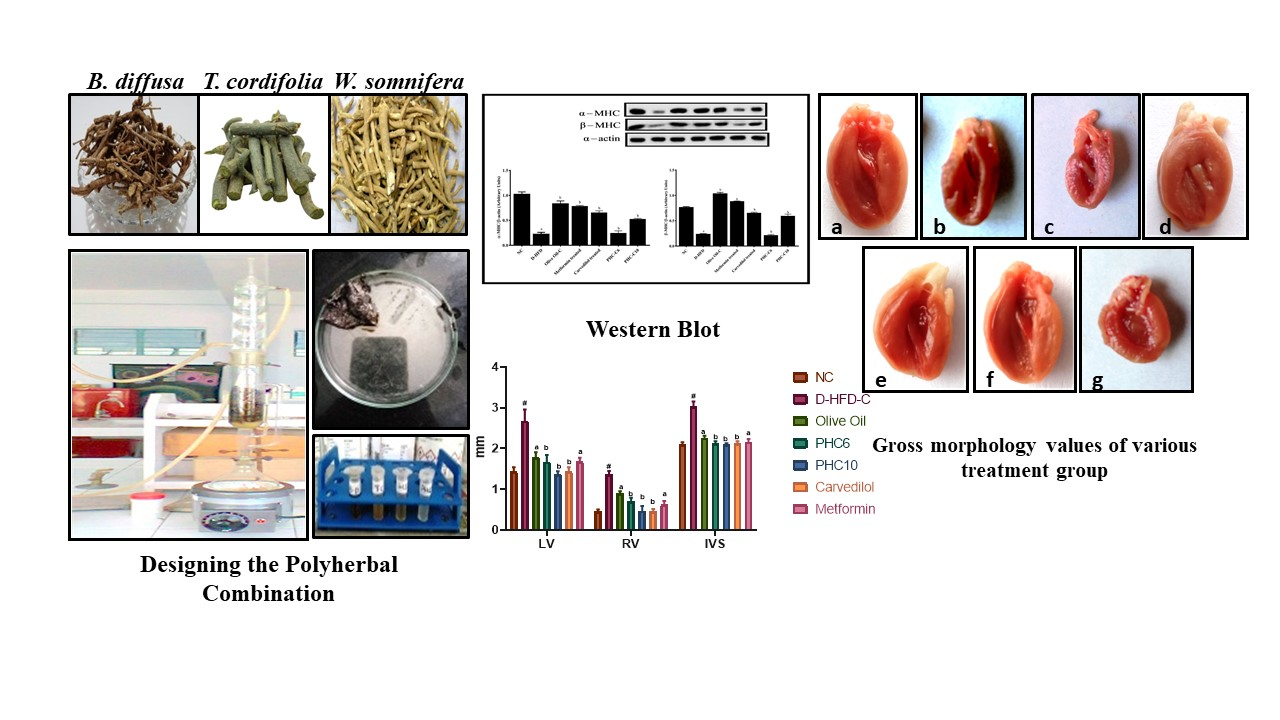
2.5.2. α/β. Myosin Heavy Chain (MHC) Expression (Western Blot)

Ventricular portion (approx. ×50 mg) from heart tissue, frozen at −80 °C, was homogenized with chilled lysis buffer (1:10 w/v containing; 62.5 mM Tris, pH 7.5, 5% beta-mercaptoethanol, and 10% glycerol) in Teflon-shrouded glass homogenizing tube with the help of polypropylene pestle at 40 °C. SDS (1% w/v) and protease inhibitor blended cocktail (Sigma, Ronkonkoma, NY, USA; 10 µL/mL) was added to the homogenate to limit any protein breakdown. The homogenate was filtered through 20 gauge needle and agitated for 30 min at 4 °C. Samples were centrifuged at 10,000× g at 4 °C for 10 min. Supernatant was taken, and protein content was evaluated (Bradford, 1976). The supernatant was mixed in with an identical volume of gel stacking support (Tris100 mM, pH 6.8; SDS (4% w/v); bromophenol blue (0.2% w/v); beta-mercaptoethanol (200 mM), and glycerol (20% v/v)] and further denatured by warming for 3 min. SDS-PAGE was performed using the strategy by Jaiswal et al. [25]. Western blot was performed using rat MHC monoclonal antibody (1:1000) (Santa Cruz Biotechnology Inc., Santa Cruz, CA, USA, Cat # sc32733) and goat anti- mouse horseradish peroxidase as secondary antibody (1:2000) (Pierce Inc., Henrietta, TX, USA, Cat # 31430). The blot was obtained by reacting with hydrogen peroxide and diaminobenzidine. The blot was dried and filtered. The groups were separated and the examined picture of Western blot and densitometry for indistinguishable area of groups was completed utilizing the ImageJ program (Version 1.42, NIHUSA) as shown in the results section.

2.5.3. Troponin-T

Troponin-T and Troponin-I are known as cardiac troponins because they are present in heart and skeletal muscle. Heart proteins are blended and released out of cardiovascular muscle. These proteins collaborate with tropomyosin to frame the principal architecture of the striated heart muscle. Cardiovascular troponin (cTn) controls myocardial constriction by monitoring the calcium-driven association/dissociation of actin and myosin filaments. cTn has numerous isoforms.
These proteins are available in myocytes, the cytosolic pool, and the contractile fibers. The concentration of cTn present in the cytosolic pool is as much as that of CK-MB, though there is a high concentration of cTn in the contractile fibers as well.
Hence, the content of cTn per gram of myocardium is 13–15 times more than that of CK-MB. This explains the higher level of cTn present in contrast to CK-MB in the early period of an infarction episode and the raised degree of cTn in fringe blood, whereas the normal level of CK-MB being expressed when myocardial tissue damage is <1 g (because of ischemia, infarct, injury, poisonous harm, or irritation), making it a highly sensitive biomarker of cardiac tissue damage. Consequently, the WHO expressed that it is a significant and highest-quality-level biochemical marker for the confirmation of heart tissue injury [26]. In this study, Roche Trop-T unit was utilized to test the blood of 244 variously treated groups, as displayed in the results section. The blood from different groups (0.1 mL–0.5 mL) was put in the well of test pack and left undisturbed for 20–30 min. The appearance of two lines on the kit plate showed positive results and indicated the presence of Troponin in the blood sample, whereas negative results were reflected as a single line only, as shown in the results section.

2.5.4. Gross Morphological Studies of Whole Heart; Grading of Heart, Ventricle Wall and Intraventricular Septum Thickness

The hearts were dissected, washed out with ice-cold saline solution, blotted with filter paper, then measured and photographed for heart grading. The cardio morphology was evaluated for inflammation, redness, capillary dilatation, development of lesions, color in all parts of the heart and grading [27]. Hearts with absolutely normal cellular architecture were graded “0”, and those showing maximum damage as a result of exposure to disturbed glycogenic metabolism were graded “4”. All of the other results were in between these grades. The heart was slit open longitudinally in two parts, followed by estimation of left ventricular muscle wall thickness (LV), right ventricular muscle wall thickness (RV) and intraventricular septum thickness (IVS), as displayed in the results section.

2.5.5. Cardiac Biomarker Enzymes

The analysis of CK-MB was performed by using a diagnostic kit manufactured by Agappe Diagnostic, Kerala, India. The analysis was performed by utilizing the immuno-inhibition methodology. Creatine kinase (CK) is present in 3 isotypes: CK-MM located predominantly in the skeletal muscle; CK-BB present in brain and smooth muscle tissues; and CK-MB found mainly in the myocardium. During a heart attack, CK-MB is released into the bloodstream and can be seen for a few days in blood. The method includes the assessment of CK activity to CK-M monomer in the presence of an antibody. We then used the CK test to quantitatively evaluate the activity of CK-B. By multiplying the value obtained for CK-B activity by two, the CK-MB activity can be obtained. The instrument was calibrated using distilled water or deionized water. Mix and incubate at 37 °C for 100 s, add sample 10 μL, mix well and incubate for 2 min at 37 °C, measure the difference in absorbance per minute (OD/min) (340 nm) at an interval of 100 s for 5 min (with a total of five readings per group taken).
Normal concentration levels of ALT in the blood are low; however, when damaged, the heart releases more ALT into the blood, causing the concentration levels to rise. When diagnosing for cardiac damage, the root cause of the damage can be established, such as disease, drug, or injury. Alanine transaminase (ALT) catalyzes the transamination of L-alanine and α-ketoglutarate to form pyruvate and L-glutamate. In subsequent reaction, lactate dehydrogenase (LDH) reduces pyruvate to lactate with simultaneous oxidation of nicotinamide adenine dinucleotide (reduced) (NADH) to nicotinamide adenine dinucleotide (NAD). The rate of oxidation of NADH was evaluated kinetically by observing the reduction in absorbance at 340 nm, and it was directly proportional to ALT action in the sample. The endogenous sample pyruvate was quickly and entirely reduced by LDH during the preliminary incubation period, so that it would not hinder the assay. The estimation protocol for alanine transaminase (ALT) is mentioned in Table 2.
Lactate dehydrogenase (LDH) is found in many body tissues, particularly heart, liver, skeletal muscle, kidney and RBCs. LDH exists in the form of isoenzymes, which are based on their electrophoretic movement, with each isoenzyme being mostly from different organs. Increased levels are found in myocardial infarction, pulmonary diseases, hepatic diseases, hemolytic anemias, renal diseases and muscular dystrophy.
Lactate dehydrogenase promotes the catalysis of pyruvate, converting it into lactate with simultaneous oxidation of reduced NADH to NAD. The degree of oxidation of NADH to NAD is measured as a decrease in absorbance due to formation of NAD, as measured at 340 nm, and is proportional to the LDH activity in the sample. The estimation protocol for lactate dehydrogenase (LDH) has been mentioned in Table 3.

3. Results

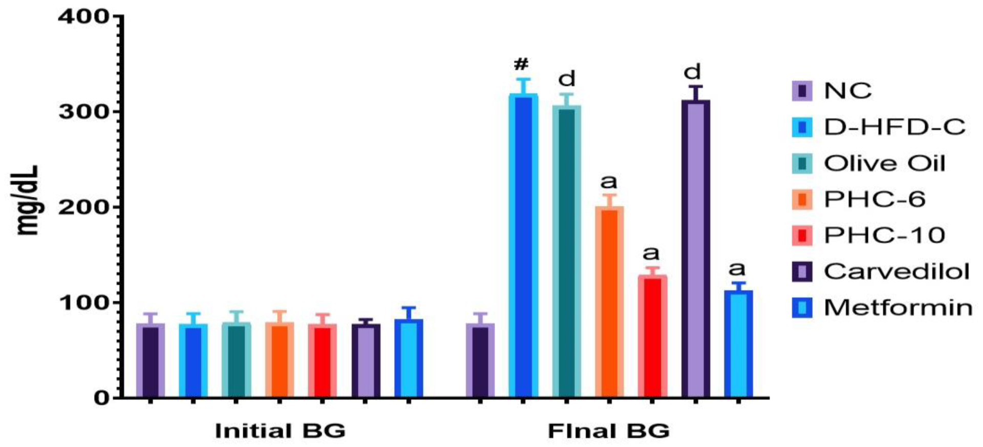
3.1. Initial and Final Blood Glucose Levels

The blood glucose levels of each treatment group were recorded in the beginning as well as at the end of the study. The blood glucose level of the D-HFD-C group was found to be elevated when compared with that of the normal control group (# p < 0.0001), indicating a successful induction of diabetes. The groups treated with the polyherbal combination showed promising results, where a very highly significant decrease in elevated glucose levels was recorded when compared with the D-HFD-C group (**** p < 0.0001). The results were comparable to the diabetic standard metformin. Results for the group treated only with carvedilol, a cardioprotective standard, reflected only a minor significant difference when compared with the D-HFD-C group, as shown in Figure 1 reflecting the influence of carvedilol on carbohydrate metabolism.

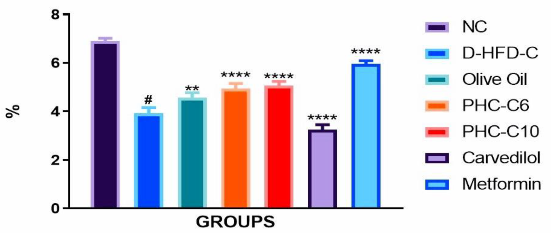
3.2. Food Efficiency Ratio

The food efficiency ratio (FER) was calculated for each treatment group as: (total body weight/total food intake during the study period) ×100. Each day, the feed to be given to the animals was weighed, and the next day the unconsumed feed was weighed again; the difference in the two values was recorded as food consumed per day by each experimental group. The average body weight of each group of animals was recorded to calculate the FER for each group. The FER was well controlled in the polyherbal combination treated groups and found to be very highly significantly different (**** p < 0.0001) from that of the D-HFD-C group. The results were found to be comparable with those of the standard metformin treated groups, as shown in Figure 2.

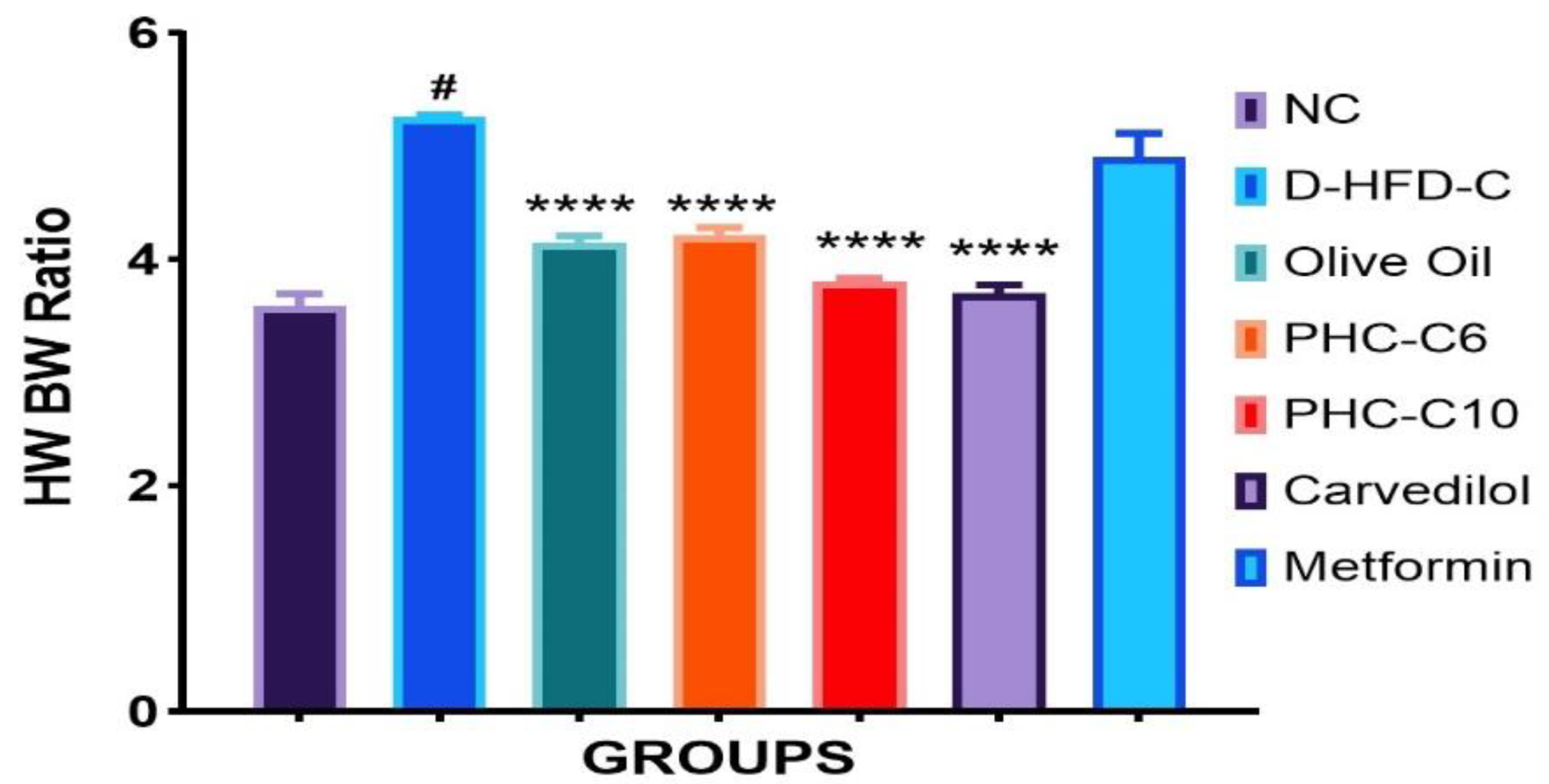
3.3. Heart Weight/Body Weight Ratio

Body weight did not differ between normal and myopathic heart rats, while the heart weight/body weight ratio was considerably different. The heart weight/body weight ratio was found to be well controlled in the polyherbal combination treated groups and found to be very highly significantly different (**** p < 0.0001) from that of the D-HFD-C group. The results were found to be comparable with those of the standard metformin treated groups, as shown in Figure 3.

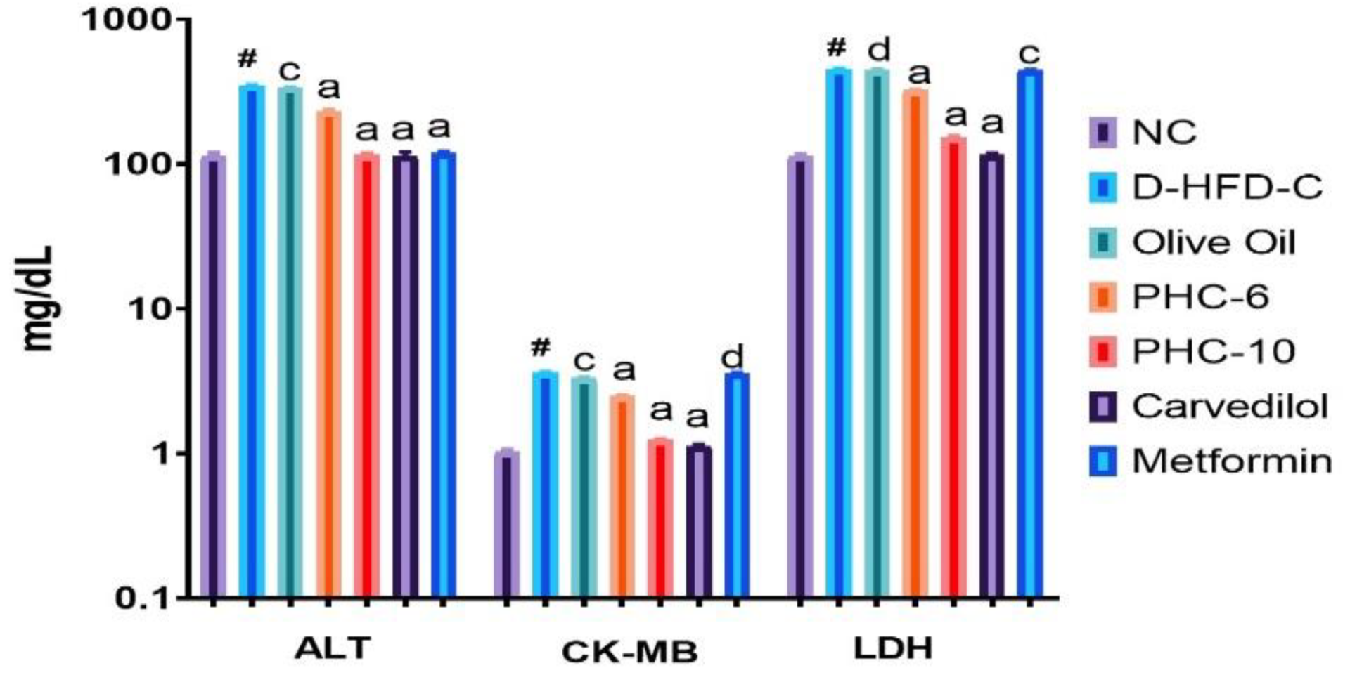
3.4. Cardiac Biomarker Enzymes

The levels of three biomarker enzymes—alanine transaminase (ALT), creatinine kinase-myoglobin (CK-MB), and lactate dehydrogenase (LDH)—in serum were assessed using UV-spectroscopic analysis following the procedures mentioned in the test kits for the respective enzymes. The results for the D-HFD-C group showed a marked rise in the blood/plasma concentration of the mentioned biomarkers when compared with the normal control group (# p < 0.0001). The controlled/decreased level of ALT in all treatment groups (PHC6 and PHC10) reflected very highly significant control over diseased conditions (Figure 4) when compared with the D-HFD-C group (a p < 0.0001). The metformin being an antidiabetic standard, the metformin-alone treatment group reflected the least significant difference (d p < 0.05) when compared with the D-HFD-C group in terms of CK-MB and LDH levels (Figure 4), showing its role only as an antidiabetic drug. However, the results for all other treatment groups, PHC6, PHC10 and carvedilol were well controlled and found to be very highly significantly different (a p < 0.0001) when compared with the D-HFD-C group.

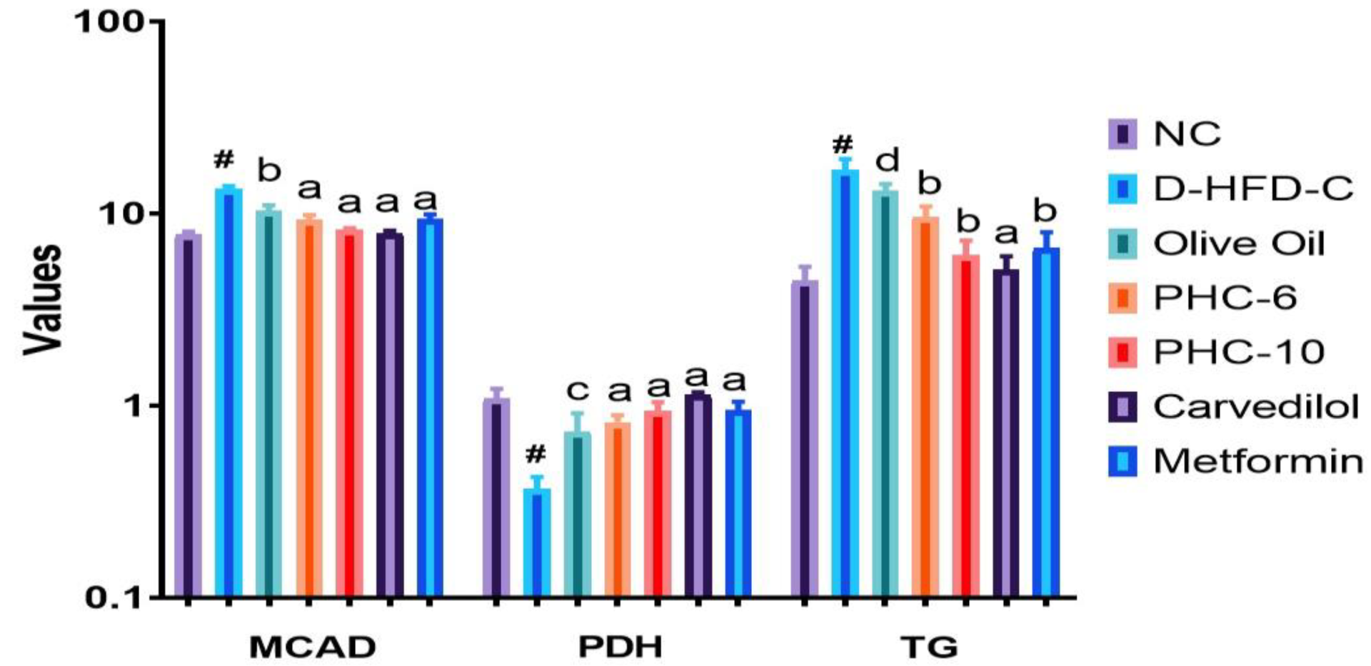
3.5. Estimation of Cardiac Markers

Cardiac intracellular substrate stores or cardiac triglyceride concentrations were highly significantly (# p < 0.0001) increased by STZ administration in the diabetic high-fat diet control group (D-HFD-C), compared with normal control rats. The olive oil treatment group showed a significant decrease in cardiac triglycerides (* p < 0.05), whereas the polyherbal combination treated groups (PHC6 and PHC10) and those treated with the standards carvedilol (2 mg/kg) or metformin (70 mg/kg) showed very significant declines (*** p < 0.001) in cardiac triglyceride content, as shown in Figure 5.
Heart pyruvate dehydrogenase (PDH) action, a vigorously controlled compound of mitochondrial glucose digestion [22], was essentially diminished in STZ tested rodents, in contrast to the normal control. The relationship observed between PDH action and STZ dosing was found to be inversely proportional. PDH concentrations were highly significantly (# p < 0.0001) decreased by STZ + HFD challenged rats (D-HFD-C) when compared with normal control rats.
The olive oil treatment group showed a significant increase in PDH activity (*p < 0.05) whereas the polyherbal combination treated groups (PHC6 and PHC10) and groups treated with the standards carvedilol (2 mg/kg) or metformin (70 mg/kg) showed very significant increases (*** p < 0.001) in PDH activity, as shown in Figure 5.
Medium chain acyl coenzyme-A dehydrogenase (MCAD), an enzyme involved in fatty acid β-oxidation [15,22], was significantly increased in STZ challenged rats, compared with normal control. These changes in MCAD activity were also assessed in polyherbal combination treated groups (PHC6 and PHC10) as well as the standard (carvedilol: 2 mg/kg or metformin: 70 mg/kg) treated groups. There was a significant correlation between MCAD activity and STZ + HFD treatment in the D-HFD-C group. MCAD concentrations were highly significantly (# p < 0.0001) increased in STZ + HFD challenged rats (D-HFD-C) when compared with normal control rats. The olive oil treatment group showed a very significant decrease (*** p < 0.001) in MCAD activity whereas the polyherbal combination treated groups (PHC6 and PHC10) and those treated with the standards carvedilol (2 mg/kg) or metformin (70 mg/kg) showed very significant declines (*** p < 0.001) in MCAD activity, as shown in Figure 5.

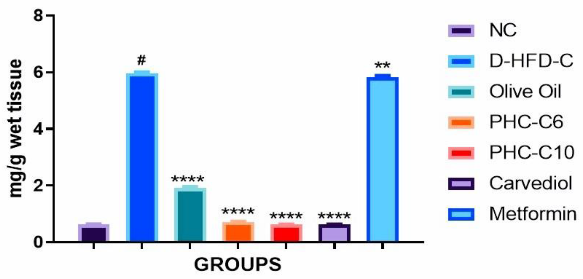
3.6. Cardiac Collagen Content

Total collagen content concentrations were highly significantly (# p < 0.0001) increased in STZ + HFD challenged rats (D-HFD-C) when compared with normal control rats. The olive oil treatment group showed a significant decrease (**** p < 0.0001) in collagen content, whereas the polyherbal combination treated groups (PHC6 and PHC10) and those treated with the standards carvedilol (2 mg/kg) or metformin (70 mg/kg) showed only a significant reduction (**p < 0.01) in cardiac collagen content. The results are shown in Figure 6.

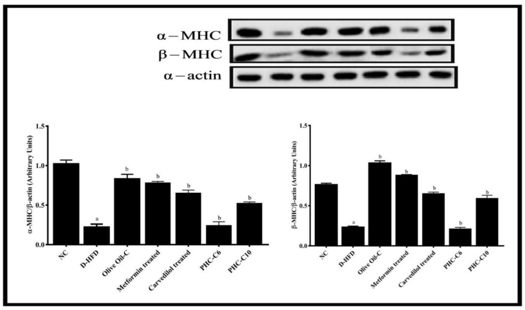
3.7. α/β. Myosin Heavy Chain (MHC) Expression (Western Blot)

The effect of polyherbal combinations in STZ + HFD challenged rats was supported by performing Western blotting analysis, and the results suggested that the expression of α-myosin heavy chain (α-MHC) and β-myosin heavy chain (β-MHC), which are major myofibrillar proteins, was highly significantly (a p < 0.001) reduced in STZ + HFD challenged rats (D-HFD-C). Thus, STZ and HFD attenuated the level of α-MHC and β-MHC in rat heart, while the levels of α-MHC and β-MHC in the standard-treated groups (carvedilol or metformin) showed highly significant increases (a p < 0.001), the level of α-MHC in polyherbal combination treated groups (PHC6 and PHC10) showed a significant (b p < 0.05) increase, the level of β-MHC in the PHC6 group showed a significant increase (b p < 0.05), and in the PHC10 group, a very significant (p < 0.01) increase when compared to the D-HFD-C group. The increase in myosin heavy chain (MHC) expression was more significant in the PHC10 than the PHC6 polyherbal combination treated group when compared to the D-HFD-C group (b p < 0.05), as shown in Figure 7.

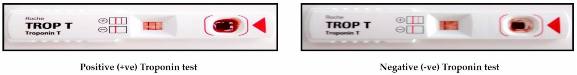
3.8. Troponin-T Test

The blood from different groups (0.1 mL–0.5 mL) was put in the well of test pack and left undisturbed for 20–30 min, reference images of test results attached as Figure 8. The appearance of two lines on the kit plate showed positive results and indicated the presence of Troponin in the blood sample, whereas negative results were reflected as a single line only, as shown in Table 4 below.

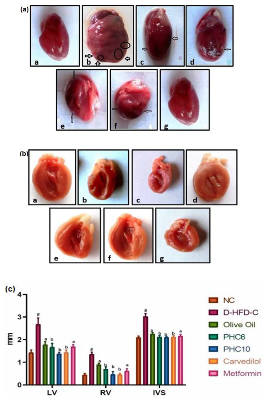
3.9. Gross Morphological Studies of Whole Heart; Grading of Heart, Ventricle Wall and Intraventricular Septum Thickness

Results showed normal morphological features in the normal control group. The morphological appearance in PHC6 and PHC10 treated groups was comparable to the that in the standard carvedilol group, while the STZ + HFD (D-HFD-C) treated rodents showed tissue damage, disaggregation, vacuolation and degenerative changes in heart tissue, as displayed in Figure 9a and accordingly the grading of cardiac tissues was done as shown in Table 5, where the highest level cardiac tissue damage corresponds to Grade 4 and least to Grade 1.
The As displayed in Figure 9b, the difference in the left ventricular wall thickness (LV) was observed to be statistically highly significant (# p < 0.001) when STZ + HFD challenged rats (D-HFD) were compared to the normal control group (NC). The polyherbal combination treated groups (PHC6 and PHC10) showed a significant improved (PHC6 and PHC10; b p < 0.0001) decrease in left ventricular wall thickness when compared to the D-HFD group, respectively. The results for the standard carvedilol treated group showed a statistically highly significant (b p < 0.0001) reduction in left ventricular wall thickness when compared to the D-HFD group, eliciting a response equivalent to those of the PHC groups.
The difference in the right ventricular wall thickness (RV) was observed to be highly significant (# p < 0.0001) in STZ + HFD challenged rats when compared to the normal control group (NC). The polyherbal combination treated groups (PHC6 and PHC10) showed a statistically very significant (a p < 0.001) decrease in RV when compared to the STZ + HFD challenged group (D-HFD), respectively, while the standard carvedilol and metformin treated groups showed statistically very highly significant (b p < 0.0001) reductions in RV when compared to the D-HFD group, eliciting responses equivalent to those of the PHC groups.
The difference in the intraventricular septum thickness (IVS) was observed to be statistically highly significant (# p < 0.0001) in the STZ + HFD challenged rats (D-HFD) when compared to the normal control group (NC). The polyherbal combination treated groups (PHC6 and PHC10) showed a statistically significant (a p < 0.001) decrease in IVS thickness when compared to the D-HFD group, while the standard carvedilol and metformin treated groups showed statistically very highly significant (b p < 0.0001) reductions in IVS when compared to the D-HFD treated group. The results for LV, RV and IVS thickness were plotted and graphically analyzed, as depicted in Figure 9c.

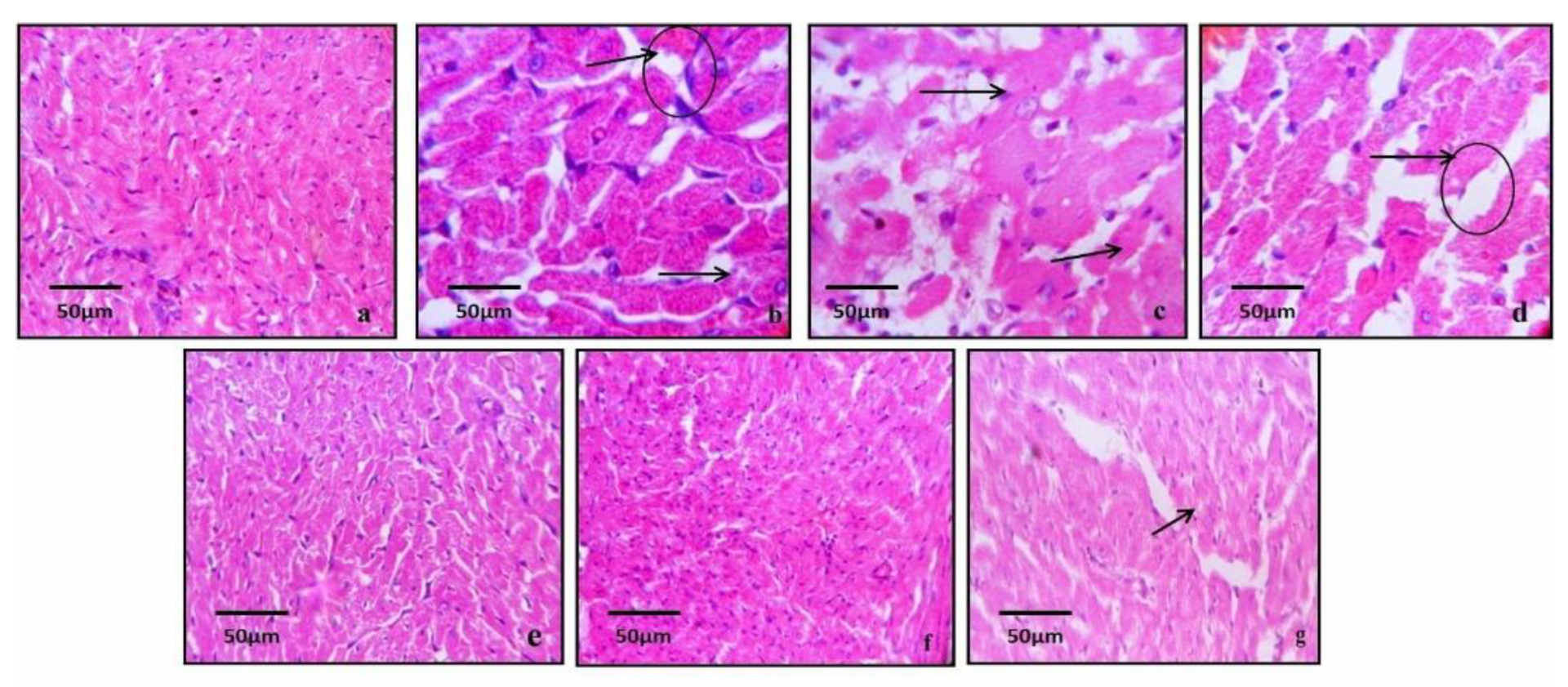
3.10. Histopathological Studies

For histopathological studies, the samples were sent to Alpine Diagnostics Centre, Lucknow. Photomicrographs of rodent hearts of the normal control group showed the least interstitial tissue, long shaft molded vascular cores, muscle striation very much stamped, hardly any small veins and the least fibro-fatty tissue, as seen in Figure 10. There were no strong signs of hypertrophy or confirmations of infarction and normal cellular architecture in normal control group rodents. Photomicrographs of rodent hearts from the HFD + Streptozotocin treated group (D-HFD-C) showed muscle strands approximately organized with broken and expanded interstitial tissue, long axle molded vascular cores and large hollow lumps. However, the carvedilol and polyherbal combination (PHC-C6 and PHC-C10) treated groups exhibited significant improvement, minimal and organized muscle strands with the least interstitial tissue, long shaft molded vascular cores, and muscle striations all around well stamped. H&E staining was used for preparing tissue slides. All photographs were taken on 40× and 50 µm scalar bar.

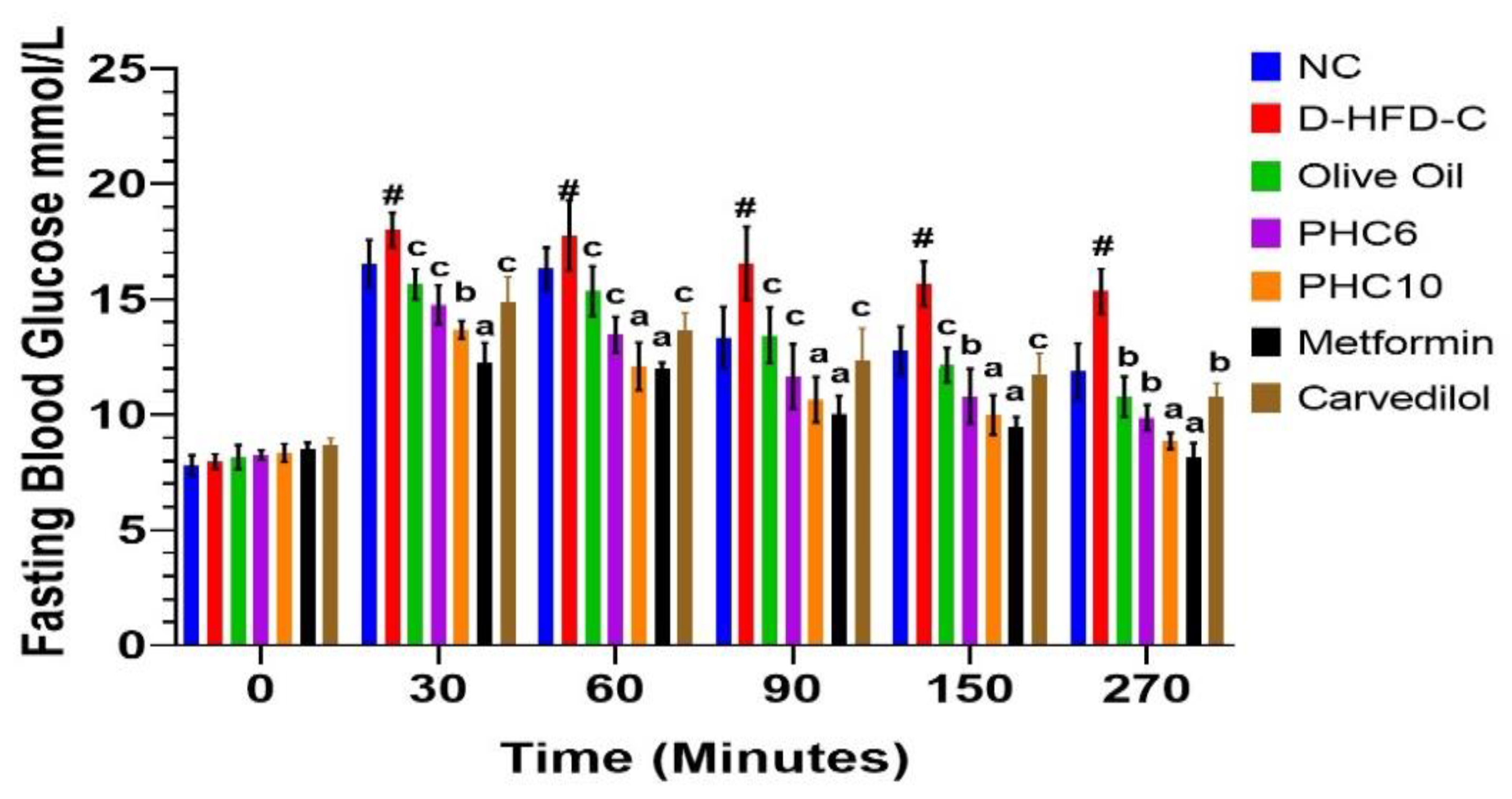
3.11. Oral Glucose Tolerance Test (OGTT)

The serum blood glucose level was obtained by pricking the tail vein using glucometer at times 0, 30, 60, 90, 150 and 270 min. In the STZ + HFD treated hyperglycemic rats, PHC6 showed maximum reduction of blood glucose level at the 60 min time point of the experiment. On the other hand, maximum reduction was observed for PHC10 at 30 min in the experiment. Metformin was used as a standard drug for comparative studies, and the results for the PHC were found quite promising in relation to the standard. Our results indicate that the PHC is relatively potent in lowering blood glucose levels, as shown in Figure 11 below.

4. Discussion

Most of the conventional Indian systems of medicine proclaim different herbs that are known to have cardioprotective and antidiabetic properties but that have unfortunately failed to attract much scientific attention aimed at pragmatically confirming their true pharmacological potential. Picking leads from this vast herbal plethora, the current study suggests and validates cardioprotective and antidiabetic properties of a newly developed polyherbal combination (PHC) comprising Tinospora cordifolia, Boerhavia diffusa and Withania somnifera based in olive oil and administered to rodents in two dose levels (PHC6 and PHC10) for assessment of their potential to alleviate the diabetic cardiomyopathy induced using an STZ + HFD screening model. The current research shows that pretreatment with the created polyherbal combination altogether forestalled diabetes-induced myocardial harm in STZ and high-fat diet (HFD) treated rodents and effectively diminished the levels of demonstrative marker proteins. The current study used the model proposed by Reed et al. [23], who recommended that if a low dose of STZ was administered and followed with a high-fat diet, the capacity of the β-cell mass would be modestly debilitated without totally compromising insulin discharge, bringing about a moderate weakness in glucose resilience. This would imitate the human diabetic condition, achieving a metabolic state like that in late-stage type II diabetic patients. This model has become progressively well-known as of late, both for exploring the complications with type II diabetes and for testing possible treatments. The model has been supplemented with several variations. For instance, Reed et al. administered 50 mg/kg STZ through an intravenous course following sedation, Srinivasan et al. [21]. utilized 35 mg/kg STZ given intraperitoneally but utilizing generally adolescent rodents, whereas Zhang et al. [22] placed the rats on a high-fat diet regimen for a very long time before STZ treatment.
In this work, rodents were separated into seven distinct groups; The normal control group was kept on a pellet diet (NPD), and all other treatment groups were kept on STZ + HFD. HFD-fed groups were administered with STZ to notice the joint impact of all of the pathogenetic potentiating factors. Each treated group, except for the normal control, followed a similar dietary routine, HFD + STZ. The D-HFD (STZ + HFD) group was contrasted to normal control (NC), and various treatment groups (test and standard) were contrasted to D-HFD. The experimental study showed that STZ lifted blood glucose levels radically in D-HFD because of the presence of all potentiating factors, i.e., STZ and HFD. The two groups treated with polyherbal combination (PHC6 and PHC10, individually) showed noticeable improvement against STZ + HFD induced diabetes (a p < 0.001), as blood glucose levels were viewed to be significantly reduced when compared to D-HFD treated animals.
The food efficiency ratio (FER) is a paradigm marker to identify metabolic deformities. It was calculated as (total body weight/total food intake during the study period) – 100 for each treatment group and normal control group, respectively. The FER of the metformin treated group was found to be highly significantly improved when compared to the D-HFD (****p < 0.0001) group. The polyherbal combination treatment (PHC-C6 and PHC-C10, respectively) groups also showed very significant improvement in FER when compared to the D-HFD (*** p < 0.001) group, almost comparable to that of the standard. This suggests that the PHC was equally as potent as the clinically used drug in improving the metabolic conditions of the rodents.
To observe the effects on cardiac glucose metabolism, we assessed various parameters that are reported to be clear markers of altered cardiac functioning due to impaired glucose metabolism. Grading of the heart is one such parameter. The heart samples of each treatment group were isolated at the end of the study by euthanizing the animals to study the impact of disease on cardiac muscles and observe the degree of improvement by subsequent treatment with PHC. STZ with HFD induced significant myocardial damage in the D-HFD group, as observed in contrast to normal rats (NC), as grading showed shifts from grade 0 to 4. The polyherbal combination PHC10 treatment showed more cardioprotective activity compared to that in the D- HFD group, as it displayed a shift from grade 4 to grade 1, which was comparable to that of the Carvedilol standard group, indicating its potent cardioprotective effect, while PHC6 showed lower cardioprotective activity compared to PHC10, with a shift from grade 4 to grade 2 compared to the D-HFD group.
The heart weight/body weight ratio is a very important parameter of cardiac hypertrophy. The polyherbal combination PHC6 showed a significant protective effect (** p < 0.01), but the polyherbal combination PHC10 showed very significant cardioprotection against STZ + HFD induced damage (*** p < 0.001), which was also comparable to that of the Carvedilol standard group, indicating its potent cardioprotective effect.
Biochemical parameters were evaluated by assessment of various enzymatic factors, such as alanine aminotransferase (ALT), creatinine kinase-myoglobin (CK-MB) and lactate dehydrogenase (LDH) [28,29] and the gold marker of cardiac damage, i.e., troponin-T. The levels of these cardiac marker enzymes were found to be most elevated in the diabetic high-fat diet group (D-HFD) clearly due to the presence of all potentiating factors i.e., STZ and HFD.
The polyherbal combinations (PHC6 and PHC10) yielded a marked improvement in the biochemical parameters, indicating cardioprotective action following treatment. The results for the PHC treated groups were found to be statistically different at various levels of significance when compared to the D-HFD group: ALT (b p < 0.001), CK-MB (a p < 0.0001).
Cardiac pyruvate dehydrogenase (PDH) activity, a heavily regulated enzyme of mitochondrial glucose metabolism, was significantly decreased in D-HFD rats, compared with that in NC rats (# p < 0.0001). The olive oil treatment group showed a significant increase in PDH activity (d p < 0.05), whereas the polyherbal combination treated groups (PHC6 and PHC10) and those treated with the standards carvedilol (2 mg/kg) or metformin (70 mg/kg) showed very significant increases (a p < 0.001) in PDH activity. This illustrates and reaffirms that PHC treatment improves cardiac glucose metabolism, and the results are comparable to those obtained with the standard carvedilol.
Cardiac triglyceride concentrations were highly significantly (# p < 0.0001) increased by STZ administration in the diabetic high-fat diet control group (D-HFD-C), compared with normal control rats. The olive oil treatment group showed a significant decrease in cardiac triglycerides (d p < 0.05), whereas the polyherbal combination treated groups (PHC6 and PHC10) and the groups treated with standards carvedilol (2 mg/kg) or metformin (70 mg/kg) showed very significant decreases (b p < 0.001) in cardiac triglyceride content, a clear indication of the cardioprotective efficacy of the PHCs.
Medium chain acyl coenzyme A dehydrogenase (MCAD) enzyme is involved in fatty acid β-oxidation and was significantly increased in STZ + HFD challenged rats when compared with normal control. These changes in MCAD activity were also assessed in the polyherbal combination treated groups (PHC6 and PHC10) as well as the standard groups (carvedilol (2 mg/kg) or metformin (70 mg/kg)). There was a significant correlation between MCAD activity and STZ + HFD treatment in the D-HFD-C group. MCAD concentrations were highly significantly (# p < 0.0001) increased in STZ + HFD challenged rats (D-HFD-C) when compared with normal control rats. The olive oil treatment group showed a very significant decrease (***p < 0.001) in MCAD activity, whereas the polyherbal combination treated groups (PHC6 and PHC10) and both standard-treated groups (carvedilol (2 mg/kg) or metformin (70 mg/kg)) showed very significant decreases (***p < 0.001) in MCAD activity.
The total cardiac collagen content was derived from hydroxyproline concentration. Total collagen content concentrations were highly significantly (# p < 0.0001) increased in STZ + HFD challenged rats (D-HFD-C) when compared with normal control rats. The olive oil treatment group showed a very significant decrease (****p < 0.0001) in collagen content, whereas the polyherbal combination treated groups (PHC6 and PHC10) and both standard treated groups (carvedilol (2 mg/kg) or metformin (70 mg/kg)) showed very highly significant reductions (***p < 0.001) in cardiac collagen content.
The effect of polyherbal combinations in STZ + HFD challenged rats was supported by Western blotting analysis, and the results suggested that the expression of α-myosin heavy chain (α-MHC) and β-myosin heavy chain (β-MHC), which are the major myofibrillar proteins, was highly significantly (p < 0.001) reduced in STZ+HFD challenged rats (D-HFD-C). Thus, STZ and HFD attenuated the level of α-MHC and β-MHC in rat heart, while the level of α-MHC and β-MHC in the standard groups (carvedilol or metformin) showed highly significant increases (p < 0.001), the level of α-MHC in polyherbal combination treated groups (PHC6 and PHC10) showed a significant (p < 0.05) increase, and the level of β-MHC in the PHC6 and PHC10 groups showed significant (p < 0.05) and very significant (p < 0.01) increases, respectively, when compared to the D-HFD-C group. The increase in myosin heavy chain (MHC) expression was more significant in the PHC10 than PHC6 combination treated group in comparison to the D-HFD-C group (p < 0.05) (Figure 7).
The diabetic groups showed positive troponin test results, whereas the pretreatment groups, namely those treated with the standard drugs and polyherbal combinations (PHC6 and PHC10), showed significant protection and negative troponin test results.
Myocardial damage was assessed by myocardial thickness evaluation, i.e., left ventricle (LV), right ventricle (RV) and intraventricular septum thickness (IVS). The increase in left ventricular wall thickness (LV) was observed to be statistically highly significant (# p < 0.001) in STZ + HFD challenged rats (D-HFD) when compared to the normal control group (NC). The polyherbal combination treated groups, PHC6 and PHC10, showed statistically very significant (a p < 0.001) and statistically highly significant (b p < 0.0001) decreases in left ventricular wall thickness when compared to the D-HFD group, respectively. The standard carvedilol treated group showed a statistically highly significant (a p < 0.001) reduction in left ventricular wall thickness when compared to the D-HFD group.
The increased right ventricular wall thickness (RV) was observed to be statistically highly significant (# p < 0.0001) in STZ + HFD challenged rats when compared to the normal control group (NC). The polyherbal combination treated groups, (PHC6 and PHC10, showed statistically very significant (a p < 0.001) and statistically highly significant (a p < 0.001) decreases, respectively, in RV when compared to the STZ + HFD challenged group (D-HFD), while the standard (carvedilol or metformin) treated groups showed statistically very highly significant (b p < 0.0001) reductions in RV when compared to the D-HFD group.
The increased intraventricular septum thickness (IVS) was observed to be statistically highly significant (# p < 0.0001) in the STZ + HFD challenged rats (D-HFD) when compared to the normal control group (NC). The polyherbal combination treated groups, PHC6 and PHC10, showed statistically significant (a p < 0.001) decreases in IVS thickness when compared to he D-HFD-C group, while the standard (carvedilol or metformin) treated groups showed statistically very highly significant (b p < 0.0001) reductions in IVS when compared to the D-HFD-C treated group.
The net morphology findings and histopathological reports convincingly demonstrated that the evaluated D-HFD model after 28 days induced strong myocardial harm, in contrast with NC.
The pretreatment groups, i.e., standard and polyherbal combination treated groups, showed strong protection against diabetes-induced cardiovascular harm. The standard medication (carvedilol (2 mg/kg) or metformin (70 mg/kg)) treated groups as well as the polyherbal blend-treated groups showed extensive improvement, minimally organized muscle strands with the least interstitial tissue, long axle molded vascular cores, and muscle striations stamped all around. This morphological architecture of cardiac tissue was equivalent to that in animals treated with the two clinically approved drugs (Carvedilol (2 mg/kg) or metformin (10 mg/kg)).

5. Statistical Analysis

The results for various parameters assessed during the study were statistically analyzed in GraphPad Prism Version 9(a) software. All values were expressed as mean ± SD (n = 6) and analyzed by one-way ANOVA followed by Tukey’s t-test.

6. Conclusions

The present study showed that pretreatment with developed polyherbal combinations significantly prevented STZ and HFD induced myocardial injury in experimental animals, lowered the levels of diagnostic marker enzymes, improved antioxidant activity and prevented cardiac tissue damage. PHC6 showed moderate protection against STZ + HFD induced damage, which was seen clearly in the gross examination of heart and histopathology. PHC10 showed marked protection against STZ + HFD induced damage, which was seen clearly in the gross examination of heart, histopathological studies as well as biochemical estimations of cardiac marker enzymes. The evaluation of various sophisticated parameters and comparison of the polyherbal combination treatment results with that of standard showed and validated the significant role of polyherbal combination as a cardioprotective preparation. The polyherbal combination was found to be not only effective as an antidiabetic but also in diabetic cardiomyopathy induced by STZ and HFD.
Thus, the present study is suggestive of certain observations. High-fat diet along with STZ induced diabetes is a very suitable, potent and short-term model for evaluating diabetic cardiomyopathy in experimental rats [30]. STZ with HFD potently induces diabetic cardiomyopathy in rats as compared to non-diabetic rats, clearly paralleling the clinical presentation. The protective effect of polyherbal combination was evident at both low and high doses (PHC6 and PHC10) against STZ + HFD induced diabetic cardiomyopathy in experimental rats, which was comparable to the effect of clinically established standard drugs, i.e., metformin (10 mg/kg) and carvedilol (2 mg/kg).
On the basis of data in hand, it was concluded that polyherbal combination produced an ameliorative effect in diabetic cardiomyopathy that may be attributed to its herbal constituents’ multiple effects, such as antioxidant, cardioprotective, antidiabetic, antistress, hepatoprotective, antifibrinolytic, and anti-inflammatory activity [31]. Therefore, the polyherbal combination comprising T. cordifolia, W. somnifera and B. diffusa plant extracts based in olive oil was found to have antidiabetic and antioxidant effects and play a prominent role in attenuation of diabetes induced cardiomyopathy and associated anomalies.

Author Contributions

A.S.: investigation, data curation, validation and analysis, original draft preparation, designing and conducting the research. H.H.S.: supervision and expert guidance. T.M.: conceptualization, methodology, supervision. M.H.S.: scientific inputs, facilitation and assistance. F.A.: literature review and collecting resources. M.S.: software handling, visualization. S.P.: editing the manuscript. T.A.W. and S.Z.: supporting the research through their inputs. A.F.: Providing laboratory infrastructure to carry out certain experiments. M.W.: Overall restructuring and analysing the manuscript. P.W. and A.V.: Providing resources for literature review. All authors have read and agreed to the published version of the manuscript.

Funding

This research received no external funding.

Institutional Review Board Statement

The manuscript communication number provided by the university’s internal manuscript review committee under the aegis of the Faculty of Doctoral Studies, Integral University is IU/R&D/2022-MCN0001482. The research protocol was affirmed by prior approval from the Institutional Animal Ethical Committee (IAEC) of Integral University, Lucknow (U.P.), India, with approval number (IU/IAEC/17/04).

Informed Consent Statement

Not Applicable.

Data Availability Statement

All data related to study have been included in the manuscript.

Acknowledgments

The authors are highly thankful to Honorable Founder and Chancellor, Syed Waseem Akhtar, Integral University and Vice-Chancellor, Javed Musarrat, Integral University, for providing an excellent research environment and facilities. The authors also express their gratitude to Syed Misbahul Hassan, Dean, Faculty of Pharmacy, Integral University for his motivation and support.

Conflicts of Interest

The authors declare no conflict of interest amongst each other.

References

  1. Andersson, C.; Olesen, J.B.; Hansen, P.R.; Weeke, P.; Norgaard, M.L.; Jørgensen, C.H.; Lange, T.; Abildstrøm, S.Z.; Schramm, T.K.; Vaag, A.; et al. Metformin treatment is associated with a low risk of mortality in diabetic patients with heart failure: A retrospective nationwide cohort study. Diabetologia 2010, 53, 2546–2553. [Google Scholar] [CrossRef] [PubMed] [Green Version]
  2. Lundbaek, K.; Ledet, T.; Neubauer, B.; Christensen, N.J. Diabetic cardiopathy. Diabetologia 1979, 16, 207–209. [Google Scholar]
  3. Rubler, S.; Dlugash, J.; Yuceoglu, Y.Z.; Kumral, T.; Branwood, A.W.; Grishman, A. New type of cardiomyopathy associated with diabetic glomerulosclerosis. Am. J. Cardiol. 1972, 30, 595–602. [Google Scholar] [CrossRef]
  4. Bertoni, A.G.; Tsai, A.; Kasper, E.K.; Brancati, F.L. Diabetes and idiopathic cardiomyopathy: A nationwide case-control study. Diabetes Care 2003, 26, 2791–2795. [Google Scholar] [CrossRef] [PubMed] [Green Version]
  5. Dwivedi, V.; Anandan, E.M.; Mony, R.S.; Muraleedharan, T.S.; Valiathan, M.S.; Mutsuddi, M.; Lakhotia, S.C. In Vivo effects of traditional Ayurvedic formulations in Drosophila melanogaster model relate with therapeutic applications. PLoS ONE 2012, 7, e37113. [Google Scholar] [CrossRef] [PubMed] [Green Version]
  6. Fossati, P.; Prencipe, L. Serum triglycerides determined colorimetrically with an enzyme that produces hydrogen peroxide. Clin. Chem. 1982, 28, 2077–2080. [Google Scholar] [CrossRef]
  7. Kuppurajan, K.; Rajgopalan, S.S.; Sitaram, R.; Rajgopalan, V.; Janaki, K.; Revathi, R.; Vekataraghavan, S. Effects of Ashwagandha on the process of ageing on human volunteers. J. Res. Ayur. Sid. 1989, 1, 247–258. [Google Scholar]
  8. Marles, R.J.; Farnsworth, N.R. Antidiabetic plants and their active constituents. Phytomedicine 1995, 2, 137–189. [Google Scholar] [CrossRef]
  9. Aphale, A.A.; Chibba, A.D.; Kumbhakarna, N.R.; Mateenuddin, M.O.; Dahat, S.H. Subacute toxicity study of the combination of ginseng (Panax ginseng) and ashwagandha (Withania somnifera) in rats: A safety assessment. Indian J. Physiol. Pharmacol. 1998, 42, 299–302. [Google Scholar]
  10. Ujowundu, C.O.; Igwe, C.U.; Enemor, V.H.; Nwaogu, L.A.; Okafor, O.E. Nutritive and anti-nutritive properties of Boerhavia diffusa and Commelina nudiflora leaves. Pak. J. Nutr. 2008, 7, 90–92. [Google Scholar] [CrossRef]
  11. Mungantiwar, A.A.; Nair, A.M.; Kamal, K.K.; Saraf, M.N. Adaptogenic activity of aqueous extract of the roots of Boerhaavia diffusa linn. Indian Drugs 1997, 34, 184–189. [Google Scholar]
  12. Vermes, E.; Ducharme, A.; Bourassa, M.G.; Lessard, M.; White, M.; Tardif, J.C. Enalapril reduces the incidence of diabetes in patients with chronic heart failure: Insight from the Studies of Left Ventricular Dysfunction (SOLVD). Circulation 2003, 107, 1291–1296. [Google Scholar] [CrossRef] [PubMed] [Green Version]
  13. Pérez, J.E.; McGill, J.B.; Santiago, J.V.; Schechtman, K.B.; Waggoner, A.D.; Miller, J.G.; Sobel, B.E. Abnormal myocardial acoustic properties in diabetic patients and their correlation with the severity of disease. J. Am. Coll. Cardiol. 1992, 19, 1154–1162. [Google Scholar] [CrossRef] [Green Version]
  14. Malik, V.S.; Popkin, B.M.; Bray, G.A.; Després, J.P.; Hu, F.B. Sugar-sweetened beverages, obesity, type II diabetes mellitus, and cardiovascular disease risk. Circulation 2010, 121, 1356–1364. [Google Scholar] [CrossRef] [PubMed]
  15. Hamdi, H.K.; Castellon, R. Oleuropein, a non-toxic olive iridoid, is an anti-tumor agent and cytoskeleton disruptor. Biochem. Biophys. Res. Commun. 2005, 334, 769–778. [Google Scholar] [CrossRef]
  16. Bisignano, G.; Tomaino, A.; Cascio, R.L.; Crisafi, G.; Uccella, N.; Saija, A. On the in-vitro antimicrobial activity of oleuropein and hydroxytyrosol. J. Pharm. Pharmacol. 1999, 51, 971–974. [Google Scholar] [CrossRef]
  17. Sharma, A.K.; Kishore, K.; Sharma, D.; Srinivasan, B.P.; Agarwal, S.S.; Sharma, A.; Singh, S.K.; Gaur, S.; Jatav, V.S. Cardioprotective activity of alcoholic extract of Tinospora cordifolia (Willd.) Miers in calcium chloride-induced cardiac ar-rhythmia in rats. J. Biomed. Res. 2011, 25, 280–286. [Google Scholar] [CrossRef] [Green Version]
  18. Sumanth, M.; Mustafa, S.S. Antistress, adoptogenic and immunopotentiating activity roots of Boerhaavia diffusa in mice. Int. J. Pharmacol. 2007, 3, 416–420. [Google Scholar] [CrossRef] [Green Version]
  19. Shamim, A.; Siddiqui, H.H.; Mahmood, T.; Siddiqui, M.H.; Bagga, P.; Ahsan, F.; Shariq, M.; Parveen, S. Pragmatic Toxicity Pro-filing of a Salubrious Polyherbal Combination of Tinospora Cordifolia, Withania Somnifera, and Boerrhavia Diffusa in Swiss Albino Mice. Int. J. Res. Pharm. Sci. 2020, 11, 4240–4252. [Google Scholar] [CrossRef]
  20. Reed, M.J.; Meszaros, K.; Entes, L.J.; Claypool, M.D.; Pinkett, J.G.; Gadbois, T.M.; Reaven, G.M. A new rat model of type 2 diabetes: The fat-fed, streptozotocin-treated rat. Metab. Clin. Exp. 2000, 49, 1390–1394. [Google Scholar] [CrossRef]
  21. Srinivasan, K.; Viswanad, B.; Asrat, L.; Kaul, C.L.; Ramarao, P. Combination of high-fat diet-fed and low-dose streptozotocin-treated rat: A model for type 2 diabetes and pharmacological screening. Pharmacol. Res. 2005, 52, 313–320. [Google Scholar] [CrossRef] [PubMed]
  22. Zhang, Y.; Proenca, R.; Maffei, M.; Barone, M.; Leopold, L.; Friedman, J.M. Positional cloning of the mouse obese gene and its human homologue. Nature 1994, 372, 425–432. [Google Scholar] [CrossRef] [PubMed]
  23. Bazoti, F.N.; Bergquist, J.; Markides, K.E.; Tsarbopoulos, A. Noncovalent interaction between amyloid-β-peptide (1–40) and oleuropein studied by electrospray ionization mass spectrometry. J. Am. Soc. Mass Spectrom. 2006, 17, 568–575. [Google Scholar] [CrossRef] [PubMed] [Green Version]
  24. Lee, E.T.; Cowan, L.D.; Welty, T.K.; Sievers, M.; Howard, W.J.; Oopik, A.; Wang, W.; Yeh, J.; Devereux, R.B.; Rhoades, E.R.; et al. All-cause morality and cardiovascular disease mortality in three American Indian populations, aged 45–74 years, 1984–1988: The Strong Heart Study. Am. J. Epidemiol. 1998, 147, 995–1008. [Google Scholar] [CrossRef] [PubMed]
  25. Kubota, N.; Tobe, K.; Terauchi, Y.; Eto, K.; Yamauchi, T.; Suzuki, R.; Tsubamoto, Y.; Komeda, K.; Nakano, R.; Miki, H.; et al. Disrup-tion of insulin receptor substrate 2 causes type II diabetes because of liver insulin resistance and lack of compensatory beta-cell hyperplasia. Diabetes 2000, 49, 1880–1889. [Google Scholar] [CrossRef] [Green Version]
  26. Mansor, L.S.; Gonzalez, E.R.; Cole, M.A.; Tyler, D.J.; Beeson, J.H.; Clarke, K.; Carr, C.A.; Heather, L.C. Cardiac metabolism in a new rat model of type 2 diabetes using high-fat diet with low dose streptozotocin. Cardiovasc. Diabetol. 2013, 12, 136. [Google Scholar] [CrossRef] [Green Version]
  27. McLaughlin, S.; McNeill, B.; Podrebarac, J.; Hosoyama, K.; Sedlakova, V.; Cron, G.; Smyth, D.; Seymour, R.; Goel, K.; Liang, W.; et al. Injectable human recombinant collagen matrices limit adverse remodeling and improve cardiac function after myocar-dial infarction. Nat. Commun. 2019, 10, 4866. [Google Scholar] [CrossRef] [Green Version]
  28. Jaiswal, A.; Kumar, S.; Enjamoori, R.; Seth, S.; Dinda, A.K.; Maulik, S.K. Peripheral benzodiazepine receptor ligand Ro5-4864 inhib-its isoprenaline-induced cardiac hypertrophy in rats. Eur. J. Pharmacol. 2010, 644, 146–153. [Google Scholar] [CrossRef]
  29. Anversa, P.; Sonnenblick, E.H. Ischemic cardiomyopathy: Pathophysiologic mechanisms. Prog. Cardiovasc. Dis. 1990, 33, 49–70. [Google Scholar] [CrossRef]
  30. Rona, G.; Chappel, C.I.; Kahn, D.S. The significance of factors modifying the development of isoproterenol-induced myocardial necrosis. Am. Heart J. 1963, 66, 389–395. [Google Scholar] [CrossRef]
  31. Patil, S.; Chaudhary, A.K. Quantitative Estimation of Guduchi Ghana obtained from different amount of water used for Kwa-tha. Int. J. Pharm. Arch. 2013, 2, 160–164. [Google Scholar]
Figure 1. Initial and final blood glucose levels. All values are expressed as mean ± SD (n = 6) analyzed by one way ANOVA followed by Tukey’s t-test. D-HFD-C was compared with NC (# p < 0.0001) and the results were found to be highly significantly different, whereas the different treatment groups were compared with D-HFD-C, and the results were found to be different at significance levels d p < 0.05; a p < 0.0001. NC is normal control; D-HFD-C is diabetic high-fat diet control group; PHC6 and PHC10 are polyherbal combination treated groups.
Diabetology 03 00043 g001
Figure 2. All values are expressed as mean ± SD (n = 6) analyzed by one-way ANOVA followed by Tukey’s t-test. D-HFD-C was compared with NC (# p < 0.0001), and the result was found to be highly significantly different. The different treatment groups were compared with D-HFD-C, and the results were found to be different at the significance levels ** p < 0.01 and **** p < 0.0001. NC is normal control; D-HFD-C is diabetic high-fat diet control group; PHC6 andPHC10 are the polyherbal combination treated groups.
Diabetology 03 00043 g002
Figure 3. Heart weight/body weight ratio. All values are expressed as mean ± SD (n = 6) analyzed by one-way ANOVA followed by Tukey’s t-test. D-HFD-C was compared with NC (# p < 0.0001), and the result was found to be highly significantly different. The different treatment groups were compared with D-HFD-C, and the results were found to be different at the significance levels and **** p < 0.0001. NC is normal control; D-HFD-C is diabetic high-fat diet control group; PHC6 and PHC10 are polyherbal combination treated groups.
Diabetology 03 00043 g003
Figure 4. Cardiac biomarker enzymes. All values are expressed as mean ± SD (n = 6) analyzed by one-way ANOVA followed by Tukey’s t-test. D-HFD-C was compared with NC (# p < 0.0001), and the values were found to be highly significantly different. The different treatment groups were compared with the D-HFD-C group, with the values expressed in log10 scale on the Y-axis, and the results for each parameter (ALT, CK-MB and LDH) were found to be different at the following significance levels: d p < 0.05; c p < 0.01; a p < 0.0001 from the respective disease control groups. NC is normal control; D-HFD-C is diabetic high-fat diet control group; PHC6 and PHC10 are polyherbal combination treated groups.
Diabetology 03 00043 g004
Figure 5. Cardiac marker activity. All values are expressed as mean ± SD (n = 6) analyzed by one-way ANOVA followed by Tukey’s t-test. D-HFD-C was compared with NC (# p < 0.0001), and the values were found to be highly significantly different. The different treatment groups were compared with D-HFD-C, with the values expressed in log10 scale on the Y-axis, and the results for each parameter (MCAD, PDH and TG) were found to be different at the following significance levels: d p < 0.05; c p < 0.01; b p < 0.001; a p < 0.0001 from the respective disease control groups. NC is normal control; D-HFD-C is diabetic high-fat diet control group; PHC6 and PHC10 are polyherbal combination treated groups.
Diabetology 03 00043 g005
Figure 6. All values are expressed as mean ± SD (n = 6) analyzed by one-way ANOVA followed by Tukey’s t-test. D-HFD-C was compared with NC (# p < 0.0001), and the result was found to be highly significantly different. The different treatment groups were compared with D-HFD-C, and the results were found to be different at the significance levels ** p < 0.01 and **** p < 0.0001. NC is normal control; D-HFD-C is diabetic high fat diet control group; PHC6 and PHC10 are polyherbal combination treated groups.
Diabetology 03 00043 g006
Figure 7. Western Blot α/β Myosin Heavy Chain (MHC) expression. All values are expressed as Mean ± SD (n = 6). The values were analysed bt one way ANOVA & followed by Tukey’s t-test and were found to be significantly different at (a,b) p < 0.05 when D-HFD-C was compared with NC & all other Treatment groups were compared with D-HFD-C. Where, NC is normal control; D-HFD-C is Diabetic High Fat Diet Control; PHC-6 & PHC-10 are polyherbal combination treated groups.
Diabetology 03 00043 g007
Figure 8. Troponin-T Test.
Diabetology 03 00043 g008
Figure 9. (a) Gross Morphology of intact heart from different treatment groups. (b) Gross Morphology of Heart section from different treatment groups. (c) Gross Morphology values of different treatment groups. All values are expressed as mean ±SD (n = 6) analyzed by one-way ANOVA followed by Tukey’s t-test. D-HFD-C was compared with NC (# p < 0.0001), and the values were found to be highly significantly different. The different treatment groups were compared with D-HFD-C, and the results for each parameter (LV, RV and IVS) were found to be different at the following significance levels: b p < 0.001; a p < 0.0001 from the respective disease control groups. NC is normal control; D-HFD-C is diabetic high-fat diet control group; PHC6 and PHC10 are polyherbal combination treated groups; LV is left ventricular wall thickness; RV is right ventricular wall thickness; IVS is intra-ventricular septum thickness).
Diabetology 03 00043 g009
Figure 10. Histopathology of cardiac tissues from different treatment groups. Where, (a). Normal Control; (b). is D-HFD-C; (c). Olive Oil-C; (d). PHC-6 treated; (e). PHC10 treated; (f). Metformin treated; (g). Carvedilol treated.
Diabetology 03 00043 g010
Figure 11. Fasting blood glucose level for OGTT. All values are expressed as mean ± SD (n = 6) analyzed by one-way ANOVA followed by Tukey’s t-test. D-HFD-C was compared with NC (# p < 0.0001), and the values were found to be highly significantly different. The different treatment groups were compared with D-HFD-C and the results were found to be different at the following significance levels: c p < 0.01; b p < 0.001; a p < 0.0001 from the respective disease control groups. NC is normal control; D-HFD-C is diabetic high-fat diet control group; PHC6 and PHC10 are the polyherbal combination treated groups.].
Diabetology 03 00043 g011
Table 1. Experimental Design.
S. No.GroupsNo. of AnimalsTreatment
1.Normal Control6Distilled water 2 mL/kg, p.o. NPD + water ad libitum for the entire period of study (28 days). On day 13, rodents from all the groups including NC were abstained from eating for the time being and given a solitary intraperitoneal infusion of water for injection (WFI)
2.STZ + HFD-C6Single i.p. injection of streptozotocin at the dose of 35 mg/kg b.w. (freshly prepared in citrate buffer). HFD + water ad libitum for the entire period of study (28 days). On day 13, rodents from all groups including NC were abstained from eating for the time being and given a solitary intraperitoneal infusion of streptozotocin (35 mg/kg STZ in citrate buffer, pH 4)
3.Olive Oil6Single i.p. injection of streptozotocin at the dose of 35 mg/kg b.w. (freshly prepared in citrate buffer). The animals were fed with 2 mL olive oil daily, NPD+ HFD + water ad libitum for the entire period of study (28 days). On day 13, rodents from all groups including NC were abstained from eating for the time being and given a solitary intraperitoneal infusion of streptozotocin (35 mg/kg STZ in citrate buffer, pH 4)
4.PHC-C6 Treated (Low dose)6Single i.p. injection of streptozotocin at the dose of 35 mg/kg b.w. (freshly prepared in citrate buffer). The animals were fed with 2 mL PHC-C 6 daily, NPD+ HFD + water ad libitum for the entire period of study (28 days). On day 13, rodents from all the groups including NC were abstained from eating for the time being and given a solitary intraperitoneal infusion of streptozotocin (35 mg/kg STZ in citrate buffer, pH 4)
5.PHC-C10 Treated (High dose)6Single i.p. injection of streptozotocin at the dose of 35 mg/kg b.w. (freshly prepared in citrate buffer). The animals were fed with 2 mL PHC-C 10 daily, NPD+ HFD + water ad libitum for the entire period of study (28 days). On day 13, rodents from all groups including NC were abstained from eating for the time being and given a solitary intraperitoneal infusion of streptozotocin (35 mg/kg STZ in citrate buffer, pH 4)
6.Metformin Treated (70 mg/kg; p.o)6Single i.p. injection of streptozotocin at the dose of 35 mg/kg b.w. (freshly prepared in citrate buffer). The animals were fed with metformin, NPD + HFD + water ad libitum for the entire period of study (28 days). On day 13, rodents from all groups including NC were abstained from eating for the time being and given a solitary intraperitoneal infusion of streptozotocin (35 mg/kg STZ in citrate buffer, pH 4)
7.Carvedilol Treated (10 mg/kg; p.o)6Single i.p. injection of streptozotocin at the dose of 35 mg/kg b.w. (freshly prepared in citrate buffer). The animals were fed with carvedilol, NPD + HFD + water ad libitum for the entire period of study (28 days). On day 13, rodents from all groups including NC were abstained from eating for the time being and given a solitary intraperitoneal infusion of streptozotocin (35 mg/kg STZ in citrate buffer, pH 4)
Table 2. Estimation protocol for alanine transaminase (ALT).
Pipette into Tube Marked Test Working Reagent PreparationRemarks
Serum/Plasma100 µLAdded reagent 2 to reagent 1 in 1:4 ratio, i.e., 1 mL of Reagent 2 + 4 mL of Reagent 1.Non-hemolyzed serum is recommended as RBCs contain ALT activity. For plasma, Heparin or EDTA can be used as anticoagulant. Frequent chilling and thawing of serum results in a rapid loss of ALT activity.
Working ALT Reagent1000 µL
Table 3. Estimation protocol for lactate dehydrogenase (LDH).
Addition Sequence(T)
25 °C/30 °C
(T)
37 °C
Working Reagent (WR)Sample Material
Pipette the following into a dry and clean test tube labeled as test (T): Sample Working reagent0.05
1.0 mL
0.02 mL
1.0 mL
The working reagent might be made as and when wanted by combining four parts L1 (i.e., buffer reagent) and one part L2 (i.e., starter reagent). On the other hand, 0.8 mL of L1 and 0.2 mL of L2 may likewise be utilized rather than 1 mL of working reagent legitimately during the analysis. Serum, free from hemolysis, total LDH is described to be steady in serum for 1–3 days at 2–8 °C. Freezing deactivates the liver isoenzyme.
Incubate the WR at specific assay temperature for about 1 min and then add. Incubate the mixture at specific assay temperature for about 1 min and mix.
Table 4. Effect of olive oil based polyherbal combination pretreatment on Troponin-T in STZ induced diabetic cardiomyopathy.
Group S. NoNCD-HFDOlive Oil-C Metformin (10 mg/kg)Carvedilol (2 mg/kg)PHC-C6PHC-C10
1−ve+ve+ve+ve−ve+ve−ve
2−ve+ve−ve+ve−ve−ve−ve
3−ve+ve−ve+ve+ve+ve−ve
4−ve+ve−ve+ve−ve−ve+ve
5−ve+ve+ve+ve−ve−ve−ve
Table 5. Grading of Heart.
GroupsGrading of Cardiac Damage
NCGrade 0
D-HFD-C
Olive Oil-C
Grade 4
Grade 3
Metformin (70 mg/kg)
Carvedilol (2 mg/kg)
Grade 3
Grade 1
PHC6Grade 2
PHC10Grade 1
Publisher’s Note: MDPI stays neutral with regard to jurisdictional claims in published maps and institutional affiliations.

