Article

Formation of OH Radicals on BiVO4–TiO2 Nanocomposite Photocatalytic Film under Visible-Light Irradiation: Roles of Photocatalytic Reduction Channels

Department of Materials Engineering, National Institute of Technology, Nagaoka College, Niigata 940-8532, Japan
*
Author to whom correspondence should be addressed.
Reactions 2024, 5(1), 98-110; https://doi.org/10.3390/reactions5010004
Submission received: 1 November 2023 / Revised: 16 December 2023 / Accepted: 22 December 2023 / Published: 22 January 2024
(This article belongs to the Special Issue Feature Papers in Reactions in 2023)

Abstract

:
In this study, we investigated the effects of H2O2 addition on OH radical formation on the surfaces of visible-light-irradiated BiVO4–TiO2 nanocomposite photocatalysts. Additionally, we examined the possible roles of OH radicals formed by the reduction reaction of H2O2 on the visible-light-irradiated surfaces of photocatalytic BiVO4–TiO2 nanocomposites. The BiVO4–TiO2 nanocomposite photocatalysts were prepared by mixing a BiVO4 photocatalytic film with commercially available semiconductor particulate TiO2 photocatalysts. By removing oxygen gas from the photocatalytic reactor, the effects of oxygen molecules on OH radical formation during the visible-light irradiation of BiVO4–TiO2 nanocomposite photocatalysts were examined. During visible-light irradiation, BiVO4 and BiVO4–TiO2 photocatalysts play different roles in OH radical formation because of two characteristic reduction reaction channels: (a) the direct reduction of H2O2 on photocatalytic surfaces and (b) the indirect reduction reaction of H2O2 by superoxide radical anions (O2).

Graphical Abstract

1. Introduction

Semiconductor photocatalysis has attracted increasing attention because of its diverse applications, including water-splitting reactions for solar energy conversion and the removal of organic pollutants from aqueous solutions or gas phases. Owing to its relatively small optical band gap of approximately 2.4 eV [1,2], monoclinic bismuth vanadate (m-BiVO4) has been recognized as a promising semiconductor photocatalyst with high photocatalytic activity in the evolution of oxygen and high visible-light absorption capability. Since the discovery of BiVO4, many new Bi-containing visible light-induced photocatalysts have been reported, including Bi2WO6 [3], Bi2O3 [4], CaBi2O4 [5], BiCu2PO6 [6], and Bi2MoO6 [7]. However, BiVO4 is known to have poor carrier transport properties owing to its short electron diffusion length [8,9]. As a result, several attempts have been made to improve the separation of photogenerated electron–hole pairs and increase the charge carrier lifetime in BiVO4 photocatalysts by coupling them with other metal-oxide semiconductors with appropriate band potentials.
Among visible-light-responsive BiVO4 photocatalysts, several studies have been conducted on TiO2/BiVO4 heterojunction or nanocomposite photocatalysts, often in combination with ultraviolet-responsive photocatalysts. Hu et al. [10] reported the enhanced heterogeneous photocatalytic oxidation of gaseous benzene using TiO2/BiVO4 heterojunction photocatalysts under visible-light irradiation (λ > 450 nm). Zhang et al. [11] reported higher decolorization rates of rhodamine B, and Wetchakun et al. [12] reported the decolorization of methylene blue under solar light irradiation. Both of these studies attributed the higher photocatalytic activity to the increased rate of separation of the photogenerated charge carriers. Son et al. [13] also observed that the photocatalytic degradation rate of gaseous ethylene for BiVO4–TiO2 (P25) nanocomposites under visible-light irradiation was significantly higher than that for both bare BiVO4 and bare TiO2 (P25) particles. They also attributed this higher photocatalytic activity to the charge transfer between the n-type BiVO4 and n-type TiO2 (P25). To identify highly efficient materials for water oxidation, several researchers [14,15,16] fabricated nanostructured composite electrodes of BiVO4–TiO2, which exhibited enhanced photocurrent efficiencies under visible light irradiation. Polo et al. [17] used an electron acceptor probe for methyl viologen and directly observed the electron transfer from photoexcited BiVO4 to the TiO2 conduction band in a TiO2/BiVO4 heterojunction. They observed the pronounced ability of the TiO2/BiVO4 heterojunction to reduce methyl viologen, indicating enhanced charge separation resulting from the transfer of photoexcited electrons in BiVO4 to the conduction band of TiO2 under visible-light irradiation.
Although improvements in the charge separation efficiency of TiO2/BiVO4 heterojunction photocatalysts have been reported, there is a lack of research clarifying the influence of OH radical formation by the heterojunction under visible-light-responsive BiVO4 in combination with other semiconductor photocatalysts. Kohtani et al. [18] investigated the photodegradation reactions of polycyclic aromatic hydrocarbons over BiVO4 and Ag-BiVO4 and indicated the crucial role of OH radicals in the degradation of polycyclic aromatic hydrocarbons. Zhang et al. [19] investigated the yield of OH radicals generated on WO3 and BiVO4 photocatalysts under 470 nm LED irradiation. They confirmed that the yield of OH radicals generated on the WO3 photocatalyst was comparable to that on anatase TiO2, whereas the yield of OH radicals on BiVO4 was much lower.
Although various types of TiO2 photocatalysts are commercially available, there is no clear understanding of their photocatalytic activities or characteristic charge carrier behaviors. In particular, crystal phase and primary particle size have some influence on the charge carrier dynamics of TiO2 particles. However, only a few studies have been carried out using commercially available TiO2 powders. The present study examined OH radical formation on BiVO4–TiO2 nanocomposite photocatalysts prepared by mixing BiVO4 photocatalytic films with commercially available semiconductor particulate TiO2 photocatalysts. We also explored the possible roles of OH radicals formed by the reduction reaction of H2O2 on the surfaces of these nanocomposite photocatalysts.

2. Experimental Section
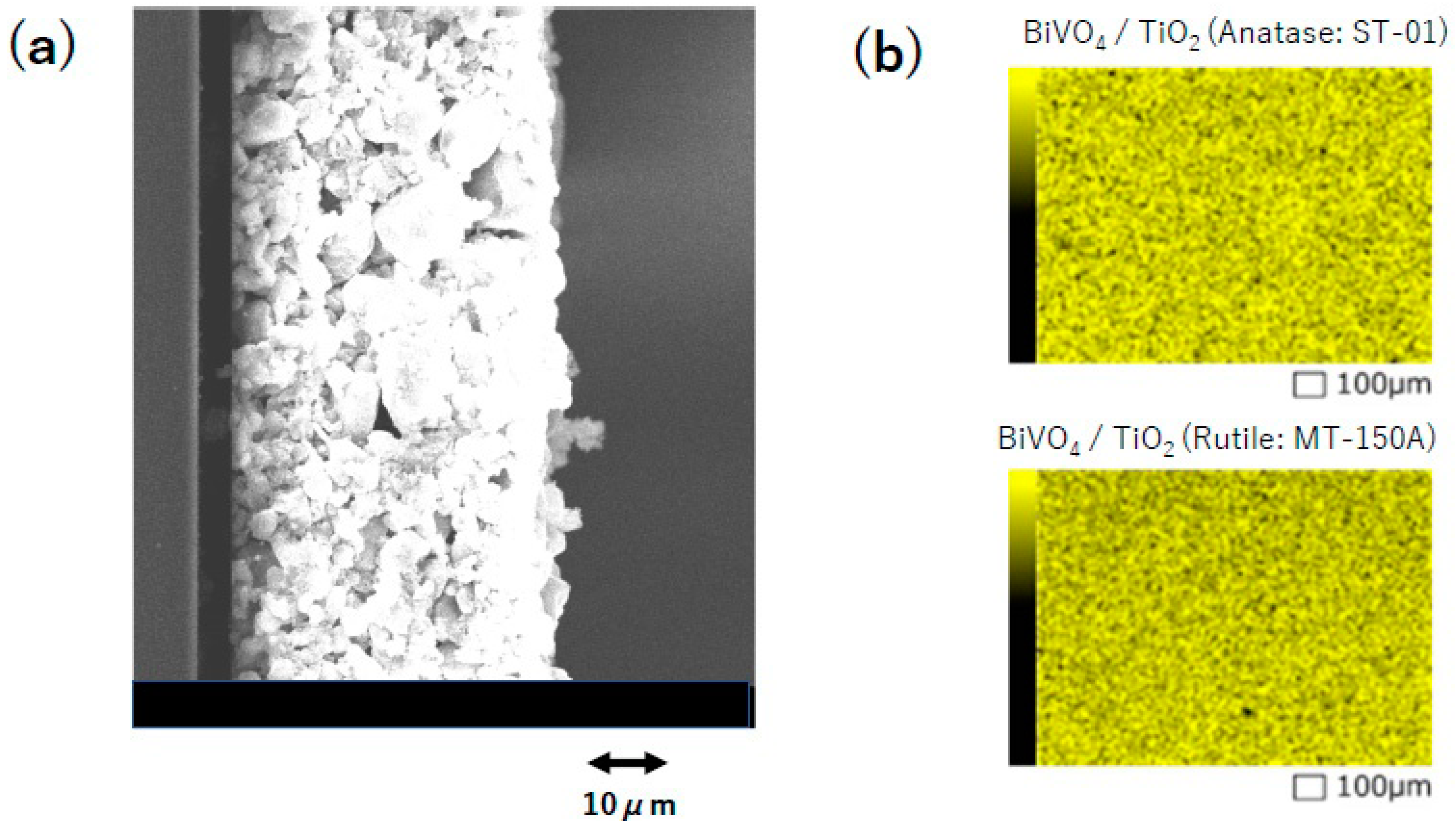
2.1. Materials

Bi(NO3)3·5H2O (0.2–1.0 mmol) and NH4VO3 (0.2–1.0 mmol) were dissolved in 10 mL HNO3 solution (1.0 mol/L) and stirred for 5 min under ambient air, which was followed by 1 min of ultrasonic irradiation. After the color of the mixed solution turned orange, it was mixed with commercially available TiO2 particles and stirred for 5 min. The orange suspension containing the TiO2 particles was then ultrasonicated for approximately 5 min and used as a solution for dip-coating the quartz glass. BiVO4/TiO2 nanocomposite photocatalytic films were obtained by dip-coating quartz glass (diameter: 30 mm) at a rate of approximately 60 cm/min. The obtained nanocomposite films were then annealed in air for 5 min at approximately 600 K (~330 °C), and these procedures were repeated three times. Finally, the films were annealed again at approximately 600 K (~330 °C) for 1 h. The TiO2 powders used in this study were generous gifts from Ishihara Sangyo (ST-01), Showa Titanium (F-1, F-2), Degussa (P25), and Tayca (AMT-100, AMT-600, MT-150A, and MT-500B), and were used without any modification. TiO2 powders purchased from Wako Co., Ltd. (Tokyo, Japan) (purity ≥ 99.9%) were used after further purification. The primary particle sizes and anatase ratios of commercially available TiO2 powders are listed in Table 1. Analytical grade Bi(NO3)3·5H2O and NH4VO3 were used without further purification. The crystal form of the prepared thin film was analyzed by X-ray diffraction (XRD, Rigaku, Ultima IV) using a Cu-Kα radiation source (λ = 1.541 Å). Characteristic XRD peaks corresponding to the diffraction patterns of the monoclinic phase of BiVO4 were observed, along with additional peaks attributed to the TiO2 photocatalytic particles. The thickness of the films was examined using scanning electron microscopy (SEM). We confirmed that all photocatalytic films prepared in this study had a thickness of approximately 50 μm (see Figure 1a). Elemental analysis of the film surface was performed using energy-dispersive X-ray spectroscopy (EDS) with a thermal emission scanning electron microscope (FESEM, JEOL, JSM-IT200LA) operating at 15 kV, as shown in Figure 1b. Subsequently, the homogeneous distribution of BiVO4 on the photocatalytic TiO2 particles was confirmed. In other words, the images presented in Figure 1 confirmed that TiO2 particles were attached to the glass plate in aggregated particulate forms; among these TiO2 particles, BiVO4 photocatalysis served as a mediator for capturing visible light between the TiO2 particles attached to the glass plate. The Bi/Ti atomic ratios of the photocatalytic films were estimated using EDS and are used in the present discussion. Finally, the optical absorption of the BiVO4–TiO2 films was examined using diffuse reflectance spectroscopy with an integrating sphere attached to a UV-VIS-NIR spectrometer (JASCO. V-770). It was confirmed that the optical absorption of these films extended to the visible region because of the absorption of BiVO4 photocatalysts.

2.2. Fluorescence Probe Detection of OH Radicals

In this study, OH radicals were detected using the coumarin fluorescence probe technique [25,26]. Briefly, this technique was used to detect 7-hydroxycoumarin, which emits stronger fluorescence than the parent coumarin molecule. By monitoring the fluorescence intensity of 7-hydroxycoumarin at approximately 450 nm (excitation wavelength of 350 nm), the relative yield of OH radicals formed by the photocatalytic reaction of BiVO4/TiO2 nanocomposite photocatalytic films was estimated. The BiVO4/TiO2 nanocomposite photocatalytic films were placed in a 0.15 mM coumarin solution and were irradiated with LED light at 470 nm (Asahi Spectra, CL-H1-470) with an average power of approximately 50 mW/cm2. After visible-light irradiation, OH radicals were measured using the coumarin fluorescence probe method after sampling the LED-light-irradiated 0.15 mM coumarin solution. All experiments using the coumarin fluorescence probe technique were performed more than three times to confirm reproducibility, and the average value was used as the present result.

3. Results and Discussion

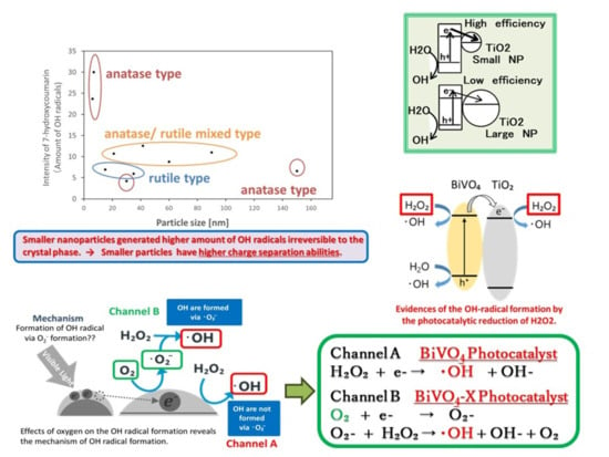
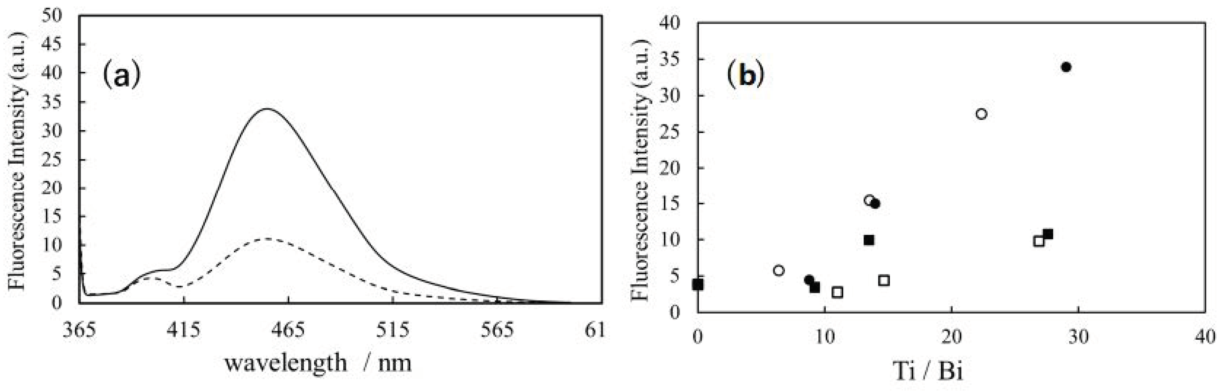
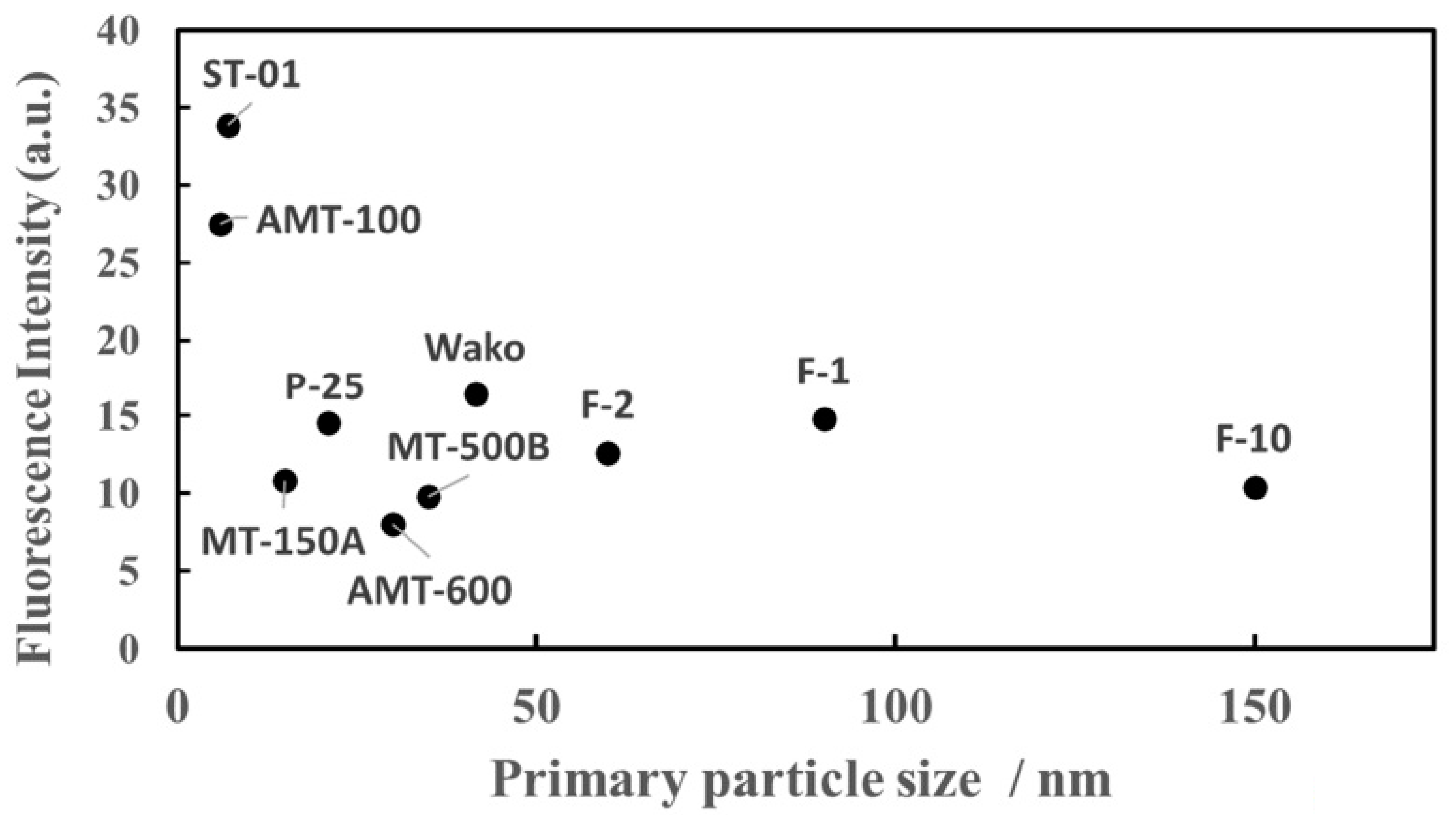
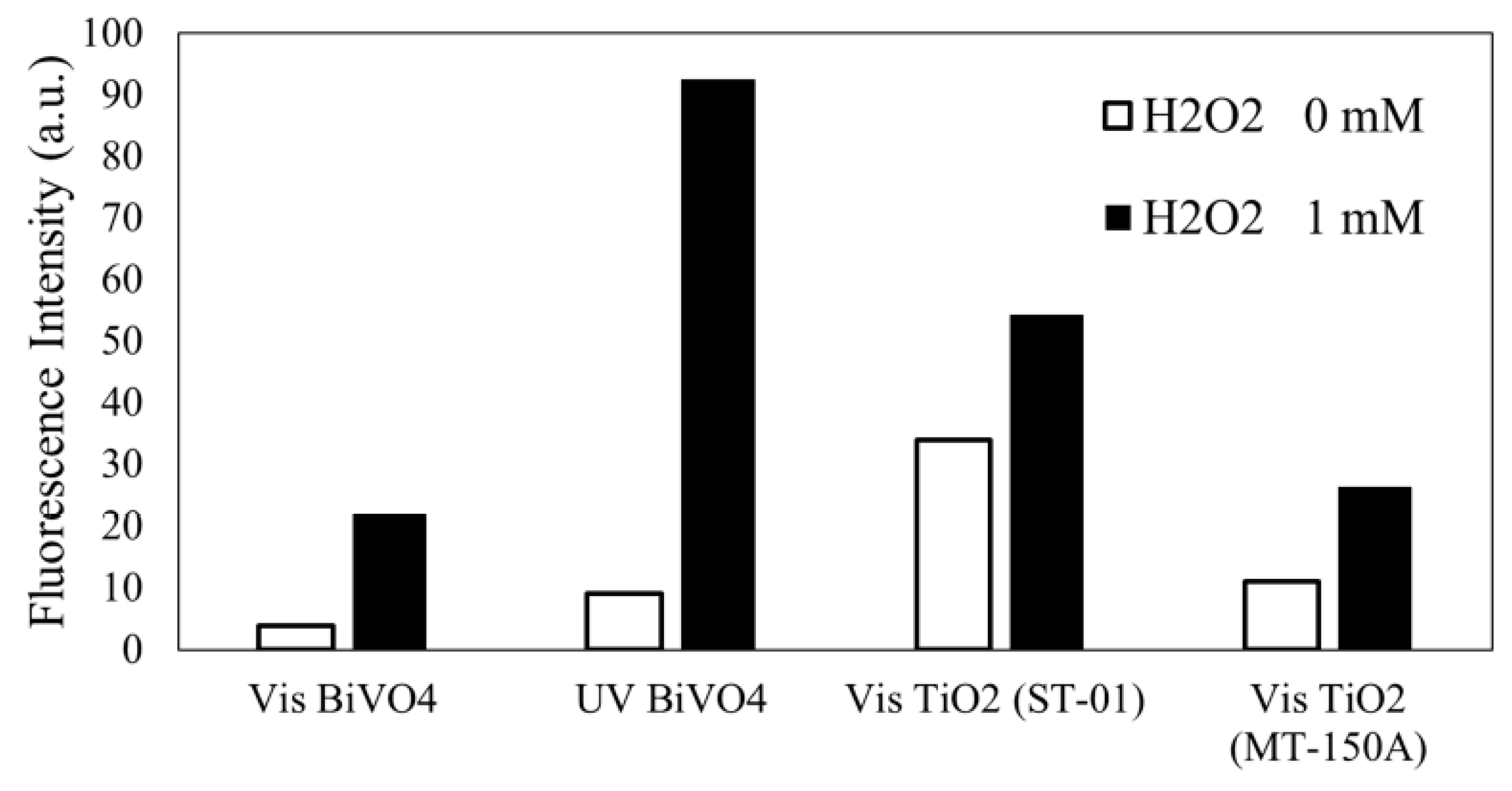
3.1. Detection of OH Radicals Formed by the BiVO4–TiO2 Photocatalyst

Figure 2a shows the fluorescence spectra obtained by the visible light irradiation of the BiVO4–TiO2 (anatase: ST-01) and BiVO4–TiO2 (rutile: MT-150A) nanocomposite photocatalysts. As shown in Figure 2a, the fluorescence of 7-hydroxycoumarin was observed at approximately 450 nm by the visible light irradiation of the BiVO4–TiO2 photocatalyst. Therefore, the formation of OH radicals on the visible-light-irradiated BiVO4–TiO2 photocatalyst was confirmed. Next, we investigated the effect of the amount of TiO2 particles on the yield of OH radicals formed by the visible-light-irradiated BiVO4–TiO2 nanocomposite photocatalysts with the irradiation time fixed at 1 h. The results are presented in Figure 2b. As shown in Figure 2b, the fluorescence intensity of 7-hydroxycoumarin increased with an increase in the amount of TiO2 particles in the BiVO4–TiO2 nanocomposite photocatalysts, which was irreversible for commercially available TiO2 powders, such as ST-01 (anatase), AMT-100 (anatase), MT-150A (rutile), and MT-500B (rutile). These results can be attributed to the suppression of the recombination of photoexcited electrons and holes in BiVO4. This is due to the enhanced transfer of photoexcited electrons from the visible-light-irradiated BiVO4 thin film to TiO2 particles, which is caused by the increase in the amount of TiO2 particles in the BiVO4 thin films. However, Figure 2b also shows differences in electron transfer and the suppression of charge recombination between TiO2 particles. This indicates that the number of OH radicals formed on the TiO2 particles of ST-01(anatase) and AMT-100 (anatase) appeared to be much higher than those on the TiO2 particles of MT-150A (rutile) and MT-500B (rutile), despite having the same amount of TiO2 in the BiVO4–TiO2 nanocomposite photocatalysts. To further investigate the differences in the charge transfer rates from BiVO4 to TiO2 particles (ST-01 (anatase), AMT-100 (anatase), MT-150A (rutile), and MT-500B (rutile)), we studied the OH radical formation on the visible-light-irradiated BiVO4 nanocomposite photocatalytic films with various other commercially available TiO2 particles. Figure 3 shows the relationship between the fluorescence intensity of 7-hydroxycoumarin and primary particle size of TiO2. As shown in Figure 3, the number of OH radicals formed through the photoexcited electron transfer from the visible-light-irradiated BiVO4 thin film to TiO2 particles was independent of the particle size and crystal phase of TiO2 except for the two commercially available TiO2 particles, ST-01 and AMT-100, which have a very small primary particle size of less than 10 nm. Dibbell et al. [27] reported the dependence of electron transfer on the distance between CdS quantum dots and TiO2 nanoparticles. They concluded that the electron injection yield decreases with increasing the chain length of bifunctional mercaptoalkanoic acid and interparticle separation. For the plasmonic excitation of Au−TiO2 photocatalysts, Du et al. [28] reported that for all TiO2 nanoparticle diameters, the plasmon-induced electron injection yields were almost the same within the experimental error with efficiencies of approximately 20–50%. However, the charge recombination decay was strongly dependent on the particle diameter of TiO2. Our results are similar to those of the plasmon-induced electron injection yields observed in the plasmonic excitation of Au−TiO2 photocatalysts, which showed almost the same yields for all TiO2 nanoparticle diameters. However, higher yields of OH radicals were obtained for the two commercially available TiO2 particles, ST-01 and AMT-100, which have small primary diameters. The reason for this has not been clarified yet. Liu et al. [29] investigated the effect of particle size on the liquid-phase photooxidation of phenol using nanometer-sized TiO2 crystals. They observed that the reaction rate constants for the photocatalytic decomposition of phenol were maximized when the anatase TiO2 particles were approximately 10 nm. The optimal particle size for the photocatalytic oxidation of phenol is attributed to the competing effects of volume recombination, surface recombination, migration of photogenerated electrons and holes, light absorption, defects, and surface area. Similar effects may be important in our present results, where nanocomposites of the BiVO4 thin films with two commercially available TiO2 particles, ST-01 and AMT-100, exhibit a higher quantum yield of OH radicals.

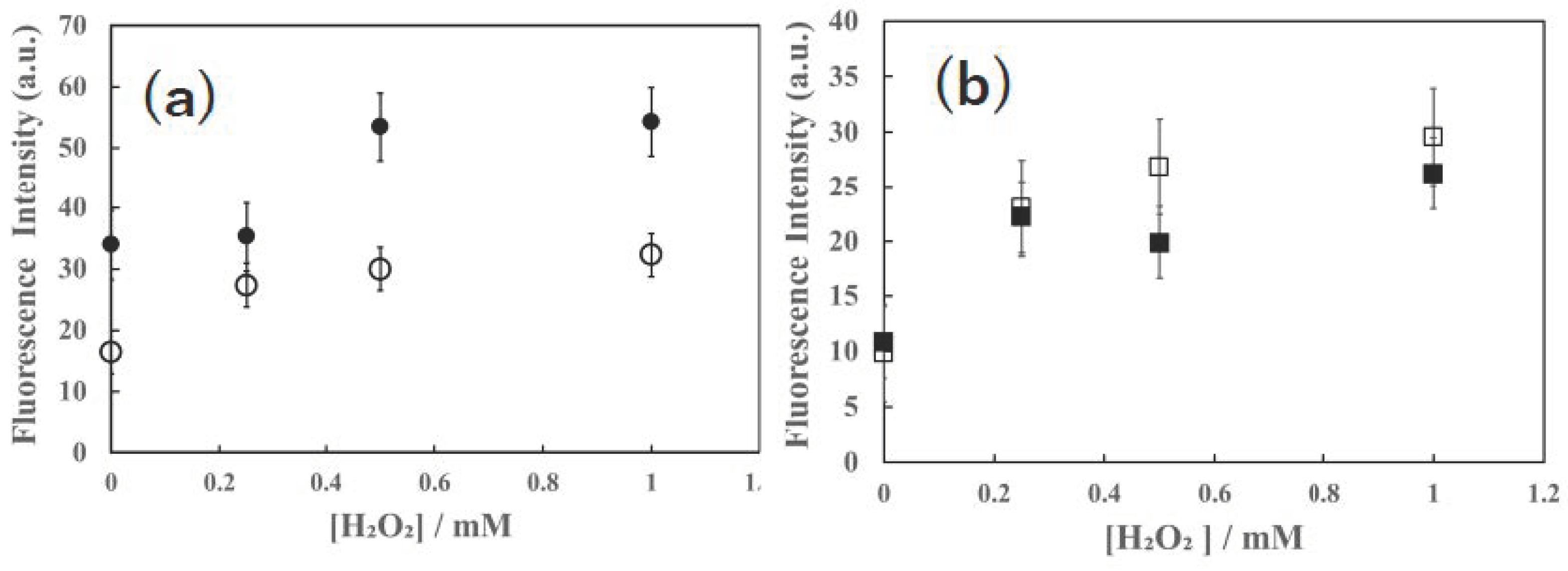
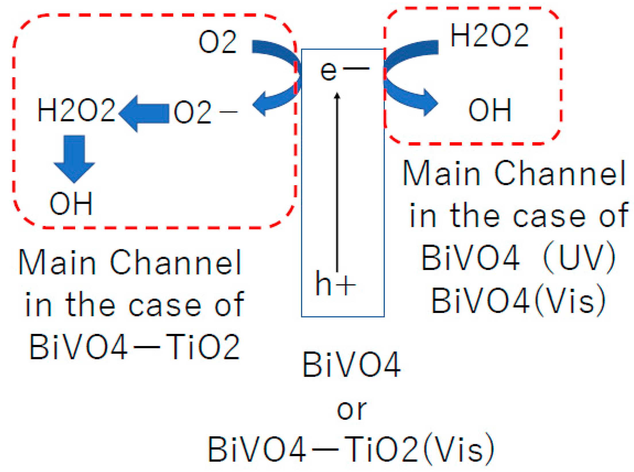
3.2. Effects of H2O2 Addition for OH Radical Formation by the Visible Light Irradiated BiVO4–TiO2 Photocatalyst

To understand the potential roles of OH radical formation through the photocatalytic reduction of H2O2 on BiVO4–TiO2 nanocomposite photocatalysts, we examined the effects of H2O2 addition on the amount of OH radicals formed on the surfaces of photocatalytic BiVO4 films mixed with several commercially available TiO2 particles (Anatase: ST-01, Wako, Rutile: MT-150A, MT-500B) under visible-light irradiation. Serpone et al. [20,30] proposed the formation of OH radicals via the photocatalytic reduction of H2O2 on the TiO2 surface. Li et al. [31] investigated the decomposition of H2O2 on TiO2 surfaces under visible-light irradiation. They observed OH radicals formed on the TiO2 surfaces by the photocatalytic decomposition of H2O2 under visible light irradiation and supported the mechanism reported by Serpone et al. [20,30]. Figure 4 shows that the amount of OH radicals increased with an increase in H2O2 concentration in the 0.15 mM coumarin solution containing the BiVO4–TiO2 nanocomposite photocatalytic film. These results indicate that OH radicals are generated during the photocatalytic reduction of H2O2 on the surfaces of BiVO4 and TiO2. It was also confirmed that the number of OH radicals increased with an increase in the concentration of H2O2, irrespective of the type of crystal phase of TiO2 (anatase or rutile) in the BiVO4–TiO2 nanocomposite photocatalytic film. These results are similar to those obtained in a previous study by Hayashi et al. [32], in which they used a visible-light-induced plasmonic Au–TiO2 photocatalyst. They also found that OH radicals were generated only in the presence of H2O2 by the visible-light plasmonic excitation of the Au–TiO2 photocatalyst. In addition, they observed an increase in the number of OH radicals in various crystal phases of TiO2 (anatase or rutile) by adding H2O2 to the Au–TiO2 plasmonic photocatalyst. From these results, they concluded that OH radicals were formed by the photocatalytic reduction of H2O2 on the surface of TiO2 by electrons in the TiO2 conduction band. These electrons migrate from the plasmonically excited Au nanoparticles. In contrast, Hirakawa et al. [33] observed a decrease in OH radical formation on the anatase form of TiO2 photocatalytic powders because of the addition of H2O2 to the TiO2 suspension. Although the reasons for such different trends—the increase or decrease in the amount of OH radicals between the photocatalytic reactions of visible-light-irradiated BiVO4–TiO2 nanocomposite or plasmonic Au–TiO2 photocatalysts and UV-irradiated TiO2 photocatalysts—have not yet been clarified, it may be attributed to the surface conditions and the presence of holes in TiO2.
To further investigate the relative roles of BiVO4 and TiO2 in OH radical formation at the reduction site of the BiVO4–TiO2 nanocomposite photocatalysts, we examined the increase in the number of OH radicals formed on the BiVO4 thin-film photocatalysts upon the addition of H2O2. The results are shown in Figure 5. First, an increase in the number of OH radicals was observed on the BiVO4 thin-film photocatalyst under visible-light irradiation. To confirm the differences in the excitation wavelengths of BiVO4, we also investigated the OH radical formation on a bare BiVO4 thin-film photocatalyst under UV light irradiation. An increase in the number of OH radicals was observed on the BiVO4 thin-film photocatalyst under UV irradiation, which was similar to the results for the visible-light-irradiated BiVO4 thin-film photocatalyst. However, the increase in the number of OH radicals on the UV-irradiated BiVO4 thin-film photocatalyst was much larger than that in the non-irradiated BiVO4 thin-film photocatalyst. This indicates that the OH radicals were formed via photocatalytic reduction at the conduction band of the BiVO4 thin-film photocatalyst. We speculated that the increase in OH radical formation for the UV-irradiated BiVO4 thin-film photocatalyst was large because the excitation energy of UV light is higher than that of visible light. Furthermore, we observed that the amount of OH radicals formed by the visible-light irradiation of the bare BiVO4 thin-film photocatalyst was much smaller than that formed by the visible-light irradiation of the BiVO4–TiO2 nanocomposite thin film photocatalyst (Figure 5). Zhang et al. [19] and Nakabayashi et al. [34] have already reported the generation of OH radical on the BiVO4 photocatalyst by photocatalytic water oxidation. The present results indicate that OH radical formation by water oxidation at the surface of the BiVO4 photocatalyst increased when the photocatalytic films were mixed with commercially available TiO2 photocatalytic powders. This supports our conclusion that the photoexcited electron transfer from the visible light-irradiated BiVO4 thin film to TiO2 particulates, along with the suppression of the recombination reaction of photoexcited electrons and holes in BiVO4, resulted in an increased amount of OH radicals formed by water oxidation at the BiVO4 surface in the BiVO4–TiO2 nanocomposite thin film.
Figure 5 shows that H2O2 addition increased the fluorescence intensity of 7-hydroxycoumarin for all the BiVO4–TiO2 photocatalytic films and bare BiVO4 photocatalytic films. As shown in Figure 5, H2O2 addition also increased the amount of OH radicals on both the BiVO4–TiO2 nanocomposite film and BiVO4 photocatalytic thin film under visible light irradiation. However, the ratio of increase in the amount of OH radicals on the BiVO4–TiO2 nanocomposite photocatalysts was smaller than that on the BiVO4 thin film after H2O2 addition under visible light irradiation (Figure 5). The variation in the ratio of the increase between the visible light-irradiated BiVO4–TiO2 nanocomposite film and BiVO4 thin film may be attributed to their different abilities for OH radical formation. During the photocatalytic reduction reaction, the BiVO4 photocatalytic surface may exhibit a greater ability for OH radical formation than the TiO2 surface. However, this is not true because the conduction band of BiVO4 is more positive than that of TiO2. When BiVO4 is in contact with TiO2, its conduction band becomes more negative than that of TiO2 because of the matching Fermi levels of both semiconductors. The band shifts of BiVO4 and TiO2 when BiVO4 and TiO2 are in contact with each other were also proposed by Wetchakun et al. [12] and Song et al. [13]. Furthermore, Shi et al. [35] and Wang et al. [36] reported band shifts of BiVO4 and TiO2 such that the conduction band edge of BiVO4 became more negative than that of TiO2. Our present experimental observation, indicating that the ability of OH radical formation on the TiO2 photocatalytic surface was smaller than that on the BiVO4 surface for the visible light-irradiated BiVO4–TiO2 nanocomposite films, could be evidence for the matching Fermi levels of the BiVO4 film and TiO2 particles in the BiVO4–TiO2 nanocomposite thin film. Thus, the conduction band edge of BiVO4 became more negative than that of TiO2 even when the BiVO4 film was in contact with commercially available TiO2 particles.

3.3. Influence of Oxygen on OH-Radicals Formation on the BiVO4 and the BiVO4–TiO2, Nanocomposite Photocatalysts

In our previous study [32], we discussed the following two plausible reaction channels for OH radical formation during the photocatalytic reduction of H2O2 on visible-light-irradiated plasmonic Au–TiO2 photocatalysts: the direct reduction channel (3) and indirect reduction channel via superoxide anion radical (O2) formation (4).
Au–TiO2 + hv → e + positive charge in Au
e + O2 → O2
H2O2 + O2 → OH + OH + O2
H2O2 + e → OH + OH
The mechanism of OH radical formation was investigated by degassing oxygen gas using nitrogen gas. The influence of the amount of OH radicals formed by the irradiation of plasmonic Au–TiO2 photocatalytic powders was also discussed. If OH radicals were generated via reaction (3), degassing oxygen would inhibit OH radical generation because reaction (3) involves the superoxide radical (O2), which is formed by the photocatalytic reduction of oxygen molecules. We observed that the degassing of oxygen reduced the amount of 7-hydroxycoumarin because coumarin reacts with OH radicals; however, the ratios of decrease were not less than half regardless of the use of commercially available TiO2 powders in the plasmonic Au–TiO2 photocatalysts. Thus, we concluded that the main channel for OH radical formation during the plasmonic Au–TiO2 photocatalyst-induced photocatalytic reduction of H2O2 is reaction (4).
In the present study, the degassing of oxygen was performed for the visible-light-irradiated bare BiVO4 photocatalysts and BiVO4–TiO2 nanocomposite photocatalysts. To remove oxygen from the photocatalytic reaction system, a flow of N2 gas was used in a closed glass flow photocatalytic reactor. To ensure the complete removal of oxygen from the reaction system, N2 gas was flowed for more than 5 min before initiating the photocatalytic reaction through irradiation. Moreover, a dissolved oxygen meter was used to determine the amount of dissolved oxygen in the coumarin solution, in which the bare BiVO4 and BiVO4–TiO2 nanocomposite photocatalytic films were immersed.
The results of these experiments are shown in Figure 6. For comparison, the visible-light-irradiated BiVO4 and the UV (365 nm)-irradiated BiVO4 photocatalytic films were investigated, and the results are shown in Figure 6.
For the UV- and visible-light-irradiated BiVO4 photocatalytic films, oxygen degassing increased the amount of OH radicals because of the inhibition of O2 formation, which facilitated the direct photocatalytic reduction of H2O on the BiVO4 surface via the reaction channel of Equation (4).
In contrast, for the visible-light-irradiated BiVO4–TiO2 nanocomposite photocatalysts, oxygen degassing decreased the amount of OH radicals. If OH radicals were formed via reaction (4), degassing oxygen would enhance OH radical formation because of the inhibition of O2 formation. Thus, for visible-light-irradiated BiVO4–TiO2 nanocomposite photocatalysts, oxygen plays an important role in OH radical formation. For BiVO4–TiO2 nanocomposite photocatalysts, OH radicals were also formed by the reaction of Equation (3). This reaction, of Equation (3), known as the Haber–Weiss Reaction [37], occurred after the photoexcitation of the BiVO4–TiO2 forming O2. During the electron migration from BiVO4 to TiO2, photoexcited electrons might be captured in the trapped sites in the BiVO4–TiO2 nanocomposite photocatalysts, and such trapping processes in the visible-light-irradiated BiVO4–TiO2 nanocomposite photocatalysts might cause such a difference in the mechanism of OH radical formation. The results are presented in Figure 7. According to our previous study [32], the main channel for OH radical formation during plasmonic Au–TiO2-induced reduction of H2O2 is the direct photocatalytic reduction of H2O on the BiVO4 surface via the reaction channel of Equation (4).
Plasmonic Au–TiO2 photocatalysts generate hot electrons in Au nanoparticles and inject them into the conduction band of TiO2, as previously investigated by several researchers [38,39]. These hot electrons play key roles in OH radical formation via reaction channel (4). One plausible explanation for the difference in the reaction channels of OH radical formation from H2O2 between BiVO4 and BiVO4–TiO2 nanocomposite photocatalysts is the distinct reactivities of the trapped and hot electrons. There are several discussions on the reaction rates [40] and reaction sites [41] of trapped electrons during molecular oxygen reduction on the conduction band of TiO2 photocatalytic reactions. However, no studies have investigated the different reactivities of free and trapped electrons in the photocatalytic reduction of H2O2. However, Losada et al. [42] used density functional theory to study the reaction of H2O2 with the surface of a transition metal oxide. There have been several previous discussions on whether OH radicals are formed by the reduction of H2O2 via reaction channel (4). Nosaka et al. [43] concluded that reaction (4) did not occur during the photocatalytic reaction of TiO2. They determined the redox potential for reaction channel (4) as +0.73 V vs SHE at pH 7. Yu et al. [44] determined the redox potential for reaction channel (4) as +0.1 V vs SHE, which was close to the value reported by Nosaka et al. [43]. Because of the potential of the conduction band edge of BiVO4, that of TiO2 is more negative than the redox potential for reaction (4). Therefore, reaction (4) could proceed in the present reaction system containing the bare BiVO4 photocatalytic film or BiVO4–TiO2 nanocomposite photocatalytic film under-visible light irradiation if only the redox potential is considered. These speculations remain unclear because of the lack of direct evidence, such as the detection of superoxide anions in the present photocatalytic reaction system. Recently, hydroxide-based co-catalysts for heterogeneous photocatalysis have been developed, and the authors have discussed enhanced electron migration and hydrogen evolution [45,46]. The use of other photocatalysts, such as hydroxide-based photocatalysts, may contribute to a better understanding of the roles of photocatalytic reduction channels in OH radical formation from H2O2 using BiVO4–TiO2 nanocomposite photocatalytic systems.
Further studies to detect other active oxygen or intermediate species, such as superoxide anion radicals, in the photocatalytic reaction system are necessary to clarify the mechanism proposed in this study.

4. Conclusions

We investigated the formation of OH radicals on visible-light-responsive BiVO4 nanocomposite films mixed with commercially available TiO2 particles. We observed that the amount of OH radicals formed by the photoexcited electron transfer from the visible-light-irradiated BiVO4 thin film to TiO2 particles was independent of the particle size and crystal phase of TiO2 except for two commercially available TiO2 particles with primary particle sizes of less than 10 nm. The effects of H2O2 addition on OH radical formation were also examined to investigate the possible role of OH radical formation in the reduction reaction of H2O2 on the surfaces of the BiVO4–TiO2 nanocomposite photocatalysts. An increase in the number of OH radicals with increasing H2O2 concentration was observed for the visible-light irradiated BiVO4–TiO2 nanocomposite photocatalyst irrespective of the crystal phase of TiO2 (anatase or rutile). The amount of OH radicals formed by the visible-light irradiation of the BiVO4 film photocatalyst was much smaller than that formed by the visible-light irradiation of the BiVO4–TiO2 nanocomposite film photocatalyst. This indicates that the electron transfer from BiVO4 to TiO2 suppressed the charge recombination reaction of electrons and holes in the BiVO4–TiO2 nanocomposite thin-film photocatalyst. In addition, we observed that the ratio of the increase in the amount of OH radicals upon adding H2O2 to the visible-light-irradiated BiVO4–TiO2 nanocomposite photocatalysts was smaller than that of the BiVO4 photocatalytic film. We also discussed the reduction potentials of the BiVO4 thin-film photocatalyst and BiVO4–TiO2 nanocomposite photocatalyst under visible-light irradiation.
Next, we confirmed the role of oxygen in OH radical formation during the photocatalytic reduction of H2O2 on the BiVO4–TiO2 nanocomposite film under visible light irradiation. Oxygen degassing was also performed for the visible-light-irradiated BiVO4 and BiVO4–TiO2 nanocomposite films. Different mechanisms were suggested for OH radical formation during the photocatalytic reduction reaction of H2O2 in the presence of visible-light-irradiated bare BiVO4 and BiVO4–TiO2 photocatalytic films. In the present study, measurements of the superoxide radicals (O2) were not carried out. As superoxide radicals are one of the key species in the photocatalytic reduction reaction, measurements of other intermediates, such as O2, would help in understanding the mechanism.

Author Contributions

Y.M., writing and editing; S.T., formal analysis and investigation. All authors have read and agreed to the published version of the manuscript.

Funding

This study received no external funding.

Data Availability Statement

The data presented in this study are available upon request from the corresponding author.

Conflicts of Interest

The authors declare no conflicts of interest.

References

  1. Kudo, A.; Omori, K.; Kato, H. A Novel Aqueous Process for Preparation of Crystal Form-Controlled and Highly Crystalline BiVO4 Powder from Layered Vanadates at Room Temperature and Its Photocatalytic and Photophysical Properties. J. Am. Chem. Soc. 1999, 121, 11459–11467. [Google Scholar] [CrossRef]
  2. Zhang, Z.; Wang, W.; Shang, M.; Yin, W. Photocatalytic degradation of rhodamine B and phenol by solution combustion synthesized BiVO4 photocatalyst. Catal. Commun. 2010, 11, 982–986. [Google Scholar] [CrossRef]
  3. Huang, C.; Chen, L.; Li, H.; Mu, Y.; Yang, Z. Synthesis and application of Bi2WO6 for the photocatalytic degradation of two typical fluoroquinolones under visible light irradiation. RSC Adv. 2019, 9, 27768–27779. [Google Scholar] [CrossRef] [PubMed]
  4. Ai, Z.; Huang, Y.; Lee, S.; Zhang, L. Monoclinic α-Bi2O3 photocatalyst for efficient removal of gaseous NO and HCHO under visible light irradiation. J. Alloys Compd. 2011, 509, 2044–2049. [Google Scholar] [CrossRef]
  5. Tang, J.; Zou, Z.; Ye, J. Efficient Photocatalytic Decomposition of Organic Contaminants over CaBi2O4 under Visible-Light Irradiation. Angew. Chem. 2004, 116, 4563–4566. [Google Scholar] [CrossRef]
  6. Murakami, Y.; Chatchai, P.; Nosaka, Y. Developments of the Efficient Water-splitting Electrodes under the Visible Light Irradiation. Electrochemistry 2009, 77, 44–50. [Google Scholar] [CrossRef]
  7. Stelo, F.; Kublik, N.; Ullah, S.; Wender, H. Recent advances in Bi2MoO6 based Z-scheme heterojunctions for photocatalytic degradation of pollutants. J. Alloys Compd. 2020, 829, 154591. [Google Scholar] [CrossRef]
  8. Abdi, F.; Savenije, T.J.; May, M.M.; Dam, B.; Krol, R. The Origin of Slow Carrier Transport in BiVO4 Thin Film Photoanodes: A Time-Resolved Microwave Conductivity Study. J. Phys. Chem. Lett. 2013, 4, 2752–2757. [Google Scholar] [CrossRef]
  9. Ziwritsch, M.; Muller, S.; Hempel, H.; Unold, T.; Abdi, F.F.; Krol, R.; Friedrich, D.; Eichberger, R. Direct Time-Resolved Observation of Carrier Trapping and Polaron Conductivity in BiVO4. ACS Energy Lett. 2016, 1, 888–894. [Google Scholar] [CrossRef]
  10. Hu, Y.; Li, D.; Zheng, Y.; Chem, W.; He, Y.; Shao, Y.; Fu, X.; Xiao, G. BiVO4/TiO2 nanocrystalline heterostructure: A wide spectrum responsive photocatalyst towards the highly efficient decomposition of gaseous benzene. Appl. Catal. B Environ. 2011, 104, 30–36. [Google Scholar] [CrossRef]
  11. Zhang, L.; Tan, G.; Wei, S.; Ren, H.; Xia, A.; Luo, Y. Microwave hydrothermal synthesis and photocatalytic properties of TiO2/BiVO4 composite photocatalysts. Ceram. Int. 2013, 39, 8597–8604. [Google Scholar] [CrossRef]
  12. Wetchakun, N.; Chainset, S.; Phanichphant, S.; Wetchakun, K. Efficient photocatalytic degradation of methylene blue over BiVO4/TiO2 nanocomposites. Ceram. Int. 2014, 41, 5999–6004. [Google Scholar] [CrossRef]
  13. Song, X.; Li, Y.; Wei, Z.; Ye, S.; Dionysiou, D. Synthesis of BiVO4/P25 composites for the photocatalytic degradation of ethylene under visible light. Chem. Eng. J. 2017, 314, 443–452. [Google Scholar] [CrossRef]
  14. Ho-Kimura, S.; Moniz, S.J.A.; Handko, A.D.; Tang, J. Enhanced photoelectrochemical water splitting by nanostructured BiVO4–TiO2 composite electrodes. J. Mater. Chem. A 2014, 2, 3948–3953. [Google Scholar] [CrossRef]
  15. Resasco, J.; Zhang, H.; Kornienko, N.; Becknell, N.; Lee, H.; Guo, J.; Briseno, A.L.; Yang, P. TiO2/BiVO4 Nanowire Heterostructure Photoanodes Based on Type II Band Alignment. ACS Cent. Sci. 2016, 2, 80–88. [Google Scholar] [CrossRef] [PubMed]
  16. Cheng, B.; Yang, J.; Cho, H.; Wu, J. Fabrication of an Efficient BiVO4–TiO2 Heterojunction Photoanode for Photoelectrochemical Water Oxidation. ACS Appl. Mater. Interfaces 2016, 8, 20032–20039. [Google Scholar] [CrossRef] [PubMed]
  17. Polo, A.; Grigioni, I.; Dozzi, M.; Selli, E. Sensitizing effects of BiVO4 and visible light-induced production of highly reductive electrons in the TiO2/BiVO4 heterojunction. Catal. Today 2020, 340, 19–25. [Google Scholar] [CrossRef]
  18. Kohtani, S.; Tomohiro, M.; Tokumura, K.; Nakagaki, R. Photooxidation reactions of polycyclic aromatic hydrocarbons over pure and Ag-loaded BiVO4 photocatalysts. Appl. Catal. B. Environ. 2005, 58, 265–272. [Google Scholar] [CrossRef]
  19. Zhang, J.; Nosaka, Y. Generation of OH radicals and oxidation mechanism in photocatalysis of WO3 and BiVO4 powders. J. Photochem. Photobiol. A Chem. 2015, 303–304, 53–58. [Google Scholar] [CrossRef]
  20. Liu, G.; Wum, T.; Zhao, J.; Hidaka, H.; Serpone, N. Photoassisted Degradation of Dye Pollutants. 8. Irreversible Degradation of Alizarin Red under Visible Light Radiation in Air-Equilibrated Aqueous TiO2 Dispersions. Environ. Sci. Technol. 1999, 33, 2081–2087. [Google Scholar] [CrossRef]
  21. Nosaka, A.Y.; Fujiwara, T.; Yagi, H.; Akutsu, H.; Nosaka, Y. Photoinduced Changes of Surface and Adsorbed Water in TiO2 Photocatalytic Systems as Studied by Solid State 1H-NMR Spectroscopy. Chem. Lett. 2002, 31, 420. [Google Scholar] [CrossRef]
  22. Zhang, J.; Nosaka, Y. Mechanism of the OH Radical generation in Photocatalysis with TiO2 of Different Crystalline Types. J. Phys. Chem. C 2014, 118, 10824–10832. [Google Scholar] [CrossRef]
  23. Murakami, Y.; Endo, K.; Ohta, I.; Nosaka, A.Y.; Nosaka, Y. Can OH Radicals Diffuse from the UV-Irradiated Photocatalytic TiO2 Surfaces? Laser-Induced-Fluorescence Study. J. Phys. Chem. C 2007, 111, 11339–11346. [Google Scholar] [CrossRef]
  24. Deguchi, S.; Shibata, N.; Takeuchi, T.; Fujiwara, Y.; Isu, N. Photocatalytic Hydrogen Production from Aqueous Solution of Various Oxidizing Sacrifice Agents. J. Jpn. Petroleum. Inst. 2010, 52, 95–100. [Google Scholar] [CrossRef]
  25. Louie, G.; Foley, S.; Cabilic, J.; Coffigny, H.; Taran, F.; Valleix, A.; Renault, J.P.; Pin, S. The reaction of coumarin with the OH radical revisited: Hydroxylation product analysis determined by fluorescence and chromatography. Radiat. Phys. Chem. 2005, 72, 119–124. [Google Scholar] [CrossRef]
  26. Zhang, J.; Nosaka, Y. Quantitative detection of OH radicals for investigating the reaction mechanism of Various visible-light TiO2 photocatalysts in aqueous suspension. J. Phys. Chem. C 2013, 117, 1383–1391. [Google Scholar] [CrossRef]
  27. Dibbel, R.S.; Watson, D.F. Distance-Dependent Electron Transfer in Tethered Assemblies of CdS Quantum Dots and TiO2 Nanoparticles. J. Phys. Chem. C 2009, 113, 3139–3149. [Google Scholar] [CrossRef]
  28. Du, L.; Furube, A.; Yamamoto, K.; Hara, K.; Katoh, R.; Tachiya, M. Plasmon-Induced Charge Separation, and Recombination Dynamics in Gold-TiO2 Nanoparticle Systems: Dependence on TiO2 Particle Size. J. Phys. Chem. C 2009, 113, 6454–6462. [Google Scholar] [CrossRef]
  29. Liu, S.; Jaffrezic, N.; Guillard, C. Size effects in liquid-phase photo-oxidation of phenol using nanometer-sized TiO2 catalysts. Appl. Surf. Sci. 2008, 255, 2704–2709. [Google Scholar] [CrossRef]
  30. Wu, T.; Liu, G.; Zhao, J.; Hidaka, H.; Serpone, N. Evidence for H2O2 Generation during the TiO2-Assisted Photodegradation of Dyes in Aqueous Dispersions under Visible Light Illumination. J. Phys. Chem. B 1999, 103, 4862–4867. [Google Scholar] [CrossRef]
  31. Li, X.; Chen, C.; Zhao, J. Mechanism of Photodecomposition of H2O2 on TiO2 surfaces under Visible Light Irradiation. Langmuir 2001, 17, 4118–4122. [Google Scholar] [CrossRef]
  32. Hayashi, T.; Nakamura, K.; Suzuki, T.; Saito, N.; Murakami, Y. OH radical formation by the photocatalytic reduction reactions of H2O2 on the surface of plasmonic excited Au-TiO2 photocatalysts. Chem. Phys. Lett. 2020, 739, 136958. [Google Scholar] [CrossRef]
  33. Hirakawa, T.; Yawata, K.; Nosaka, Y. Photocatalytic reactivity for O2 and OH radical formation in anatase and rutile TiO2 suspension as the effect of H2O2 addition. Appl. Catal. A Gen. 2007, 325, 105–111. [Google Scholar] [CrossRef]
  34. Nakabayashi, Y.; Nishikawa, M.; Saito, N.; Terashima, C.; Fujishima, A. Significance of Hydroxyl Radical in Photoinduced Oxygen Evolution in Water on Monoclinic Bismuth Vanadate. J. Phys. Chem. C 2017, 121, 25624–25631. [Google Scholar] [CrossRef]
  35. Shi, L.; Xu, C.; Zhang, H.; Liu, Z.; Qu, X.; Du, F. Facile fabrication of hierarchical BiVO4/TiO2 heterostructures for enhanced photocatalytic activities under visible-light irradiation. J. Mater. Sci. 2018, 53, 11329–11342. [Google Scholar] [CrossRef]
  36. Wang, Y.; Lu, N.; Luo, M.; Fan, L.; Zhao, K.; Qu, J.; Guan, J.; Yuan, X. Enhancement mechanism of fiddlehead-shaped TiO2-BiVO4 type II heterojunction in SPEC towards RhB degradation and detoxification. Appl. Surf. Sci. 2019, 463, 234–243. [Google Scholar] [CrossRef]
  37. Kehrer, J.P. The Haber–Weiss reaction and mechanisms of toxicity. Toxicology 2000, 149, 43–50. [Google Scholar] [CrossRef] [PubMed]
  38. Furube, A.; Du, L.; Hara, K.; Kato, R.; Tachiya, M. Ultrafast Plasmon-Induced Electron Transfer from Gold Nanodots into TiO2 Nanoparticles. J. Am. Chem. Soc. 2007, 129, 14852–14853. [Google Scholar] [CrossRef]
  39. Du, L.; Furube, A.; Hara, K.; Kato, R.; Tachiya, M. Ultrafast plasmon-induced electron injection mechanism in gold–TiO2 nanoparticle system. J. Photochem. Photobiol. C 2013, 15, 21–30. [Google Scholar] [CrossRef]
  40. Bahnemann, D.W.; Hilgendorff, M.; Memming, R. Charge Carrier Dynamics at TiO2 Particles: Reactivity of Free and Trapped Holes. J. Phys. Chem. B 1997, 101, 4265–4275. [Google Scholar] [CrossRef]
  41. Henderson, M.; Epling, W.S.; Peden, C.H.; Perkins, C.L. Insights into Photoexcited Electron Scavenging Processes on TiO2 Obtained from Studies of the Reaction of O2 with OH Groups Adsorbed at Electronic Defects on TiO2(110). J. Phys. Chem. B 2003, 107, 534–545. [Google Scholar] [CrossRef]
  42. Losada, C.M.; Johansson, J.; Brinck, T.; Jonsson, M. Mechanism of H2O2 Decomposition on Transition Metal Oxide Surfaces. J. Phys. Chem. C 2012, 116, 9533–9543. [Google Scholar] [CrossRef]
  43. Nosaka, Y.; Nosaka, A.Y. Understanding Hydroxyl Radical (•OH) Generation Processes in Photocatalysis. ACS Energy Lett. 2016, 1, 356–359. [Google Scholar] [CrossRef]
  44. Yu, T.; Breslin, C.B. Review—2D Graphene and Graphene-Like Materials and Their Promising Applications in the Generation of Hydrogen Peroxide. J. Electrochem. Soc. 2020, 167, 126502. [Google Scholar] [CrossRef]
  45. Xie, L.; Hao, J.-G.; Chen, H.-Q.; Li, Z.-X.; Ge, S.-Y.; Mi, Y.; Yang, K.; Lu, K. Recent advances of nickel hydroxide-based cocatalysts in heterogeneous photocatalysis. Catal. Commun. 2022, 162, 106371. [Google Scholar] [CrossRef]
  46. Wei, Y.; Hao, J.-G.; Zhang, J.-L.; Huang, W.-Y.; Ouyang, S.-B.; Yang, K.; Lu, K.-Q. Integrating Co(OH)2 nanosheet arrays on graphene for efficient noble-metal-free EY-sensitized photocatalytic H2 evolution. Dalton. Trans. 2023, 52, 13923–13929. [Google Scholar] [CrossRef]
Figure 1. (a) Scanning electron micrograph of the cross-section of a BiVO4–TiO2 (ST-01) nanocomposite film. (b) Energy-dispersive spectroscopy (EDS) element mapping image of BiVO4–TiO2 nanocomposite films containing ST-01 (anatase, upper figure) and MT-150A (rutile, lower figure) TiO2 powders.
Reactions 05 00004 g001
Figure 2. (a) Fluorescence spectra of 7-hydroxycoumarin obtained after 1 h visible LED light irradiation of the BiVO4–TiO2 (anatase: ST-01) (solid line) and BiVO4–TiO2 (rutile: MT-150A) (dashed line) nanocomposite photocatalysts at 470 nm. (b) Dependence of the Ti/Bi ratio in the BiVO4–TiO2 nanocomposite film on the fluorescence intensity of 7-hydroxycoumarin formed during 1 h visible LED light irradiation at 470 nm. This BiVO4–TiO2 nanocomposite film was placed in a 0.15 mM coumarin aqueous solution. The commercially available TiO2 particles used for the BiVO4–TiO2 nanocomposite film were ST-01; anatase (●), AMT-100; anatase (◦), MT-150A, rutile (▪), and MT-500B, rutile (□). For all these experiments, the excitation wavelength was set to 350 nm. Additionally, the Ti/Bi ratios estimated through the EDS measurements were used in the figure.
Reactions 05 00004 g002
Figure 3. Relationship between the fluorescence intensity of 7-hydroxycoumarin formed during 1-h visible LED light irradiation at 470 nm and the primary particle sizes of the TiO2 particles. For this purpose, the BiVO4–TiO2 nanocomposite film (Ti/Bi ratio was fixed at 20:1) was placed in a 0.15 mM coumarin aqueous solution. Various commercially available TiO2 powders listed in Table 1 were used. The names of the commercially available TiO2 powders have also been indicated in the above figure, and their primary particle sizes were taken from Table 1. For all these fluorescence measurements, the excitation wavelength was set to 350 nm.
Reactions 05 00004 g003
Figure 4. Dependence of H2O2 concentration on the fluorescence intensity of 7-hydroxycoumarin formed by the 1 h visible LED light irradiation (470 nm) of the BiVO4–TiO2 nanocomposite photocatalytic film suspended in a 0.15 mM coumarin aqueous solution. (a) Anatase form of TiO2 particles: ST-01 (●), Wako (◦). (b) rutile form of TiO2 particles: MT-150A (▪), MT-500B (□). Experiments were repeated more than three times, and the error bars for the data are also illustrated in the figure. For these experiments, the Ti/Bi ratio for the BiVO4–TiO2 nanocomposite film used was fixed at 20:1, and the excitation wavelength was set to 350 nm.
Reactions 05 00004 g004
Figure 5. Fluorescence intensity of 7-hydroxycoumarin formed by the 1 h visible LED light irradiation (470 nm) of the bare BiVO4 photocatalytic film (the first left bar) and the 1 h UV LED light irradiation (350 nm) of the bare BiVO4 (the second left bar) and BiVO4–TiO2 (Anatase: ST-01, Anatase: MT-150A, Rutile) nanocomposite photocatalytic films (the second right and right bars). These films were suspended in 0.15 mM coumarin aqueous solutions with 1 mM H2O2 (▪) and without H2O2 (□). The commercially available TiO2 particles (Anatase: ST-01, Anatase: MT-150A, Rutile) listed in Table 1 were used. For all measurements, the Ti/Bi ratio for the BiVO4–TiO2 nanocomposite film used was fixed at 20:1. The excitation wavelength for the fluorescence measurements was set to 350 nm.
Reactions 05 00004 g005
Figure 6. Effect of oxygen degassing on the fluorescence intensity of 7-hydroxycoumarin formed during the photocatalytic reaction on the visible-light-irradiated (LED, λ = 470 nm) BiVO4–TiO2 (ST-01: Anatase) and BiVO4–TiO2 (MT-150A: Rutile) nanocomposite photocatalytic films. These films were immersed in 0.15 mM coumarin aqueous solution. For comparison, the effect of oxygen degassing on the fluorescence intensity of 7-hydroxycoumarin formed during the photocatalytic reactions on visible-light-irradiated BiVO4 films (LED, λ = 470 nm) and UV-light-irradiated BiVO4 films (LED, λ = 365 nm) in 0.15 mM coumarin aqueous solution was also investigated. For these fluorescence measurements, the Ti/Bi ratio for the BiVO4–TiO2 nanocomposite film used was fixed at 20:1. The concentration of H2O2 was fixed at 1 mM, and the excitation wavelength was set to 350 nm ((□) without O2 (▪) with O2).
Reactions 05 00004 g006
Figure 7. Schematic for the proposed mechanism for OH radical formation by H2O2 reduction on the BiVO4 photocatalyst and the indirect mechanism for the reaction of O2 and H2O2 on the BiVO4–TiO2 nanocomposite photocatalyst.
Reactions 05 00004 g007
Table 1. Primary particle sizes and anatase ratios for TiO2 powders used in the present study. All data regarding the TiO2 powders were obtained from the suppliers. BET surface area measuremnents were taken from references [20,21,22,23,24].
NameAnatase Content (%)Rutile Content (%)Primary Particle Size (nm)BET Surface Area (m2 g−1)Supplier
ST-0110007320 [21,22]Ishihara Sangyo
F190105026 [23]Showa Titanuim
F2604040-Showa Titanuim
P2570 [20]302132 [21], 49 [22]Degussa
AMT-100100066 [21]Tayca
AMT-60010003030 [21], 49 [22]Tayca
MT-150A01001588 [22]Tayca
MT-500B01003538 [23]Tayca
Wako100041.58.3 [24]Wako Chemicals
Disclaimer/Publisher’s Note: The statements, opinions and data contained in all publications are solely those of the individual author(s) and contributor(s) and not of MDPI and/or the editor(s). MDPI and/or the editor(s) disclaim responsibility for any injury to people or property resulting from any ideas, methods, instructions or products referred to in the content.

