Accuracy of the GENEActiv Device for Measuring Light Exposure in Sleep and Circadian Research

 , , ,
, , ,
Abstract
:1. Introduction
2. Materials and Methods
2.1. Devices
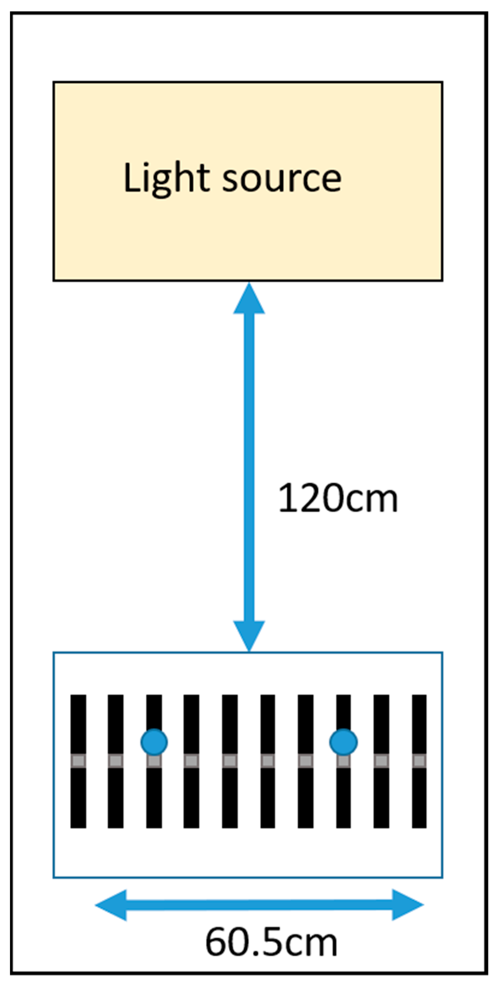
2.2. Light Stimuli
2.3. Protocol
2.4. Data Analysis
3. Results
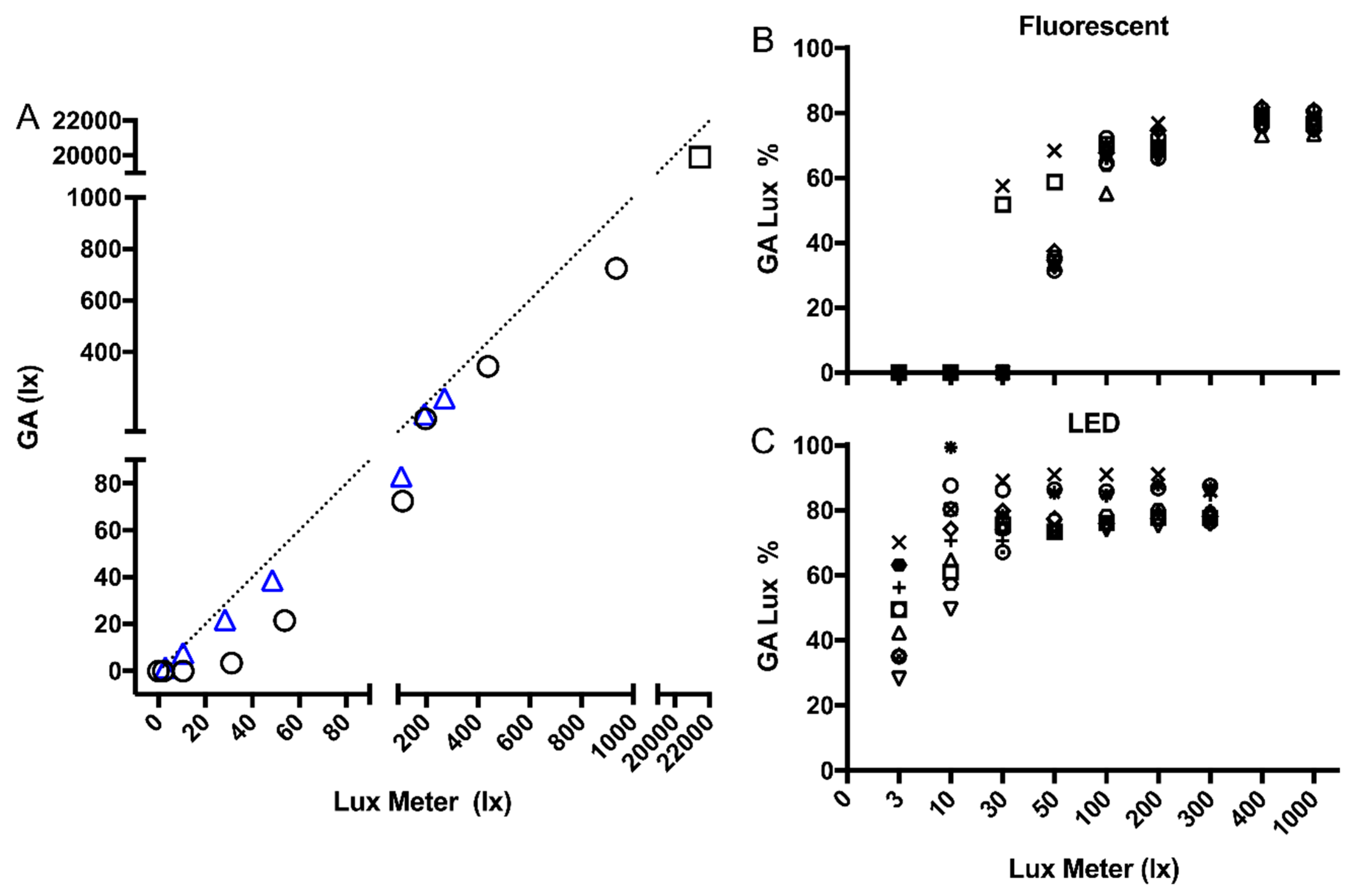
3.1. Linearity Test (LED and Fluorescent Light Sources)
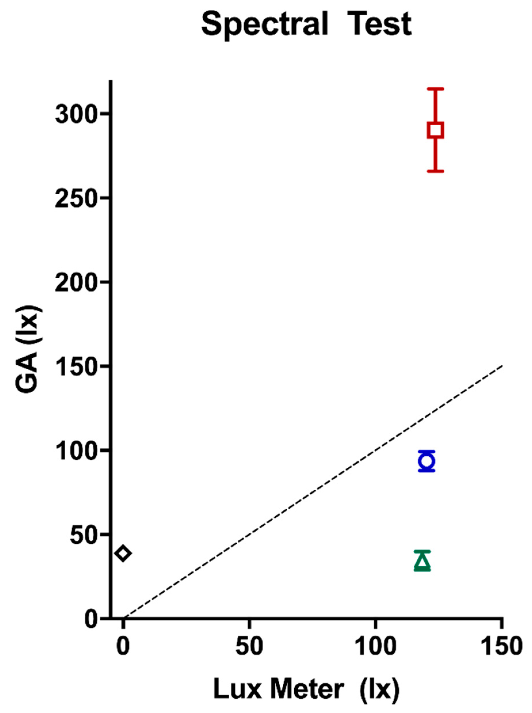
3.2. Variation in Spectral Composition
4. Discussion
5. Conclusions
Author Contributions
Funding
Acknowledgments
Conflicts of Interest
References
- Wright, K.P.; Gronfier, C.; Duffy, J.F.; Czeisler, C.A. Intrinsic period and light intensity determine the phase relationship between melatonin and sleep in humans. J. Biol. Rhythm. 2005, 20, 168–177. [Google Scholar] [CrossRef] [PubMed] [Green Version]
- Zeitzer, J.M.; Dijk, D.J.; Kronauer, R.E.; Brown, E.N.; Czeisler, C.A. Sensitivity of the human circadian pacemaker to nocturnal light: Melatonin phase resetting and suppression. J. Physiol. 2000, 526, 695–702. [Google Scholar] [CrossRef] [PubMed]
- Phillips, A.J.; Vidafar, P.; Burns, A.C.; McGlashan, E.M.; Anderson, C.; Rajaratnam, S.M.; Lockley, S.W.; Cain, S.W. High sensitivity and interindividual variability in the response of the human circadian system to evening light. Proc. Natl. Acad. Sci. USA 2019, 116, 12019–12024. [Google Scholar] [CrossRef] [PubMed] [Green Version]
- Lockley, S.W.; Brainard, G.C.; Czeisler, C.A. High sensitivity of the human circadian melatonin rhythm to resetting by short wavelength light. J. Clin. Endocrinol. Metab. 2003, 88, 4502–4505. [Google Scholar] [CrossRef]
- Thapan, K.; Arendt, J.; Skene, D.J. An action spectrum for melatonin suppression: Evidence for a novel non-rod, non-cone photoreceptor system in humans. J. Physiol. 2001, 535, 261–267. [Google Scholar] [CrossRef]
- Dumont, M.; Benhaberou-Brun, D.; Paquet, J. Profile of 24-h light exposure and circadian phase of melatonin secretion in night workers. J. Biol. Rhythm. 2001, 16, 502–511. [Google Scholar] [CrossRef]
- Stone, J.E.; Sletten, T.L.; Magee, M.; Ganesan, S.; Mulhall, M.D.; Collins, A.; Howard, M.E.; Lockley, S.W.; Rajaratnam, S.M. Temporal dynamics of circadian phase shifting response to consecutive night shifts in healthcare workers: Role of light-dark exposure. J. Physiol. 2018, 596, 2381–2395. [Google Scholar] [CrossRef]
- Martin, J.S.; Gaudreault, M.M.; Perron, M.; Laberge, L. Chronotype, light exposure, sleep, and daytime functioning in high school students attending morning or afternoon school shifts: An actigraphic study. J. Biol. Rhythm. 2016, 31, 205–217. [Google Scholar] [CrossRef] [Green Version]
- Obayashi, K.; Saeki, K.; Kurumatani, N. Association between light exposure at night and insomnia in the general elderly population: The HEIJO-KYO cohort. Chronobiol. Int. 2014, 31, 976–982. [Google Scholar] [CrossRef]
- Scheuermaier, K.; Laffan, A.M.; Duffy, J.F. Light exposure patterns in healthy older and young adults. J. Biol. Rhythm. 2010, 25, 113–122. [Google Scholar] [CrossRef]
- Migueles, J.H.; Rowlands, A.V.; Huber, F.; Sabia, S.; van Hees, V.T. GGIR: A Research Community–Driven Open Source R Package for Generating Physical Activity and Sleep Outcomes From Multi-Day Raw Accelerometer Data. J. Meas. Phys. Behav. 2019, 2, 188–196. [Google Scholar] [CrossRef] [Green Version]
- Van Hees, V.T.; Sabia, S.; Anderson, K.N.; Denton, S.J.; Oliver, J.; Catt, M.; Abell, J.G.; Kivimäki, M.; Trenell, M.I.; Singh-Manoux, A. A novel, open access method to assess sleep duration using a wrist-worn accelerometer. PLoS ONE 2015, 10, e0142533. [Google Scholar] [CrossRef] [PubMed] [Green Version]
- Van Hees, V.T.; Sabia, S.; Jones, S.E.; Wood, A.R.; Anderson, K.N.; Kivimäki, M.; Frayling, T.M.; Pack, A.I.; Bucan, M.; Trenell, M. Estimating sleep parameters using an accelerometer without sleep diary. Sci. Rep. 2018, 8, 12975. [Google Scholar] [CrossRef] [PubMed]
- Te Lindert, B.H.; Van Someren, E.J. Sleep estimates using microelectromechanical systems (MEMS). Sleep 2013, 36, 781–789. [Google Scholar] [CrossRef] [Green Version]
- Joyce, D.S.; Zele, A.J.; Feigl, B.; Adhikari, P. The accuracy of artificial and natural light measurements by actigraphs. J. Sleep Res. 2019, e12963. [Google Scholar] [CrossRef] [Green Version]
- Price, L.L.; Lyachev, A.; Khazova, M. Optical performance characterization of light-logging actigraphy dosimeters. JOSA A 2017, 34, 545–557. [Google Scholar] [CrossRef]
- Lucas, R.J.; Peirson, S.N.; Berson, D.M.; Brown, T.M.; Cooper, H.M.; Czeisler, C.A.; Figueiro, M.G.; Gamlin, P.D.; Lockley, S.W.; O’Hagan, J.B. Measuring and using light in the melanopsin age. Trends Neurosci. 2014, 37, 1–9. [Google Scholar] [CrossRef]
- Dontje, M.L.; Dall, P.M.; Skelton, D.A.; Gill, J.M.; Chastin, S.F.; Team, S.U. Reliability, minimal detectable change and responsiveness to change: Indicators to select the best method to measure sedentary behaviour in older adults in different study designs. PLoS ONE 2018, 13, e0195424. [Google Scholar] [CrossRef] [Green Version]
- Prayag, A.S.; Najjar, R.P.; Gronfier, C. Melatonin suppression is exquisitely sensitive to light and primarily driven by melanopsin in humans. J. Pineal Res. 2019, 66, e12562. [Google Scholar] [CrossRef]
- Gooley, J.J.; Lu, J.; Fischer, D.; Saper, C.B. A broad role for melanopsin in nonvisual photoreception. J. Neurosci. 2003, 23, 7093–7106. [Google Scholar] [CrossRef] [Green Version]
- Graham, D.M.; Wong, K.Y. Melanopsin-expressing, intrinsically photosensitive retinal ganglion cells (ipRGCs). In Webvision: The Organization of the Retina and Visual System; University of Utah Health Sciences Center: Salt Lake City, UT, USA, 2016. [Google Scholar]
- Hattar, S.; Liao, H.-W.; Takao, M.; Berson, D.M.; Yau, K.-W. Melanopsin-containing retinal ganglion cells: Architecture, projections, and intrinsic photosensitivity. Science 2002, 295, 1065–1070. [Google Scholar] [CrossRef] [PubMed] [Green Version]


| Peak (nm) | Photopic Illuminance (lux) | Irradiance (µW/cm2) | S Cone | Melanopsin ipRGC | Rod | M Cone | L Cone | |
|---|---|---|---|---|---|---|---|---|
| Fluorescent | 545 | 100.33 | 36.30 | 66.99 | 59.40 | 73.04 | 89.04 | 94.81 |
| LED | 450 | 100.00 | 31.48 | 64.54 | 72.75 | 80.06 | 92.38 | 97.25 |
| Red | 630 | 111.00 | 57.31 | 1.57 | 1.82 | 4.11 | 41.09 | 141.72 |
| Green | 520 | 114.78 | 25.76 | 8.28 | 119.59 | 140.84 | 127.00 | 97.71 |
| Blue | 470 | 106.56 | 86.88 | 464.61 | 605.06 | 454.05 | 253.92 | 142.74 |
| Light Source | Target Condition (lux) | Lux Meter | Mean ± SD Lux | SE Lux | MDC | Mean ± SD Error | Mean ± SD % of Lux Meter |
|---|---|---|---|---|---|---|---|
| Dark | |||||||
| 0 | 0.00 | 0.00 ± 0.00 | 0.00 | 0.00 | 0.00 ± 0.00 | 100 ± 0.00 | |
| Fluorescent | |||||||
| 3 | 2.10 | 0.00 ± 0.00 | 0.00 | 0.00 | −2.10 ± 0.00 | 0.00 ± 0.00 | |
| 10 | 10.60 | 0.00 ± 0.00 | 0.00 | 0.00 | −10.60 ± 0.00 | 0.00 ± 0.00 | |
| 30 | 31.10 | 3.09 ± 6.89 | 2.18 | 6.04 | −27.90 ± 7.18 | 9.94 ± 22.16 | |
| 50 | 53.80 | 21.36 ± 6.54 | 2.07 | 5.73 | −32.60 ± 6.87 | 39.71 ± 12.15 | |
| 100 | 108.15 | 72.53 ± 5.07 | 1.60 | 4.44 | −36.02 ± 5.34 | 67.06 ± 4.69 | |
| 200 | 197.30 | 140.40 ± 7.32 | 2.31 | 6.41 | −59.16 ± 7.46 | 71.16 ± 3.71 | |
| 400 | 438.75 | 345.24 ± 12.91 | 4.08 | 11.32 | −97.24 ± 12.97 | 78.69 ± 2.94 | |
| 1000 | 935.55 | 726.82 ± 27.14 | 8.58 | 23.79 | −218.76 ± 27.37 | 77.69 ± 2.90 | |
| LED | |||||||
| 3 | 2.84 | 1.36 ± 0.40 | 0.13 | 0.35 | −1.45 ± 0.40 | 48.02 ± 14.02 | |
| 10 | 10.48 | 7.62 ± 1.49 | 0.47 | 1.31 | −2.86 ± 1.57 | 72.69 ± 14.22 | |
| 30 | 28.30 | 21.87 ± 1.81 | 0.57 | 1.58 | −6.50 ± 1.89 | 77.29 ± 6.38 | |
| 50 | 48.46 | 38.56 ± 2.90 | 0.92 | 2.54 | −9.28 ± 3.05 | 79.58 ± 5.99 | |
| 100 | 103.35 | 82.58 ± 5.44 | 1.72 | 4.77 | −19.48 ± 5.61 | 79.91 ±5.26 | |
| 200 | 193.60 | 158.16 ± 10.16 | 3.21 | 8.91 | −33.12 ± 10.41 | 81.70 ± 5.25 | |
| 300 | 270.20 | 218.62 ± 12.17 | 3.85 | 10.66 | −51.40 ± 12.53 | 80.91 ± 4.50 | |
| Outdoor | |||||||
| 21 500 | 21437 | 19909.70 ± 709.74 | 224.44 | 622.12 | −1527.30 ± 709.74 | 92.88 ± 3.31 | |
| Lux Meter | Mean ± SD Lux | SE Lux | MDC | Mean ± SD Error | |
|---|---|---|---|---|---|
| Blue | 120 | 93.56 ± 5.66 | 1.79 | 4.97 | −26.69 ± 5.66 |
| Red | 124 | 290.28 ± 24.58 | 7.77 | 21.55 | 170.03 ± 24.58 |
| Green | 119 | 34.38 ± 5.61 | 1.77 | 4.92 | −85.87 ± 5.61 |
| Infrared | 0 | 39.15 ± 3.70 | 1.17 | 3.24 | 36.86 ± 3.84 |
© 2020 by the authors. Licensee MDPI, Basel, Switzerland. This article is an open access article distributed under the terms and conditions of the Creative Commons Attribution (CC BY) license (http://creativecommons.org/licenses/by/4.0/).
Share and Cite
Stone, J.E.; McGlashan, E.M.; Facer-Childs, E.R.; Cain, S.W.; Phillips, A.J.K. Accuracy of the GENEActiv Device for Measuring Light Exposure in Sleep and Circadian Research. Clocks & Sleep 2020, 2, 143-152. https://doi.org/10.3390/clockssleep2020012
Stone JE, McGlashan EM, Facer-Childs ER, Cain SW, Phillips AJK. Accuracy of the GENEActiv Device for Measuring Light Exposure in Sleep and Circadian Research. Clocks & Sleep. 2020; 2(2):143-152. https://doi.org/10.3390/clockssleep2020012
Chicago/Turabian StyleStone, Julia E., Elise M. McGlashan, Elise R. Facer-Childs, Sean W. Cain, and Andrew J. K. Phillips. 2020. "Accuracy of the GENEActiv Device for Measuring Light Exposure in Sleep and Circadian Research" Clocks & Sleep 2, no. 2: 143-152. https://doi.org/10.3390/clockssleep2020012