
Polymer-Based Materials Built with Additive Manufacturing Methods for Orthopedic Applications: A Review


Abstract
:1. Introduction
2. Methods
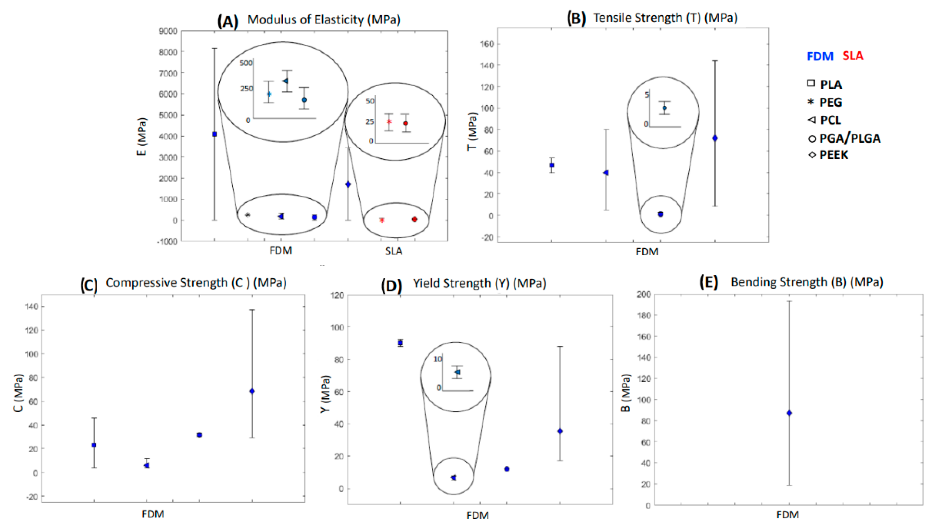
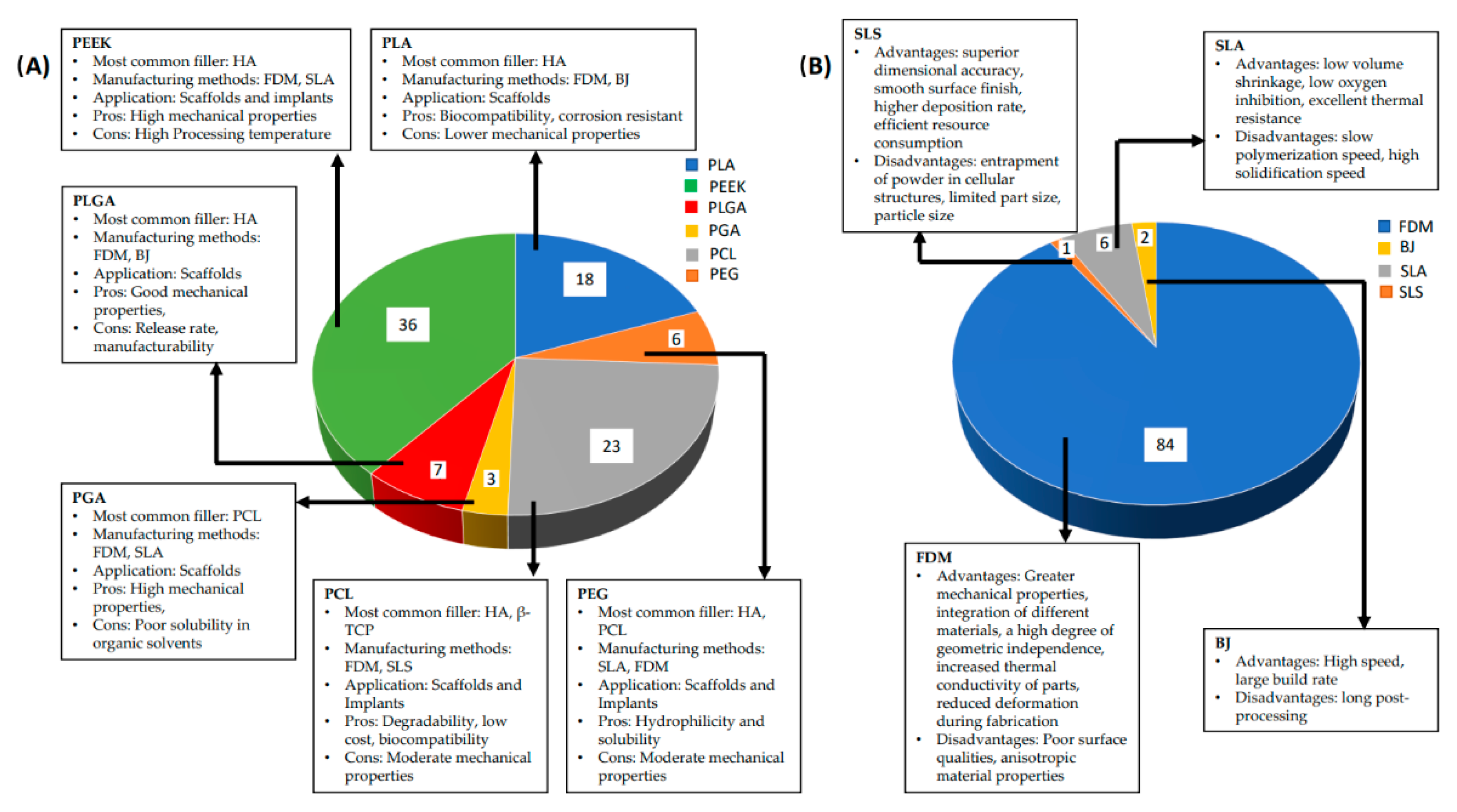
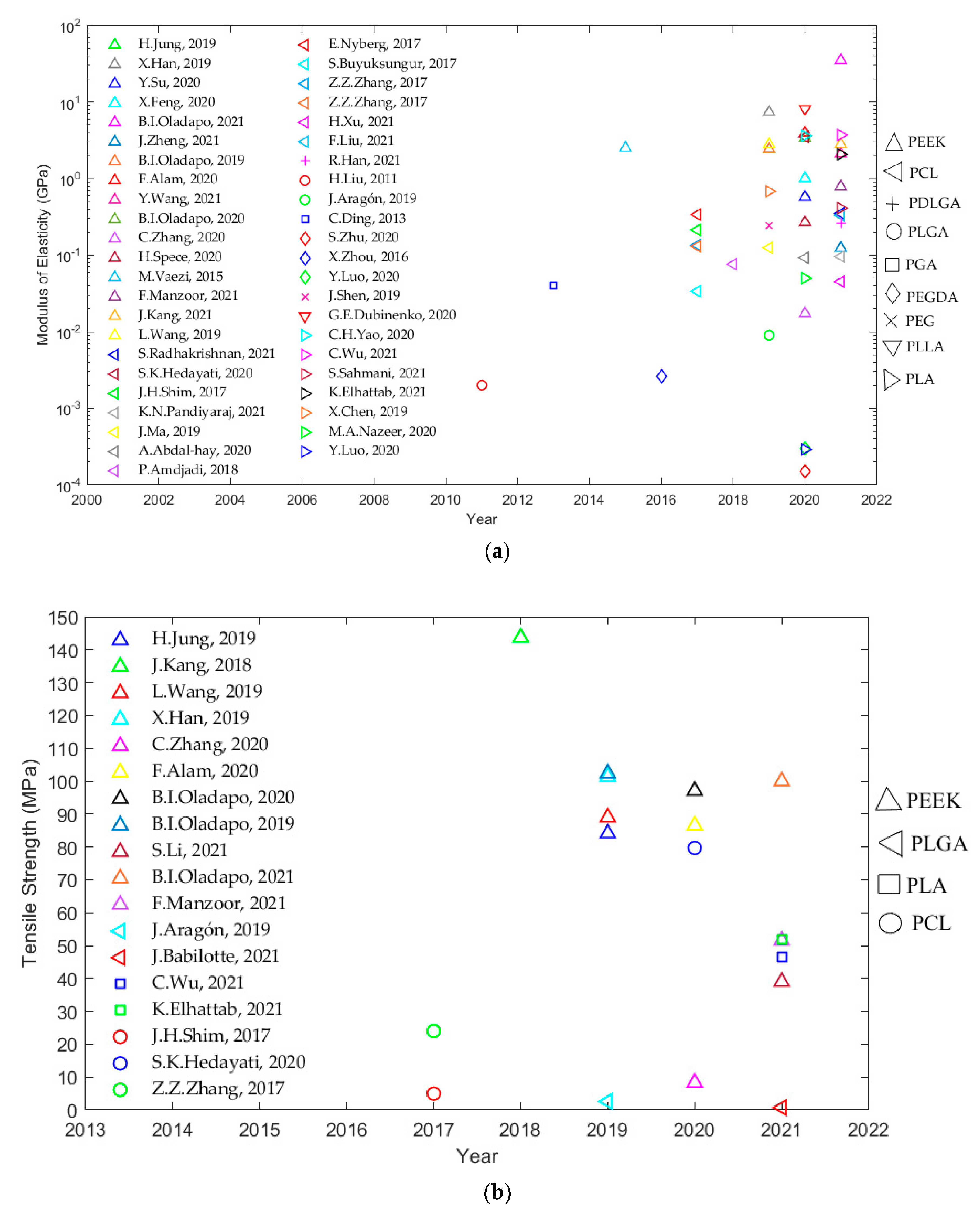
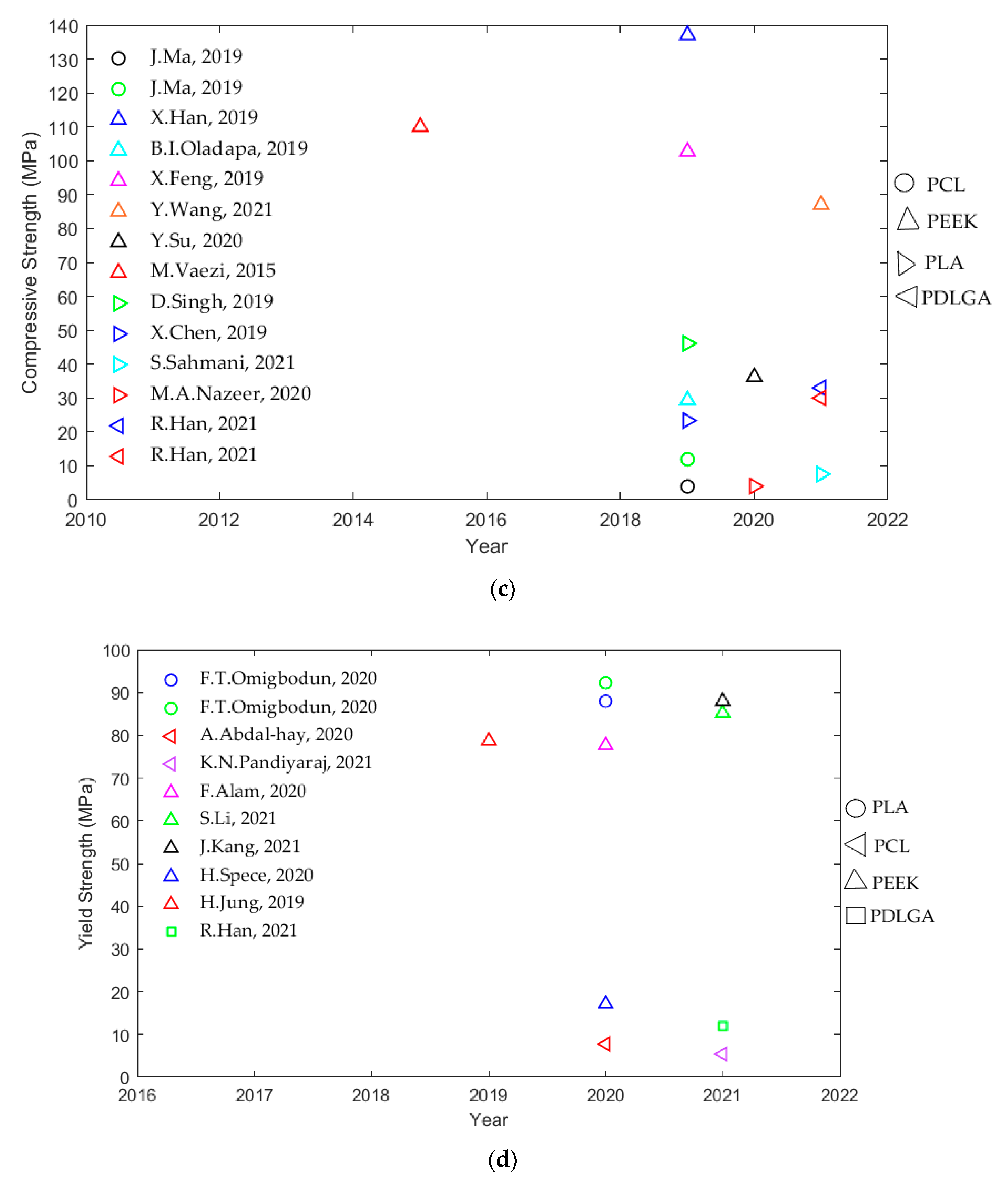
3. Results
3.1. Polylactic Acid (PLA)
3.2. Polyethylene Glycol (PEG)
3.3. Polycaprolactone (PCL)

| Material Properties | Value Range | Ref |
|---|---|---|
| Melting temperature | 52.9–72.3 °C | [43,155] |
| Tensile strength | 4.96–79.7 MPa | [155,156] |
| Elongation at break | 50–1342% | [151] |
| Modulus of elasticity | 0.03374–3.5 GPa | [145,155] |
| Yield strength | 5.44–7.8 MPa | [146,151] |
| Compressive strength | 3.9–11.9 MPa | [148] |
| Mechanical Studies | |||||
| Ref | Filler | Manufacturing Methods | Type | Tests | Results |
| [164] | HA | SLS | Experimental | Compression test Compression–shear test Torsion test | Compressive load: 650 N Compressive–shear load: 395 N Tr: 0.25 Nm |
| Biological Studies | |||||
| Ref | Filler | Manufacturing Methods | Type | Tests | Results |
| [42] | Aln | FDM | Experimental | In vivo In vitro | Bone tissue regeneration observed Cell proliferation and differentiation observed |
| [147] | Ta | FDM | Experimental | In vitro | Cell proliferation and bone formation observed |
| [157] | β-TCP | FDM | Experimental | In vivo In vitro | Improved bone tissue regeneration observed Cell proliferation observed |
| [158] | Platelet-rich plasma (PRP) | FDM | Experimental | In vivo In vitro | New bone formation observed Cell attachment, migration, proliferation increased |
| [159] | PLGA/PDA | FDM | Experimental | In vivo In vitro | New bone formation observed Cell proliferation increased |
| [161] | β-TCP | FDM | Experimental | In vivo | New bone formation observed |
| [163] | Calcium silicate (CS) | FDM | Experimental | In vivo In vitro | New bone formation observed Cell proliferation, adhesion, differentiation increased |
| [149] | Mg | FDM | Experimental | Water-contact-angle analysis In vivo In vitro | WCA: 75° Bone tissue regeneration observed Cell proliferation and differentiation observed |
| [160] | HA | FDM | Experimental | Water-contact-angle analysis In vitro | WCA: 62.2° Cell proliferation and differentiation increased |
| Mechanobiological Studies | |||||
| Ref | Filler | Manufacturing Methods | Type | Tests | Results |
| [144] | HA | FDM | Experimental | Compression test In vivo In vitro | E: 330 MPa New bone formation observed Cell proliferation observed |
| [152] | PEEK | FDM | Experimental | Compression test Water-contact-angle analysis In vitro | E: 76 MPa WCA: 69.4° Enhanced adhesion and growth observed |
| [145] | HA/PPF | FDM | Experimental | Compression test Tension test In vivo In vitro | Compressive Stiffness: 394 N/mm E: 33.74 MPa Tensile Stiffness: 463 N/mm WCA:65° New bone formation observed Cell Proliferation observed |
| [146] | TIPP | FDM | Experimental | - Water-contact-angle measurement In vitro | E: 96.64 MPa Y: 5.44 MPa WCA: 11.5° Apatite formation observed |
| [148] | PVAc/HA | FDM | Experimental | Compression test In vivo In vitro | C: 3.9–11.9 MPa E: 125.4 MPa Cell proliferation and osteogenic activity observed |
| [43] | MSCs | FDM | Experimental | Compression test Tensile test In vivo In vitro | E(C): 135 MPa T: 24 MPa E(T): 130 MPa Bone tissue regeneration observed Cell proliferation and differentiation observed |
| [153] | MgPSr | FDM | Experimental | Compression test In vivo In vitro | Compressive Toughness: 375.5 kJ/m3 Bone tissue regeneration observed Cell proliferation and differentiation observed |
| [150] | AgNps | FDM | Experimental | Tensile test In vitro | E: 0.35 GPa Degradation observed |
| [151] | MH | FDM | Experimental | Tensile test In vitro | E: 92.3 MPa Y: 7.8 MPa Increased degradation and enhanced osteoblastic activity observed |
| [155] | PGA | FDM | Experimental | Tensile test In vitro | T: 79.7 MPa E: 3.5 GPa 20% higher degradation observed |
| [156] | β-TCP | FDM | Experimental | Tensile test In vivo In vitro | T: 4.96 MPa E: 213.1 MPa Guided bone regeneration observed Cell proliferation observed Inhibition of external tissue ingrowth observed |
| [162] | TCP, HA, Bio-Oss (BO) (DCB) | FDM | Experimental | Compression test In vivo | E: 338 MPa Increased cell seeding and growth observed |
| [165] | BMSC/SAP | - | Experimental | Compression test In vivo In vitro | E: 45 MPa New bone formation observed Cell proliferation and differentiation increased |
3.4. Polyglycolic Acid (PGA) and Poly (Lactic-co-glycolic Acid) (PLGA)
| Material Properties | Value Range | Ref |
|---|---|---|
| Modulus of elasticity | 40 MPa | [182] |
| Material Properties | Value Range | Ref |
|---|---|---|
| Tensile strength | 0.7–2.6 MPa | [172,175] |
| Elongation at break | 120–180% | [172] |
| Modulus of elasticity | 2–260 MPa | [176,177] |
| Compressive strength | 30–33 MPa | [177] |
| Yield strength | 12 MPa | [177] |
| Biological Studies | |||||
|---|---|---|---|---|---|
| Ref | Filler | Manufacturing Methods | Type | Tests | Results |
| [180] | PLA | FDM | Experimental | In vivo | Connective tissue formation and bone regeneration observed |
| [183] | PLLA/PCL | FDM | Experimental | In vivo In vitro | New bone formation observed Cell proliferation observed |
| Mechanobiological Studies | |||||
| Ref | Filler | Manufacturing Methods | Type | Tests | Results |
| [182] | PLA/PCL/HA | SLA/FDM | Experimental | Compression test In vivo | E: 40 MPa Cell proliferation, adhesion, differentiation increased New bone formation observed |
| Biological Studies | |||||
|---|---|---|---|---|---|
| Ref | Filler | Manufacturing Methods | Type | Tests | Results |
| [178] | HA/Chitosan | FDM | Experimental | In vitro | Cell proliferation and differentiation increased |
| [181] | TCP/Mg | FDM | Experimental | In vivo In vitro | New bone formation observed Cell proliferation, viability increased |
| [179] | PGA | Binder jetting | Experimental | In vitro | Cell attachment, tissue growth affected by degradation |
| Mechanobiological Studies | |||||
| Ref | Filler | Manufacturing Methods | Type | Tests | Results |
| [172] | PCL | FDM | Experimental | Tensile test In vitro | T: 2.6 MPa E: 9 ± 4 MPa Cell proliferation, viability observed |
| [175] | HA | FDM | Experimental | Tensile test In vitro In vivo | T: 0.7 MPa New bone formation observed Cell proliferation, viability, differentiation increased |
| [176] | Nano-Titania | FDM | Experimental | Roughness analysis Tensile test In vitro | R: 100 nm E: 2 MPa Osteoblast adhesion increased |
| [177] | Bioglass/ Biosilica | FDM | Experimental | Compression test In vitro | C: 30–33 Mpa Y: 12 Mpa E: 190–260 Mpa Increased degradation observed |
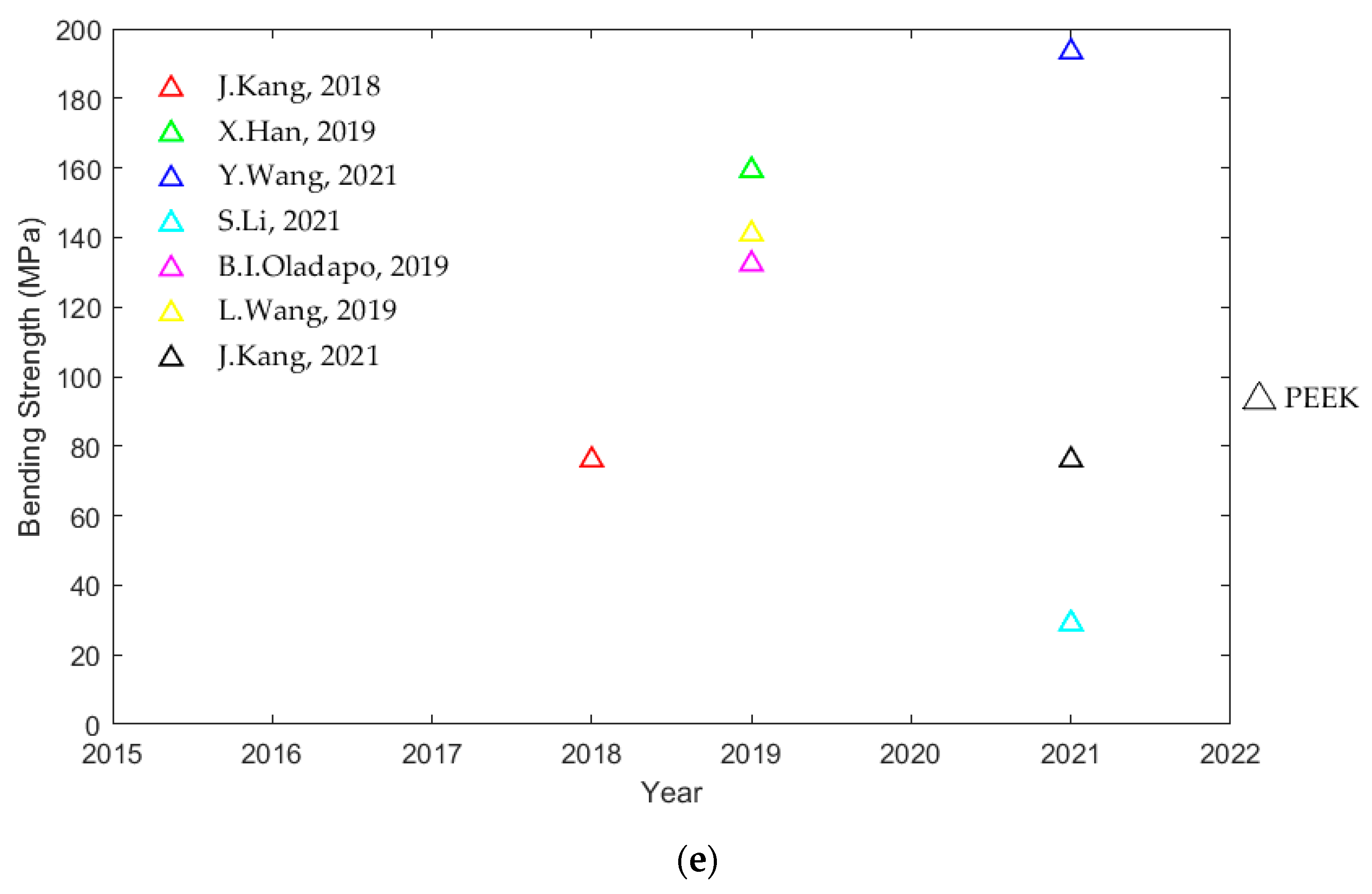
3.5. Polyetheretherketone (PEEK)
| Material Properties | Value Range | Ref |
|---|---|---|
| Density | 1.181–1.868 g/cm3 | [40,215] |
| Melting temperature | 334–400 °C | [40,209] |
| Glass transition temperature | 139–149 °C | [216] |
| Tensile strength | 8.3–143.7 MPa | [200,205] |
| Modulus of elasticity | 0.017–34.96 GPa | [205,215] |
| Bending strength | 19–193.33 MPa | [40,208] |
| Compressive strength | 29.34–137.1 MPa | [21,209] |
| Yield strength | 17.1–88 MPa | [206,207] |
| Mechanical Studies | |||||
|---|---|---|---|---|---|
| Ref | Filler | Methods | Type | Tests | Results |
| [41] | N/A | FDM | Experimental | Tensile test Bending test | T: 89 MPa B: 141 MPa E: 2.8 GPa |
| [40] | N/A | FDM | Experimental | Compression test Bending test | C: 87 MPa E: 2.098 GPa B: 193.33 MPa |
| [204] | N/A | FDM | Experimental | Compression test Compression–shear test | Compressive stiffness: 8874 N/mm Compressive–shear Stiffness: 1335 N/mm |
| [192] | N/A | FDM | Experimental | Compression test | Ultimate load for C: 11,686 N |
| [196] | N/A | FDM | Experimental | Compression test Compression–shear test Torsion test | Compressive stiffness: 9324 N/mm Compressive–shear stiffness: 929 N/mm Tr: 1.37 Nm/deg |
| [205] | N/A | FDM | Experimental FEA analysis | Tensile test | T: 8.3 MPa E: 17.3 MPa |
| [207] | N/A | FDM | Experimental FEA analysis | Tensile test Flexure test | E: 2.8 GPa Von Mises: 366.5 MPa Y: 88 MPa B: 76 MPa |
| [197] | N/A | FDM | Experimental FEA analysis | Compression test | Von Mises (screws): 9.71 MPa Von Mises (condyle): 10.33 MPa |
| [200] | N/A | FDM | Experimental FEA analysis | Tensile test Bending test | T: 143.7 MPa B: 76.0 ± 23.7 MPa |
| [209] | HA/GO | FDM | Experimental FEA analysis | Compression test Tensile test | C: 29.34 MPa T: 102.38 MPa E: 2.43 GPa B: 132.37 MPa |
| Biological Studies | |||||
| Ref | Filler | Methods | Type | Tests | Results |
| [199] | N/A | FDM | Experimental | In vitro | R: 26.7 µm Cell attachment, growth, and proliferation observed |
| [185] | AMP | FDM | Experimental | In vivo In vitro | Enhanced bioactivity and superior pre-osteoblast cell function observed Enhanced osseointegration observed |
| [188] | AgNPs | FDM | Experimental | In vitro | Cell attachment, growth, and proliferation observed |
| [210] | Graphene/HA | FDM | Experimental | In vivo In vitro | New bone growth observed Cell attachment, growth, and proliferation observed |
| [211] | AgNPs/pDA | FDM | Experimental | In vivo In vitro | New bone growth observed Cell attachment, growth, and proliferation observed |
| [201] | N/A | FDM | Experimental | Water-contact-angle analysis In vitro | WCA: 39° Cell attachment, growth, and proliferation observed |
| Mechanobiological Studies | |||||
| Ref | Filler | Methods | Type | Tests | Results |
| [190] | HA | FDM | Experimental | Compression test In vitro | E: 112 MPa (Z-axis) 124 MPa (X-axis) Increased cell attachment and mineralization observed |
| [186] | GO/HA | FDM | Experimental FEA analysis | Tensile test In vitro | - Von-Mises stress: 25.32 GPa Cell attachment, growth, and proliferation observed |
| [213] | Ti | FDM | Experimental | Tensile test In vivo In vitro | T: 84.1 MPa Y: 78.7 MPa E: 2.42 GPa New bone growth observed Cell attachment, growth, and proliferation observed |
| [202] | N/A | FDM | Experimental | Compression test In vivo In vitro | C: 36.20 MPa E: 575 MPa Newly-regenerated soft tissues adhesion observed Cell attachment, growth, and proliferation observed |
| [203] | N/A | FDM | FEA analysis | Compression test Tensile test Shear test Bone tissue-strain study | 82.3% load shared Majority of the load is carried by the bone with peek Favorable bone ingrowth properties found |
| [206] | N/A | FDM | Experimental | Compression test In vitro | Y: 17.1 MPa E: 210–268 MPa Cell attachment, growth, and proliferation observed |
| [208] | N/A | FDM | Experimental | Tensile test Bending test In vitro | T: 29–39 MPa Y: 85.23 MPa B: 19–29 MPa Cell attachment, growth, and proliferation observed |
| [195] | N/A | FDM | Experimental | Compression test In vivo In vitro | C: 102.7 MPa E: 1006.5 MPa New bone formation observed Cell attachment, growth, and proliferation observed |
| [214] | cHA | FDM | Experimental | Tensile test In vitro | T: 97.08 MPa E: 3.4 GPa Cell attachment, growth, and proliferation observed |
| [215] | rGO/cHA | FDM | Experimental FEA analysis | Compression test Tensile test In vivo In vitro | Von Mises stress: 25,000 MPa T: 100 MPa E: 34.96 GPa Osseointegration activity observed Cell attachment, growth, and proliferation observed |
| [216] | HA/Sr/Zn | FDM | Experimental | Tensile test In vitro | T: 51.5 MPa E: 785.9 MPa Cell attachment, growth, and proliferation observed |
| [61] | HA | FDM/compression molding | Experimental | Compression test In vitro | C: 110 MPa E: 2.5 GPa Good biocompatibility and cell attachment |
| [212] | CNS/GNPs | FDM | Experimental | Tensile test In vitro | T: 86.54 MPa Y: 77.69 MPa E: 3.96 GPa Cell attachment, growth, and proliferation observed |
| [21] | CFR | FDM | Experimental | Compression test Tensile test Bending test In vitro | C: 137.1 MPa T: 101.41 MPa E: 7.37 GPa B: 159.25 MPa Cell attachment, growth, and proliferation observed |
| [198] | N/A | SLA/injection molding | Experimental | In vitro | Cell attachment, growth, and proliferation observed |
4. Discussion
5. Conclusions
- The first way is to examine high-performance materials for various medical-oriented 3D-printing techniques.
- The next approach requires using composite techniques for the production of durable polymer-based composites with superior mechanobiological performance.
- Another method involves developing new AM technologies that enable the fabrication of a complex structure with a controlled microarchitecture with high dimensional precision.
- Lastly, it would be helpful to create universal standards for 3D-printed implants and scaffold fabrication and testing.
Author Contributions
Funding
Institutional Review Board Statement
Informed Consent Statement
Data Availability Statement
Acknowledgments
Conflicts of Interest
References
- Laurencin, C.T.; Ambrosio, A.M.A.; Borden, M.D.; Cooper, J.A. Tissue engineering: Orthopedic applications. Annu. Rev. Biomed. Eng. 1999, 19–46. [Google Scholar] [CrossRef] [PubMed]
- Woolf, A.D.; Pfleger, B. Burden of major musculoskeletal conditions.pdf. Bull. World Health Organ. 2003, 81, 646–656. [Google Scholar] [PubMed]
- Navarro, M.; Michiardi, A.; Castano, O.; Planell, J.A. Biomaterials in Orthopaedics. J. R. Soc. Interface 2008, 5, 1137–1158. [Google Scholar] [CrossRef]
- Johnell, O.; Cooper, C.; Cummings, S.; Slemenda, C.; Seeman, E. The socioeconomic burden of fractures: Today and in the 21st century. Am. J. Med. 1997, 103, S20–S26. [Google Scholar] [CrossRef]
- Kay, H.F.; Sathiyakumar, V.; Yoneda, Z.T.; Lee, Y.M.; Jahangir, A.A.; Ehrenfeld, J.M.; Obremskey, W.T.; Apfeld, J.C.; Sethi, M.K. The effects of American society of anesthesiologists physical status on length of stay and inpatient cost in the surgical treatment of isolated orthopaedic fractures. J. Orthop. Trauma 2014, 28, 153–159. [Google Scholar] [CrossRef]
- Yan, Q.; Dong, H.; Su, J.; Han, J.; Song, B.; Wei, Q.; Shi, Y. A Review of 3D Printing Technology for Medical Applications. Engineering 2018, 4, 729–742. [Google Scholar] [CrossRef]
- Harsini, S.M.; Oryan, A. Bone Grafting and the Materials for Using in Orthopedics. EC Orthop. 2018, 9, 822–833. [Google Scholar]
- Qin, C.; Lu, T.; Yang, B.; Wang, R. Xenotransplantation: Current Status in Preclinical Research. Front. Immunol. 2020, 1, 3060. [Google Scholar] [CrossRef]
- Mitsuo, N.; Narushima, T.; Nakai, M. Advances in Metallic Biomaterials: Tissues, Materials and Biological Reactions; Springer: Berlin/Heidelberg, Germany, 2015. [Google Scholar] [CrossRef]
- Walley, K.C.; Bajraliu, M.; Gonzalez, T.; Nazarian, A. The Chronicle of a Stainless Steel Orthopaedic Implant. Orthop. J. Harvard Med. Sch. 2016, 17, 68–74. [Google Scholar]
- Kanchanomai, C.; Phiphobmongkol, V.; Muanjan, P. Fatigue failure of an orthopedic implant—A locking compression plate. Eng. Fail. Anal. 2008, 15, 521–530. [Google Scholar] [CrossRef]
- Patel, B.; Favaro, G.; Inam, F.; Reece, M.J.; Angadji, A.; Bonfield, W.; Huang, J.; Edirisinghe, M. Cobalt-based orthopaedic alloys: Relationship between forming route, microstructure and tribological performance. Mater. Sci. Eng. C 2012, 32, 1222–1229. [Google Scholar] [CrossRef]
- Limmahakhun, S.; Oloyede, A.; Chantarapanich, N.; Jiamwatthanachai, P.; Sitthiseripratip, K.; Xiao, Y.; Yan, C. Alternative designs of load−sharing cobalt chromium graded femoral stems. Mater. Today Commun. 2017, 12, 1–10. [Google Scholar] [CrossRef]
- Geetha, M.; Singh, A.K.; Asokamani, R.; Gogia, A.K. Ti based biomaterials, the ultimate choice for orthopaedic implants—A review. Prog. Mater. Sci. 2009, 54, 397–425. [Google Scholar] [CrossRef]
- Lewallen, E.A.; Riester, S.M.; Bonin, C.A.; Kremers, H.M.; Dudakovic, A.; Kakar, S.; Cohen, R.C.; Westendorf, J.J.; Lewallen, D.G.; Van Wijnen, A.J. Biological strategies for improved osseointegration and osteoinduction of porous metal orthopedic implants. Tissue Eng. Part B Rev. 2015, 21, 218–230. [Google Scholar] [CrossRef] [PubMed]
- Semlitsch, M.F.; Weber, H.; Streicher, R.M.; Schön, R. Joint replacement components made of hot-forged and surface-treated Ti-6Al-7Nb alloy. Biomaterials 1992, 13, 781–788. [Google Scholar] [CrossRef]
- Vendittoli, P.A.; Mottard, S.; Roy, A.G.; Dupont, C.; Lavigne, M. Chromium and cobalt ion release following the Durom high carbon content, forged metal-on metal surface replacement of the hip. J. Bone Jt. Surg. Ser. B 2007, 89, 441–448. [Google Scholar] [CrossRef] [PubMed]
- Jaiswal, S.; Agrawal, S.; Dubey, A.; Lahiri, D. Effect of multi-axial hot forging process on mechanical, andcorrosion resistance behavior of Mg-3Zn alloy for temporaryorthopedic implants. Eng. Rep. 2020, 3, e12286. [Google Scholar]
- Harris, K.; Sikkenga, S. Investment Cast Cobalt Alloys. Cobalt News 1999, 4, 3–7. [Google Scholar]
- Babaie, E.; Bhaduri, S.B. Fabrication Aspects of porous biomaterials in orthopedic applications: A Review. ACS Biomater. Sci. Eng. 2018, 4, 1–39. [Google Scholar] [CrossRef]
- Han, X.; Yang, D.; Yang, C.; Spintzyk, S.; Scheideler, L.; Li, P.; Li, D.; Geis-Gerstorfer, J.; Rupp, F. Carbon Fiber Reinforced PEEK Composites Based on 3D-Printing Technology for Orthopedic and Dental Applications. J. Clin. Med. 2019, 8, 240. [Google Scholar] [CrossRef]
- Ridzwan, M.I.Z.; Shuib, S.; Hassan, A.Y.; Shokri, A.A.; Mohammad Ibrahim, M.N. Problem of stress shielding and improvement to the hip implant designs: A review. J. Med. Sci. 2007, 7, 460–467. [Google Scholar] [CrossRef]
- Sumner, D.R. Long-term implant fixation and stress-shielding in total hip replacement. J. Biomech. 2015, 48, 797–800. [Google Scholar] [CrossRef]
- Sedel, L. Evolution of Alumina-on-Alumina Implants. Clin. Orthop. Relat. Res. 2000, 379, 48–54. [Google Scholar] [CrossRef]
- D’Antonio, J.; Capello, W.; Manley, M.; Bierbaum, B. New experience with alumina-on-alumina ceramic bearings for total hip arthroplasty. J. Arthroplasty 2002, 17, 390–397. [Google Scholar] [CrossRef]
- LeGeros, R.Z. Calcium phosphate-based osteoinductive materials. Chem. Rev. 2008, 108, 4742–4753. [Google Scholar] [CrossRef]
- Daculsi, G.; Laboux, O.; Malard, O.; Weiss, P. Current Current state of the art of biphasic calcium phosphate bioceramics. J. Mater. Sci. Mater. Med. 2003, 14, 195–200. [Google Scholar] [CrossRef]
- Clarke, I.C.; Manaka, M.; Green, D.D.; Williams, P.; Pezzotti, G.; Kim, Y.H.; Ries, M.; Sugano, N.; Sedel, L.; Delauney, C.; et al. Current status of zirconia used in total hip implants. J. Bone Jt. Surg. Ser. A 2003, 85, 73–84. [Google Scholar] [CrossRef]
- Kohal, R.J.; Bächle, M.; Att, W.; Chaar, S.; Altmann, B.; Renz, A.; Butz, F. Osteoblast and bone tissue response to surface modified zirconia and titanium implant materials. Dent. Mater. 2013, 29, 763–776. [Google Scholar] [CrossRef]
- Arita, M.; Takahashi, Y.; Pezzotti, G.; Shishido, T.; Masaoka, T.; Sano, K.; Yamamoto, K. Environmental Stability and Residual Stresses in Zirconia Femoral Head for Total Hip Arthroplasty: In Vitro Aging versus Retrieval Studies. Biomed Res. Int. 2015, 2015, 638502. [Google Scholar] [CrossRef]
- Rizwan, M.; Hamdi, M.; Basirun, W.J. Bioglass® 45S5-based composites for bone tissue engineering and functional applications. J. Biomed. Mater. Res. Part A 2017, 105, 3197–3223. [Google Scholar] [CrossRef]
- Ma, R.; Tang, T. Current strategies to improve the bioactivity of PEEK. Int. J. Mol. Sci. 2014, 15, 5426–5445. [Google Scholar] [CrossRef]
- Senra, M.R.; Marques, M.D.F.V. Synthetic Polymeric Materials for Bone Replacement. J. Compos. Sci. 2020, 4, 191. [Google Scholar] [CrossRef]
- Tappa, K.; Jammalamadaka, U. Novel biomaterials used in medical 3D printing techniques. J. Funct. Biomater. 2018, 9, 17. [Google Scholar] [CrossRef] [Green Version]
- D’Alessio, J.; Christensen, A. 3D Printing for Commercial Orthopedic Applications; Elsevier Inc.: Amsterdam, The Netherlands, 2019. [Google Scholar] [CrossRef]
- Bourell, D.; Espalin, D.; Arcaute, K.; Rodriguez, D.; Medina, F.; Posner, M.; Wicker, R. Fused deposition modeling of patient specific polymethylmethacrylate implants. Rapid Prototyp. J. 2010, 16, 164–173. [Google Scholar] [CrossRef]
- Yarimitsu, S.; Sasaki, S.; Murakami, T.; Suzuki, A. Evaluation of lubrication properties of hydrogel artificial cartilage materials for joint prosthesis. Biosurf. Biotribol. 2016, 2, 40–47. [Google Scholar] [CrossRef]
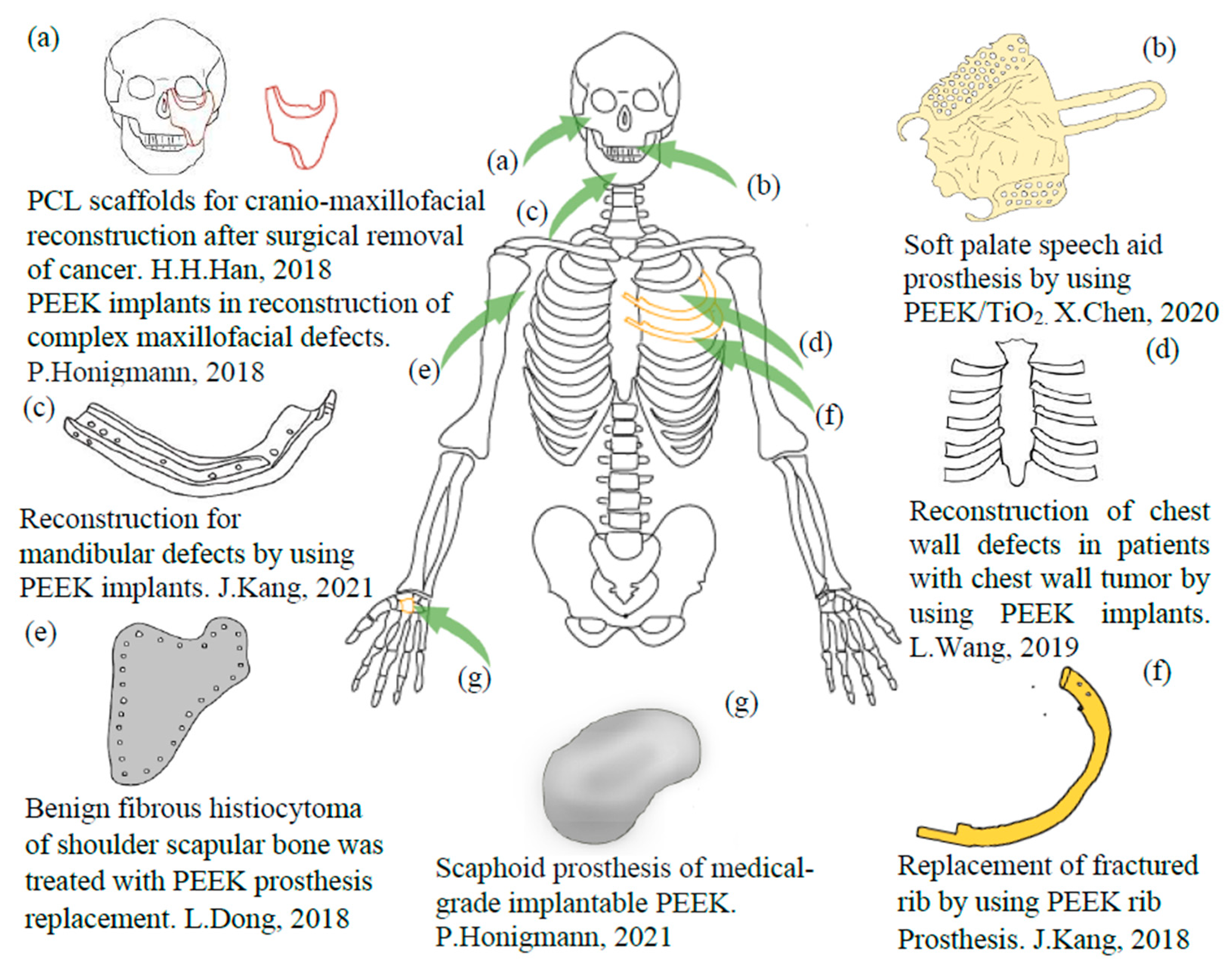
- Han, H.H.; Shim, J.H.; Lee, H.; Kim, B.Y.; Lee, J.S.; Jung, J.W.; Yun, W.S.; Baek, C.H.; Rhie, J.W.; Cho, D.W. Reconstruction of complex maxillary defects using patient-specific 3D-printed biodegradable scaffolds. Plast. Reconstr. Surg. Glob. Open 2018, 6, e1975. [Google Scholar] [CrossRef] [PubMed]
- Singh, S.; Prakash, C.; Ramakrishna, S. 3D printing of polyether-ether-ketone for biomedical applications. Eur. Polym. J. 2019, 114, 234–248. [Google Scholar] [CrossRef]
- Wang, Y.; Müller, W.; Rumjahn, A.; Schmidt, F.; Schwitalla, D. Mechanical properties of fused filament fabricated PEEK for biomedical applications depending on additive manufacturing parameters. J. Mech. Behav. Biomed. Mater. 2021, 115, 104250. [Google Scholar] [CrossRef]
- Wang, L.; Huang, L.; Li, X.; Zhong, D.; Li, D. Three-Dimensional Printing PEEK Implant: A Novel Choice for the Reconstruction of Chest Wall Defect. Ann. Thorac. Surg. 2019, 107, 921–928. [Google Scholar] [CrossRef]
- Kim, S.E.; Yun, Y.P.; Shim, K.S.; Kim, H.J.; Park, K.; Song, H.R. 3D printed alendronate-releasing poly(caprolactone) porous scaffolds enhance osteogenic differentiation and bone formation in rat tibial defects. Biomed. Mater. 2016, 11, 055005. [Google Scholar] [CrossRef]
- Zhang, Z.Z.; Wang, S.J.; Zhang, J.Y.; Jiang, W.B.; Huang, A.B.; Qi, Y.S.; Ding, J.X.; Chen, X.S.; Jiang, D.; Yu, J.K. 3D-Printed Poly(∈-caprolactone) Scaffold Augmented with Mesenchymal Stem Cells for Total Meniscal Substitution: A 12- and 24-Week Animal Study in a Rabbit Model. Am. J. Sports Med. 2017, 45, 1497–1511. [Google Scholar] [CrossRef] [PubMed]
- Brach del Prever, E.M.; Bistolfi, A.; Bracco, P.; Costa, L. UHMWPE for arthroplasty: Past or future? J. Orthop. Traumatol. 2009, 10, 1–8. [Google Scholar] [CrossRef] [PubMed] [Green Version]
- Saito, N.; Aoki, K.; Usui, Y.; Shimizu, M.; Hara, K.; Narita, N.; Ogihara, N.; Nakamura, K.; Ishigaki, N.; Kato, H.; et al. Application of carbon fibers to biomaterials: A new era of nano-level control of carbon fibers after 30-years of development. Chem. Soc. Rev. 2011, 40, 3824–3834. [Google Scholar] [CrossRef] [PubMed]
- Ramakrishna, S.; Mayer, J.; Wintermantel, E.; Leong, K. Biomedical applications of polymer-composite material: A review. Compos. Sci. Technol. 2001, 61, 1189–1224. [Google Scholar] [CrossRef]
- Cheung, H.Y.; Lau, K.T.; Lu, T.P.; Hui, D. A critical review on polymer-based bio-engineered materials for scaffold development. Compos. Part B Eng. 2007, 38, 291–300. [Google Scholar] [CrossRef]
- Katti, K.S.; Verma, D.; Katti, D.R. Materials for Joint Replacement; Elsevier: Amsterdam, The Netherlands, 2008; pp. 81–104. [Google Scholar] [CrossRef]
- Liu, H.; Webster, T.J. Bioinspired Nanocomposites for Orthopedic Applications. In Nanotechnology for the Regeneration of Hard and Soft Tissues; World Scientific: Singapore, 2007. [Google Scholar] [CrossRef]
- Steinberg, E.L.; Rath, E.; Shlaifer, A.; Chechik, O.; Maman, E.; Salai, M. Carbon fiber reinforced PEEK Optima-A composite material biomechanical properties and wear/debris characteristics of CF-PEEK composites for orthopedic trauma implants. J. Mech. Behav. Biomed. Mater. 2013, 17, 221–228. [Google Scholar] [CrossRef]
- Ghanbari, A.; Kargar, S. Preparation of Ni-P-Al2O3-TiO2 Nano composite coating by Electroless. In Proceedings of the 7th International Conference on Nanostructures (ICNS7), Tehran, Iran, 27 February–1 March 2018; p. ADV-41. [Google Scholar]
- Kargar, S.; Ghanbari, A.; Moosavi, A.; Seyedraoufi, Z. Simulation of Flow Ni-P-Al2O3-TiO2 nano composite coating by Prediction (ANN). In Proceedings of the 7th International Conference on Nanostructures (ICNS7), Tehran, Iran, 27 February–1 March 2018; p. BLK-19. [Google Scholar]
- Chen, X.; Gao, C.; Jiang, J.; Wu, Y.; Zhu, P.; Chen, G. 3D printed porous PLA/nHA composite scaffolds with enhanced osteogenesis and osteoconductivityin vivo for bone regeneration. Biomed. Mater. 2019, 14, 065003. [Google Scholar] [CrossRef]
- Tanodekaew, S.; Channasanon, S.; Kaewkong, P.; Uppanan, P. PLA-HA scaffolds: Preparation and bioactivity. Procedia Eng. 2013, 59, 144–149. [Google Scholar] [CrossRef]
- Jeong, S.I.; Ko, E.K.; Yum, J.; Jung, C.H.; Lee, Y.M.; Shin, H. Nanofibrous poly(lactic acid)/hydroxyapatite composite scaffolds for guided tissue regeneration. Macromol. Biosci. 2008, 8, 328–338. [Google Scholar] [CrossRef]
- Lui, H.; Webster, T.J. Mechanical properties of dispersed ceramic nanoparticles in polymer composites for orthopedic applications. Int. J. Nanomed. 2010, 5, 299. [Google Scholar]
- Jose, M.V.; Thomas, V.; Johnson, K.T.; Dean, D.R.; Nyairo, E. Aligned PLGA/HA nanofibrous nanocomposite scaffolds for bone tissue engineering. Acta Biomater. 2009, 5, 305–315. [Google Scholar] [CrossRef] [PubMed]
- Kang, Y.; Scully, A.; Young, D.A.; Kim, S.; Tsao, H.; Sen, M.; Yang, Y. Enhanced mechanical performance and biological evaluation of a PLGA coated β-TCP composite scaffold for load-bearing applications. Eur. Polym. J. 2011, 47, 1569–1577. [Google Scholar] [CrossRef] [PubMed]
- Ehrahimian-Hosseinabadi, M.; Ashrafizadeh, F.; Etemadifar, M.; Venkatraman, S.S. Evaluating and Modeling the Mechanical Properties of the Prepared PLGA/nano-BCP Composite Scaffolds for Bone Tissue Engineering. J. Mater. Sci. Technol. 2011, 27, 1105–1112. [Google Scholar] [CrossRef]
- Vaezi, M.; Black, C.; Gibbs, D.M.R.; Oreffo, R.O.C.; Brady, M.; Moshrefi-Torbati, M.; Yang, S. Characterization of New PEEK/HA composites with 3D HA network fabricated by extrusion freeforming. Molecules 2016, 21, 687. [Google Scholar] [CrossRef] [PubMed]
- Vaezi, M.; Yang, S. A novel bioactive PEEK/HA composite with controlled 3D interconnected HA network. Int. J. Bioprint. 2015, 1, 66–76. [Google Scholar] [CrossRef]
- Uddin, M.N.; Dhanasekaran, P.S.; Asmatulu, R. Mechanical properties of highly porous PEEK bionanocomposites incorporated with carbon and hydroxyapatite nanoparticles for scaffold applications. Prog. Biomater. 2019, 8, 211–221. [Google Scholar] [CrossRef]
- Haider, A.; Haider, S.; Rao Kummara, M.; Kamal, T.; Alghyamah, A.A.A.; Jan Iftikhar, F.; Bano, B.; Khan, N.; Amjid Afridi, M.; Soo Han, S.; et al. Advances in the scaffolds fabrication techniques using biocompatible polymers and their biomedical application: A technical and statistical review. J. Saudi Chem. Soc. 2020, 24, 186–215. [Google Scholar] [CrossRef]
- Asefnejad, A.; Mohammad, T.K.; Aliasghar, B.; Babak, F.; Shahin, B. Manufacturing of biodegradable polyurethane scaffolds based on polycaprolactone using a phase separation method: Physical properties and in vitro assay. Int. J. Nanomed. 2011, 6, 2375–2384. [Google Scholar] [CrossRef]
- Nam, Y.S.; Park, T.G. Porous biodegradable polymeric scaffolds prepared by thermally induced phase separation. J. Biomed. Mater. Res. 1999, 47, 8–17. [Google Scholar] [CrossRef]
- Gautam, S.; Dinda, A.K.; Mishra, N.C. Fabrication and characterization of PCL/gelatin composite nanofibrous scaffold for tissue engineering applications by electrospinning method. Mater. Sci. Eng. C 2013, 33, 1228–1235. [Google Scholar] [CrossRef]
- Zhao, L.; He, C.; Gao, Y.; Cen, L.; Cui, L.; Cao, Y. Preparation and cytocompatibility of PLGA scaffolds with controllable fiber morphology and diameter using electrospinning method. J. Biomed. Mater. Res. Part B Appl. Biomater. 2008, 87, 26–34. [Google Scholar] [CrossRef] [PubMed]
- Hou, Q.; Grijpma, D.W.; Feijen, J. Preparation of Interconnected Highly Porous Polymeric Structures by a Replication and Freeze-Drying Process. J. Biomed. Mater. Res. Part B Appl. Biomater. 2003, 67, 732–740. [Google Scholar] [CrossRef] [PubMed]
- Hou, Q.; Grijpma, D.W.; Feijen, J. Porous polymeric structures for tissue engineering prepared by a coagulation, compression moulding and salt leaching technique. Biomaterials 2003, 24, 1937–1947. [Google Scholar] [CrossRef]
- Liao, C.J.; Chen, C.F.; Chen, J.H.; Chiang, S.F.; Lin, Y.J.; Chang, K.Y. Fabrication of porous biodegradable polymer scaffolds using a solvent merging/particulate leaching method. J. Biomed. Mater. Res. 2002, 59, 676–681. [Google Scholar] [CrossRef] [PubMed]
- Oh, S.H.; Kang, S.G.; Kim, E.S.; Cho, S.H.; Lee, J.H. Fabrication and characterization of hydrophilic poly(lactic-co-glycolic acid)/poly(vinyl alcohol) blend cell scaffolds by melt-molding particulate-leaching method. Biomaterials 2003, 24, 4011–4021. [Google Scholar] [CrossRef]
- Young, M.J.; Park, K.; Jun, S.S.; Kim, J.J.; Rhie, J.W.; Han, D.K. Beneficial effect of hydrophilized porous polymer scaffolds in tissue-engineered cartilage formation. J. Biomed. Mater. Res. Part B Appl. Biomater. 2008, 85, 252–260. [Google Scholar] [CrossRef]
- Moghadam, M.Z.; Hassanajili, S.; Esmaeilzadeh, F.; Ayatollahi, M.; Ahmadi, M. Formation of porous HPCL/LPCL/HA scaffolds with supercritical CO2 gas foaming method. J. Mech. Behav. Biomed. Mater. 2017, 69, 115–127. [Google Scholar] [CrossRef] [PubMed]
- Lyons, R.; Newell, A.; Ghadimi, P.; Papakostas, N. Environmental impacts of conventional and additive manufacturing for the production of Ti-6Al-4V knee implant: A life cycle approach. Int. J. Adv. Manuf. Technol. 2020, 112, 787–801. [Google Scholar] [CrossRef]
- Cronskär, M.; Bäckström, M.; Rännar, L.E. Production of customized hip stem prostheses - A comparison between conventional machining and electron beam melting (EBM). Rapid Prototyp. J. 2013, 19, 365–372. [Google Scholar] [CrossRef]
- Costantini, M.; Barbetta, A. Gas Foaming Technologies for 3D scaffold Engineering; Elsevier Ltd.: Amsterdam, The Netherlands, 2018. [Google Scholar] [CrossRef]
- Boudriot, U.; Dersch, R.; Greiner, A.; Wendorff, J.H. Electrospinning approaches toward scaffold engineering—A brief overview. Artif. Organs 2006, 30, 785–792. [Google Scholar] [CrossRef]
- Bagaria, V.; Bhansali, R.; Pawar, P. 3D printing- creating a blueprint for the future of orthopedics: Current concept review and the road ahead! J. Clin. Orthop. Trauma 2018, 9, 207–212. [Google Scholar] [CrossRef] [PubMed]
- Zhang, B.; Wang, L.; Song, P.; Pei, X.; Sun, H.; Wu, L.; Zhou, C.; Wang, K.; Fan, Y.; Zhang, X. 3D printed bone tissue regenerative PLA/HA scaffolds with comprehensive performance optimizations. Mater. Des. 2021, 201, 109490. [Google Scholar] [CrossRef]
- Fan, D.; Li, Y.; Wang, X.; Zhu, T.; Wang, Q.; Cai, H.; Li, W.; Tian, Y.; Liu, Z. Progressive 3D Printing Technology and Its Application in Medical Materials. Front. Pharmacol. 2020, 11, 122. [Google Scholar] [CrossRef] [PubMed]
- Du, W.; Ren, X.; Pei, Z.; Ma, C. Ceramic Binder Jetting Additive Manufacturing: A Literature Review on Density. J. Manuf. Sci. Eng. Trans. ASME 2020, 142, 040801. [Google Scholar] [CrossRef]
- Zhou, Z.; Lennon, A.; Buchanan, F.; McCarthy, H.O.; Dunne, N. Binder jetting additive manufacturing of hydroxyapatite powders: Effects of adhesives on geometrical accuracy and green compressive strength. Addit. Manuf. 2020, 36, 101645. [Google Scholar] [CrossRef]
- Szymczyk-Ziółkowska, P.; Łabowska, M.B.; Detyna, J.; Michalak, I.; Gruber, P. A review of fabrication polymer scaffolds for biomedical applications using additive manufacturing techniques. Biocybern. Biomed. Eng. 2020, 40, 624–638. [Google Scholar] [CrossRef]
- Abdulhameed, O.; Al-Ahmari, A.; Ameen, W.; Mian, S.H. Additive manufacturing: Challenges, trends, and applications. Adv. Mech. Eng. 2019, 11, 1–27. [Google Scholar] [CrossRef]
- Kumar, M.B.; Sathiya, P. Methods and materials for additive manufacturing: A critical review on advancements and challenges. Thin-Walled Struct. 2021, 159, 107228. [Google Scholar] [CrossRef]
- Jared, B.H.; Aguilo, M.A.; Beghini, L.L.; Boyce, B.L.; Clark, B.W.; Cook, A.; Kaehr, B.J.; Robbins, J. Additive manufacturing: Toward holistic design. Scr. Mater. 2017, 135, 141–147. [Google Scholar] [CrossRef]
- Guessasma, S.; Zhang, W.; Zhu, J.; Belhabib, S.; Nouri, H. Challenges of additive manufacturing technologies from an optimisation perspective. Int. J. Simul. Multidiscip. Des. Optim. 2015, 6, A9. [Google Scholar] [CrossRef] [Green Version]
- Somireddy, M. Fabrication of Composite Structures via 3D Printing. In Fused Deposition Modeling Based 3D Printing; Springer: Berlin/Heidelberg, Germany, 2021; pp. 255–276. [Google Scholar] [CrossRef]
- Melancon, D.; Bagheri, Z.S.; Johnston, R.B.; Liu, L.; Tanzer, M.; Pasini, D. Mechanical characterization of structurally porous biomaterials built via additive manufacturing: Experiments, predictive models, and design maps for load-bearing bone replacement implants. Acta Biomater. 2017, 63, 350–368. [Google Scholar] [CrossRef] [PubMed]
- Bagheri, Z.S.; Melancon, D.; Liu, L.; Johnston, R.B.; Pasini, D. Compensation strategy to reduce geometry and mechanics mismatches in porous biomaterials built with Selective Laser Melting. J. Mech. Behav. Biomed. Mater. 2017, 70, 17–27. [Google Scholar] [CrossRef] [PubMed]
- Piat, R.; Sinchuk, Y.; Vasoya, M.; Sigmund, O. Minimal compliance design for metal-ceramic composites with lamellar microstructures. Acta Mater. 2011, 59, 4835–4846. [Google Scholar] [CrossRef]
- Kum, S.G.; Laurencin, C.; Deng, M. Natural and Synthetic Biomedical Polymers; Elsevier Science & Technology: Saint Louis, MI, USA, 2014. [Google Scholar]
- Allan, B.; Ruan, R.; Landao-Bassonga, E.; Gillman, N.; Wang, T.; Gao, J.; Ruan, Y.; Xu, Y.; Lee, C.; Goonewardene, M.; et al. Collagen membrane for guided bone regeneration in dental and orthopedic applications. Tissue Eng. Part A 2021, 27, 372–381. [Google Scholar] [CrossRef]
- Cunniffe, G.M.; O’Brien, F.J. Collagen scaffolds for orthopedic regenerative medicine. JOM 2011, 63, 66–73. [Google Scholar] [CrossRef]
- Alghamdi, H.S.; Bosco, R.; van den Beucken, J.J.J.P.; Walboomers, X.F.; Jansen, J.A. Osteogenicity of titanium implants coated with calcium phosphate or collagen type-I in osteoporotic rats. Biomaterials 2013, 34, 3747–3757. [Google Scholar] [CrossRef]
- Lee, Y.H.; Lee, B.W.; Jung, Y.C.; Yoon, B.I.; Woo, H.M.; Kang, B.J. Application of alginate microbeads as a carrier of bone morphogenetic protein-2 for bone regeneration. J. Biomed. Mater. Res. Part B Appl. Biomater. 2019, 107, 286–294. [Google Scholar] [CrossRef]
- Chen, Q.; Cordero-Arias, L.; Roether, J.A.; Cabanas-Polo, S.; Virtanen, S.; Boccaccini, A.R. Alginate/Bioglass® composite coatings on stainless steel deposited by direct current and alternating current electrophoretic deposition. Surf. Coat. Technol. 2013, 233, 49–56. [Google Scholar] [CrossRef]
- Ho, H.V.; Tripathi, G.; Gwon, J.; Lee, S.Y.; Lee, B.T. Novel TOCNF reinforced injectable alginate β-tricalcium phosphate microspheres for bone regeneration. Mater. Des. 2020, 194, 108892. [Google Scholar] [CrossRef]
- Arjmandi, M.; Ramezani, M.; Nand, A.; Neitzert, T. Experimental study on friction and wear properties of interpenetrating polymer network alginate-polyacrylamide hydrogels for use in minimally-invasive joint implants. Wear 2018, 406–407, 194–204. [Google Scholar] [CrossRef]
- Varoni, E.; Tschon, M.; Palazzo, B.; Nitti, P.; Martini, L.; Rimondini, L. Agarose Gel as Biomaterial or Scaffold for Implantation Surgery: Characterization, Histological and Histomorphometric Study on Soft Tissue Response. Connect. Tissue Res. 2012, 53, 548–554. [Google Scholar] [CrossRef] [PubMed]
- Melis Soylu, H.; Chevallier, P.; Copes, F.; Ponti, F.; Candiani, G.; Yurt, F.; Mantovani, D.; Li, Y.; Nan, K. A Novel Strategy to Coat Dopamine-Functionalized Titanium Surfaces With Agarose-Based Hydrogels for the Controlled Release of Gentamicin. Article 2021, 11, 1. [Google Scholar] [CrossRef]
- Gupta, A.; Bhat, S.; Jagdale, P.R.; Chaudhari, B.P.; Lidgren, L.; Gupta, K.C.; Kumar, A. Evaluation of three-dimensional chitosan-agarose-gelatin cryogel scaffold for the repair of subchondral cartilage defects: An in vivo study in a rabbit model. Tissue Eng. Part A 2014, 20, 3101–3111. [Google Scholar] [CrossRef]
- Figueiredo, L.; Fonseca, R.; Pinto, L.F.V.; Ferreira, F.C.; Almeida, A.; Rodrigues, A. Strategy to improve the mechanical properties of bioabsorbable materials based on chitosan for orthopedic fixation applications. J. Mech. Behav. Biomed. Mater. 2020, 103, 103572. [Google Scholar] [CrossRef] [PubMed]
- Francis, A.; Yang, Y.; Boccaccini, A.R. A new strategy for developing chitosan conversion coating on magnesium substrates for orthopedic implants. Appl. Surf. Sci. 2019, 466, 854–862. [Google Scholar] [CrossRef]
- Pighinelli, L.; Kucharska, M. Chitosan-hydroxyapatite composites. Carbohydr. Polym. 2013, 93, 256–262. [Google Scholar] [CrossRef]
- Tuzlakoglu, K.; Reis, R.L. Introduction: Chemical and Physical Structure of Chitosan and Its. In Woodhead Publishing Series in Biomaterials; Woodhead Publishing: Sawston, UK, 2008; pp. 357–373. [Google Scholar] [CrossRef]
- Qin, L.; Dong, H.; Mu, Z.; Zhang, Y.; Dong, G. Preparation and bioactive properties of chitosan and casein phosphopeptides composite coatings for orthopedic implants. Carbohydr. Polym. 2015, 133, 236–244. [Google Scholar] [CrossRef] [PubMed]
- Ahmed, R.A.; Fekry, A.M.; Farghali, R.A. A study of calcium carbonate/multiwalled-carbon nanotubes/chitosan composite coatings on Ti-6Al-4V alloy for orthopedic implants. Appl. Surf. Sci. 2013, 285, 309–316. [Google Scholar] [CrossRef]
- Deepachitra, R.; Nigam, R.; Purohit, S.D.; Kumar, B.S.; Hemalatha, T.; Sastry, T.P. In Vitro Study of Hydroxyapatite Coatings on Fibrin Functionalized/Pristine Graphene Oxide for Bone Grafting. Mater. Manuf. Process. 2015, 30, 804–811. [Google Scholar] [CrossRef]
- Noori, A.; Ashrafi, S.J.; Vaez-Ghaemi, R.; Hatamian-Zaremi, A.; Webster, T.J. A review of fibrin and fibrin composites for bone tissue engineering. Int. J. Nanomed. 2017, 12, 4937. [Google Scholar] [CrossRef]
- Kobayashi, M.; Hyu, H.S. Development and Evaluation of Polyvinyl Alcohol-Hydrogels as an Artificial Atrticular Cartilage for Orthopedic Implants. Materials (Basel) 2010, 3, 2753–2771. [Google Scholar] [CrossRef]
- Pitarresi, G.; Palumbo, F.S.; Calascibetta, F.; Fiorica, C.; Di Stefano, M.; Giammona, G. Medicated hydrogels of hyaluronic acid derivatives for use in orthopedic field. Int. J. Pharm. 2013, 449, 84–94. [Google Scholar] [CrossRef] [PubMed]
- Zhai, P.; Peng, X.; Li, B.; Liu, Y.; Sun, H.; Li, X. The application of hyaluronic acid in bone regeneration. Int. J. Biol. Macromol. 2020, 151, 1224–1239. [Google Scholar] [CrossRef] [PubMed]
- Martínez-Sanz, E.; Ossipov, D.A.; Hilborn, J.; Larsson, S.; Jonsson, K.B.; Varghese, O.P. Bone reservoir: Injectable hyaluronic acid hydrogel for minimal invasive bone augmentation. J. Control. Release 2011, 152, 232–240. [Google Scholar] [CrossRef]
- Peppas, N.A.; Hoffman, A.S. Hydrogels. Elsevier 2020, 153–166. [Google Scholar] [CrossRef]
- Maitz, M.F. Applications of synthetic polymers in clinical medicine. Biosurf. Biotribol. 2015, 1, 161–176. [Google Scholar] [CrossRef]
- Velu, R.; Calais, T.; Jayakumar, A.; Raspall, F. A comprehensive review on bio-nanomaterials for medical implants and feasibility studies on fabrication of such implants by additive manufacturing technique. Materials 2020, 13, 92. [Google Scholar] [CrossRef]
- Yao, C.H.; Lai, Y.H.; Chen, Y.W.; Cheng, C.H. Bone Morphogenetic Protein-2-Activated 3D-Printed Polylactic Acid Scaffolds to Promote Bone Regrowth and Repair. Macromol. Biosci. 2020, 20, 1–15. [Google Scholar] [CrossRef]
- Singhvi, M.S.; Zinjarde, S.S.; Gokhale, D.V. Polylactic acid: Synthesis and biomedical applications. J. Appl. Microbiol. 2019, 127, 1612–1626. [Google Scholar] [CrossRef]
- Burge, G.; Aytac, E.; Evcil, A.; Savas, M.A. An Investigation on Mechanical Properties of PLA Produced by 3D Printing as an Implant Material. In Proceedings of the 2020 4th International Symposium on Multidisciplinary Studies and Innovative Technologies (ISMSIT), Istanbul, Turkey, 22–24 October 2020. [Google Scholar] [CrossRef]
- Chen, X.; Chen, G.; Wang, G.; Zhu, P.; Gao, C. Recent Progress on 3D-Printed Polylactic Acid and Its Applications in Bone Repair. Adv. Eng. Mater. 2020, 22, 1–19. [Google Scholar] [CrossRef]
- Wu, C.; Wang, S.; Wu, D.; Shih, W. Novel composite 3D-printed filament made from fish scale-derived hydroxyapatite, eggshell and polylactic acid via a fused fabrication approach. Addit. Manuf. 2021, 46, 102169. [Google Scholar] [CrossRef]
- Alksne, M.; Kalvaityte, M.; Simoliunas, E.; Rinkunaite, I.; Gendviliene, I.; Locs, J.; Rutkunas, V.; Bukelskiene, V. In vitro comparison of 3D printed polylactic acid/hydroxyapatite and polylactic acid/bioglass composite scaffolds: Insights into materials for bone regeneration. J. Mech. Behav. Biomed. Mater. 2020, 104, 103641. [Google Scholar] [CrossRef] [PubMed]
- Nazeer, M.A.; Onder, O.C.; Sevgili, I.; Yilgor, E.; Kavakli, I.H.; Yilgor, I. 3D printed poly(lactic acid) scaffolds modified with chitosan and hydroxyapatite for bone repair applications. Mater. Today Commun. 2020, 25, 101515. [Google Scholar] [CrossRef]
- Luo, Y.; Humayun, A.; Mills, D.K. Surface Modification of 3D Printed PLA / Halloysite Composite Sca ff olds with Antibacterial and Osteogenic Capabilities. Appl. Sci. 2020, 10, 3971. [Google Scholar] [CrossRef]
- Singh, D.; Babbar, A.; Jain, V.; Gupta, D.; Saxena, S.; Dwibedi, V. Synthesis, characterization, and bioactivity investigation of biomimetic biodegradable PLA scaffold fabricated by fused filament fabrication process. J. Brazilian Soc. Mech. Sci. Eng. 2019, 41, 1–13. [Google Scholar] [CrossRef]
- Wang, P.; Yin, H.M.; Li, X.; Liu, W.; Chu, Y.X.; Wang, Y.; Wang, Y.; Xu, J.Z.; Li, Z.M.; Li, J.H. Simultaneously constructing nanotopographical and chemical cues in 3D-printed polylactic acid scaffolds to promote bone regeneration. Mater. Sci. Eng. C 2021, 118, 111457. [Google Scholar] [CrossRef] [PubMed]
- Wang, M.; Favi, P.; Cheng, X.; Golshan, N.H.; Ziemer, K.S.; Keidar, M.; Webster, T.J. Cold atmospheric plasma (CAP) surface nanomodified 3D printed polylactic acid (PLA) scaffolds for bone regeneration. Acta Biomater. 2016, 46, 256–265. [Google Scholar] [CrossRef]
- Dubinenko, G.E.; Zinoviev, A.L.; Bolbasov, E.N.; Novikov, V.T.; Tverdokhlebov, S.I. Preparation of Poly(L-lactic acid)/Hydroxyapatite composite scaffolds by fused deposit modeling 3D printing. Mater. Today Proc. 2020, 22, 228–234. [Google Scholar] [CrossRef]
- Omigbodun, F.T.; Engstrom, D.S.; Mele, E. Improving Mechanical strength of bone-implant with primitive and gyroid lattice of PLA/cHAP and rGO composites. J. Hazard. Mater. 2020, 124370. [Google Scholar] [CrossRef]
- Sahmani, S.; Khandan, A.; Saber-Samandari, S.; Esmaeili, S.; Aghdam, M.M. Fabrication and resonance simulation of 3D-printed biocomposite mesoporous implants with different periodic cellular topologies. Bioprinting 2021, 22, e00138. [Google Scholar] [CrossRef]
- Elhattab, K.; Bhaduri, S.B.; Lawrence, J.G.; Sikder, P. Fused Filament Fabrication (Three-Dimensional Printing) of Amorphous Magnesium Phosphate/Polylactic Acid Macroporous Biocomposite Scaffolds. ACS Appl. Bio Mater. 2021, 4, 3276–3286. [Google Scholar] [CrossRef] [PubMed]
- Ranjan, N.; Singh, R.; Ahuja, I.P.S.; Kumar, R.; Singh, J.; Verma, A.K.; Leekha, A. On 3D printed scaffolds for orthopedic tissue engineering applications. SN Appl. Sci. 2020, 2, 192. [Google Scholar] [CrossRef]
- Tcacencu, I.; Rodrigues, N.; Alharbi, N.; Benning, M.; Toumpaniari, S.; Mancuso, E.; Marshall, M.; Bretcanu, O.; Birch, M.; McCaskie, A.; et al. Osseointegration of porous apatite-wollastonite and poly(lactic acid) composite structures created using 3D printing techniques. Mater. Sci. Eng. C 2018, 90, 1–7. [Google Scholar] [CrossRef] [PubMed]
- Zhang, H.; Mao, X.; Zhao, D.; Jiang, W.; Du, Z.; Li, Q.; Jiang, C.; Han, D. Three dimensional printed polylactic acid-hydroxyapatite composite scaffolds for prefabricating vascularized tissue engineered bone: An in vivo bioreactor model. Sci. Rep. 2017, 7, 15255. [Google Scholar] [CrossRef]
- Liu, Z.; Ge, Y.; Zhang, L.; Wang, Y.; Guo, C.; Feng, K.; Yang, S.; Zhai, Z.; Chi, Y.; Zhao, J.; et al. The effect of induced membranes combined with enhanced bone marrow and 3D PLA-HA on repairing long bone defects in vivo. J. Tissue Eng. Regen. Med. 2020, 14, 1403–1414. [Google Scholar] [CrossRef]
- Shen, J.; Wang, W.; Zhai, X.; Chen, B.; Qiao, W.; Li, W.; Li, P.; Zhao, Y.; Meng, Y.; Qian, S.; et al. 3D-printed nanocomposite scaffolds with tunable magnesium ionic microenvironment induce in situ bone tissue regeneration. Appl. Mater. Today 2019, 16, 493–507. [Google Scholar] [CrossRef]
- Haverová, L.; Oriňaková, R.; Oriňak, A.; Gorejová, R.; Baláž, M.; Vanýsek, P.; Kupková, M.; Hrubovčáková, M.; Mudroň, P.; Radoňák, J.; et al. An in vitro corrosion study of open cell Iron structures with PEG coating for bone replacement applications. Metals (Basel) 2018, 8, 499. [Google Scholar] [CrossRef] [Green Version]
- Luo, Y.; Pan, H.; Jiang, J.; Zhao, C.; Zhang, J.; Chen, P.; Lin, X.; Fan, S. Desktop-Stereolithography 3D Printing of a Polyporous Extracellular Matrix Bioink for Bone Defect Regeneration. Front. Bioeng. Biotechnol. 2020, 8, 1–13. [Google Scholar] [CrossRef]
- Zhou, X.; Esworthy, T.; Lee, S.J.; Miao, S.; Cui, H.; Plesiniak, M.; Fenniri, H.; Webster, T.; Rao, R.D.; Zhang, L.G. 3D Printed scaffolds with hierarchical biomimetic structure for osteochondral regeneration. Nanomed. Nanotechnol. Biol. Med. 2019, 19, 58–70. [Google Scholar] [CrossRef]
- Zhou, X.; Castro, N.J.; Zhu, W.; Cui, H.; Aliabouzar, M.; Sarkar, K.; Zhang, L.G. Improved Human Bone Marrow Mesenchymal Stem Cell Osteogenesis in 3D Bioprinted Tissue Scaffolds with Low Intensity Pulsed Ultrasound Stimulation. Sci. Rep. 2016, 6, 32876. [Google Scholar] [CrossRef]
- Zhu, S.; Chen, P.; Chen, Y.; Li, M.; Chen, C.; Lu, H. 3D-Printed Extracellular Matrix/Polyethylene Glycol Diacrylate Hydrogel Incorporating the Anti-inflammatory Phytomolecule Honokiol for Regeneration of Osteochondral Defects. Am. J. Sports Med. 2020, 48, 2808–2818. [Google Scholar] [CrossRef] [PubMed]
- Bai, J.; Wang, H.; Gao, W.; Liang, F.; Wang, Z.; Zhou, Y.; Lan, X.; Chen, X.; Cai, N.; Huang, W.; et al. Melt electrohydrodynamic 3D printed poly (ε-caprolactone)/polyethylene glycol/roxithromycin scaffold as a potential anti-infective implant in bone repair. Int. J. Pharm. 2020, 576, 118941. [Google Scholar] [CrossRef] [PubMed]
- Liu, F.; Kang, H.; Liu, Z.; Jin, S.; Yan, G.; Sun, Y.; Li, F.; Zhan, H.; Gu, Y. 3D Printed Multi-Functional Scaffolds Based on Poly(ε-Caprolactone) and Hydroxyapatite Composites. Nanomaterials 2021, 11, 2456. [Google Scholar] [CrossRef] [PubMed]
- Buyuksungur, S.; Endogan Tanir, T.; Buyuksungur, A.; Bektas, E.I.; Torun Kose, G.; Yucel, D.; Beyzadeoglu, T.; Cetinkaya, E.; Yenigun, C.; Tönük, E.; et al. 3D printed poly(ϵ-caprolactone) scaffolds modified with hydroxyapatite and poly(propylene fumarate) and their effects on the healing of rabbit femur defects. Biomater. Sci. 2017, 5, 2144–2158. [Google Scholar] [CrossRef]
- Pandiyaraj, K.N.; Ghobeira, R.; Esbah Tabaei, P.S.; Cools, P.; De Geyter, N.; Morent, R.; Deshmukh, R.R. Non-thermal plasma jet-assisted development of phosphorus-containing functional coatings on 3D-printed PCL scaffolds intended for bone tissue engineering. J. Phys. Chem. Solids 2021, 154, 110025. [Google Scholar] [CrossRef]
- Xiong, Z.; Liu, W.; Qian, H.; Lei, T.; He, X.; Hu, Y.; Lei, P. Tantalum Nanoparticles Reinforced PCL Scaffolds Using Direct 3D Printing for Bone Tissue Engineering. Front. Mater. 2021, 8, 1–7. [Google Scholar] [CrossRef]
- Ma, J.; Lin, L.; Zuo, Y.; Zou, Q.; Ren, X.; Li, J.; Li, Y. Modification of 3D printed PCL scaffolds by PVAc and HA to enhance cytocompatibility and osteogenesis. RSC Adv. 2019, 9, 5338–5346. [Google Scholar] [CrossRef] [Green Version]
- Zhao, S.; Xie, K.; Guo, Y.; Tan, J.; Wu, J.; Yang, Y.; Fu, P.; Wang, L.; Jiang, W.; Hao, Y. Fabrication and Biological Activity of 3D-Printed Polycaprolactone/Magnesium Porous Scaffolds for Critical Size Bone Defect Repair. ACS Biomater. Sci. Eng. 2020, 6, 5120–5131. [Google Scholar] [CrossRef]
- Radhakrishnan, S.; Nagarajan, S.; Belaid, H.; Farha, C.; Iatsunskyi, I.; Coy, E.; Soussan, L.; Huon, V.; Bares, J.; Belkacemi, K.; et al. Fabrication of 3D printed antimicrobial polycaprolactone scaffolds for tissue engineering applications. Mater. Sci. Eng. C 2021, 118, 111525. [Google Scholar] [CrossRef]
- Abdal-hay, A.; Raveendran, N.T.; Fournier, B.; Ivanovski, S. Fabrication of biocompatible and bioabsorbable polycaprolactone/ magnesium hydroxide 3D printed scaffolds: Degradation and in vitro osteoblasts interactions. Compos. Part B Eng. 2020, 197, 108158. [Google Scholar] [CrossRef]
- Amdjadi, P.; Khoshroo, K.; Seifi, M.; Tahriri, M.; Tayebi, L. Mechanical Properties of 3D Printed reinforced Polycaprolactone Composite Scaffolds. J. Dent. Sch. Shahid Beheshti Univ. Med. Sci. 2018, 38, 7. [Google Scholar] [CrossRef]
- Golafshan, N.; Vorndran, E.; Zaharievski, S.; Brommer, H.; Kadumudi, F.B.; Dolatshahi-Pirouz, A.; Gbureck, U.; van Weeren, R.; Castilho, M.; Malda, J. Tough magnesium phosphate-based 3D-printed implants induce bone regeneration in an equine defect model. Biomaterials 2020, 261, 120302. [Google Scholar] [CrossRef] [PubMed]
- Xu, Y.; Peng, J.; Richards, G.; Lu, S.; Eglin, D. Optimization of electrospray fabrication of stem cell–embedded alginate–gelatin microspheres and their assembly in 3D-printed poly(ε-caprolactone) scaffold for cartilage tissue engineering. J. Orthop. Transl. 2019, 18, 128–141. [Google Scholar] [CrossRef]
- Hedayati, S.K.; Behravesh, A.H.; Hasannia, S.; Bagheri Saed, A.; Akhoundi, B. 3D printed PCL scaffold reinforced with continuous biodegradable fiber yarn: A study on mechanical and cell viability properties. Polym. Test. 2020, 83, 106347. [Google Scholar] [CrossRef]
- Shim, J.H.; Won, J.Y.; Park, J.H.; Bae, J.H.; Ahn, G.; Kim, C.H.; Lim, D.H.; Cho, D.W.; Yun, W.S.; Bae, E.B.; et al. Effects of 3D-printed polycaprolactone/β-tricalcium phosphate membranes on guided bone regeneration. Int. J. Mol. Sci. 2017, 18, 899. [Google Scholar] [CrossRef]
- DeBaun, M.R.; Stahl, A.M.; Daoud, A.I.; Pan, C.C.; Bishop, J.A.; Gardner, M.J.; Yang, Y.P. Preclinical induced membrane model to evaluate synthetic implants for healing critical bone defects without autograft. J. Orthop. Res. 2019, 37, 60–68. [Google Scholar] [CrossRef]
- Li, J.; Chen, M.; Wei, X.; Hao, Y.; Wang, J. Evaluation of 3D-printed polycaprolactone scaffolds coated with freeze-dried platelet-rich plasma for bone regeneration. Materials (Basel) 2017, 10, 831. [Google Scholar] [CrossRef] [Green Version]
- Zhou, Z.; Yao, Q.; Li, L.; Zhang, X.; Wei, B.; Yuan, L.; Wang, L. Antimicrobial activity of 3D-printed poly(ε-Caprolactone) (PCL) composite scaffolds presenting vancomycin-loaded polylactic acid-glycolic acid (PLGA) microspheres. Med. Sci. Monit. 2018, 24, 6934–6945. [Google Scholar] [CrossRef]
- Park, S.; Kim, J.E.; Han, J.; Jeong, S.; Lim, J.W.; Lee, M.C.; Son, H.; Kim, H.B.; Choung, Y.H.; Seonwoo, H.; et al. 3D-Printed Poly(Ε-Caprolactone)/Hydroxyapatite Scaffolds Modified With Alkaline Hydrolysis Enhance Osteogenesis in Vitro. Polymers 2021, 13, 257. [Google Scholar] [CrossRef]
- Lee, S.; Choi, D.; Shim, J.H.; Nam, W. Efficacy of three-dimensionally printed polycaprolactone/beta tricalcium phosphate scaffold on mandibular reconstruction. Sci. Rep. 2020, 10, 4979. [Google Scholar] [CrossRef]
- Nyberg, E.; Rindone, A.; Dorafshar, A.; Grayson, W.L. Comparison of 3D-Printed Poly-ϵ-Caprolactone Scaffolds Functionalized with Tricalcium Phosphate, Hydroxyapatite, Bio-Oss, or Decellularized Bone Matrix. Tissue Eng. Part A 2017, 23, 503–514. [Google Scholar] [CrossRef] [PubMed]
- Andrew Wu, Y.H.; Chiu, Y.C.; Lin, Y.H.; Ho, C.C.; Shie, M.Y.; Chen, Y.W. 3D-Printed bioactive calcium silicate/poly-ε-Caprolactone bioscaffolds modified with biomimetic extracellular matrices for bone regeneration. Int. J. Mol. Sci. 2019, 20, 942. [Google Scholar] [CrossRef]
- Knutsen, A.R.; Borkowski, S.L.; Ebramzadeh, E.; Flanagan, C.L.; Hollister, S.J.; Sangiorgio, S.N. Static and dynamic fatigue behavior of topology designed and conventional 3D printed bioresorbable PCL cervical interbody fusion devices. J. Mech. Behav. Biomed. Mater. 2015, 49, 332–342. [Google Scholar] [CrossRef] [PubMed]
- Xu, H.; Wang, C.; Liu, C.; Li, J.; Peng, Z.; Guo, J.; Zhu, L. Stem cell-seeded 3D-printed scaffolds combined with self-assembling peptides for bone defect repair. Tissue Eng. Part A 2021, 28, 111–124. [Google Scholar] [CrossRef]
- Kalluri, L.; Duan, Y. Advances in Dental Implantology using Nanomaterials and Allied Technology Applications. In Advances in Dental Implantology Using Nanomaterials and Allied Technology Applications; Chaughule, R.S., Dashaputra, R., Eds.; Springer: Berlin/Heidelberg, Germany, 2021. [Google Scholar] [CrossRef]
- Botlhoko, O.J. Preparation Characterization and Properties of Bionanohybrids Based on Biocompatible Poly (GlycolicAcid)/Polylactide Blends and Carbon Nanotubes—Towards Orthopaedic Applications by orebotse Joseph Botlhoko. Ph.D. Thesis, University of Johannesburg, Johannesburg, South Africa, 2012. [Google Scholar]
- Prabhu, B.; Karau, A.; Wood, A.; Dadsetan, M.; Liedtke, H.; DeWitt, T. Orthopedic Biomaterials; Li, B., Webster, T., Eds.; Springer: Berlin/Heidelberg, Germany, 2018. [Google Scholar]
- Benatti, A.C.B.; Pattaro, A.F.; Rodrigues, A.A.; Xavier, M.V.; Kaasi, A.; Barbosa, M.I.R.; Jardini, A.L.; Filho, R.M.; Kharmandayan, P. Bioreabsorbable polymers for tissue engineering: PLA, PGA, and their copolymers. In Materials for Biomedical Engineering; Elsevier: Amsterdam, The Netherlands, 2019. [Google Scholar] [CrossRef]
- Naseem, R.; Tzivelekis, C.; German, M.; Gentile, P.; Ferreira, A.; Dalgarno, K. Strategies for Enhancing Polyester-Based Materials for Bone Fixation Applications. Molecules 2021, 26, 992. [Google Scholar] [CrossRef] [PubMed]
- Ødegaard, K.S.; Torgersen, J.; Elverum, C.W. Structural and biomedical properties of common additively manufactured biomaterials: A concise review. Metals (Basel) 2020, 10, 1677. [Google Scholar] [CrossRef]
- Aragón, J.; Feoli, S.; Irusta, S.; Mendoza, G. Composite scaffold obtained by electro-hydrodynamic technique for infection prevention and treatment in bone repair. Int. J. Pharm. 2019, 557, 162–169. [Google Scholar] [CrossRef]
- Liu, C.G.; Zeng, Y.T.; Kankala, R.K.; Zhang, S.S.; Chen, A.Z.; Wang, S. Bin Characterization and preliminary biological evaluation of 3D-printed porous scaffolds for engineering bone tissues. Materials (Basel) 2018, 11, 1832. [Google Scholar] [CrossRef]
- Zhao, D.; Zhu, T.; Li, J.; Cui, L.; Zhang, Z.; Zhuang, X.; Ding, J. Poly(lactic-co-glycolic acid)-based composite bone-substitute materials. Bioact. Mater. 2021, 6, 346–360. [Google Scholar] [CrossRef]
- Babilotte, J.; Martin, B.; Guduric, V.; Bareille, R.; Agniel, R.; Roques, S.; Héroguez, V.; Dussauze, M.; Gaudon, M.; Le Nihouannen, D.; et al. Development and characterization of a PLGA-HA composite material to fabricate 3D-printed scaffolds for bone tissue engineering. Mater. Sci. Eng. C 2021, 118, 111334. [Google Scholar] [CrossRef]
- Liu, H.; Webster, T.J. Enhanced biological and mechanical properties of well-dispersed nanophase ceramics in polymer composites: From 2D to 3D printed structures. Mater. Sci. Eng. C 2011, 31, 77–89. [Google Scholar] [CrossRef]
- Han, R.; Buchanan, F.; Ford, L.; Julius, M.; Walsh, P.J. A comparison of the degradation behaviour of 3D printed PDLGA scaffolds incorporating bioglass or biosilica. Mater. Sci. Eng. C 2021, 120, 111755. [Google Scholar] [CrossRef] [PubMed]
- Yang, Y.; Yang, S.; Wang, Y.; Yu, Z.; Ao, H.; Zhang, H.; Qin, L.; Guillaume, O.; Eglin, D.; Richards, R.G.; et al. Anti-infective efficacy, cytocompatibility and biocompatibility of a 3D-printed osteoconductive composite scaffold functionalized with quaternized chitosan. Acta Biomater. 2016, 46, 112–128. [Google Scholar] [CrossRef] [PubMed]
- Wiria, F.E.; Tay, B.Y.; Chandrasekaran, M.; Zhang, S.X.; Maleksaeedi, S.; He, Z. 3D Printing of customized biomedical scaffolds and implants. In Proceedings of the 1st International Conference on Progress in Additive Manufacturing, Singapore, 26–28 May 2014; pp. 411–416. [Google Scholar] [CrossRef]
- Kim, E.V.; Petronyuk, Y.S.; Guseynov, N.A.; Tereshchuk, S.V.; Popov, A.A.; Volkov, A.V.; Gorshenev, V.N.; Olkhov, A.A.; Levin, V.M.; Dymnikov, A.B.; et al. Biocompatibility and Bioresorption of 3D-Printed Polylactide and Polyglycolide Tissue Membranes. Bull. Exp. Biol. Med. 2021, 170, 356–359. [Google Scholar] [CrossRef] [PubMed]
- Ma, R.; Lai, Y.-X.; Li, L.; Tan, H.-L.; Wang, J.-L.; Li, Y.; Tang, T.-T.; Qin, L. Bacterial inhibition potential of 3D rapid-prototyped magnesium-based porous composite scaffolds–an in vitro efficacy study. Sci. Rep. 2015, 5, 13775. [Google Scholar] [CrossRef]
- Ding, C.; Qiao, Z.; Jiang, W.; Li, H.; Wei, J.; Zhou, G.; Dai, K. Regeneration of a goat femoral head using a tissue-specific, biphasic scaffold fabricated with CAD/CAM technology. Biomaterials 2013, 34, 6706–6716. [Google Scholar] [CrossRef]
- Kim, S.H.; Park, J.H.; Kwon, J.S.; Cho, J.G.; Park, K.G.; Park, C.H.; Yoo, J.J.; Atala, A.; Choi, H.S.; Kim, M.S.; et al. NIR fluorescence for monitoring in vivo scaffold degradation along with stem cell tracking in bone tissue engineering. Biomaterials 2020, 258, 120267. [Google Scholar] [CrossRef]
- Panayotov, I.V.; Orti, V.; Cuisinier, F.; Yachouh, J. Polyetheretherketone (PEEK) for medical applications. J. Mater. Sci. Mater. Med. 2016, 27, 118. [Google Scholar] [CrossRef]
- Sikder, P.; Ferreira, J.A.; Fakhrabadi, E.A.; Kantorski, K.Z.; Liberatore, M.W.; Bottino, M.C.; Bhaduri, S.B. Bioactive amorphous magnesium phosphate-polyetheretherketone composite filaments for 3D printing. Dent. Mater. 2020, 36, 865–883. [Google Scholar] [CrossRef]
- Oladapo, B.I.; Zahedi, S.A.; Ismail, S.O. Mechanical performances of hip implant design and fabrication with PEEK composite. Polymer (Guildf) 2021, 227, 123865. [Google Scholar] [CrossRef]
- Chen, X.; Wang, F.; Sun, F.; Zhang, L.; Wu, G. Digital fabrication of an adult speech aid prosthesis by using a 3-dimensionally printed polyetheretherketone framework. J. Prosthet. Dent. 2020, 127, 358–361. [Google Scholar] [CrossRef] [PubMed]
- Deng, L.; Deng, Y.; Xie, K. AgNPs-decorated 3D printed PEEK implant for infection control and bone repair. Colloids Surfaces B Biointerfaces 2017, 160, 483–492. [Google Scholar] [CrossRef]
- Oladapo, B.I.; Zahedi, S.A.; Ismail, S.O.; Omigbodun, F.T. 3D printing of PEEK and its composite to increase biointerfaces as a biomedical material- A review. Colloids Surfaces B Biointerfaces 2021, 203, 111726. [Google Scholar] [CrossRef] [PubMed]
- Zheng, J.; Zhao, H.; Dong, E.; Kang, J.; Liu, C.; Sun, C.; Li, D.; Wang, L. Additively-manufactured PEEK/HA porous scaffolds with highly-controllable mechanical properties and excellent biocompatibility. Mater. Sci. Eng. C 2021, 128, 112333. [Google Scholar] [CrossRef] [PubMed]
- Liu, D.; Fu, J.; Fan, H.; Li, D.; Dong, E.; Xiao, X.; Wang, L.; Guo, Z. Application of 3D-printed PEEK scapula prosthesis in the treatment of scapular benign fibrous histiocytoma: A case report. J. Bone Oncol. 2018, 12, 78–82. [Google Scholar] [CrossRef]
- Basgul, C.; MacDonald, D.W.; Siskey, R.; Kurtz, S.M. Thermal localization improves the interlayer adhesion and structural integrity of 3D printed PEEK lumbar spinal cages. Materialia 2020, 10, 100650. [Google Scholar] [CrossRef] [PubMed]
- Honigmann, P.; Sharma, N.; Schumacher, R.; Rueegg, J.; Haefeli, M.; Thieringer, F. In-Hospital 3D Printed Scaphoid Prosthesis Using Medical-Grade. Biomed Res. Int. 2021, 2021, 1301028. [Google Scholar] [CrossRef]
- Honigmann, P.; Sharma, N.; Okolo, B.; Popp, U.; Msallem, B.; Thieringer, F.M. Patient-Specific Surgical Implants Made of 3D Printed PEEK: Material, Technology, and Scope of Surgical Application. Biomed Res. Int. 2018, 2018, 4520636. [Google Scholar] [CrossRef]
- Feng, X.; Ma, L.; Liang, H.; Liu, X.; Lei, J.; Li, W.; Wang, K.; Song, Y.; Wang, B.; Li, G.; et al. Osteointegration of 3D-Printed Fully Porous Polyetheretherketone Scaffolds with Different Pore Sizes. ACS Omega 2020, 5, 26655–26666. [Google Scholar] [CrossRef]
- Basgul, C.; Yu, T.; Macdonald, D.W.; Siskey, R.; Marcolongo, M.; Kurtz, S.M. Structure-property relationships for 3d-printed PEEK intervertebral lumbar cages produced using fused filament fabrication. Mater. Res. Soc. 2018, 33, 2040–2051. [Google Scholar] [CrossRef]
- Guo, F.; Huang, S.; Hu, M.; Yang, C.; Li, D.; Liu, C. Biomechanical evaluation of a customized 3D-printed polyetheretherketone condylar prosthesis. Exp. Ther. Med. 2021, 21, 348. [Google Scholar] [CrossRef] [PubMed]
- Feng, X.; Yu, H.; Liu, H.; Yu, X.; Feng, Z.; Bai, S.; Zhao, Y. Three-Dimensionally-Printed Polyether-Ether-Ketone Implant with a Cross-Linked Structure and Acid-Etched Microporous Surface Promotes Integration with Soft Tissue. Int. J. Mol. Sci. 2019, 20, 3811. [Google Scholar] [CrossRef] [PubMed]
- Han, X.; Sharma, N.; Xu, Z.; Scheideler, L.; Geis-Gerstorfer, J.; Rupp, F.; Thieringer, F.M.; Spintzyk, S. An In Vitro Study of Osteoblast Response on Fused-Filament Fabrication 3D Printed PEEK for Dental and Cranio-Maxillofacial Implants. J. Clin. Med. 2019, 8, 771. [Google Scholar] [CrossRef] [PubMed]
- Kang, J.; Wang, L.; Yang, C.; Wang, L.; Yi, C.; He, J.; Li, D. Custom design and biomechanical analysis of 3D-printed PEEK rib prostheses. Biomech. Model. Mechanobiol. 2018, 17, 1083–1092. [Google Scholar] [CrossRef] [PubMed] [Green Version]
- Elhattab, K.; Sikder, P.; Walker, J.M.; Bottino, M.C.; Bhaduri, S.B. Fabrication and evaluation of 3-D printed PEEK scaffolds containing Macropores by design. Mater. Lett. 2020, 263, 127227. [Google Scholar] [CrossRef]
- Su, Y.; He, J.; Jiang, N.; Zhang, H.; Wang, L.; Liu, X.; Li, D.; Yin, Z. Additively-manufactured poly-ether-ether-ketone (PEEK) lattice scaffolds with uniform microporous architectures for enhanced cellular response and soft tissue adhesion. Mater. Des. 2020, 191, 108671. [Google Scholar] [CrossRef]
- Carpenter, R.D.; Klosterho, B.S.; Torstrick, F.B.; Foley, K.T.; Burkus, J.K.; Lee, C.S.D.; Gall, K.; Guldberg, R.E.; Safranski, D.L. Effect of porous orthopaedic implant material and structure on load sharing with simulated bone ingrowth: A fi nite element analysis comparing titanium and PEEK. Behav. Biomed. Mater. 2018, 80, 68–76. [Google Scholar] [CrossRef]
- Basgul, C.; Yu, T.; Macdonald, D.W.; Siskey, R.; Marcolongo, M.; Kurtz, S.M. Does annealing improve the interlayer adhesion and structural integrity of FFF 3D printed PEEK lumbar spinal cages? J. Mech. Behav. Biomed. Mater. 2020, 102, 103455. [Google Scholar] [CrossRef] [PubMed]
- Zhang, C.; Wang, L.; Kang, J.; Martel, O.; Li, D. Bionic design and verification of 3D printed PEEK costal cartilage prosthesis. J. Mech. Behav. Biomed. Mater. 2020, 103, 103561. [Google Scholar] [CrossRef]
- Spece, H.; Yu, T.; Law, A.; Marcolongo, M.; Kurtz, S. 3D printed porous PEEK created via fused filament fabrication for osteoconductive orthopaedic surfaces. J. Mech. Behav. Biomed. Mater. 2020, 109, 103850. [Google Scholar] [CrossRef]
- Kang, J.; Zhang, J.; Zheng, J.; Wang, L.; Li, D.; Liu, S. 3D PEEK implant for mandibular defects repair—A new method. J. Mech. Behav. Biomed. Mater. 2021, 116, 104335. [Google Scholar] [CrossRef] [PubMed]
- Li, S.; Wang, T.; Hu, J.; Li, Z.; Wang, B.; Wang, L. Surface porous poly-ether-ether-ketone based on three-dimensional printing for load-bearing orthopedic implant. J. Mech. Behav. Biomed. Mater. 2021, 120, 104561. [Google Scholar] [CrossRef] [PubMed]
- Oladapo, B.I.; Zahedi, S.A.; Chong, S.; Omigbodun, F.T.; Malachi, I.O. 3D printing of surface characterisation and finite element analysis improvement of tensile properties for PEEK-HAP-GO in bone implant. Int. J. Adv. Manuf. Technol. 2019, 1–3. [Google Scholar]
- Zhu, C.; He, M.; Sun, D.; Huang, Y.; Huang, L. 3D printed multifunctional PEEK bone scaffold for multimodal treatment of osteosarcoma and osteomyelitis. ACS Appl. Mater. Interfaces 2021, 40, 47327–47340. [Google Scholar] [CrossRef] [PubMed]
- Deng, Y.; Shi, X.; Chen, Y.; Yang, W.; Ma, Y.; Shi, X.-L.; Song, P.; Dargusch, M.S.; Chen, Z.-G. Bacteria-Triggered pH-Responsive Osteopotentiating Coating on 3D-Printed Polyetheretherketone Scaffolds for Infective Bone Defect Repair. Ind. Eng. Chem. Res. 2020, 59, 12123–12135. [Google Scholar] [CrossRef]
- Alam, F.; Varadarajan, K.M.; Koo, J.H.; Wardle, B.L.; Kumar, S. Additively Manufactured Polyetheretherketone (PEEK) with Carbon Nanostructure Reinforcement for Biomedical Structural Applications. Adv. Eng. Mater. 2020, 22, 2000483. [Google Scholar] [CrossRef]
- Jung, H.; Jang, T.; Lee, J.E.; Park, S.J.; Son, Y.; Park, S. Enhanced bioactivity of titanium-coated polyetheretherketone implants created by a high- temperature 3D printing process. Biofabrication 2019, 11, 045014. [Google Scholar] [CrossRef]
- Oladapo, B.I.; Ismail, S.O.; Bowoto, O.K.; Omigbodun, F.T.; Olawumi, M.A.; Muhammad, M.A. Lattice design and 3D-printing of PEEK with Ca 10 ( OH )( PO 4 ) 3 and in-vitro bio-composite for bone implant. Int. J. Biol. Macromol. 2020, 165, 50–62. [Google Scholar] [CrossRef]
- Oladapo, B.I.; Zahedi, S.A. Improving bioactivity and strength of PEEK composite polymer for bone application. Mater. Chem. Phys. 2021, 266, 124485. [Google Scholar] [CrossRef]
- Manzoor, F.; Golbang, A.; Jindal, S.; Dixon, D.; Mcilhagger, A.; Harkin-jones, E.; Crawford, D.; Mancuso, E. 3D printed PEEK/HA composites for bone tissue engineering applications: Effect of material formulation on mechanical performance and bioactive potential. J. Mech. Behav. Biomed. Mater. 2021, 121, 104601. [Google Scholar] [CrossRef]


| Method | Raw Material | Working Principle | Material Variety | Surface Finish | Processing Temperature | Production Speed | Part Property | Cost | Applications |
|---|---|---|---|---|---|---|---|---|---|
| FDM | Filament based | Filament extrusion | Polymer | Good accuracy | Low | Low | Moderate | Low | Automobile, aerospace applications, medical, education, piezo actuators, micro batteries |
| SLA | Liquid resin | Photopolymerisation | Polymer/resins | High accuracy | Very low | Low | Moderate | High | Architecture, bioengineering, jewelry industry, dental, education |
| SLS | Powder based | Powder bed fusion | Polymer/Ceramics/metals | Limited accuracy | High | High | High | High | Design prototype, structural components in aircraft, satellites, micro pumps |
| BJ | Liquid binding/powder based | Chemical bonding | Polymer/Ceramics/metals | Good accuracy | Low | High | Moderate | High | Figurines, sand-casting cores, molds, automotive, aerospace, art and design, architecture |
| Material Properties | Value Range | Ref |
|---|---|---|
| Density | 1.022–2.9 g/cm3 | [53,131] |
| Melting temperature | 150–220 °C | [120,126,132] |
| Glass transition temperature | 50–65 °C | [120,122] |
| Tensile strength | 46.5–52 ± 6.7 MPa | [122,132] |
| Elongation at break | 2.8–56% | [122,126] |
| Modulus of elasticity | 0.00029–8.1 GPa | [125,129] |
| Yield strength | 88–92.23 MPa | [130] |
| Compressive strength | 4–46.11 MPa | [124,126] |
| Mechanical Studies | |||||
| Ref | Filler | Manufacturing Methods | Type | Tests | Results |
| [120] | N/A | FDM | Experimental | Tensile test Bending test | Ultimate load (tension): 572 N Ultimate load (bending): 97 N |
| [129] | HA | FDM | Experimental | Nanoindentation test | E: 8.111 ± 0.714 GPa |
| [131] | HA | FDM | Experimental FEA analysis | Compression test | C: 7.55 MPa E: 0.410 GPa Cylindrical cell structure prevented stress and crack propagation more effectively than the others |
| Biological Studies | |||||
| Ref | Filler | Manufacturing Methods | Type | Tests | Results |
| [128] | N/A | FDM | Experimental | In vitro | WCA: 24° R: 27.60 nm Increased cell adhesion and proliferation observed |
| [127] | PDA | FDM | Experimental | In vivo In vitro | WCA: 11.2° Bone formation observed Good cell viability |
| [134] | AW | Binder jetting | Experimental | In vivo In vitro | Cytocompatibility and new bone formation observed |
| [123] | HA/BG | FDM | Experimental | In vitro | Cell adhesion and proliferation observed |
| [135] | HA | FDM | Experimental | In vivo | Vascularized bone tissues prefabricated |
| [136] | HA/eBM/IM | FDM | Experimental | In vivo | Enhancing bone repair and new bone formation observed |
| Mechanobiological Studies | |||||
| Ref | Filler | Manufacturing Methods | Type | Tests | Results |
| [126] | N/A | FDM | Experimental | Compression test pH meter In vitro | C: 46.11 MPa pH: 5.27 Cell adhesion and proliferation observed |
| [118] | PDA/ BMP-2 | FDM and Freeze-drying | Experimental | Nanoindentation test In vivo | E: 3.62 GPa Increased rate of bone repair was observed |
| [122] | EFHA/FHA | FDM | Experimental | Tensile test In vivo In vitro | T: 46.5 ± 2.4 MPa E: 3.70 GPa WCA: 66.4 ± 4.3° Improved cell adhesion and proliferation observed |
| [130] | cHA/ rGO | FDM | Experimental FEA analysis | Compression test In vitro | Y: 88–92.23 MPa Nontoxic with good biocompatibility and biodegradability |
| [132] | AMP | FDM | Experimental | Tensile test In vitro | T:52 ± 6.7 MPa E: 2.07 GPa Enhanced cell adhesion and proliferation observed |
| [133] | HA/CS | FDM | Experimental | Bending test In vitro | - Rapid growth in cell culture and proliferation observed |
| [124] | HA/CS | FDM | Experimental | Compression test In vitro | C: 4 MPa E: 50 MPa WCA: 38.1 ± 2.9° Cytocompatibility and proliferation observed |
| [125] | Halloysite/Zn | FDM | Experimental | Compression test In vitro | E: 0.29 MPa Enhanced cell adhesion observed |
| [53] | HA | FDM | Experimental | Compression Test In vivo In vitro | C: 23.36 MPa E: 0.6809 GPa New bone formation observed Cell adhesion and proliferation observed |
| Material Properties | Value Range | Ref |
|---|---|---|
| Melting temperature | 56.5 °C | [142] |
| Modulus of elasticity | 0.150–241.8 MPa | [137,142] |
| Biological Studies | |||||
| Ref | Filler | Manufacturing Methods | Type | Tests | Results |
| [143] | PCL/roxithromycin (ROX) | FDM | Experimental | Water-contact-angle analysis In vitro | WCA: 100.6 ± 3.7° Favorable for the prevention and treatment of bone infection |
| Mechanobiological Studies | |||||
| Ref | Filler | Manufacturing Methods | Type | Tests | Results |
| [139] | tECM | SLA | Experimental | Compression test In vivo In vitro | E: 0.3 MPa New bone formation observed Cell proliferation observed |
| [137] | PCL/MgO | FDM | Experimental | Compression test In vivo In vitro | E: 241.8 ± 16.6 MPa Early bone formation observed Cell proliferation observed |
| [140] | GelMA/HA/PLGA/TGF-β1 | SLA | Experimental | Compression test In vitro study | - Enhanced cell proliferation observed |
| [141] | nHA/RGDS | SLA | Experimental | Tensile test In vitro | E: 2.633 ± 0.218 MPa Increased cell proliferation and differentiation |
| [142] | ECM/phytomolecule Honokiol | SLA | Experimental | Compression test In vivo In vitro | E: 0.150 MPa New bone and cartilage regeneration observed Suppress the release of proinflammatory cytokines from macrophages |
Publisher’s Note: MDPI stays neutral with regard to jurisdictional claims in published maps and institutional affiliations. |
© 2022 by the authors. Licensee MDPI, Basel, Switzerland. This article is an open access article distributed under the terms and conditions of the Creative Commons Attribution (CC BY) license (https://creativecommons.org/licenses/by/4.0/).
Share and Cite
Gide, K.M.; Islam, S.; Bagheri, Z.S. Polymer-Based Materials Built with Additive Manufacturing Methods for Orthopedic Applications: A Review. J. Compos. Sci. 2022, 6, 262. https://doi.org/10.3390/jcs6090262
Gide KM, Islam S, Bagheri ZS. Polymer-Based Materials Built with Additive Manufacturing Methods for Orthopedic Applications: A Review. Journal of Composites Science. 2022; 6(9):262. https://doi.org/10.3390/jcs6090262
Chicago/Turabian StyleGide, Kunal Manoj, Sabrina Islam, and Z. Shaghayegh Bagheri. 2022. "Polymer-Based Materials Built with Additive Manufacturing Methods for Orthopedic Applications: A Review" Journal of Composites Science 6, no. 9: 262. https://doi.org/10.3390/jcs6090262