Article

In Vitro Characterization of Limosilactobacillus reuteri Lac Ib01 (OL468126.1) Isolated from Traditional Sheep Dry Sausage and Evaluation of the Activity of Arthrospira platensis or Phycocyanin on Its Growth-Promoting Ability

1
Laboratory of Analysis, Treatment and Valorisation of Environment Polluants and Products, Faculty of Pharmacy, University of Monastir, Monastir 5000, Tunisia
2
Department of Biology, Faculty of Sciences, University of Ha’il, Ha’il 55436, Saudi Arabia
3
Bioalgae Tunisia Society, Ksour Essef, Mahdia 5180, Tunisia
*
Author to whom correspondence should be addressed.
Fermentation 2023, 9(3), 248; https://doi.org/10.3390/fermentation9030248
Submission received: 20 December 2022 / Revised: 6 February 2023 / Accepted: 22 February 2023 / Published: 3 March 2023
(This article belongs to the Special Issue Fermentation and Bioactive Metabolites 4.0)

Abstract

:
The positive impact of probiotic strains on human health is more evident than ever. To achieve the beneficial health effects and desirable functional properties of probiotics, sufficient numbers of these microorganisms must reach the intestinal tract with high survival rates. The purpose of this study was to identify and characterize a novel strain of Limosilactobacillus reuteri isolated from traditional sheep dry sausage and evaluate its growth-promoting ability with the addition of Arthrospira platensis or phycocyanin extract. In vitro experimental approaches were conducted to determine the physiological features of the candidate probiotic isolate, including biochemical identification, 16S rRNA gene sequencing, tolerance assays to acid and bile salts, antimicrobial activities, adherence ability, and antiproliferative assays. The effects of A. platensis or phycocyanin (0, 1, 5, and 8 mg/mL) on the growth of probiotic cultures were studied after 0, 24, 48, and 72 h. Our results showed that the isolated Limosilactobacillus reuteri (OL468126.1) possesses desirable characteristics as a probiotic candidate and can, therefore, be used as an ingredient in functional foods. Furthermore, A. platensis and phycocyanin extract have great potential for enhancing the growth and prolonging the stationary phase of isolated probiotics. Our findings showed that phycocyanin extract not only plays the role of a natural pigment but also acts as a growth promoter of probiotics.

1. Introduction

In recent years, awareness of health and nutrition has become more popular among consumers [1]. The beneficial effects of probiotic strains on human health and nutrition have become more evident than ever before [2]. According to the Food and Agriculture Organization of the United Nations (FAO) and the World Health Organization (WHO), probiotics are live microorganisms that are administered in adequate amounts and confer health benefits to the host (FAO/WHO, 2006). Many bacterial strains, in particular Lactobacillus and Bifidobacterium, are currently marketed as probiotics [3]. The genus Lactobacillus belongs to a group of lactic acid bacteria (LAB) and includes a variety of species, such as Lactiplantibacillus plantarum, Limosilactobacillus fermentum, Lacticaseibacillus rhamnosus, Limosilactobacillus reuteri, Lactobacillus acidophilus, and Lactobacillus delbrueckii subsp. bulgaricus [4]. Probiotic strains are selected for potential applications based on their particular physiologic and functional properties [5]. Importantly, probiotics must be tested for their pathogenicity, tolerance to bile and acids present in the gastrointestinal tract, and ability to adhere to the intestinal mucosa [6]. When administered, these microorganisms must be alive at an adequate number. They must be genetically identified, designated, and classified using the latest terminology [7].
Unlike probiotics, which designate living microorganisms, prebiotics are defined as nondigestible food ingredients that beneficially affect the host by stimulating the growth and/or activity of one or a limited number of bacterial species already residing in the colon and thus attempt to improve host health [8].
Arthrospira platensis is a prokaryotic photosynthetic microbe that belongs to the phylum Cyanophyta, is widely distributed in nature, and has been consumed as a human food supplement for centuries because of its well-known nutritional value. Spray-dried microalgal biomass typically contains 3–7% moisture, 46–63% protein, 8–17% carbohydrates, 4–22% lipids, 2–4% nucleic acid, 7% to 10% ash, and other biologically active substances [9,10]. It also contains vitamins (B1, B2, B12, E, and provitamin A), minerals (Fe, Mg, Ca, P, Cr, Cu, Na, and Zn), and essential fatty acids (γ-linolenic acid) [11,12]. This cyanobacterium possesses a rich metabolite profile, including high amounts of natural pigments [13]. Phycobiliproteins are found in very high abundance (approximately 60% of the total protein content and 20% of the dry cell weight) in cyanobacteria [12]. The principal phycobiliproteins present in A. platensis are C-phycocyanin and allophycocyanin, which are composed of dissimilar α and β polypeptide subunits [13].
Because of the presence of these phytonutrients, they have numerous health benefits, including antihypertensive, hypolipidemic, anticancer, and antioxidant properties [14].
Since the human gut microbiota plays an important role in health, there is currently some interest in functional food ingredients that can stimulate endogenous or exogenous beneficial lactic acid bacteria. Several studies reported that A. platensis stimulated intestinal Lactobacillus growth [15,16,17] and promoted acid production of Lactococci [10]. Moreover, in dairy products, A. platensis contributed to the preservation of LAB viability during storage [18,19,20]. After ingestion, this aspect could be very important, particularly for probiotic cultures that are believed to play a significant role in the intestinal tract toward pathogenic microorganisms such as Helicobacter pylori, Salmonella Typhi, and Yersinia enterocolitica [19,21], To perform this activity, a sufficient number of viable microorganisms must be present throughout the shelf life of the product [22].
Interestingly, cyanobacterial extracts such as phycocyanin have nutraceutical and pharmaceutical potential in the area of immunomodulation, anticancer, antiviral, and cholesterol-reduction effects [23]. It is clear that undigested proteins can be fermented by the intestinal microbiota [24]. Previous studies have reported that proteins promote the growth of intestinal bacteria, and many of the nutrients available to these bacteria in the intestine are derived from undigested proteins from the host’s diet [25].
The present study aimed to analyze the probiotic properties of Limosilactobacillus reuteri Lac Ib01 (OL468126.1) isolated from traditional sheep dry sausage and evaluate its growth-promoting ability after the addition of Arthrospira platensis or phycocyanin.

2. Material and Methods

2.1. Isolation and Identification of Limosilactobacillus reuteri

The Limosilactobacillus reuteri strain was isolated from traditional Tunisian sheep dry sausage. Briefly, “Mirqaz dawwara” is a sun-dried sausage prepared with sheep offal (tripe, kidneys, heart, lung, and liver), seasoned with preserved lemon, harissa, tabil, aniseed, salt, and black pepper, and processed by long, continuous air-drying. For microbiological analysis of the dried sausages, 25 g of the sample was homogenized into 225 mL of buffered peptone water (Oxid). Furthermore, a decimal solution was made, and plate count analysis was carried out on duplicate agar plates. The total populations of the following bacteria were counted (a) lactic acid bacteria on MRS agar (Oxoid) incubated at 37 °C for 48 h; (b) Staphylococcus aureus on Baird Parker medium (Oxoid) with the addition of yolk tellurite emulsion (Oxoid) incubated at 37 °C for 24–48 h; (c) Escherichia coli on eosin methylene blue agar (Oxoid) incubated at 37 °C for 24–48 h; (d) Salmonella spp. was qualitatively determined on serial media, including XLD agar (Oxoid) incubated at 37 °C for 48 h. Then, the purified Limosilactobacillus reuteri was biochemically identified using the Api-50CHL system (BioMerieux, Marcy-l’Étoile, France) according to the manufacturer’s recommendations. The results were observed with a microbiological mini-Api automate (bioMerieux). Molecular confirmation of the selected probiotic (Lac Ib01) was performed by 16S rDNA sequence analysis. The chromosomal DNA of the selected bacteria was extracted and purified as suggested earlier [26]. A forward primer (27F 5′-AGAGTTTGATCCTGGCTCAG-3′) and a reverse primer (1492R 5′-GGTTACCTTGTTACGACTT-3′) were used for the amplification of the Lactobacillus 16S rRNA gene [27]. The amplified fragments were sequenced using Applied Biosystems according to the instructions of the manufacturers. These sequences were compared and submitted to the GenBank database. For routine use, a primary culture from stocks stored at −20 °C was grown overnight at 37 °C in plates of MRS agar. From a single isolated colony, 100 mL of each specific broth was inoculated and grown overnight under static conditions for 24 h. The bacterial biomass was harvested and washed in phosphate-saline buffer (PBS; 145 mM NaCl, 2.87 125 mM KH2PO4, and 6.95 mM K2HPO4, pH 7.2) and centrifuged (3500× g, 10 min, room temperature).

2.2. Characteristic Features of Isolated Probiotic

Acid and Bile Tolerance Tests

The bile and acid tolerance of the isolated Limosilactobacillus reuteri (OL468126.1) and two reference strains (Lactiplantibacillus plantarum ATCC 8014 and Lacticaseibacillus casei ATCC 334) were analyzed as previously described with slight modifications [28,29]. Briefly, the acid resistance of the organisms was examined in MRS broth adjusted to pH 5.8, 3.0, and 2.0 with 5 N HCl and incubated at 37 °C for 3 h. Samples were taken every hour for 3 h, and the viable number of bacteria was enumerated by plate counts of all samples using 10-fold serial dilutions prepared in 0.1% peptone water. For the bile tolerance assay, strains were grown in MRS broth at 37 °C overnight; saturated bile solution was prepared separately by dissolving powdered bile extract (Oxoid). The bile solution was then filtered and sterilized by an se4 micron filter and was added to two of the cultures to achieve a final concentration of 0.3% and the second culture with 0% bile served as a control sample. The cultures were incubated at 37 °C for 3 h and then every hour for 3 h. Viable counts of Lactobacillus strains were determined by pour plate counts of all the samples using 10-fold serial dilutions prepared in 0.1% peptone water.

2.3. Antimicrobial Assay

2.3.1. Agar Well Diffusion Method

Antimicrobial activity was assessed according to the agar well diffusion assay as described previously [30], with slight modifications. The pathogenic bacterial strains, including Escherichia coli (ATCC 35218), Salmonella Typhimurium (ATCC 14028), Pseudomonas aeruginosa (ATCC 27853), Staphylococcus aureus (ATCC 25923), and Micrococcus luteus NCIMB 8166, were cultured in Mueller–Hinton broth (Merck, Darmstadt, Germany) at 37 °C until they reached 0.5 McFarland standard turbidity (approximately 108 colony forming units, CFU/mL). A hole with a diameter of 6 mm was punched aseptically using the reverse end of a sterile 1 mL pipette tip onto Mueller–Hinton agar (Oxoid, Basingstoke, UK).
The Limosilactobacillus reuteri strain was grown in MRS broth under anaerobic conditions at 37 °C for 24 h. Cell-free culture supernatants (CFCS) were obtained by centrifuging the MRS broth (10,000× g, 10 min).
The supernatants were filtered under sterile conditions at 0.22 µm (Millex GS Millipore). One hundred microliters of the CFCS was placed into the wells. Gentamycine (30 µg/mL) was used as a positive control. The diameter of the clear zone was measured after 24 h at 37 °C.

2.3.2. Antimicrobial Agent Characterization

The antimicrobial agents synthesized by the L. reuteri (OL468126.1) strain, such as bacteriocin, hydrogen peroxide, and organic acid, were determined as described previously by Shokryazdan et al. [31]. The isolated strain was cultured in 50 mL of MRS medium, and the sample was centrifuged for 10 min at 5000× g at 4 °C. The cell-free extract was used as an example for all experiments. For the determination of bacteriocin, the culture supernatant (10 mL) was treated with 2 mg/mL trypsin (Sigma, St. Louis, MO, USA). For the analysis of organic acids, the pH of the culture supernatant (10 mL) was adjusted to 6.5 using 1 N NaOH, and for the determination of hydrogen peroxide, the cell-free sample (10 mL) was incubated with 1 mg/mL catalase.

2.4. Antibacterial Activity of Reuterin Extracts

Reuterin extracts were assessed as described by [24,25,26], with slight modifications. MRS broth supplemented with 0.1% thioglycolic acid (Sigma Aldrich, St. Louis, MO, USA) and 300 mM glycerol (Sigma Aldrich) was inoculated (1%) with L. reuteri culture to be tested and incubated at 37 °C for 20 h. Following incubation, the culture was placed in ice water to stop further growth. Each sample was centrifuged for 20 min at 12,000× g, and cell pellets were washed once with 20 mL of 50 mM potassium phosphate buffer (pH 7.5). Washed cells were resuspended in 10 mL of the buffer solution. A total of 5 mL of resuspended cells were added to 5 mL of buffer solution containing 200 mM, 300 mM, and 400 mM glycerol (pH 6.5). Each tube was incubated for 2 h in a 37 °C water bath followed by centrifugation for 20 min at 12,000× g to remove the cells. The supernatant was recovered and filtered (0.22 µm Millex GS Millipore). The filtrate was considered the reuterin aqueous extract. The antibacterial activity of reuterin extracts was determined as described previously against Escherichia coli (ATCC 35218), Salmonella Typhimurium (ATCC 14028), Staphylococcus aureus (ATCC 25923), and Micrococcus luteus NCIMB 8166 as described previously (Section 2.3.1) [32].

2.5. Antiproliferative Assay

2.5.1. Cell Lines and Culture Medium

Human colon carcinoma cells (HCT-116) and epidermoid carcinoma epithelial cells (Hep-2) were cultured in Roswell Park Memorial Institute (RPMI) 1640 medium supplemented with 10% fetal bovine serum, 1% nonessential amino acids, and 1% penicillin/streptomycin. At 85–90% confluence, cells were harvested using 0.25% trypsin/EDTA solution and subcultured onto 96-well plates according to the experimental requirements.

2.5.2. Cell Viability Screening Assays

The percentage of cytotoxicity was determined using the 3-(4,5-dimethylthiazol-2-yl)-2,5-diphenyl tetrazoliumbromide (MTT) colorimetric assay as described previously [33], with some modifications. Briefly, the HCT-116 and Hep-2 cell lines (1 × 10⁴ cells/well) were grown overnight on 24-well plates and incubated for 24 h at 37 °C in a CO2 incubator. The cells were gently washed twice with 1× PBS. Then, 1 mL of L. reuteri (OL468126.1) and two reference strains (L. casei ATCC 334 and L. plantarum ATCC 8014) culture supernatants and cell culture media were added to the respective wells. The results were subsequently analyzed after 24 h. For the analysis, first, the solution was aspirated from the 24-well plate, and the cells were gently washed with 1× PBS twice, followed by the removal of PBS by suction. Second, 300 µL of MTT solution was added to the cells. After a 1 h culture at 37 °C in a CO2 incubator, the supernatants were removed and 200 µL of dimethyl sulfoxide was added to the wells, which was followed by continuous shaking for 10 min to solubilize the purple formazan crystals. An ELISA reader (Model 680, BIO-RAD, Hercules, CA, USA) was used to read the absorbance at 570 nm. The formula to calculate the inhibitory rate is as follows:
Inhibition ratio (%) = [(ODcontrol − ODtreated)/(ODcontrol)] × 100%
OD, optical density.

2.6. Limosilactobacillus reuteri Adhesion Assay

The ability of L. reuteri (OL468126.1) and two reference strains (L. casei ATCC 334 and L. plantarum ATCC 8014) to adhere to the HCT-116 and Hep-2 cell lines was evaluated according to the method described by Verdenelli et al. (2009) [34]. For adhesion assays, monolayers of HCT-116 and Hep-2 cells were prepared on tissue culture plates. After incubation at 37 °C under a 5% CO2 atmosphere for 24 h, the cell cultures were washed twice with PBS and 10 mL of a bacterial suspension at a concentration of 108 cells/mL was applied to each plate. The plates were incubated at 37 °C for 2 h followed by washing three times with PBS to collect non-adherent bacteria. The adherent bacteria were released by applying a solution of PBS and EDTA (0.2%) and resuspended in 10 mL of saline solution. After centrifugation for 5 min at 3000 rpm, the cells were suspended in 5 mL of saline solution, and a series of tenfold dilutions (10−1 to 10−5) was prepared. A given amount of each dilution (50 µL) was plated on MRS agar and incubated anaerobically at the temperature for each cell line for 24–48 h. The adhesion percentage was calculated by comparing the number of adhered cells to the total cells of the bacterial suspension used. Each adherence assay was conducted in triplicate.

2.7. Growth Kinetics of L. reuteri Supplemented with Arthrospira platensis or Phycocyanin

Dried A. platensis and C-phycocyanin extract were obtained from Bioalgae Tunisia Society.
An overnight culture of L. reuteri (OL468126.1) and two reference strains (L. casei ATCC 334 and L. plantarum ATCC 8014) (106 CFU/mL) were inoculated in MRS broth medium. A. platensis suspensions at concentrations of 1, 5, and 8 mg/mL and phycocyanin at concentrations of 1, 5, and 8 mg/mL were prepared in sterilized distilled water and added to MRS broth medium containing bacterial cultures (50% A. platensis or phycocyanin + 50% probiotic (v/v)). The cells were then incubated at 37 °C for 24, 48, and 72 h.
For growth measurements, a bacterial suspension (0.5 mL) was poured on previously prepared MRS agar media and incubated at 37 °C for 48 h. All experiments were performed in triplicate. Growth kinetics were established by counting colonies on MRS agar medium, and the results were expressed as CFU/mL [35].

2.8. Statistical Analysis

The results are expressed as the mean ± SEM. Data were statistically analyzed by one-way analysis of variance (ANOVA) to determine differences among groups and Tukey’s test as a post hoc test. All statistical analyses were conducted using Statistical Package for Social Science (SPSS for Windows; v19.0, USA), and differences were considered statistically significant when p < 0.05.

3. Results

3.1. Metabolic Profile and Molecular Characterization of Limosilactobacillus reuteri (OL468126.1)

The fermentative profile of the Limosilactobacillus reuteri isolate was identified based on biochemical procedures using the API 50 CHL (BioMerieux, Marcy-l’Étoile, France) as shown in Table 1. This bacterium ferments arabinose, ribose, xylose, galactose, glucose, maltose, lactose, melibiose, saccharose, and raffinose. Molecular confirmation of the selected probiotic was performed by 16S rDNA sequence analysis (Sanger dideoxy sequencing). The 16S rRNA sequences were screened for chimeras using ChromasPro and finally deposited in GenBank under accession number OL468126.1.

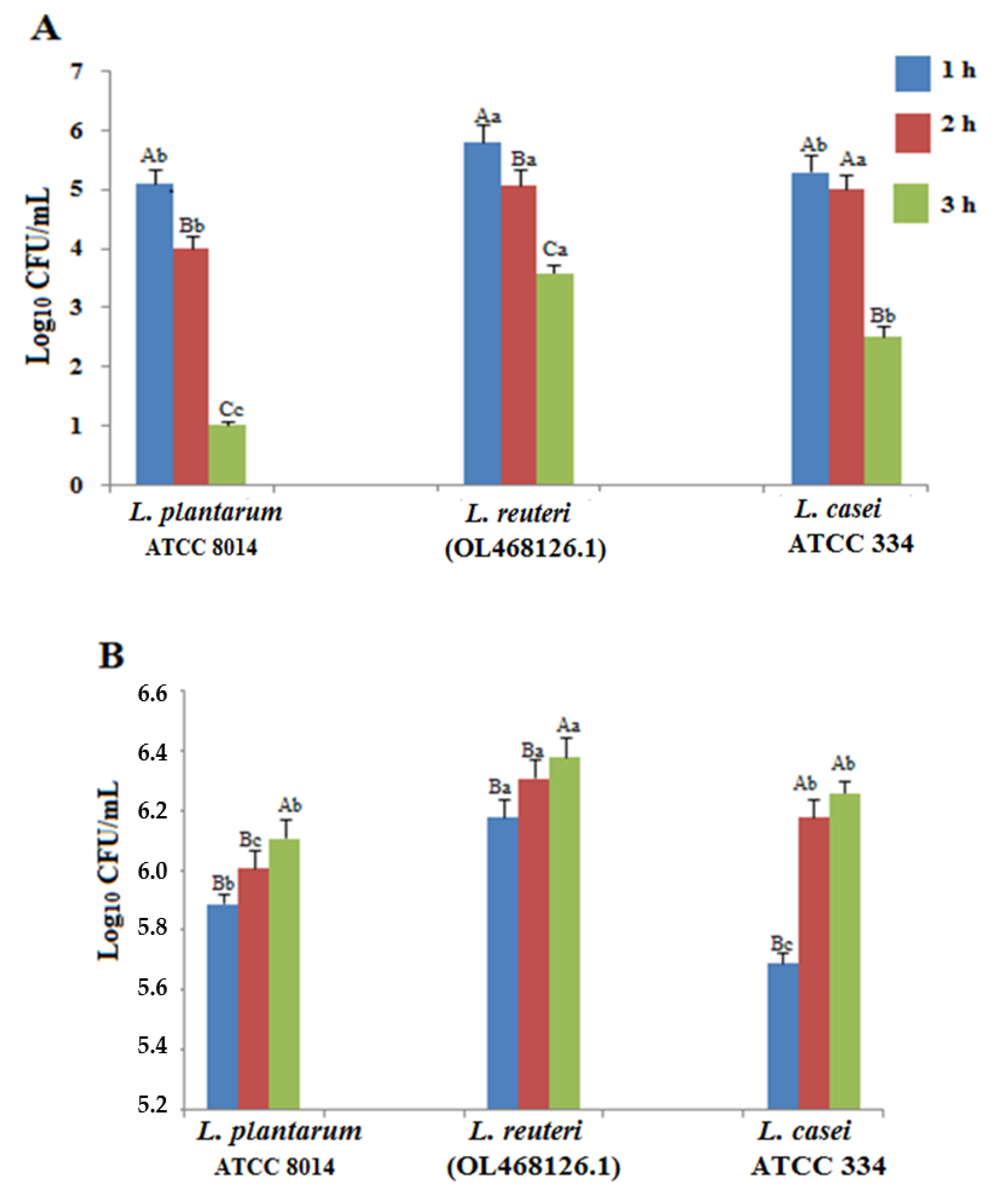
3.2. Acid and Bile Tolerances

The results of the viability of the isolated L. reuteri (OL468126.1) and two reference strains (L. casei ATCC 334 and L. plantarum ATCC 8014) against acid (pH 5.8, 3.0, and 2.0) and bile tolerance are shown in Figure 1 and Figure 2. At pH = 2 (Figure 1A), the cell viability after 3 h of treatment decreased significantly because of the loss of viable bacterial cells in the medium. L. reuteri (OL468126.1) and the reference strains exhibited tolerance to bile salts (0.3%). Bile salts present in the bacterial cultures were much more effective on bacterial viability than pH = 3.0. However, the degrees of tolerance varied among the strains. Interestingly, the isolated L. reuteri (OL468126.1) showed potent tolerance against acid and bile salts.

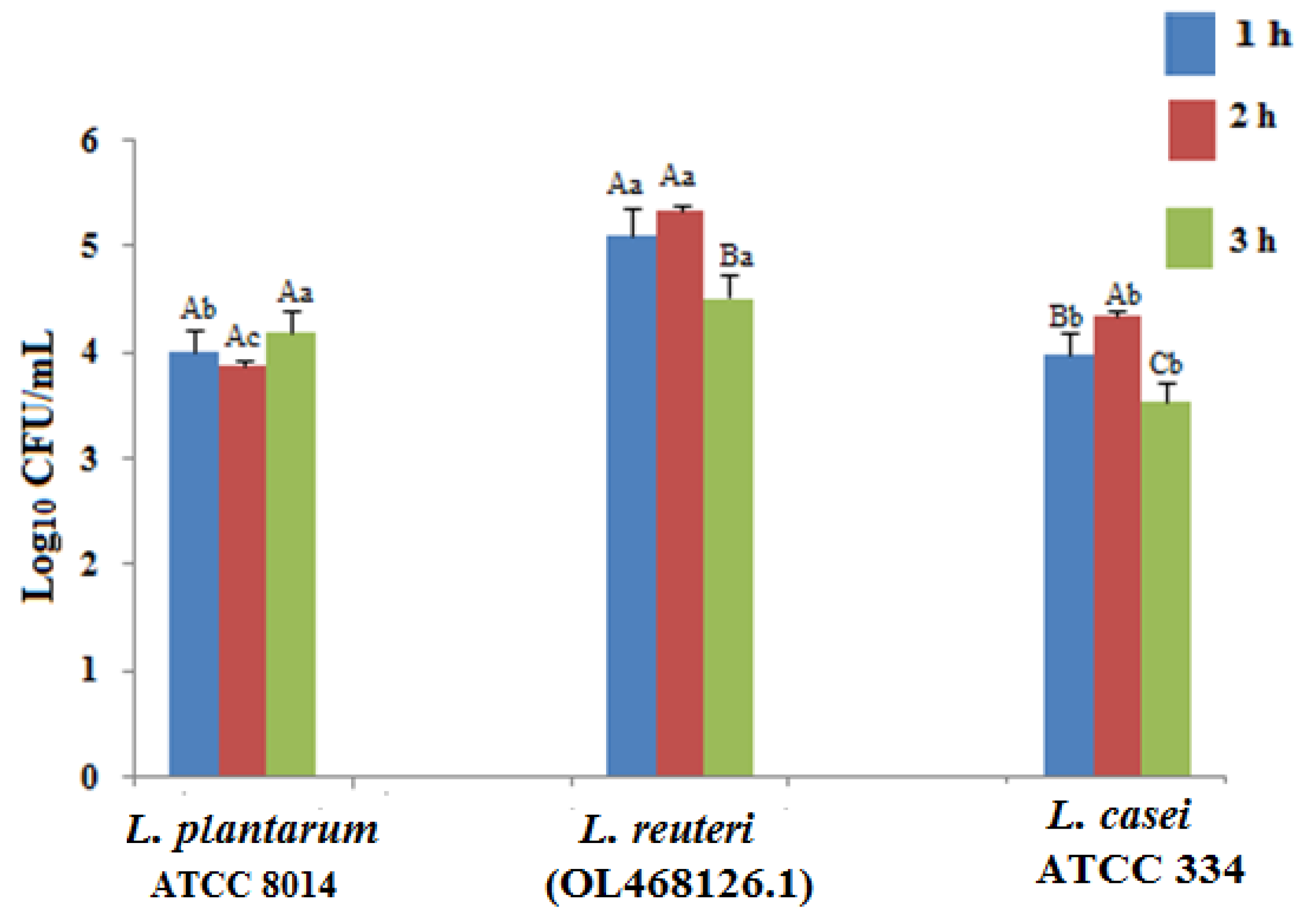
3.3. Antimicrobial Characterization of Limosilactobacillus reuteri (OL468126.1)

The antagonistic effects and the antimicrobial substances produced by the isolated L. reuteri (OL468126.1) were characterized by the agar well diffusion assay against five pathogens. As shown in Table 2, the inhibitory activity of the culture supernatant was observed, and the zone of diameter ranged between 7 ± 0.9 mm and 13.3 ± 0.6 mm. Trypsin and catalase-treated samples showed low antagonistic activities compared to the control experiments. At an increased pH of the sample (pH = 6.5), the antibacterial activity decreased significantly compared with the other treatments. The results revealed no activity of the culture supernatant treated with trypsin, catalase and pH against Pseudomonas aeruginosa (ATCC 27853).

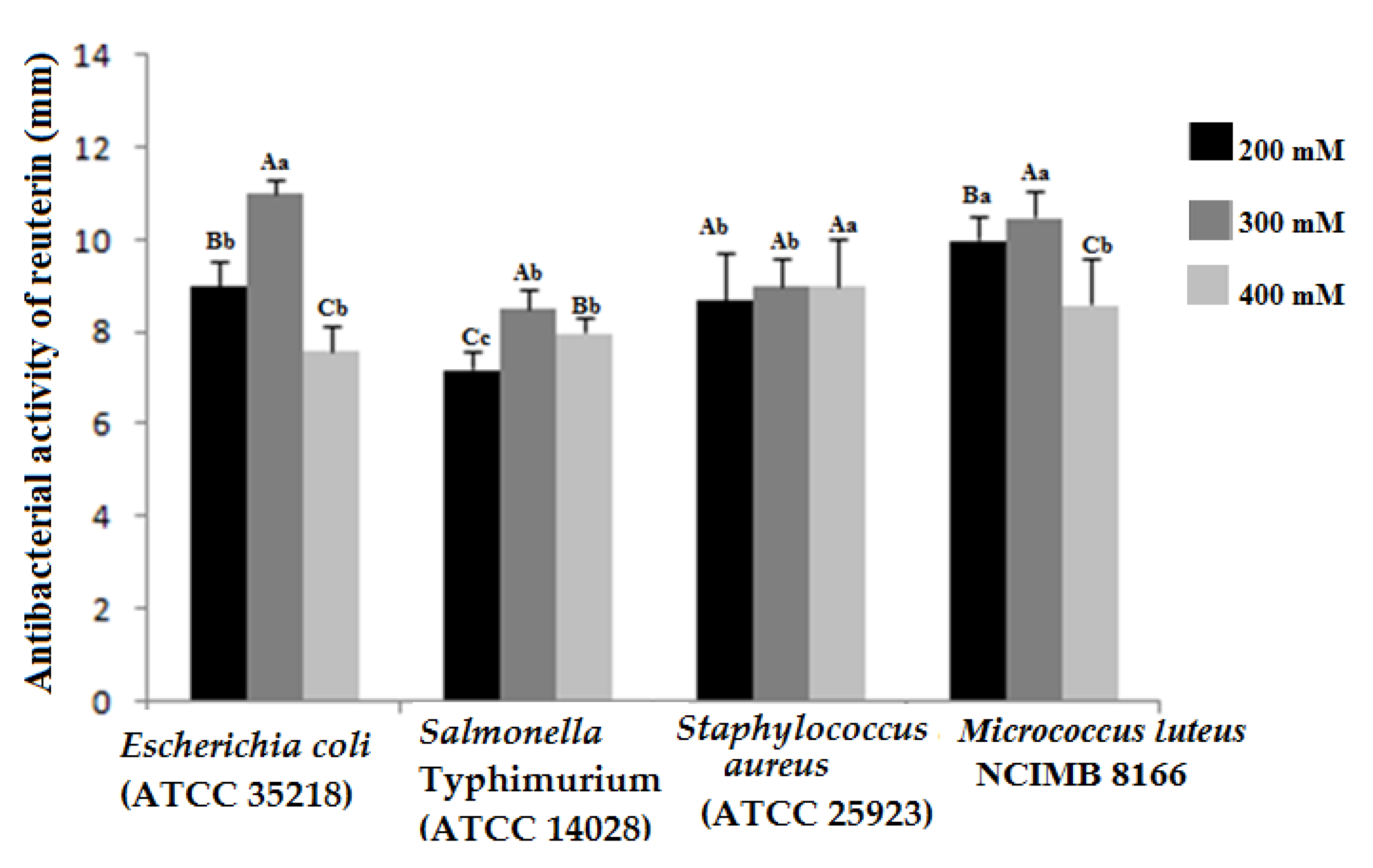
3.4. Antibacterial Activity of Reuterin Extracts

The antibacterial activity of reuterin produced by L. reuteri (OL468126.1) was studied using different concentrations of glycerol solutions (200 mM, 300 mM, and 400 mM) (Figure 3). The results show that reuterin extracts exhibited antimicrobial activity against the tested pathogens. The most important antagonistic effect was observed against Escherichia coli (ATCC 35218). Regardless of the pathogenic strain used, the degree of antagonism decreased significantly when the concentration of glycerol was 400 mM. Overall, the optimum initial glycerol concentration for the conversion to reuterin was 300 mM to obtain the highest bacterial inhibition.

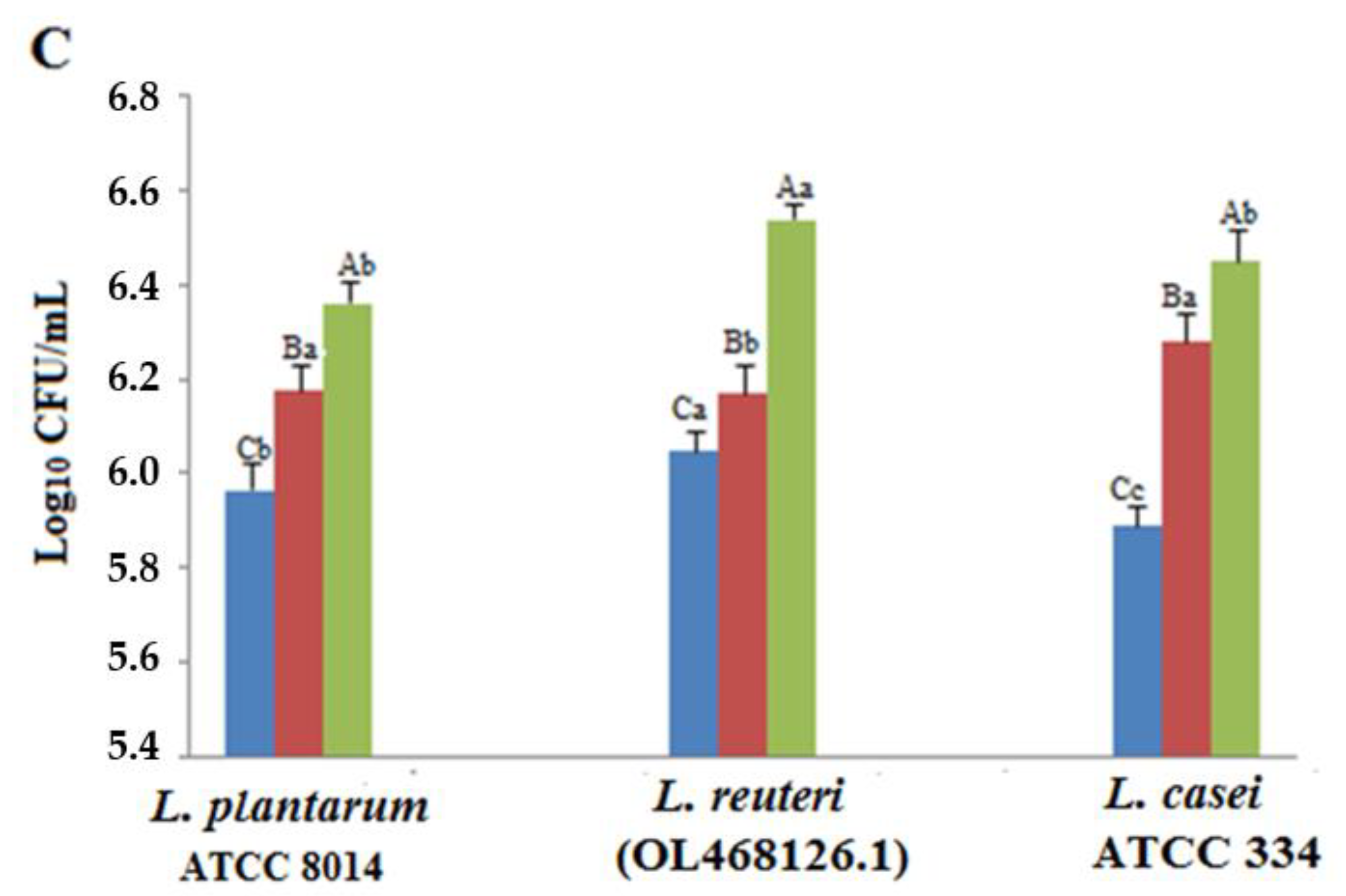
3.5. Adhesion Ability and Antiproliferative Activity

The isolated L. reuteri (OL468126.1) and two reference strains (L. casei ATCC 334 and L. plantarum ATCC 8014) were subjected to HCT-116 and Hep-2 cells for adhesion and antiproliferative activity analyses (Table 3). The tested strains showed varying adhesion abilities and among all strains, L. plantarum ATCC 8014 showed maximum adhesion to HCT-116 cells (80.1 ± 2.5%). Moreover, L. reuteri demonstrated high adhesion activities to HCT-116 (73.12 ± 3.02%) and Hep-2 (60.7 ± 3.02) cells indicating its potential as a good probiotic strain.
HCT-116 and Hep-2 cells were incubated with MTT solution after treatment with the bacterial supernatant for each strain. As shown in Table 3, all the tested Lactobacillus strains exhibited antiproliferative activity.

3.6. Growth Promotion of Isolated Limosilactobacillus reuteri with A. platensis or Phycocyanin

The growth stimulation of L. reuteri and the two reference strains (L. casei ATCC 334 and L. plantarum ATCC 8014) was evaluated in the presence of A. platensis or phycocyanin (Table 4). The addition of dry biomass of A. platensis at various concentrations of 1, 5, and 8 mg/mL enhanced the growth of L. reuteri. Maximum growth was promoted at an 8 mg/mL concentration of A. platensis after 48 h. Likewise, the addition of different concentrations of phycocyanin had a boosting effect on the growth of L. reuteri (OL468126.1), L. casei ATCC 334, and L. plantarum ATCC 8014. Interestingly, the results revealed that the addition of these supplements prolonged the stationary phase of the tested probiotics. After 72 h of incubation, the viability significantly decreased for all tested strains without A. platensis or phycocyanin extract.

4. Discussion

Lactobacilli play an important role in food fermentation and are part of the human gut microbiota, with numbers varying by species, host age, and gut location [36]. To be considered an effective probiotic, these microorganisms used in food preparation or supplementation must demonstrate utility in the host [37]. One species of Lactobacillus, Limosilactobacillus reuteri, first isolated in 1962, is thought to be a heterofermentative bacterium that grows in anoxic atmospheres and colonizes the gastrointestinal tract of humans and animals [38]. In this study, we characterized L. reuteri Lac Ib01 (OL468126.1) isolated from traditional sheep dry sausage and optimized its culture using A. platensis or phycocyanin extract. It is a well-studied probiotic with health benefits due to multiple metabolic mechanisms [39]. The fermentative profile of the isolated strain was tested using API50CHL. According to our results, this bacterium ferments arabinose, ribose, xylose, galactose, glucose, maltose, lactose, melibiose, succharose, and raffinose. Similar results were reported by Sulemankhil et al. [40]. L. reuteri was previously known as L. fermentum Type II because it resembles it physiologically [41]. The identity of the strain was then investigated using 16S rRNA gene sequencing. It has been determined that the capacity to withstand acid, bile, and pancreatic enzymes as well as the ability to adhere to intestinal epithelial cells are good indicators for a bacterial strain’s survival in the gastrointestinal tract (GIT) [31]. After 3 h of treatment, the isolated bacterium and the two reference strains (L. casei ATCC 334 and L. plantarum ATCC 8014) were tested for bile and acid tolerance in terms of cell viability, which revealed variance in pH tolerance. Importantly, the isolated L. reuteri (OL468126.1) showed potent tolerance to acid (pH = 3) and bile salts (0.3%). At low pH, the medium lost viable bacterial cells, resulting in a reduction in the bacterial population. Previous research has also demonstrated that exposing Lactobacilli to gastric acid for three hours resulted in a significant decrease in the bacterial population [42]. Bile acids, which are amphipathic molecules made from cholesterol, are crucial for the breakdown of fats and the assimilation of fat-soluble vitamins [43,44]. The amount of dietary fat consumed affects bile acid concentration in the human small intestine, which varies from 0.2 to 2% [37]. We were interested in determining whether L. reuteri can flourish in the presence of bile because it has been demonstrated that this strain may colonize, at least briefly, the small intestine. Our findings showed that L. reuteri can continue to grow even at bile concentrations as high as 0.3%. Whitehead et al. [45] provided insight into the possible mechanisms that L. reuteri ATCC 55730 may use to survive and grow in the presence of bile in the small intestine. These qualities are frequently evaluated in vitro for the initial detection of probiotic strains. Although in vitro tests might not be completely able to replicate the in situ circumstances in the gut environment, they are effective methods for quickly screening prospective strains [46]. Lactobacillus bacteria produce numerous antimicrobial compounds such as bacteriocin, hydrogen peroxide, organic acids, and reuterin, which can be used to prevent the growth of pathogenic bacteria and improve overall food sustainability [47].
The inhibitory activity of the culture supernatants and bioactive substances present (after different types of treatments) was determined against five pathogens through disk diffusion assays following 24 h incubation at 37 °C (Table 2). The results revealed that the cell-free supernatant of the isolated strain exhibited an average inhibition (7–13.3 mm) on the growth of Escherichia coli (ATCC 35218), Salmonella Typhimurium (ATCC 14028), Staphylococcus aureus (ATCC 25923), and Micrococcus luteus NCIMB 8166 but had no activity against Pseudomonas aeruginosa (ATCC 27853). Several previous studies have demonstrated the antibacterial effects of L. reuteri strains. Singh et al. [48] reported the antagonistic activity of L. reuteri strains against Escherichia coli ATCC25922, Salmonella Typhi NCDC113, Listeria monocytogenes ATCC 53135, and Enterococcus faecalis NCDC115. Moreover, Yang et al. [49] have shown the antimicrobial effects of cell-free culture supernatant from L. reuteri strain AN417 (LRS), a strain isolated from the porcine small intestine, against oral pathogenic bacteria. The production of organic acids has been proven to promote an antibacterial effect against Gram-negative pathogenic bacteria by way of the front of and next acidification of the pathogen cytoplasm, leading to cellular death [50]. It was also found that the production of H2O2 during metabolic processes critically inhibits the growth of several pathogenic microorganisms [51]. The antimicrobial activity of L. reuteri has been attributed to its production of organic acids, hydrogen peroxide, and bacteriocin-like compounds, which include reuterin, reuteran, and reutericyclin [52,53,54,55]. In this study, we tested the antibacterial activity of reuterin produced by L. reuteri (OL468126.1) using different concentrations of glycerol solutions (200 mM, 300 mM, and 400 mM). Our findings showed that the reuterin extracts exhibited antimicrobial activity against the tested pathogens. Previous studies have reported that reuterin exhibits strong antimicrobial properties against a wide range of Gram-positive and Gram-negative bacteria [48,49]. The principle mechanism of the antimicrobial effect of reuterin is causing a redoxim balance in the cellular redox state by reacting with free thiol groups of proteins causing their loss of function [56]. Apart from being able to survive the stressful gastric environment of the gastrointestinal tract, every potential probiotic strain is expected to adhere to and subsequently colonize the intestines [57]. Furthermore, the isolated L. reuteri showed a good adherence ability and antiproliferative activity in HCT-116 and Hep-2 cells.
A. platensis represents a valuable source of bioactive compounds. It is known for its high protein, γ-linolenic acid, and phycocyanin contents [58]. In this study, we investigated the effect of the addition of different concentrations of A. platensis or phycocyanin on the growth of L. reuteri (OL468126.1), L. casei ATCC 334, and L. plantarum ATCC 8014. Interestingly, the addition of these supplements promoted the ability of probiotic bacteria, specifically isolated L. reuteri. A. platensis increases the amount of amino acids in samples and is considered a nutrient for probiotic bacteria [19]. Moreover, proteins, free amino acids, and peptides can provide nitrogen sources for microbial growth [59]. A. platensis positively influences the growth of Lactobacillus in the intestinal system [16], synthetic media, and dairy [60]. The dry biomass of this cyanobacteria has been extensively studied to determine its effect on the growth of various lactic acid strains, such as Lactobacillus and Bifidobacterium [61]. Bhomwik et al. [35] reported that the growth of L. casei MTCC 1423, L. acidophilus MTCC 447, and S. thermophilus MTCC 1938 was stimulated by the dry biomass of algal products derived from the late log phase of growth.
A current tendency has been noted to incorporate microalgal biomass into fermented milk to improve the therapeutic and nutritional properties by enhancing probiotic stability [17,21,62]. The addition of A. platensis biomass to yogurt, cheese, and fermented milk has been tested by several authors [9,17,60], with promising results, including an increase in the number of lactic acid bacteria and an improvement in the nutritional quality of the fermented product during storage. Niccolai et al. [63] showed that A. platensis F&M-C256 is a suitable substrate for L. plantarum ATCC 8014 growth. After 72 h of fermentation, the broth contained a high concentration of LAB 8014, which could be responsible, together with phenolic and phycocyanin released from A. platensis F&M-C256, for the increase in radical scavenging capacity.
Although much is known about the ability of A. platensis to improve the activity of probiotics and support health-promoting dairy products, little is known about phycocyanin, which is one of the most important bioactive compounds. Our results showed that phycocyanin extract acts not only as a natural pigment but also as a growth promoter of probiotics. Phycocyanin may increase carbohydrate decomposition and stimulate the production of short-chain fatty acids [24]. According to previous studies, proteins promote the growth of intestinal bacteria, and many of the nutrients available to these bacteria in the intestine are derived from undigested proteins from the host’s diet [25]. Xie et al. [24] evaluated the effects of phycocyanin treatment on the intestinal microbiota and gut permeability in mice. The administration of phycocyanin significantly increased the abundance of the family Lactobacillaceae. Phycocyanin intervention regulated the composition of the lung and gut microbiota, transformed them into a normal state, and reduced the level of lipopolysaccharide (LPS), which reduced the abundance of inflammation-related bacteria and increased the abundance of probiotics that produce short-chain fatty acids [64].

5. Conclusions

According to our results, the bacterium Limosilactobacillus reuteri (OL468126.1) possesses desirable characteristics as a probiotic candidate; therefore, it can be used as an ingredient in functional foods or as a dietary supplement. An in vivo feeding study of an isolated probiotic must be conducted to establish its safety and effectiveness. Moreover, our findings suggested that A. platensis and phycocyanin extract stimulated the growth of probiotics, which resulted in improved viability of these microorganisms.

Author Contributions

Conceptualization, I.C. and Y.K.; methodology, I.C., N.H., Y.K. and K.F.; software, I.C. and M.M.; validation, I.C., Y.K., N.H., K.F., R.M. and A.B. formal analysis, I.C.; investigation, I.C.; resources, I.C., N.H., K.F., Y.K. and A.B.; data curation, I.C., N.H. and K.F.; writing—original draft preparation, I.C.; writing—review and editing, I.C. and N.H.; visualization, I.C.; supervision, A.B.; project administration, R.M. and Y.C.; funding acquisition, I.C. and N.H. All authors have read and agreed to the published version of the manuscript.

Funding

This research received no external funding.

Acknowledgments

We are grateful to the Bioalgae Tunisia Society, who gave us the samples free of charge to carry out the different experiments.

Conflicts of Interest

The authors declare that they do not have any conflicts of interest.

References

  1. Topolska, K.; Florkiewicz, A.; Filipiak-Florkiewicz, A. Functional Food—Consumer Motivations and Expectations. Int. J. Environ. Res. Public Health 2021, 18, 5327. [Google Scholar] [CrossRef]
  2. Sharifi-Rad, J.; Rodrigues, C.F.; Stojanović-Radić, Z.; Dimitrijević, M.; Aleksić, A.; Neffe-Skocińska, K.; Zielińska, D.; Kołożyn-Krajewska, D.; Salehi, B.; Milton Prabu, S.; et al. Probiotics: Versatile Bioactive Components in Promoting Human Health. Medicina 2020, 56, 433. [Google Scholar] [CrossRef]
  3. Torres-Maravilla, E.; Reyes-Pavón, D.; Benítez-Cabello, A.; González-Vázquez, R.; Ramírez-Chamorro, L.M.; Langella, P.; Bermúdez-Humarán, L.G. Strategies for the Identification and Assessment of Bacterial Strains with Specific Probiotic Traits. Microorganisms 2022, 10, 1389. [Google Scholar] [CrossRef] [PubMed]
  4. Alizadeh Behbahani, B.; Noshad, M.; Falah, F. Inhibition of Escherichia coli Adhesion to Human Intestinal Caco-2 Cells by Probiotic Candidate Lactobacillus Plantarum Strain L15. Microb. Pathog. 2019, 136, 103677. [Google Scholar] [CrossRef] [PubMed]
  5. Holzapfel, W.H.; Haberer, P.; Geisen, R.; Björkroth, J.; Schillinger, U. Taxonomy and Important Features of Probiotic Microorganisms in Food and Nutrition. Am. J. Clin. Nutr. 2001, 73, 365s–373s. [Google Scholar] [CrossRef] [PubMed] [Green Version]
  6. Darmastuti, A.; Hasan, P.N.; Wikandari, R.; Utami, T.; Rahayu, E.S.; Suroto, D.A. Adhesion Properties of Lactobacillus Plantarum Dad-13 and Lactobacillus Plantarum Mut-7 on Sprague Dawley Rat Intestine. Microorganisms 2021, 9, 2336. [Google Scholar] [CrossRef]
  7. Reid, G.; Gadir, A.A.; Dhir, R. Probiotics: Reiterating What They Are and What They Are Not. Front. Microbiol. 2019, 10, 424. [Google Scholar] [CrossRef] [Green Version]
  8. Gibson, G.R.; Roberfroid, M.B. Dietary Modulation of the Human Colonic Microbiota: Introducing the Concept of Prebiotics. J. Nutr. 1995, 125, 1401–1412. [Google Scholar] [CrossRef]
  9. Beheshtipour, H.; Mortazavian, A.M.; Mohammadi, R.; Sohrabvandi, S.; Khosravi-Darani, K. Supplementation of Spirulina Platensis and Chlorella Vulgaris Algae into Probiotic Fermented Milks: Algae Addition into Probiotic Fermented Milks…. Compr. Rev. Food Sci. Food Saf. 2013, 12, 144–154. [Google Scholar] [CrossRef]
  10. Molnár, N.; Gyenis, B.; Varga, L. Influence of a Powdered Spirulina Platensis Biomass on Acid Production of Lactococci in Milk. Milchwissenschaft 2005, 60, 380–382. [Google Scholar]
  11. Sommella, E.; Conte, G.; Salviati, E.; Pepe, G.; Bertamino, A.; Ostacolo, C.; Sansone, F.; Prete, F.; Aquino, R.; Campiglia, P. Fast Profiling of Natural Pigments in Different Spirulina (Arthrospira Platensis) Dietary Supplements by DI-FT-ICR and Evaluation of Their Antioxidant Potential by Pre-Column DPPH-UHPLC Assay. Molecules 2018, 23, 1152. [Google Scholar] [CrossRef] [Green Version]
  12. Safari, R.; Raftani Amiri, Z.; Esmaeilzadeh Kenari, R. Antioxidant and Antibacterial Activities of C-Phycocyanin from Common Name Spirulina Platensis. Iran. J. Fish. Sci. 2019, 19, 1911–1927. [Google Scholar] [CrossRef]
  13. Wang, X.-Q.; Li, L.-N.; Chang, W.-R.; Zhang, J.-P.; Gui, L.-L.; Guo, B.-J.; Liang, D.-C. Structure of C-Phycocyanin from Spirulina Platensis at 2.2 AÊ Resolution: A Novel Monoclinic Crystal Form for Phycobiliproteins in Phycobilisomes. Acta Crystallogr. Sect. D: Biol. Crystallogr. 2001, 57, 784–792. [Google Scholar] [CrossRef]
  14. Abdel-Moneim, A.-M.E.; El-Saadony, M.T.; Shehata, A.M.; Saad, A.M.; Aldhumri, S.A.; Ouda, S.M.; Mesalam, N.M. Antioxidant and Antimicrobial Activities of Spirulina Platensis Extracts and Biogenic Selenium Nanoparticles against Selected Pathogenic Bacteria and Fungi. Saudi J. Biol. Sci. 2021, 29, 1197–1209. [Google Scholar] [CrossRef] [PubMed]
  15. Barros de Medeiros, V.P.; Salgaço, M.K.; Pimentel, T.C.; Rodrigues da Silva, T.C.; Sartoratto, A.; dos Lima, M.S.; da Sassi, C.F.C.; Mesa, V.; Magnani, M.; Sivieri, K. Spirulina Platensis Biomass Enhances the Proliferation Rate of Lactobacillus Acidophilus 5 (La-5) and Combined with La-5 Impact the Gut Microbiota of Medium-Age Healthy Individuals through an in Vitro Gut Microbiome Model. Food Res. Int. 2022, 154, 110880. [Google Scholar] [CrossRef] [PubMed]
  16. Shimamatsu, H. Mass Production of Spirulina, an Edible Microalga. Hydrobiologia 2004, 512, 39–44. [Google Scholar] [CrossRef]
  17. Çelekli, A.; Alslibi, Z.A.; Bozkurt, H. Boosting Effects of Spirulina Platensis, Whey Protein, and Probiotics on the Growth of Microflora and the Nutritional Value of Ayran. Eng. Rep. 2020, 2, e12235. [Google Scholar] [CrossRef]
  18. Barkallah, M.; Dammak, M.; Louati, I.; Hentati, F.; Hadrich, B.; Mechichi, T.; Ayadi, M.A.; Fendri, I.; Attia, H.; Abdelkafi, S. Effect of Spirulina Platensis Fortification on Physicochemical, Textural, Antioxidant and Sensory Properties of Yogurt during Fermentation and Storage. LWT 2017, 84, 323–330. [Google Scholar] [CrossRef]
  19. Çelekli, A.; Alslibi, Z.A.; Bozkurt, H. Influence of Incorporated Spirulina Platensis on the Growth of Microflora and Physicochemical Properties of Ayran as a Functional Food. Algal Res. 2019, 44, 101710. [Google Scholar] [CrossRef]
  20. Patel, P.; Jethani, H.; Radha, C.; Vijayendra, S.V.N.; Mudliar, S.N.; Sarada, R.; Chauhan, V.S. Development of a Carotenoid Enriched Probiotic Yogurt from Fresh Biomass of Spirulina and Its Characterization. J. Food Sci. Technol. 2019, 56, 3721–3731. [Google Scholar] [CrossRef]
  21. Martelli, F.; Alinovi, M.; Bernini, V.; Gatti, M.; Bancalari, E. Arthrospira Platensis as Natural Fermentation Booster for Milk and Soy Fermented Beverages. Foods 2020, 9, 350. [Google Scholar] [CrossRef] [PubMed] [Green Version]
  22. Plaza, M.; Herrero, M.; Cifuentes, A.; Ibáñez, E. Innovative Natural Functional Ingredients from Microalgae. J. Agric. Food Chem. 2009, 57, 7159–7170. [Google Scholar] [CrossRef] [PubMed]
  23. Chiong, T.; Acquah, C.; Lau, S.Y.; Khor, E.H.; Danquah, M.K. Microalgal-Based Protein By-Products: Extraction, Purification, and Applications. In Protein Byproducts; Elsevier: Amsterdam, The Netherlands, 2016; pp. 213–234. [Google Scholar] [CrossRef]
  24. Xie, Y.; Li, W.; Zhu, L.; Zhai, S.; Qin, S.; Du, Z. Effects of Phycocyanin in Modulating the Intestinal Microbiota of Mice. Microbiol. Open 2019, 8, e00825. [Google Scholar] [CrossRef] [Green Version]
  25. The Human Microbiome Project Consortium. Structure, Function and Diversity of the Healthy Human Microbiome. Nature 2012, 486, 207–214. [Google Scholar] [CrossRef] [PubMed] [Green Version]
  26. Heilig, H.G.H.J.; Zoetendal, E.G.; Vaughan, E.E.; Marteau, P.; Akkermans, A.D.L.; de Vos, W.M. Molecular Diversity of Lactobacillus Spp. and Other Lactic Acid Bacteria in the Human Intestine as Determined by Specific Amplification of 16S Ribosomal DNA. Appl. Environ. Microbiol. 2002, 68, 114–123. [Google Scholar] [CrossRef] [Green Version]
  27. Schumann, P. E. Stackebrandt and M. Goodfellow (Editors), Nucleic Acid Techniques in Bacterial Systematics (Modern Microbiological Methods). XXIX + 329 S., 46 Abb., 28 Tab. Chichester—New York—Brisbane—Toronto—Singapore 1991. John Wiley & Sons. $ 55.00. ISBN: 0-471-92906-9. J. Basic Microbiol. 1991, 31, 479–480. [Google Scholar] [CrossRef]
  28. Kaboré, D.; Sawadogo-Lingani, H.; Dicko, M.H.; Diawara, B.; Jakobsen, M. Acid Resistance, Bile Tolerance and Antimicrobial Properties of Dominant Lactic Acid Bacteria Isolated from Traditional Maari Baobab Seeds Fermented Condiment. Afr. J. Biotechnol. 2012, 11, 1197–1205. [Google Scholar] [CrossRef]
  29. Klingberg, T.D.; Axelsson, L.; Naterstad, K.; Elsser, D.; Budde, B.B. Identification of Potential Probiotic Starter Cultures for Scandinavian-Type Fermented Sausages. Int. J. Food Microbiol. 2005, 105, 419–431. [Google Scholar] [CrossRef]
  30. Lamari, F.; Mahdhi, A.; Chakroun, I.; Esteban, M.A.; Mazurais, D.; Amina, B.; Gatesoupe, F.-J. Interactions between Candidate Probiotics and the Immune and Antioxidative Responses of European Sea Bass (Dicentrarchus Labrax) Larvae. J. Fish Dis. 2016, 39, 1421–1432. [Google Scholar] [CrossRef]
  31. Shokryazdan, P.; Sieo, C.C.; Kalavathy, R.; Liang, J.B.; Alitheen, N.B.; Faseleh Jahromi, M.; Ho, Y.W. Probiotic Potential of Lactobacillus Strains with Antimicrobial Activity against Some Human Pathogenic Strains. BioMed Res. Int. 2014, 2014, 927268. [Google Scholar] [CrossRef] [Green Version]
  32. Doleyres, Y.; Beck, P.; Vollenweider, S.; Lacroix, C. Production of 3-Hydroxypropionaldehyde Using a Two-Step Process with Lactobacillus Reuteri. Appl. Microbiol. Biotechnol. 2005, 68, 467–474. [Google Scholar] [CrossRef] [PubMed] [Green Version]
  33. Mosmann, T. Rapid Colorimetric Assay for Cellular Growth and Survival: Application to Proliferation and Cytotoxicity Assays. J. Immunol. Methods 1983, 65, 55–63. [Google Scholar] [CrossRef] [PubMed]
  34. Verdenelli, M.C.; Ghelfi, F.; Silvi, S.; Orpianesi, C.; Cecchini, C.; Cresci, A. Probiotic Properties of Lactobacillus Rhamnosus and Lactobacillus Paracasei Isolated from Human Faeces. Eur. J. Nutr. 2009, 48, 355–363. [Google Scholar] [CrossRef]
  35. Bhowmik, D.; Dubey, J.; Mehra, S. Probiotic Efficiency of Spirulina Platensis—Stimulating Growth of Lactic Acid Bacteria. World J. Dairy Food Sci. 2009, 4, 160–163. [Google Scholar]
  36. De Filippis, F.; Pasolli, E.; Ercolini, D. The Food-Gut Axis: Lactic Acid Bacteria and Their Link to Food, the Gut Microbiome and Human Health. FEMS Microbiol. Rev. 2020, 44, 454–489. [Google Scholar] [CrossRef]
  37. Zommiti, M.; Feuilloley, M.G.J.; Connil, N. Update of Probiotics in Human World: A Nonstop Source of Benefactions till the End of Time. Microorganisms 2020, 8, 1907. [Google Scholar] [CrossRef] [PubMed]
  38. Mu, Q.; Tavella, V.J.; Luo, X.M. Role of Lactobacillus Reuteri in Human Health and Diseases. Front. Microbiol. 2018, 9, 757. [Google Scholar] [CrossRef] [Green Version]
  39. Abuqwider, J.; Altamimi, M.; Mauriello, G. Limosilactobacillus Reuteri in Health and Disease. Microorganisms 2022, 10, 522. [Google Scholar] [CrossRef] [PubMed]
  40. Sulemankhil, I.; Parent, M.; Jones, M.L.; Feng, Z.; Labbé, A.; Prakash, S. In Vitro and in Vivo Characterization and Strain Safety of Lactobacillus Reuteri NCIMB 30253 for Probiotic Applications. Can. J. Microbiol. 2012, 58, 776–787. [Google Scholar] [CrossRef]
  41. Hou, C.; Zeng, X.; Yang, F.; Liu, H.; Qiao, S. Study and Use of the Probiotic Lactobacillus Reuteri in Pigs: A Review. J. Anim. Sci. Biotechnol. 2015, 6, 14. [Google Scholar] [CrossRef] [Green Version]
  42. Zhang, X.; Ali Esmail, G.; Fahad Alzeer, A.; Valan Arasu, M.; Vijayaraghavan, P.; Choon Choi, K.; Abdullah Al-Dhabi, N. Probiotic Characteristics of Lactobacillus Strains Isolated from Cheese and Their Antibacterial Properties against Gastrointestinal Tract Pathogens. Saudi J. Biol. Sci. 2020, 27, 3505–3513. [Google Scholar] [CrossRef] [PubMed]
  43. Mandal, S.; Puniya, A.K.; Singh, K. Effect of Alginate Concentrations on Survival of Microencapsulated Lactobacillus Casei NCDC-298. Int. Dairy J. 2006, 16, 1190–1195. [Google Scholar] [CrossRef]
  44. Hofmann, A.F. Chemistry and Enterohepatic Circulation of Bile Acids. Hepatology 1984, 4 (Suppl. S2), 4S–14S. [Google Scholar] [CrossRef]
  45. Whitehead, K.; Versalovic, J.; Roos, S.; Britton, R.A. Genomic and Genetic Characterization of the Bile Stress Response of Probiotic Lactobacillus Reuteri ATCC 55730. Appl. Environ. Microbiol. 2008, 74, 1812–1819. [Google Scholar] [CrossRef] [PubMed] [Green Version]
  46. Dunne, C.; O’Mahony, L.; Murphy, L.; Thornton, G.; Morrissey, D.; O’Halloran, S.; Feeney, M.; Flynn, S.; Fitzgerald, G.; Daly, C.; et al. In Vitro Selection Criteria for Probiotic Bacteria of Human Origin: Correlation with in Vivo Findings. Am. J. Clin. Nutr. 2001, 73, 386s–392s. [Google Scholar] [CrossRef] [Green Version]
  47. Agriopoulou, S.; Stamatelopoulou, E.; Sachadyn-Król, M.; Varzakas, T. Lactic Acid Bacteria as Antibacterial Agents to Extend the Shelf Life of Fresh and Minimally Processed Fruits and Vegetables: Quality and Safety Aspects. Microorganisms 2020, 8, 952. [Google Scholar] [CrossRef]
  48. Singh, T.P.; Kaur, G.; Kapila, S.; Malik, R.K. Antagonistic Activity of Lactobacillus Reuteri Strains on the Adhesion Characteristics of Selected Pathogens. Front. Microbiol. 2017, 8, 486. [Google Scholar] [CrossRef] [Green Version]
  49. Yang, K.M.; Kim, J.-S.; Kim, H.-S.; Kim, Y.-Y.; Oh, J.-K.; Jung, H.-W.; Park, D.-S.; Bae, K.-H. Lactobacillus Reuteri AN417 Cell-Free Culture Supernatant as a Novel Antibacterial Agent Targeting Oral Pathogenic Bacteria. Sci. Rep. 2021, 11, 1631. [Google Scholar] [CrossRef]
  50. Makras, L.; De Vuyst, L. The in Vitro Inhibition of Gram-Negative Pathogenic Bacteria by Bifidobacteria Is Caused by the Production of Organic Acids. Int. Dairy J. 2006, 16, 1049–1057. [Google Scholar] [CrossRef]
  51. Ilavenil, S.; Vijayakumar, M.; Kim, D.H.; Valan Arasu, M.; Park, H.S.; Ravikumar, S.; Choi, K.C. Assessment of Probiotic, Antifungal and Cholesterol Lowering Properties of Pediococcus Pentosaceus KCC-23 Isolated from Italian Ryegrass: Properties of P. Pentosaceus from Ryegrass. J. Sci. Food Agric. 2016, 96, 593–601. [Google Scholar] [CrossRef]
  52. Kang, M.-S.; Oh, J.-S.; Lee, H.-C.; Lim, H.-S.; Lee, S.-W.; Yang, K.-H.; Choi, N.-K.; Kim, S.-M. Inhibitory Effect of Lactobacillus Reuteri on Periodontopathic and Cariogenic Bacteria. J. Microbiol. 2011, 49, 193–199. [Google Scholar] [CrossRef] [PubMed]
  53. Cleusix, V.; Lacroix, C.; Vollenweider, S.; Duboux, M.; Le Blay, G. Inhibitory Activity Spectrum of Reuterin Produced by Lactobacillus Reuteri against Intestinal Bacteria. BMC Microbiol. 2007, 7, 101. [Google Scholar] [CrossRef] [PubMed] [Green Version]
  54. Asare, P.T.; Zurfluh, K.; Greppi, A.; Lynch, D.; Schwab, C.; Stephan, R.; Lacroix, C. Reuterin Demonstrates Potent Antimicrobial Activity against a Broad Panel of Human and Poultry Meat Campylobacter Spp. Isolates. Microorganisms 2020, 8, 78. [Google Scholar] [CrossRef] [PubMed] [Green Version]
  55. Zhao, Q.; Maddox, I.S.; Mutukumira, A.; Lee, S.J.; Shu, Q. The Effect of Cell Immobilization on the Antibacterial Activity of Lactobacillus Reuteri DPC16 Cells during Passage through a Simulated Gastrointestinal Tract System. World J. Microbiol. Biotechnol. 2012, 28, 3025–3037. [Google Scholar] [CrossRef]
  56. Schaefer, L.; Auchtung, T.A.; Hermans, K.E.; Whitehead, D.; Borhan, B.; Britton, R.A. The Antimicrobial Compound Reuterin (3-Hydroxypropionaldehyde) Induces Oxidative Stress via Interaction with Thiol Groups. Microbiology 2010, 156, 1589–1599. [Google Scholar] [CrossRef] [Green Version]
  57. Han, S.; Lu, Y.; Xie, J.; Fei, Y.; Zheng, G.; Wang, Z.; Liu, J.; Lv, L.; Ling, Z.; Berglund, B.; et al. Probiotic Gastrointestinal Transit and Colonization After Oral Administration: A Long Journey. Front. Cell. Infect. Microbiol. 2021, 11, 609722. [Google Scholar] [CrossRef]
  58. Fradinho, P.; Niccolai, A.; Soares, R.; Rodolfi, L.; Biondi, N.; Tredici, M.R.; Sousa, I.; Raymundo, A. Effect of Arthrospira Platensis (Spirulina) Incorporation on the Rheological and Bioactive Properties of Gluten-Free Fresh Pasta. Algal Res. 2020, 45, 101743. [Google Scholar] [CrossRef]
  59. Wang, Y.; Tibbetts, S.; McGinn, P. Microalgae as Sources of High-Quality Protein for Human Food and Protein Supplements. Foods 2021, 10, 3002. [Google Scholar] [CrossRef]
  60. Varga, L.; Szigeti, J.; Kovács, R.; Földes, T.; Buti, S. Influence of a Spirulina Platensis Biomass on the Microflora of Fermented ABT Milks During Storage (R1). J. Dairy Sci. 2002, 85, 1031–1038. [Google Scholar] [CrossRef]
  61. Perković, L.; Djedović, E.; Vujović, T.; Baković, M.; Paradžik, T.; Čož-Rakovac, R. Biotechnological Enhancement of Probiotics through Co-Cultivation with Algae: Future or a Trend? Mar. Drugs 2022, 20, 142. [Google Scholar] [CrossRef]
  62. Bhalamurugan, G.L.; Valerie, O.; Mark, L. Valuable Bioproducts Obtained from Microalgal Biomass and Their Commercial Applications: A Review. Environ. Eng. Res. 2018, 23, 229–241. [Google Scholar] [CrossRef] [Green Version]
  63. Niccolai, A.; Shannon, E.; Abu-Ghannam, N.; Biondi, N.; Rodolfi, L.; Tredici, M.R. Lactic Acid Fermentation of Arthrospira Platensis (Spirulina) Biomass for Probiotic-Based Products. J. Appl. Phycol. 2019, 31, 1077–1083. [Google Scholar] [CrossRef] [Green Version]
  64. Li, W.; Lu, L.; Liu, B.; Qin, S. Effects of Phycocyanin on Pulmonary and Gut Microbiota in a Radiation-Induced Pulmonary Fibrosis Model. Biomed. Pharmacother. 2020, 132, 110826. [Google Scholar] [CrossRef] [PubMed]
Figure 1. Tolerance of the isolated Limosilactobacillus reuteri (OL468126.1), Lactiplantibacillus plantarum ATCC 8014, and Lacticaseibacillus casei ATCC 334 to pH = 2 (A), pH = 3 (B), and pH = 5.8 (C) after 1, 2, and 3 h of incubation. Data are the mean values of three replicates, and the error bars indicate the standard deviation (n = 3). Capital letters are used to indicate the significant differences observed for the same strain after different periods (1, 2, and 3 h). Different small letters indicate significant differences between the strains in the same condition (p < 0.05).
Fermentation 09 00248 g001aFermentation 09 00248 g001b
Figure 2. Tolerance of the isolated Limosilactobacillus reuteri (OL468126.1), Lactiplantibacillus plantarum ATCC 8014, and Lacticaseibacillus casei ATCC 334 to 0.3% bile concentration after 1, 2, and 3 h of incubation. Data are the mean values of three replicates, and the error bars indicate the standard deviation (n = 3). Capital letters are used to indicate the significant differences observed for the same strain after different periods (1, 2, and 3 h). Different small letters indicate significant differences between the strains in the same condition (p < 0.05).
Fermentation 09 00248 g002
Figure 3. Antibacterial activity of reuterin extracts produced by Limosilactobacillus reuteri (OL468126.1) using different concentrations of glycerol solutions (200 mM, 300 mM, and 400 mM) against tested pathogens. Data are the mean values of three replicates, and the error bars indicate the standard deviation (n = 3). Capital letters are used to indicate significant differences observed for the same strain at different concentrations of glycerol. Different small letters indicate significant differences between the strains under the same conditions (p < 0.05).
Fermentation 09 00248 g003
Table 1. Biochemical profile of the API 50 CHL of Limosilactobacillus reuteri (OL468126.1).
TubeBiochemical TestsResults
0Temoin-
1Glycerol-
2Erythritol-
3D-Arabinose-
4L-Arabinose+
5D-Ribose+
6D-Xylose+
7L-Xylose-
8D-Adonitol-
9Methyl-b-D-xylopyranoside-
10D-Galactose+
11D-Glucose+
12D-Fructose-
13D-Mannose-
14L-Sorbose-
15L-Rhamnose-
16Dulcitol-
17Inositol-
18D-Mannitol-
19D-Sorbitol-
20Methyl-a-D-mannopyroside-
21Methyl-a-D-glucopyranoside-
22N-Acetylglucosamine-
23Amygdalin-
24Arbutin-
25Esculinferriccitrate-
26Salicin-
27D-Cellobiose-
28D-Maltose+
29D-Lactose+
30D-Melibiose+
31D-Saccharose+
32D-Trehalose-
33Inulin-
34D-Melezitose-
35D-Raffinose+
36Amidon-
37Glycogen-
38Xylitol-
39Gentibiose-
40D-Turanose-
41D-Lyxose-
42D-Tagatose-
43D-Fucose-
44L-Fucose-
45D-Arabitol-
46L-Arabitol-
47Potassiumgluconate±
48Potassium2-ketogluconate-
49Potassium5-ketogluconate-
Note: (-): negative reaction; (+): positive reaction; (±): unclear result.
Table 2. Antimicrobial activity of Limosilactobacillus reuteri (Lac Ib01) against the tested pathogens. Diameter of the inhibition zone (mm) after incubation at 37 °C for 24 h.
StrainsEscherichia coli (ATCC 35218)Salmonella Typhimurium (ATCC 14028)Pseudomonas
aeruginosa
(ATCC 27853)
Staphylococcus
aureus
(ATCC 25923)
Micrococcus luteus NCIMB 8166
Treatment
CFCS: control12.3 ± 0.6 Ba11.6 ± 1.0 Ca-10.76 ± 1.2 Ca13.3 ± 0.6 Aa
Trypsin10.6 ± 0.5 Ab8.3 ± 0.6 Cb-9 ± 1 Cb10.6 ± 1 Bb
Catalase9.5 ± 1 Ab8 ± 1.02 Bb-9 ± 0.6 Ab8 ± 1.2 Bc
pH = 6.58 ± 0.0 Ac7 ± 0.9 Bc-8.3 ± 1.3 Ab7.2 ± 1.1 Bc
Antibacterial activity of fermentation broth of Limosilactobacillus reuteri strain after being subjected to various treatments. Cell-free culture supernatants (CFCS): Control sample: filtered, untreated sample; Trypsin: samples treated with trypsin at 2 mg/mL concentration; pH: samples adjusted the pH to 6.5; Catalase: sample treated with catalase at 1 mg/mL. Lac Ib01: Limosilactobacillus reuteri (OL468126.1). The results are expressed as the mean ± standard deviation; n = 3. (-): no activity. Different capital letters in each row and different small letters in each column indicate significant differences (p < 0.05).
Table 3. Antiproliferative activity and adhesion ability of Limosilactobacillus reuteri (Lac Ib01) selected as candidate probiotics.
StrainAntiproliferative Activity (%)Adhesion (%)
HCT-116Hep-2HCT-116Hep-2
Lac Ib0160.3 ± 2.1 Ab57.06 ± 2.8 Aa73.12 ± 3.02 Ab60.7 ± 3.02 Aa
L. casei ATCC 33476.25 ± 0.9 Aa40.18 ± 1.4 Bb78.08 ± 1.8 Ab53.64 ± 2.1 Bb
L. plantarum ATCC 801471.3 ± 1.1 Aa50.2 ± 2.2 Ba80.1 ± 2.5 Aa42.15 ± 2.8 Bc
The results are expressed as the mean ± standard deviation; n = 3. Different capital letters in each row indicate significant differences for the same strain using different cells and different small letters in each column indicate significant differences between strains for the same test (p < 0.05).
Table 4. Growth kinetics of the tested Lactobacillus strains (log10 CFU/mL) supplemented with A. platensis or phycocyanin extract.
L. reuteri
OL468126.1
L. casei ATCC 334L. plantarum ATCC 8014
0 h24 h48 h72 h0 h24 h48 h72 h0 h24 h48 h72 h
Control3.51 ± 0.25 Ca6.42 ± 0.3 Bc8.41± 1.5 Ac7.05 ± 1.12 Bc3.6 ± 0.52 Ca7.02 ± 0.2Bc9.1 ± 0.37 Ac8.79 ± 0.5 Ac4.2 ± 0.6 Ca7.02 ± 0.73 Bc9.4 ± 0.25 Ab8.15 ± 0.5 Ac
Concentration of A. platensis (mg/mL)013.58 ± 0.58 Ca7.18 ± 0.63 Bc8.7 ± 1.1 Ac8.21 ± 1.03 Ac3.82 ± 0.9 Ca7.53 ± 0.5 Bc8.6 ± 0.21 Ac8.01 ± 1.05 Ac3.9 ± 0.83 Ca7.6 ± 0.78 Bc9.12 ± 0.97 Ab9.05 ± 0.62 Ab
043.7 ± 0.12 Ca7.9 ± 0.7 Bb9.92 ± 0.73 Ab11.0 ± 0.94 Aa3.7 ± 0.6 Ca8.23 ± 1.1 Bb10.33 ± 0.5 Aa10.8 ± 0.9 Aa4 ± 0.2 Ca8.04 ± 0.35Bb10.58 ± 1 Aa10.6 ± 0.4 Aa
083.52 ± 0.26 Ca9.17 ± 0.41 Ba12.0 ± 0.6 Aa8.12 ± 0.92 Bb3.47 ± 0.3 Ca11.0 ± 0.91 Ba13.1 ± 0.84 Aa10.21 ± 0.9 Aa3.8 ± 0.15 Ca10.5 ± 0.5 Ba12.6 ± 0.2 Aa10.12 ± 0.7 Ba
Concentration of phycocyanin extract (mg/mL)013.81 ± 0.95 Ca6.78 ± 0.5 Bc8.1 ± 0.2 Ac7.1 ± 1.2 Ac3.65 ± 0.47 Ca7.3 ± 0.83 Bc9.51 ± 0.9 Ab8.32 ± 0.7 Ac3.6 ± 0.5 Ca6.58 ± 1.02 Bc8.67 ± 1.Ab7.9 ± 0.81 Ac
043.57 ± 0.2 Ca7.5 ± 0.9 Bc8.62 ± 0.15 Ac8.54 ± 1.0Ac3.75 ± 0.9 Ca8.02 ± 0.1 Bb10 ± 1.06 Ab10.6 ± 0.2Aa4.1 ± 0.7 Ca7.05 ± 0.3 Bc9.1 ± 0.Ab9.37 ± 0.61 Ab
083.64 ± 0.7 Ca8.03 ± 0.21 Bb9.51 ± 0.33 Ab9.05 ± 0.6Ab3.56 ± 1.02 Ca9.7 ± 0.5 Bb11.06 ± 0.8 Aa9.5 ± 0.34 Bb3.81 ± 0.92 Ca8.9 ± 0.57 Bb10.84 ± 0.92 Aa9.25 ± 0.7 Ab
The results are expressed as the mean ± standard deviation; n = 3. Different capital letters in each row indicate significant differences for each strain at different periods (0, 24, 48, and 72 h) and different small letters in each column indicate significant differences at different concentrations of A. platensis or phycocyanin extract (p < 0.05).
Disclaimer/Publisher’s Note: The statements, opinions and data contained in all publications are solely those of the individual author(s) and contributor(s) and not of MDPI and/or the editor(s). MDPI and/or the editor(s) disclaim responsibility for any injury to people or property resulting from any ideas, methods, instructions or products referred to in the content.

